Fast and Efficient Peptide Mapping of a Monoclonal … Performance with Superficially Porous...
10
Fast and Efficient Peptide Mapping of a Monoclonal Antibody (mAb): UHPLC Performance with Superficially Porous Particles Authors James Martosella, Alex Zhu Agilent Technologies, Inc. Wilmington, DE 19808 Ning Tang Agilent Technologies, Inc. Santa Clara, CA 95052 Application Note Biotherapeutics and Biosimilars Introduction Peptide mapping by reversed-phase (RP) chromatography is the mainstay technique in biotherapeutic analysis, delivering comprehensive characterization of biopharmaceutical products. When interfaced with a mass spectrometer (MS), it can deliver the identification of proteins and their variants, determine post-translational modifications (PTMs) and locations, and confirm protein sequences. However, peptide mapping represents a significant chromatographic challenge due to the inherent complexity of protein digests. As a result, many organizations struggle with developing robust and reliable peptide maps. In general, peptide maps have suffered from low sensitivity, poor peak shapes, and very long separation times to achieve the desired resolution. More recently, Ultra High Performance Liquid Chromatography (UHPLC) has been employed to overcome these challenges, demonstrating superior resolution, higher sensitivity, and much shorter analysis times compared to traditional HPLC. UHPLC technology can provide the basis for a more detailed characterization of protein biotherapeutics and takes advantage of faster flow rates, smaller particles, and shorter column lengths to achieve high separation performance during much faster run times. However, the pressure requirement becomes increasingly high for these analyses and prevents operation on traditional 400 and 600 bar HPLC instruments, thus severely limiting its broader use.
Transcript of Fast and Efficient Peptide Mapping of a Monoclonal … Performance with Superficially Porous...

Fast and Efficient Peptide Mappingof a Monoclonal Antibody (mAb):UHPLC Performance withSuperficially Porous Particles
Authors
James Martosella, Alex Zhu
Agilent Technologies, Inc.
Wilmington, DE 19808
Ning Tang
Agilent Technologies, Inc.
Santa Clara, CA 95052
Application Note
Biotherapeutics and Biosimilars
Introduction
Peptide mapping by reversed-phase (RP) chromatography is the mainstay techniquein biotherapeutic analysis, delivering comprehensive characterization ofbiopharmaceutical products. When interfaced with a mass spectrometer (MS), itcan deliver the identification of proteins and their variants, determinepost-translational modifications (PTMs) and locations, and confirm proteinsequences. However, peptide mapping represents a significant chromatographicchallenge due to the inherent complexity of protein digests. As a result, manyorganizations struggle with developing robust and reliable peptide maps. In general,peptide maps have suffered from low sensitivity, poor peak shapes, and very longseparation times to achieve the desired resolution.
More recently, Ultra High Performance Liquid Chromatography (UHPLC) has beenemployed to overcome these challenges, demonstrating superior resolution, highersensitivity, and much shorter analysis times compared to traditional HPLC. UHPLCtechnology can provide the basis for a more detailed characterization of proteinbiotherapeutics and takes advantage of faster flow rates, smaller particles, andshorter column lengths to achieve high separation performance during much fasterrun times. However, the pressure requirement becomes increasingly high for theseanalyses and prevents operation on traditional 400 and 600 bar HPLC instruments,thus severely limiting its broader use.

2
To address this limitation, Agilent has introduced a 2.7 µmAdvanceBio Peptide Mapping column to fill a critical gap inbiotherapeutics characterizations, for generating both rapidand highly efficient peptide maps at traditional LC-systempressures. Using superficially porous chromatographic media,AdvanceBio Peptide Mapping columns achieve substantialimprovements in peptide mapping during very fast run timesand low system pressures, while still maintaining high peak-performance efficiency. In this work, an AdvanceBio PeptideMapping column was used for the LC/MS peptide mappinganalysis of a monoclonal antibody (mAb) tryptic digest withreduced analysis times. Additionally, the AdvanceBio columnand methodology was compared to a sub-2 µm non-AgilentUHPLC peptide mapping column for performance comparisons.
Materials and Methods
Sample preparation
Fifty microliters mAb IgG1 (30 mg/mL) were first mixed with75 µL 100 mM ammonium bicarbonate (pH 8). Seventy-fivemicroliters of trifluoroethanol and 3 µL 200 mM dithiothreitol(DTT) were added to the protein sample and heated at 60 °Cfor 1 h to denature and reduce the protein. After the proteinhad cooled to room temperature, 12 µL 200 mMiodoacetamide (IAM) was added to the sample. The samplewas kept at room temperature in the dark for 1 h.Subsequently, 3 µL DTT was added in the sample to reactwith the excess IAM for 1 h. The sample was diluted with900 µL water and 300 µL 100 mM ammonium bicarbonate.Trypsin solution (75 µL) was added to the sample andincubated at 37 °C for 20 h. After overnight incubation, 3 µL ofneat formic acid was added to quench the digest.
ConditionsColumns: AdvanceBio Peptide Mapping 2.1 × 100 mm
(p/n 655750-902), AdvanceBio Peptide Mapping, 2.1 × 150 mm (p/n 653750-902), non-Agilent UHPLC column, 2.1 × 100 mm
Eluent: A: H2O + 0.1% FA (v/v)B: 90% ACN + 0.1% FA (v/v)
Injection volume: 15 µL
Flow rates: various
Temperature: 40 °C
Detection: UV, 215/220 nm
Instrumentation: Agilent 1290 Infinity LC System and an Agilent 6530Accurate-Mass Quadrupole Time-of-Flight (Q-TOF)
Q-TOF MS parametersIon mode: Positive
Source: Agilent Dual Jet Stream
Drying gas temperature: 250 °C
Drying gas flow: 10 L/min
Sheath gas temperature: 250 °C
Sheath gas flow: 12 L/min
Nebulizer: 35 psi
Capillary voltage: 3,500 V
Fragmentor: 200 V
Skimmer: 65 V
Oct 1 RF: 750 V
Nozzle: 0 V
MS range (m/z): 100 to 1600
MS/MS range (m/z): 100 to 1600
MS scan rate (spectra/second): 8
MS/MS scan rate (spectra/second): 3
LC/MS results were analyzed using Agilent MassHunterQualitative Analysis Software B.06 and Agilent MassHunterBioConfirm Software B.06.

3
Figure 1. AdvanceBio Peptide Mapping column optimization for achieveing a faster peptide mapping analysis. Gradient 10-40% B,DAD: 215 nm, 40 °C. Top panel, 75 minute separation on a 2.1 × 150 mm column generated 59 peptide peaks (flow rate 0.2 mL/min,211 bar). Bottom panel, optimized 14 minute separation on a 2.1 × 100 mm column generated 57 peptide peaks (flow rate 0.6 mL/min, 433 bar).
min0 10 20 30 40 50 60
mAU2.1 × 150 mm AdvanceBio Peptide Mapping column
2.1 × 100 mm AdvanceBio Peptide Mapping column
0
20
40
60
80
100
120
140
160%B-50%
%B-50%
min0 2 4 6 8 10 12
mAU
0
20
40
60
80
100
120
140
160
Results and Discussion
Fast peptide mapping optimization Optimization of peptide maps to resolve and analyzepotentially hundreds of peaks is a significant task. Performingthese optimizations with high resolution during shortenedanalysis times can be even more challenging and timeconsuming. Employing long gradients of mAb tryptic peptidemaps to achieve the desired resolution can take 120 minutesor longer, especially for large proteins like mAbs. While fastermaps are highly desirable, resolution must not becompromised, while end test results need to provide the samelevel of information found during an extended run.
To demonstrate the AdvanceBio Peptide Mapping column’sflexibility for increasing analysis speed without sacrificingseparation performance, two UV separations are compared inFigure 1. In this comparison, a mAb tryptic peptide map wasfully optimized at 75 minutes on a 2.1 × 150 mm AdvanceBioPeptide Mapping column (top chromatogram) with an
efficient baseline-resolved peptide map across the entiregradient profile. The 75 minute separation is an excellentexample of the column’s ability to enable faster separationperformance for a 150 mm length column (traditional columnlength), compared to conventional peptide mapping times atthis dimension. However, it also provides a better example forcomparing the column’s flexibility for further increasinganalysis speed without sacrificing separation performancewhen moving to a shorter gradient.
In the bottom 14 minute chromatogram in Figure 1, columnflow rate was increased from 0.2 to 0.6 mL/min while columnlength was decreased from 150 to 100 mm. Volumetric flowduring this change was carefully maintained, keeping thegradient change per column volume equal to ensure similarselectivity. The 14 minute peptide map details excellentseparation performance, maintaining the baseline resolutionand sensitivity while keeping resolving power unchanged. The14 minute separation demonstrates UHPLC- type speed andperformance, yet keeps this rapid analysis attainable on600 bar HPLC instrumentation.

4
Figure 2. Comparison of Agilent AdvanceBio Peptide Mapping column total ion chromatograms (TICs), highlighting peptidemapping performance between 40-min and 14-min runs for an mAb tryptic digest. Left panel, TIC collected during 40-minanalysis (flow rate 0.2 mL/min, 140 bar). Right panel, TIC collected during 14-min analysis. (flow rate 0.6 mL/min, 433 bar).
0
0.6
1.6
2.6
3.6
4.6
1 2 3 4 5 6 7 8 9 10 11 12 13
×108×108
0
0.75
1.75
2.75
3.75
Acquisition time (min)
Counts
Acquisition time (min)
Counts
2 4 6 8 10 12 14 16 18 20 22 24 26 28 30 32 34 36 38
40-min run140 bar0.2 mL/min
14-min run433 bar0.6 mL/min
Gradient, 40 minute Gradient, 14 minute
Time (min) %B Time (min) %B0 3 0 335 35 10 3537 90 12 9040 90 14 90
LC/MS peptide mapping with an AdvanceBio Peptide
Mapping column: steep versus long gradient
In a typical biopharmaceutical peptide mapping workflow,reversed-phase LC is combined with mass spectrometrydetection. Compared with the more traditional approach of UVonly detection, RP LC/MS significantly enhances theinformation content available from the peptide-mappingexperiment. Measurement by RP LC/MS can differentiatecoeluting peptides, locate and identify peptide modifications,and determine the sequence coverage. High sequencecoverage is an indicator that optimal separation of the trypticdigest has been achieved.
During RP LC/MS, long gradients are typically used to enablehigher resolution and ensure modified peptides (referred to aspost translational modifications or PTMs) are detectable fromtheir unmodified (native) forms as well as other close eluting
peptides within the overall digest profile. To reduce run timessteeper gradients are employed, however, this is typically atthe cost of lost resolution and overall mapping quality. It is,therefore, important to not sacrifice separation quality orcompromise mass spectral information when run times arereduced.
Figure 2 compares peptide mapping total ion chromatograms(TIC) from an AdvancedBio Peptide Mapping column during40-min and 14-min runs. In this comparison, columndimensions remained constant, and the gradient slope wasadjusted to keep gradient change/column volume betweenthe columns equal, ensuring that the chromatographicselectivity remained similar. The TIC comparisons showedthat resolution, selectivity, and separation performance hadnot been compromised during the reduced run time of 14 min,while the increase in pressure at the higher flow rate did notrequire > 600 bar UHPLC instrumentation.

5
Additionally, sequence coverage between the 40-min and14-min run was also evaluated using the Molecular FeatureExtractor (MFE) in the MassHunter Qualitative AnalysisSoftware. MFE is an algorithm that finds and extracts a list ofcompounds from complex separation data, such as peptidemaps. The list of compounds is then matched back to themAb protein sequence and provides the sequence coverage.Figure 3 displays the extracted compound chromatograms(ECCs) for the matched peptides and the sequence coverage.
All matched peptides were acquired with at least one MS/MSspectrum for confirmation. The 40-min and the 14-min runsremain relatively unchanged in coverage. The 14-min LC/MSrun resulted in 99.63% sequence coverage of mAb while the40-min analysis resulted in 99.84%. This comparison providesfurther confidence that peptide characterization information isnot sacrificed when peptide mapping analyses times areshortened on the AdvanceBio Peptide Mapping column.
Figure 3. Agilent AdvanceBio Peptide Mapping column extracted compound chromatograms (ECC) to compare mAb tryptic digestresults. The top panel shows an ECC for peptides identified from the 40-min run, resulting in 99.84% sequence coverage of mAb.The bottom panel shows an ECC for peptides identified from the 14-min run, resulting in 99.63% sequence coverage of mAb.
00.40.81.21.6
22.42.83.23.6
44.44.8
Cpd
702
: B(2
98-3
13)
Cpd
384
: B(3
57-3
66)
Cpd
445
: B(2
52-2
70)
Cpd
345
: B(1
30-1
43)
Cpd
424
: B(1
18-1
29)
Cpd
302
: A(4
8-63
)
Cpd
524
: B(4
5-65
)
Cpd
731
: B(2
98-3
13)
Cpd
660
: B(2
19-2
44)
Cpd
775
: A(1
36-1
55)
Cpd
156
: A(1
96-2
10)
Cpd
624
: B(3
89-4
05)
Cpd
106
: A(1
63-1
72)
Cpd
205
: A(1
09-1
16)
Cpd
275
: B(4
36-4
42)
Cpd
240
: B(2
45-2
51)
Cpd
236
: B(3
23-3
30)
Cpd
395
: B(4
13-4
35)
Cpd
483
: A(1
17-1
35)
Cpd
499
: A(1
8-47
)
Cpd
133
: A(2
11-2
18)
Cpd
561
: A(1
78-1
92)
Cpd
120
: B(6
8-74
)
Cpd
291
: A(1
-17)
Cpd
60:
A(1
56-1
62)
Cpd
330
: A(4
8-63
)
Cpd
580
: B(3
67-3
88)
Cpd
454
: B(3
57-3
66)
Cpd
639
: B(2
98-3
18)
Cpd
31:
A(6
4-68
)
Cpd
16:
B(2
10-2
14)
Cpd
749
: A(6
9-10
8)
Cpd
549
: B(4
5-65
)
Cpd
132
: B(2
89-2
97)
Cpd
175
: A(1
96-2
10)
Cpd
221
: A(1
56-1
77)
Cpd
599
: B(2
52-2
88)
Cpd
686
: B(2
98-3
13)
Cpd
768
: B(4
13-4
35)
Cpd
741
: A(1
73-1
95)
Cpd
43:
B(3
37-3
40)
Cpd
780
: A(1
8-47
)
0.6 1 1.4 1.8 2.2 2.6 3 3.4 3.8 4.2 4.6 5 5.4 5.8 6.2 6.6 7 7.4 7.8 8.2 8.6 9 9.4 9.8 10.2 10.6 11
×107
40-min run
14-min run×107
00.40.81.21.6
22.42.83.23.6
44.4 C
pd 9
34: B
(252
-270
)
Cpd
147
9: B
(298
-313
)
Cpd
827
: B(3
57-3
66)
Cpd
626
: A(4
8-63
)
Cpd
741
: B(1
30-1
43)
Cpd
170
9: A
(136
-155
)
Cpd
110
6: B
(45-
65)
Cpd
153
6: B
(298
-313
)
Cpd
871
: B(1
18-1
29)
Cpd
135
0: B
(219
-244
)
Cpd
312
: A(1
96-2
10)
Cpd
128
3: B
(389
-405
)
Cpd
167
: A(1
63-1
72)
Cpd
105
6: A
(18-
47)
Cpd
428
: A(1
09-1
16)
Cpd
494
: B(2
45-2
51)
Cpd
118
3: A
(178
-192
)
Cpd
562
: B(4
36-4
42)
Cpd
475
: B(3
23-3
30)
Cpd
102
7: A
(117
-135
)
Cpd
704
: A(4
8-63
)
Cpd
121
5: B
(367
-388
)
Cpd
187
: B(6
8-74
)
Cpd
230
: A(2
11-2
18)
Cpd
94:
A(1
56-1
62)
Cpd
131
0: B
(298
-318
)
Cpd
675
: A(4
8-63
)
Cpd
44:
A(6
4-68
)
Cpd
157
3: A
(69-
108)
Cpd
963
: B(3
57-3
66)
Cpd
167
3: B
(144
-206
)
Cpd
124
0: B
(367
-388
)
Cpd
104
7: A
(48-
63)
Cpd
25:
B(2
10-2
14)
Cpd
608
: B(3
41-3
51)
Cpd
162
0: B
(1-4
4)
Cpd
137
8: B
(77-
98)
Cpd
100
5: B
(252
-270
)
Cpd
156
2: B
(289
-313
)
Cpd
112
8: A
(18-
47)
Cpd
127
7: B
(99-
117)
Cpd
120
4: B
(252
-288
)
Cpd
141
4: B
(99-
129)
Cpd
147
0: B
(289
-313
)
Cpd
260
: B(2
89-2
97)
Cpd
60:
B(3
37-3
40)
Cpd
365
: B(6
6-67
)
Cpd
792
: B(2
52-2
70)
Cpd
466
: A(1
09-1
16)
Cpd
349
: B(2
45-2
51)
Cpd
108
5: A
(163
-177
)
Cpd
816
: A(4
8-63
)
Acquisition time (min)
Acquisition time (min)
Cou
nts
Cou
nts
0 1 2 3 4 5 6 7 8 9 10 11 12 13 14 15 16 17 18 19 20 21 22 23 24 25 26 27 28 29 30 31 32 33 34

6
Rapid PTM profiling
Deamidation can cause structural and functional changes andis an important post translational modification to monitorduring mAb discovery, development, and manufacturing.Figure 4 displays an example of how peptide deamidation wasmonitored and conserved during a fast run time of14 minutes, in comparison to a longer run time of 40 minutes.Heavy chain peptide 357-366, which contains Asn 357, wasidentified in both peptide mapping separations. In the top andmiddle ECC figures, the native peptide peak was fullyseparated from its two deamidated forms, displaying as atotal of three separate peaks in the overlaid ECCs. Incomparison to the 40-min run (top), the native anddeamidated species in the 14-min run (bottom) were still wellresolved and readily identified by the QTOF analysis.
In the bottom figure, MS/MS of the native peptide (precursorat m/z = 581.32) and two deamidated forms (precursor atm/z = 581.81) are shown. In this spectra, the y and b-seriesfragments are the predominant ions, and all three peptidesshow the same y4-y8 ions, however the b2 and b3 ions(circled) are 0.98 Da higher for peak 2 and 3 thus validatingthe modified forms from the native species.
The automatic compound extraction (in MFE) also adds all theion intensities (isotopes, charge states, adducts, and so forth)belonging to the peptides. These are listed under the Volumetab in peptide Table 1. The comparative percentage ofmodified peptides can then be easily derived from the twodata sets in Table 1. In summary, the rapid 14 minute peptidemap generated from the AdvanceBio Peptide Mappingcolumn did not compromise PTM (deamidation) informationor the sequence coverage in comparison to the longer run.Using a steeper gradient at increased flow, the columnmaintained excellent separation performance and delivered ahigh degree of confidence in a complete peptide mappingcharacterization.
Figure 4. Overlaid extracted compound chromatograms (ECC)and MS/MS spectra (bottom) of native peptide and itsdeamidated forms. Upper panel displays results from the40-min analysis. Middle panel displays results from the 14-minanalysis. Bottom panel provides MS/MS spectra of the nativeand two deamidated forms shown above.
Acquisition time (min)
Cou
nts
×106
00.5
11.5
22.5
33.5
44.5
55.5
66.5
77.5 Cpd 154: B(357-366)
18.5 19 19.5 20 20.5 21 21.5 22 22.5 23 23.5 24 24.5
Native peptide
Deamidatedform 2
Deamidatedform 1
40 min ECC
00.05
0.10.150.2
0.250.3
0.350.4
0.450.5
0.550.6
0.650.7
0.750.8
0.850.9
0.951
Cpd 132: B(357-366)
Acquisition time (min)
Cou
nts
6.1 6.3 6.5 6.7 6.9 7.1 7.3 7.5 7.7 7.9 8.1 8.3 8.5
Native peptide
×107
Deamidatedform 2
Deamidatedform 1
14 min ECC
×103
×103
×103
012345
820.4576y7
919.5266y8
243.1073
132.0761 342.1757620.3438
y5 733.4290y6
519.2975y4429.2099
0
1
2
3 244.0939 820.4583y7 919.5293
y8133.0605733.4298
y6620.3407
y5343.1553
88.0400
0
1
2
3244.0906b2
b2
b2
820.4558y7 919.5275
y8133.0594 620.3403
y5 733.4199y6343.1612
b3
b3
b3
469.1724
Mass-to-charge (m/z)
Coun
tsCo
unts
Coun
ts
50 100 150 200 250 300 350 400 450 500 550 600 650 700 750 800 850 900 950 10001050
Native
Deamidated
Deamidated

7
Peptide mapping with steep gradients:
HPLC (AdvanceBio Peptide column) versus
UHPLC (non-Agilent UHPLC peptide column)
With the advancement of UHPLC peptide mapping, separationperformance has been significantly improved and analysistime shortened. With sub-2 µm particle technology, peptideresolving power can now be vastly improved during shortenedruntimes; a consequence of reduced diffusional distance insmall particles. This higher resolution and increasedsensitivity becomes particularly important in peptide mapping,especially for the detection of modified peptides. In directcomparison to sub-2 µm column performance, 2.7 µmsuperficially porous particles with a thin porous outer shelland solid inner core can offer exceptional resolving power,which also results in fewer backpressure constraints ascolumn flow rates are increased. This pressure and speedadvantage can obviate the requirement for > 600 bar UHPLCinstrumentation to perform these fast and highly efficientpeptide mapping analyses.
The UV chromatographic comparison for an IgG tryptic digestin Figure 5 highlights the AdvanceBio Peptide Mappingcolumn’s speed and resolving power in direct comparison to atop industry performing UHPLC peptide mapping column. Inthe top chromatogram, the AdvanceBio Peptide Mappingcolumn was optimized for a rapid and highly efficient analysisusing 2.1 × 100 mm dimensions. In this separation, thepeptide peaks were well resolved across the entire gradientprofile, delivering 56 peaks at a pressure of 433 bar. In thebottom chromatogram, a 2.1 × 100 mm UHPLCpeptide-mapping column was compared under the samechromatographic conditions and column dimensions.However, the critical aspect of the peptide-mapping results ishighlighted by the low pressure requirement from theAdvanceBio Peptide Mapping column to achieve the sameseparation results. The UHPLC peptide column resulted in700 bar backpressure and required the use of UHPLCinstrumentation, thus limiting its broader application.
Table 1. Extracted compound list showing the native peptide HC (357-366) and two deamidated forms with their sequence,modification, retention time, mass and volume for the 40-min analysis (top) compared to the 14-min analysis (bottom).
14-min run
40-min run

8
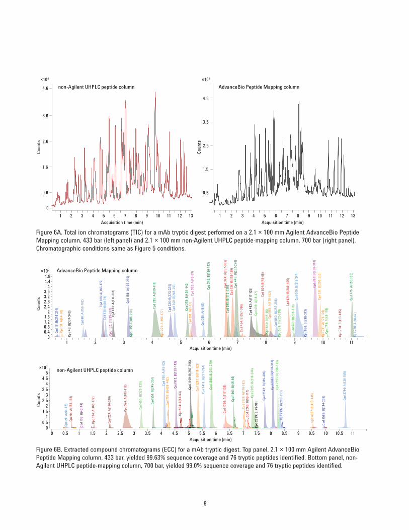
The ability to quickly, routinely and comprehensively runLC/MS analyses for mAb mapping helps to accelerate allfacets of the therapeutic development process. However,these separations are typically performed on high pressureLCs to take full advantage of maximum efficiency and timesavings. Superficially porous separations can offer theflexibility to operate on either UHPLC/MS or HPLC/MSinstrumentation coupled to MS, to deliver rapid and highefficiency separations. To contrast the LC/MS peptidemapping performance, and pressure requirements betweensub-2 µm UHPLC and superficially porous HPLC peptide-mapping columns, a 2.1 × 100 mm AdvanceBio PeptideMapping column was directly compared to a non-AgientUHPLC peptide mapping column of the same dimension.Figure 6 displays the RP LC/MS TIC and ECC performanceresults between these two columns. Again all the identified
peptides were acquired with at least one MS/MS spectrumas confirmation.
In the TIC and ECC comparisons, the AdvanceBio PeptideMapping LC/MS analysis delivered excellent separation andsequence coverage in comparison to the non-Agilent UHPLCpeptide-mapping column. Sequence coverage for theAdvanceBio peptide Mapping column was 99.63%, while theUHPLC coverage was comparable at 99.0%. Both columnsdelivered identity for 76 total tryptic peptides. However, theUHPLC analysis resulted in 700 bar backpressure, while theAdvanceBio separation operated 500 bar. The ability to obtainUHPLC like results on the AdvanceBio Peptide Mappingcolumn can make this column more attractive for obtainingrapid and highly efficient peptide maps with the flexibility ofusing traditional HPLC instrumentation.
min0 2 4 6 8 10 12
mAU
0
20
40
60
80
100
120
140
160AdvanceBio Peptide Mapping column433 bar
non-Agilent UHPLC peptide column700 bar
min0 2 4 6 8 10 12
mAU
0
20
40
60
80
100
120
140
160
Figure 5. 2.1 × 100 mm Agilent and non-Agilent UHPLC peptide maps of an mAb tryptic digest. Gradient: 10-40% B at 0.6 mL/min,DAD: 215 nm, temperature: 40 °C, flow: 0.6 mL/min. Top, HPLC separation with an Agilent AdvanceBio Peptide Mapping columngenerated 56 peptide peaks at 433 bar. Bottom, UHPLC separation with a non-Agilent UHPLC peptide column generated 52 peptidefragment peaks at 700 bar.
9
0
0.6
1.6
2.6
3.6
4.6 non-Agilent UHPLC peptide column AdvanceBio Peptide Mapping column
×108 ×108
0.5
1.5
2.5
3.5
4.5
1 2 3 4 5 6 7 8 9 10 11 12 13
Acquisition time (min)
Cou
nts
Acquisition time (min)
Cou
nts
1 2 3 4 5 6 7 8 9 10 11 12 13
Cpd
702
: B(2
98-3
13)
Cpd
384
: B(3
57-3
66)
Cpd
445
: B(2
52-2
70)
Cpd
345
: B(1
30-1
43)
Cpd
424
: B(1
18-1
29)
Cpd
302
: A(4
8-63
)
Cpd
524
: B(4
5-65
)
Cpd
731
: B(2
98-3
13)
Cpd
660
: B(2
19-2
44)
Cpd
775
: A(1
36-1
55)
Cpd
156
: A(1
96-2
10)
Cpd
624
: B(3
89-4
05)
Cpd
106
: A(1
63-1
72)
Cpd
205
: A(1
09-1
16)
Cpd
275
: B(4
36-4
42)
Cpd
240
: B(2
45-2
51)
Cpd
236
: B(3
23-3
30)
Cpd
395
: B(4
13-4
35)
Cpd
483
: A(1
17-1
35)
Cpd
499
: A(1
8-47
)
Cpd
133
: A(2
11-2
18)
Cpd
561
: A(1
78-1
92)
Cpd
120
: B(6
8-74
)
Cpd
291
: A(1
-17)
Cpd
60:
A(1
56-1
62)
Cpd
330:
A(4
8-63
)
Cpd
580
: B(3
67-3
88)
Cpd
454
: B(3
57-3
66)
Cpd
639
: B(2
98-3
18)
Cpd
31:
A(6
4-68
)
Cpd
16:
B(2
10-2
14)
Cpd
749
: A(6
9-10
8)
Cpd
549
: B(4
5-65
)
Cpd
132
: B(2
89-2
97)
Cpd
175
: A(1
96-2
10)
Cpd
221
: A(1
56-1
77)
Cpd
599
: B(2
52-2
88)
Cpd
686
: B(2
98-3
13)
Cpd
768
: B(4
13-4
35)
Cpd
741
: A(1
73-1
95)
Cpd
43:
B(3
37-3
40)
Cpd
780
: A(1
8-47
)
Cpd
269
2: B
(298
-313
)
Cpd
156
5: B
(252
-270
)
Cpd
114
9: B
(357
-366
)
Cpd
706
: A(4
8-63
)
Cpd
913
: B(1
30-1
43)
Cpd
279
5: B
(298
-313
)
Cpd
128
7: B
(118
-129
)
Cpd
141
6: B
(271
-284
)
Cpd
232
5: B
(219
-244
)
Cpd
374
6: A
(136
-155
)
Cpd
254
2: B
(389
-405
)
Cpd
190
1: B
(45-
65)
Cpd
551
: B(2
45-2
51)
Cpd
797
: B(4
36-4
42)
Cpd
212
7: A
(178
-192
)
Cpd
455
: B(3
23-3
30)
Cpd
324
: A(1
09-1
16)
Cpd
179
0: A
(117
-135
)
Cpd
994
: A(4
8-63
)
Cpd
220
0: B
(99-
117)
Cpd
293
2: B
(298
-313
)
Cpd
94:
A(1
56-1
62)
Cpd
338
7: B
(413
-435
)
Cpd
224
: A(1
96-2
10)
Cpd
164
: A(1
63-1
72)
Cpd
358
2: B
(144
-206
)
Cpd
26:
A(6
4-68
)
Cpd
133
: B(4
0-44
)
Cpd
239
9: B
(75-
98)
×107
00.40.81.21.6
22.42.83.23.6
44.44.8
AdvanceBio Peptide Mapping column
non-Agilent UHPLC peptide column
Acquisition time (min)
Acquisition time (min)
Cou
nts
Cou
nts
1 2 3 4 5 6 7 8 9 10 11
×107
00.5
11.5
22.5
33.5
44.5
5
0.5 1 1.50 2 2.5 3 3.5 4 4.5 5 5.5 6 6.5 7 7.5 8 8.5 9 9.5 10 10.5 11
Figure 6A. Total ion chromatograms (TIC) for a mAb tryptic digest performed on a 2.1 × 100 mm Agilent AdvanceBio PeptideMapping column, 433 bar (left panel) and 2.1 × 100 mm non-Agilent UHPLC peptide-mapping column, 700 bar (right panel).Chromatographic conditions same as Figure 5 conditions.
Figure 6B. Extracted compound chromatograms (ECC) for a mAb tryptic digest. Top panel, 2.1 × 100 mm Agilent AdvanceBioPeptide Mapping column, 433 bar, yielded 99.63% sequence coverage and 76 tryptic peptides identified. Bottom panel, non-Agilent UHPLC peptide-mapping column, 700 bar, yielded 99.0% sequence coverage and 76 tryptic peptides identified.

www.agilent.com/chem
Agilent shall not be liable for errors contained herein or for incidental or consequentialdamages in connection with the furnishing, performance, or use of this material.
Information, descriptions, and specifications in this publication are subject to changewithout notice.
© Agilent Technologies, Inc., 2013Printed in the USADecember 6, 20135991-3585EN
Conclusions
The AdvanceBio Peptide Mapping column greatly reducedthe analysis time for RP LC/MS peptide mapping, deliveringhigh resolution separations during fast runtimes. Incombination with optimized gradient conditions, thisapplication provides examples of highly efficient peptidemapping analysis for a monoclonal antibody tryptic digest in14 minutes with low LC operating pressures (< 450 bar). Incontrast to a non-Agilent UHPLC peptide-mapping columnand fast analysis conditions, the AdvanceBio PeptideMapping column generated rapid and well resolved peptidepeaks across the entire gradient profile and resulted in highsequence coverage of mAb. Most importantly, theAdvanceBio Peptide Mapping separations were generated atlow system pressures, highlighting the flexibility to run theseseparations on HPLC or UHPLC instrumentation.
For More Information
These data represent typical results. For more information onour products and services, visit our Web site atwww.agilent.com/chem.