Familial gigantiform cementoma with Ehlers - Danlos ... · 178 Familial gigantiform cementoma with...
5
178 Familial gigantiform cementoma with Ehlers - Danlos syndrome: A report of 2 cases Olcay Şakar 1 , Gamze Aren 2 , Zeynep Mumcu 1 *, Fatma Ünalan 1 , Nihan Aksakallı 3 , Ceren Güney Tolgay 2 1 Department of Prosthodontics, School of Dentistry, İstanbul University, İstanbul, Turkey 2 Department of Pedodontics, Faculty of Dentistry, İstanbul University, İstanbul, Turkey 3 Institute of Oncology, Faculty of Medicine, İstanbul University, İstanbul, Turkey Ehlers-Danlos syndrome is an autosomal dominant hereditary disorder of connective tissue, while familial gigantiform cementoma is a condition that usually manifests as multiple radiopaque cementum-like masses throughout the jaws. This case report discusses the oral management and prosthetic rehabilitation of two patients presenting familial gigantiform cementoma with Ehlers-Danlos Syndrome. [ J Adv Prosthodont 2015;7:178-82] KEY WORDS: Ehlers-Danlos syndrome; Familial gigantiform cementoma; Overlay removable partial dentures; Tooth supported complete dentures http://dx.doi.org/10.4047/jap.2015.7.2.178 http://jap.or.kr J Adv Prosthodont 2015;7:178-82 INTRODUCTION Bone dysplasias are characterized by the replacement of normal bone with fibrous tissue containing abnormal bone or cementum. 1 Familial gigantiform cementoma, which is a subgroup of osseous dysplasias, is a rare condition of the jaw (Table 1). 2-4 Its true incidence is unknown, as is the gen- der and ethnic predispositions. The etiology is also unclear, but it is believed to have a genetic component. The familial form is reported as an autosomal dominant trait with vari- able expression. 5,6 The condition may be asymptomatic; in these cases, the lesions are detected radiographically as an incidental finding. The familial form presumably differs from the non-familial form clinically and pathologically. 7 Ehlers-Danlos syndrome (EDS) is a group of disorders affecting connective tissues, causing primarily dermatologi- cal and joint disorders. The prevalence of the condition varies between 1:10,000 8 and 1:150,000. 9 EDS is an autoso- mal dominant inherited disorder, which can be primarily diagnosed on clinical findings and family history. 10-13 The classical symptoms of EDS are joint hypermobility, skin hyperextensibility, fragile and soft skin, the presence of atrophic scars, and easy bruising. 10 At least 15 subtypes of the syndrome have been described to date. EDS type VIII (periodontitis type) is characterized with severe periodonti- tis leading to precocious loss of permanent teeth and alveo- lar bone resorption. 11 The periodontal problems begin with puberty and mostly lead to loss of teeth before the age of 30. 10 The facies characteristics are hypertelorism, widening of nasal bridge, and narrow face. 12 Corresponding author: Zeynep Mumcu Department of Prosthodontics Istanbul University Faculty of Dentistry Millet Cad. No:1 Fatih Istanbul 34093, Turkey Tel. 902124142020: e-mail, [email protected] Received August 27, 2014 / Last Revision December 29, 2014 / Accepted March 5, 2015 © 2015 The Korean Academy of Prosthodontics This is an Open Access article distributed under the terms of the Creative Commons Attribution Non-Commercial License (http://creativecommons. org/licenses/by-nc/3.0) which permits unrestricted non-commercial use, distribution, and reproduction in any medium, provided the original work is properly cited. pISSN 2005-7806, eISSN 2005-7814 Table 1. Classification of odontogenic tumors of the jaws Bone-related lesions 1. Ossifying fibroma 2. Fibrous dysplasia 3. Osseous dysplasia 3.1. Periapical osseous dysplasia 3.2. Focal osseous dysplasia 3.3. Florid osseous dysplasia 3.4. Familial gigantiform cementoma 4. Central giant cell lesion (granuloma) 5. Cherubism 6. Aneurysmal bone cyst 7. Simple bone cyst
Transcript of Familial gigantiform cementoma with Ehlers - Danlos ... · 178 Familial gigantiform cementoma with...

178
Familial gigantiform cementoma with Ehlers - Danlos syndrome: A report of 2 cases
Olcay Şakar1, Gamze Aren2, Zeynep Mumcu1*, Fatma Ünalan1, Nihan Aksakallı3, Ceren Güney Tolgay2
1Department of Prosthodontics, School of Dentistry, İstanbul University, İstanbul, Turkey2Department of Pedodontics, Faculty of Dentistry, İstanbul University, İstanbul, Turkey3Institute of Oncology, Faculty of Medicine, İstanbul University, İstanbul, Turkey
Ehlers-Danlos syndrome is an autosomal dominant hereditary disorder of connective tissue, while familial gigantiform cementoma is a condition that usually manifests as multiple radiopaque cementum-like masses throughout the jaws. This case report discusses the oral management and prosthetic rehabilitation of two patients presenting familial gigantiform cementoma with Ehlers-Danlos Syndrome. [ J Adv Prosthodont 2015;7:178-82]
KEY WORDS: Ehlers-Danlos syndrome; Familial gigantiform cementoma; Overlay removable partial dentures; Tooth supported complete dentures
http://dx.doi.org/10.4047/jap.2015.7.2.178http://jap.or.kr J Adv Prosthodont 2015;7:178-82
INTRODUCTION
Bone dysplasias are characterized by the replacement of normal bone with fibrous tissue containing abnormal bone or cementum.1 Familial gigantiform cementoma, which is a subgroup of osseous dysplasias, is a rare condition of the jaw (Table 1).2-4 Its true incidence is unknown, as is the gen-der and ethnic predispositions. The etiology is also unclear, but it is believed to have a genetic component. The familial form is reported as an autosomal dominant trait with vari-able expression.5,6 The condition may be asymptomatic; in these cases, the lesions are detected radiographically as an incidental finding. The familial form presumably differs from the non-familial form clinically and pathologically.7
Ehlers-Danlos syndrome (EDS) is a group of disorders affecting connective tissues, causing primarily dermatologi-cal and joint disorders. The prevalence of the condition varies between 1:10,0008 and 1:150,000.9 EDS is an autoso-
mal dominant inherited disorder, which can be primarily diagnosed on clinical findings and family history.10-13 The classical symptoms of EDS are joint hypermobility, skin hyperextensibility, fragile and soft skin, the presence of atrophic scars, and easy bruising.10 At least 15 subtypes of the syndrome have been described to date. EDS type VIII (periodontitis type) is characterized with severe periodonti-tis leading to precocious loss of permanent teeth and alveo-lar bone resorption.11 The periodontal problems begin with puberty and mostly lead to loss of teeth before the age of 30.10 The facies characteristics are hypertelorism, widening of nasal bridge, and narrow face.12
Corresponding author: Zeynep MumcuDepartment of Prosthodontics Istanbul University Faculty of Dentistry Millet Cad. No:1 Fatih Istanbul 34093, TurkeyTel. 902124142020: e-mail, [email protected] August 27, 2014 / Last Revision December 29, 2014 / Accepted March 5, 2015
© 2015 The Korean Academy of ProsthodonticsThis is an Open Access article distributed under the terms of the Creative Commons Attribution Non-Commercial License (http://creativecommons.org/licenses/by-nc/3.0) which permits unrestricted non-commercial use, distribution, and reproduction in any medium, provided the original work is properly cited.
pISSN 2005-7806, eISSN 2005-7814
Table 1. Classification of odontogenic tumors of the jaws
Bone-related lesions
1. Ossifying fibroma
2. Fibrous dysplasia
3. Osseous dysplasia
3.1. Periapical osseous dysplasia
3.2. Focal osseous dysplasia
3.3. Florid osseous dysplasia
3.4. Familial gigantiform cementoma
4. Central giant cell lesion (granuloma)
5. Cherubism
6. Aneurysmal bone cyst
7. Simple bone cyst

The Journal of Advanced Prosthodontics 179
The present case report is the first known describing concurrent familial gigantiform cementoma and EDS in a single patient. The aim of this report is to discuss the oral management of patients diagnosed with both familial gigantiform cementoma and EDS.
CLINICAL REPORT
Case 1
A 34-year-old man self-referred to the Istanbul University Faculty of Dentistry. His chief complaints were tooth loss, difficult mastication, poor esthetics, and periodontal dis-ease. The extraoral clinical examination showed a difference between the proportions of upper and lower facial height and Class III malocclusion was also present (Fig. 1A, Fig. 1B). The examination also revealed temporomandibular joint hypermobility. Teeth 11, 15, 17, 25, 27 and 28 were missing. Tooth 12 was replaced with a premolar, and occlu-sal contact existed only between teeth 26 to 36. A reduced occlusal vertical dimension was observed by evaluating closest speaking space, proportional face measurement and
interocclusal rest space. Panoramic radiographs showed multiple sclerotic masses with radiolucent borders in the mandible (Fig. 1C).
After performing dental prophylaxis, teeth 36 and 46 were extracted because of severe infection and were evalu-ated histologically. The mineralized material showed apposi-tion and resorption lines, and spherical mineralized tissue (large pulp stones) were free-floating within the pulp cham-ber (Fig. 2). Based on the clinical and histological findings, the patient was diagnosed with familial gigantiform cemen-toma with Ehlers-Danlos syndrome type VIII.
The patient had a previous composite restoration on tooth 11 and mottled enamel on teeth 35 and 45. The old restorations were repaired, and the defects were restored with new composite restorations. After healing, irreversible hydrocolloid impressions were made; diagnostic casts and record bases with wax occlusion rims were also fabricated.
The maxillary record base covering all teeth was designed to assess lip support. The occlusal vertical dimension was established using visual observation of the space between the rims when the mandible is in its physiological rest posi-tion, judgment of the overall esthetic facial support and
Fig. 2. (A) Extracted mandibular left first molar. (B) Microscopic view of tooth (loop magnification H&E). (C) Mineralized material (M) is observed surrounding the root and at the root apex (Ra) (×40, H&E).
A B C
Fig. 1. (A) Extraoral view of the patient, (B) Intraoral view of the patient, (C) Panoramic radiography of the patient show-ing periapical lesions of teeth 36 and 46. Absence of teeth 12, 15, 17, 25, 27, 28 and transposition of maxillary right premolar can be seen.
A B C
Familial gigantiform cementoma with Ehlers - Danlos syndrome: A report of 2 cases

180
phonetic tests.13,14 The tests revealed that the occlusal verti-cal dimension needed to be increased approximately 9 mm to satisfy esthetic and functional requirements. The treat-ment options were discussed with the patient. Orthodontic treatment was not recommended because of possible root resorption, and orthognathic surgery was not recommend-ed due to a surgical contraindication. In addition, it was impossible to restore the mouth with fixed prostheses.
A tooth-supported maxillary complete denture15 to pro-vide lip support and a mandibular overlay removable partial denture (ORPD) were selected as the treatment alternative. ORPDs, a subset of overdentures, are often referred to as a removable partial denture that has part of their compo-nents covering the occlusal surface of the abutment teeth to restore them into a functional occlusion.16 The mandibu-lar framework was cast in a Cr-Co alloy with retention beads on the occlusal surfaces for the veneering material. (Fig. 3A).
After intraoral evaluation of the framework, veneering material was placed, and artificial teeth were arranged. Occlusal vertical dimension, esthetics, and maxillomandibular relationship were examined. A heat-processed silicone liner (Molloplast-B) served as the retainer for the tooth support-ed complete denture (Fig. 3B). It also compensated for the resilience difference between the teeth and soft tissues. The patient used the overdentures for one year with regular re-evaluations (Fig. 4). No muscle tenderness, tooth sensitivity, or temporomandibular dysfunction was observed during this period.
Fig. 3. (A) Mandibular frame-work was prepared using Cr-Co alloy. (B) Maxillary overdentures were fabricated using a heat-processed silicone liner (Molloplast-B).
A B
Fig. 4. (A) Intraoral view after prosthodontic treatment, (B) Extraoral view after prosthodon-tic treatment.
A B
Case 2
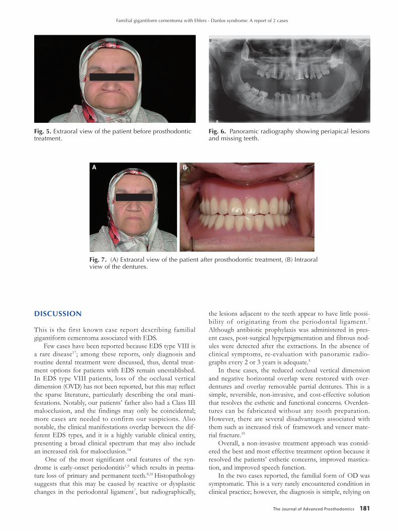
The 38-year-old sister of the first patient also self-referred to the Istanbul University Faculty of Dentistry. Unlike her brother, she did not have skin hyperelasticity on clinical examination. Head and neck examination showed no abnormalities or facial asymmetry, but the temporoman-diular joint did demonstrate hypermobility. A difference between the upper and lower facial height and Class III malocclusion were diagnosed (Fig. 5). Early-onset peri-odontitis was also noted. Teeth 14, 17, 18, 26, 38, 41 and 45 were missing, besides teeth 36 and 37 were impacted (Fig. 6). The incisors and canines had mottled enamel, and there was only occlusal contact between teeth 15 and 46. Panoramic radiography showed radio-opaque masses scattered through-out the mandible and maxilla. Like her brother, she was diagnosed with familial gigantiform cementoma with EDS type VIII.
All tooth defects were treated with composite restora-tions. The occlusal vertical dimension needed to be increased by approximately 12 mm. to satisfy esthetic and functional requirements. New tooth-supported complete dentures pro-viding lip support and rehabilitating the decreased occlusal vertical dimension were fabricated (Fig. 7).
In this case, acceptable retention was achieved without requiring soft liner; the dentures were fabricated as described above. The patient used the dentures for one year with regular re-evaluations. No muscle tenderness, tooth sensitivity, or tem-poromandibular dysfunction was observed during this period.
J Adv Prosthodont 2015;7:178-82
The Journal of Advanced Prosthodontics 181
DISCUSSION
This is the first known case report describing familial gigantiform cementoma associated with EDS.
Few cases have been reported because EDS type VIII is a rare disease17; among these reports, only diagnosis and routine dental treatment were discussed, thus, dental treat-ment options for patients with EDS remain unestablished. In EDS type VIII patients, loss of the occlusal vertical dimension (OVD) has not been reported, but this may reflect the sparse literature, particularly describing the oral mani-festations. Notably, our patients’ father also had a Class III malocclusion, and the findings may only be coincidental; more cases are needed to confirm our suspicions. Also notable, the clinical manifestations overlap between the dif-ferent EDS types, and it is a highly variable clinical entity, presenting a broad clinical spectrum that may also include an increased risk for malocclusion.18
One of the most significant oral features of the syn-drome is early-onset periodontitis1,9 which results in prema-ture loss of primary and permanent teeth.8,10 Histopathology suggests that this may be caused by reactive or dysplastic changes in the periodontal ligament7, but radiographically,
the lesions adjacent to the teeth appear to have little possi-bility of originating from the periodontal ligament.7 Although antibiotic prophylaxis was administered in pres-ent cases, post-surgical hyperpigmentation and fibrous nod-ules were detected after the extractions. In the absence of clinical symptoms, re-evaluation with panoramic radio-graphs every 2 or 3 years is adequate.5
In these cases, the reduced occlusal vertical dimension and negative horizontal overlap were restored with over-dentures and overlay removable partial dentures. This is a simple, reversible, non-invasive, and cost-effective solution that resolves the esthetic and functional concerns. Overden-tures can be fabricated without any tooth preparation. However, there are several disadvantages associated with them such as increased risk of framework and veneer mate-rial fracture.19
Overall, a non-invasive treatment approach was consid-ered the best and most effective treatment option because it resolved the patients’ esthetic concerns, improved mastica-tion, and improved speech function.
In the two cases reported, the familial form of OD was symptomatic. This is a very rarely encountered condition in clinical practice; however, the diagnosis is simple, relying on
Fig. 7. (A) Extraoral view of the patient after prosthodontic treatment, (B) Intraoral view of the dentures.
A B
Fig. 6. Panoramic radiography showing periapical lesions and missing teeth.
Fig. 5. Extraoral view of the patient before prosthodontic treatment.
Familial gigantiform cementoma with Ehlers - Danlos syndrome: A report of 2 cases

182
adequate clinical, histological and radiographic examination. The dentist should be able to easily diagnose in order to manage treatment satisfactorily.
ORCID
OlcayŞakarhttp://orcid.org/0000-0003-1513-4186Gamze Aren http://orcid.org/0000-0002-1479-0723Zeynep Mumcu http://orcid.org/0000-0002-2534-1499FatmaÜnalanhttp://orcid.org/0000-0002-6417-2915NihanAksakallıhttp://orcid.org/0000-0002-2429-9196Ceren Güney Tolgay http://orcid.org/0000-0001-5521-9016
REFERENCES
1. White SC, Pharoah MJ. Oral radiology: principles and inter-pretation. 6th ed. St. Louis, CV; Mosby; 2008. p. 96.
2. Barnes L, Eveson J, Reichart P, et al. World Health Organi-zation Classification of Tumours: Pathology and Genetics of Head and Neck Tumours. Lyon; IARC; 2005. p. 284.
3. Speight PM, Carlos R. Maxillofacial fibro-osseous lesions. Current Diagnostic Pathology 2006;12:1-10.
4. Slootweg PJ. Bone diseases of the jaws. Int J Dent 2010; 2010:702314.
5. Beylouni I, Farge P, Mazoyer JF, Coudert JL. Florid cemento-osseous dysplasia: Report of a case documented with com-puted tomography and 3D imaging. Oral Surg Oral Med Oral Pathol Oral Radiol Endod 1998;85:707-11.
6. Young SK, Markowitz NR, Sullivan S, Seale TW, Hirschi R. Familial gigantiform cementoma: classification and presenta-tion of a large pedigree. Oral Surg Oral Med Oral Pathol 1989;68:740-7.
7. Coleman H, Altini M, Kieser J, Nissenbaum M. Familial flor-id cemento-osseous dysplasia-a case report and review of the literature. J Dent Assoc S Afr 1996;51:766-70.
8. Gorlin RJ, Cohen MM, Levi LS. Syndrome of the head and neck. 3rd ed. Oxford; Pergamon; 1990. p. 156-60.
9. Beighton P. McKusick’s heritable disorders of connective tis-sue. 5th ed. St. Louis; Mosby, 1993. p. 267-89.
10. Létourneau Y, Pérusse R, Buithieu H. Oral manifestations of Ehlers-Danlos syndrome. J Can Dent Assoc 2001;67:330-4.
11. Reinstein E, Wang RY, Zhan L, Rimoin DL, Wilcox WR. Ehlers-Danlos type VIII, periodontitis-type: further delinea-tion of the syndrome in a four-generation pedigree. Am J Med Genet A 2011;155A:742-7.
12. Norton LA. Orthodontic tooth movement response in Ehlers-Danlos syndrome: report of case. J Am Dent Assoc 1984;109:259-62.
13. Stewart RE, Hollister DW, Rimoin DL. A new variant of Ehlers-Danlos syndrome: an autosomal dominant disorder of fragile skin, abnormal scarring, and generalized periodon-titis. Birth Defects Orig Artic Ser 1977;13:85-93.
14. Zarb GA, Hobkirk JA, Eckert SE, et al. Prosthodontic treat-ment for edentulous patients, vol 1. 13th ed. Elsevier; Mosby; 2013. p. 190.
15. Morrow RM, Feldmann EE, Rudd KD, Trovillion HM. Tooth-supported complete dentures: an approach to preven-
tive prosthodontics. J Prosthet Dent 1969;21:513-22.16. Patel MB, Bencharit S. A treatment protocol for restoring oc-
clusal vertical dimension using an overlay removable partial denture as an alternative to extensive fixed restorations: a clinical report. Open Dent J 2009;3:213-8.
17. Karrer S, Landthaler M, Schmalz G. Ehlers-Danlos type VIII. Review of the literature. Clin Oral Investig 2000;4:66-9.
18. Kakadia N, Kanaki NS. Ehlers-Danlos syndrome: an over-view. J Chem Pharm Res 2011;3:98-107.
19. Graser GN, Rogoff GS. Removable partial overdentures for special patients. Dent Clin North Am 1990;34:741-58.
J Adv Prosthodont 2015;7:178-82