
F LUIDS AND E LECTROLYTE DISORDERS IN NEONATES M. Saboute M.D. Neonatologist Assistant Professor Of...
33
FLUIDS AND ELECTROLYTE DISORDERS IN NEONATES M. Saboute M.D. Neonatologist Assistant Professor Of Iranian Univercity
-
Upload
bernadette-weaver -
Category
Documents
-
view
213 -
download
0
Transcript of F LUIDS AND E LECTROLYTE DISORDERS IN NEONATES M. Saboute M.D. Neonatologist Assistant Professor Of...

FLUIDS AND ELECTROLYTE DISORDERS IN NEONATESM. Saboute M.D.
Neonatologist
Assistant Professor Of Iranian Univercity

ASSESSMENT OF FLUID AND ELECTROLYTE STATUS

PHYSICAL EXAMINATION
1. Change in body weight2. Skin and mucosal manifestations
skin turgor
sunken antenor fontanelle
dry mucous membranes
3. Cardiovascular :Tachycardia
Capillary refill time
Hepatomegaly
Blood pressuer

LABORATORY STUDIES
Serum electrolytes and plasma
osmolarity
Input and output measurements
Urine electrolytes and specific gravity

LABORATORY STUDIES
Fractional excretion of Na (FENa)
FENa : (urine Na x plasma creatinine)/(plasma Na x urine creatinine)x 100
Level of <1% indicates prerenal factors reducing renal blood flow.
Level of 2.5% occurs wiih acute renal failure (ARF).
Urea nitrogen (BUN) and serum crArterial pH, pco2,and Na
bicarbonate

DEHYDRATION
Losses of na and water Through thoracostomy, nasogastric, or
ventriculostomy drainage) Third space Renal water losses in the VLBV

DEHYDRATION
Weight loss Decreased urine output Increased urine SG Poor skin turgor Tachycardia Hypotension Metabolic acidosis Increasing BUN FENa <1%

THERAPY
Administer Na and water to first correct deficits and then adjust to equal maintenance needs plus ongoing losses.
Acute isonatremic dehydration may require IV infusion of 10 cc/kg of NS if acute weight loss is >10% of body weight with signs of poor cardiac output.

EDEMA
a. Predisposing factors : excessive isotonic fluid administration, heart failure, sepsis, and neuromuscular paralysis.
b. Diagnosis. Clinical signs include periorbital and extremity edema, increased weight, and hepatomegaly.
c. Therapy: Na restriction and water restriction

COMMON ELECTROLYTE ABNORMALITIES:
Hypo/hypernatremia
Hypo/hyperkalemia

SODIUM ABNORMALITIES:
Hyponatremia: Na < 130 mEq/L
Hypernatremia: Na > 150 mEq/L

HYPONATREMIA DISORDERS
1. Exclude "pseudohyponatremia“ (hyperlipidemia, hyperproteinemia)
2. Exclude hypertonic hyponatremia (↑ECF osmolality due primarily to
hyperglycosemia)3. Evaluate ECF volume status (clinical and laboratory indicators)
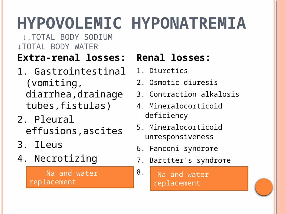
HYPOVOLEMIC HYPONATREMIA ↓↓TOTAL BODY SODIUM↓TOTAL BODY WATER
Extra-renal losses:1. Gastrointestinal
(vomiting, diarrhea,drainage tubes,fistulas)
2. Pleural effusions,ascites
3. ILeus4. Necrotizing
enterocolitis
Renal losses:1. Diuretics
2. Osmotic diuresis
3. Contraction alkalosis
4. Mineralocorticoid deficiency
5. Mineralocorticoid unresponsiveness
6. Fanconi syndrome
7. Barttter's syndrome
8. Obstructive uropathy Na and water replacement
Na and water replacement

EUVOLEMIC HYPONATREMIA ± TOTAL BODY SODIUM↑ TOTAL BODY WATER
1. excess fluid administration2.SIADH

SIADH Predisposing
factors pain, opiate, IVH,
asphyxia, meningitis, pneumothorax, and positive-pressure ventilation
Weight gain without edema
↓urine output - ↑ urine osmolarity
Treatment :Water restriction is
therapeutic unless :(i) serum Na ≤120
mEq/L (ii) neurologic signs :
obtundation or seizure
In these instances furosemide 1 mg/kg
IV q6h + hypertonic Nacl (3%)

HYPERVOLEMIC HYPONATREMIA ↑ TOTAL BODY SODIUM↑ ↑ TOTAL BODY WATER
Edema-forming states:
1. Congestive heartfailure2. Liver
failure/cirrhosis3. Nephrosis
syndrome4. Indomethacin
therapy
Sodium and water restriction
Renal failure:1. Acute2. Chronic
Sodium and water restriction
Sodium and water restriction
Sodium and water restriction


TREATMENT OF HYPONATREMIA
If Na ≤120 Mev/L,or Hyponatremia With Seizures Or Coma Correction Of Hyponatremia Is Recommended With 3% Saline Solution (Up To 120 Meq/L Of Serum Sodium On Centration Over 4 To 6 Hours
Rapid And Complete Correction Of Low Serum Sodium Concentration In Adults ,With Chronic Hyponatremia Has Been Shown To Be Associated With Pontine And Xtrapontine Myelinolysis.
When Na Has Reached 120 Meqil, Complete Correction Of Hyponatremia Should Be Performed More Slowly, Over 24 To 48 Hours. The Use Of 5% Dextrose In Water With 0.45% To 0.9% Saline Is Reasonable.

HYPERNATREMIA(SERUM SODIUM> 150 MEQ/L)
Hypovolemic hypernatremia
Inadequate breast milk intake
Diarrhea Radiant warmers Excessive sweating Renal dysplasia Osmotic diuresisHypervolemic
hypenatrenia: Improperily mixed formula NaHC03 administration NaCI administration Primary hyperaldosteronism
Euvolemic hypernatremia:Decreased production of
antidiuretic hormone: Central diabetes insipidus,
head trauma, central nervous
system tumors (craniopharyngioma), meningitis, or encephalitis
Decrease or absence of renal responsiveness:
Nephrogenic diabetes insipidus, extreme immaturity,
renal insult and medications such as amphotericin, hydantoin, aminoglycosides
HYPERNATREMIA DUE TO EXCLUSIVELY BREAST-FED NEONATE
Infant sucks poorly, breast milk production drops and sodium concentration rises, and the infant becomes increasingly
dehydrated, hypenatremic, and lethargic. Usually manifests at the end of the first to the
third week after : seizures, DIC, and permanent neurologic and vascular injury.
HYPERNATREMIA IN BREAST-FED TERM NEONATE Inadequate breast milk intake. Usually occurring over 7 to 14 days Clinical presentation : lethargy, iritability,
abnormal muscle tone with or without seizures, and
cardiovascular collapse with renal failure. Significant central nervous system
morbidity from both the hypertonicity (saggital or other venous sinus thrombosis, subdural capillary hemorrhage, white matter injury) and inappropriately rapid rehydration therapy (brain edema, myelinolysis)
HYPERNATREMIA IN EXTREMELY IMMATURE NEONATE
From excessive transepidermal free water losses developed rapidly, within 24 to 48 hours after birth
Prevention with antenatal steroids and humidified incubators.
Decrease in body weight and clinical signs of extracellular volume contraction.
POTASSIUM
Serum k level : 3.5 to 5.5 meq/l Potassium is mostly intracellular: blood levels do
not usually indicate total body potassium Ph affects K+: 0.1 ph change=>0.3-0.6 K+
change (more acid, more K; less acid, less K) ECG affected by both hypok and hyperk:
Hypok : flat T, prolonged QT, U wavesHyperk : peaked T waves, widened QRS,
bradycardia, tachycardia, SVT, V tach, V fib

HYPOKALEMIA ST depressions with prominent U waves and
prolonged repolarization

HYPERKALEMIA
Note the “tented” or “pinched” shape to Twaves

HYPOKALEMIA ( HYPOKALEMIA: K < 3.5 MEQ/L)
1. Predisposing factors : nasogastric tube,diuretic use, and renal tubular defects.
2. Diagnosis. Obtain serum and urine electrolytes, ph, and ECG to detect possible conduction defects : prolonged QT interval and U waves
3. Therapy reduce renal or gastrointestinal losses of K. Gradually increase intake of K as needed

HYPERKALEMIA
Symptomatic hyperkalemia may begin at a serum K level >6 mEq/L.

PREDISPOSING FACTORS HYPERKALEMIA
a. Increased K release secondary to tissue destruction, trauma, cephalhematoma ,hypothermia, bleeding, intravascular or extravascular hemolysis,asphyxia/ischemia, and IVH.

PREDISPOSING FACTORS HYPERKALEMIA
b. Decreased K clearance due to renal failure, oliguria,, and congenital adrenal hyperplasia.
c. Miscellaneous associations including dehydration, birth weight <1,500 g blood transfusion, inadvertenr excess (KCl) administration and exchange transfusion
d. Up to 50 % of VLBW infants born before 25 weeks' gestation manifest serum K levels >6 mEq/L in the first 48 hours of life.
The most common cause of sudden unexpected hyperkalemia in the neonatal intensive care unit (NICU) is medication error


THERAPY
Remove all sources of exogenous K Goal 1: stabilizaiion of conducting tissues Calcium gluconate 1-2 cc/kg (10%) IV Goal 2: dilution and intracellular shifting of K
Sodium bicarbonate 1-2 meq/kg IVGlucose-insulin combination
2 adrenergic stimulation

THERAPY
enhanced K excretion Lasix (increases excretion over hours) kayexalate, potassium exchange resin
1g/kg/dose po/rectally (slow action) Dialysis/ Exchange transfusion


