Excretion and Waste
15
51 Objective biology INTRODUCTION Excretion is the elimination to waste products from the body. Was te pr oducts are unwanted and toxic by-products which are removed to maintain homeostasis and protect the body from their toxicity , e.g., jaundice in case of bile pigments, uremia in case of urea. Waste products (which do not accumulate in the body) are nitrogenous materials, CO 2 , pigments, excess of water , inorganic salts, vitamins and hormones. Carbon dioxide and some water are excreted by lungs. Other waste materials are r emoved in the urine. The organs and tissues parti cipating in the excretion of waste products constitute the excretory system. A major function of the excretory system is the excretion of nitrogenous waste. These include ammonia, urea and uric acid. Excretion also helps to maintain a constant body temperature by removing excess heat and maintain a constant internal environment in association with the other system of the body. Defaecation is elimination of undigested food residue from alimentary canal while secretion is the discharge of specially synthesised product, e.g., hormone by endocrine gland, saliva from salivary gland. Osmoregulation (term coined by Haber) is the regulation of osmotic concentration, that is, water contect and salt and cells. Both excretion and osmoregulation are helpful in maintaining homeostasis or constant favourable • • • • • • • • internal environment of the body . In vertebrates the two functions of excretion and osmoregulation are performed by kidneys and their associated structures in urinary system. The organs which form, store and void the urine constitute urinary system. Perspiration is another excretory process which removes salts and water although the primary purpose is cooling. EXCRETORY PRODUCTS AND THEIR TYPES Excretory substances are produced during metabolism of nitrogeneous substances like amino acids and nucleic acids. Metabolism of carbohydrate s and fats produces CO 2 and H 2 O which are easy to remove. Their excretion is effected through lungs (expired air), skin (sweat) or kidneys (urine). Other excretory products are pigments, mostly formed by the breakdown of haemoglobin; drugs etc. Protein metabolism produces nitrogenous waste material such as ammonia, which is the basic nitrogenous catabolite of protein, formed by breakdown of amino acids. Removal of the amino group (NH 2 ) is known as deamination and it converts the amino acid into a keto acid. In vertebrates, deamination takes place in the liver . Ammonia thus produced is highly toxic and cannot be stored within the body. It needs to be eliminated immediately. • • • • • • CHAPTER 22 Excretion & Osmoregulation
-
Upload
clayanne-knott -
Category
Documents
-
view
218 -
download
0
Transcript of Excretion and Waste

8/16/2019 Excretion and Waste
http://slidepdf.com/reader/full/excretion-and-waste 1/15
51Objective biology
INTRODUCTIONExcretion is the elimination to waste products fromthe body.
Waste products are unwanted and toxic by-products which are removed to maintain homeostasis andprotect the body from their toxicity, e.g., jaundice incase of bile pigments, uremia in case of urea.
Waste products (which do not accumulate in the body)are nitrogenous materials, CO
2, pigments, excess of
water, inorganic salts, vitamins and hormones. Carbon
dioxide and some water are excreted by lungs. Other waste materials are removed in the urine.
The organs and tissues participating in the excretionof waste products constitute the excretory system. A major function of the excretory system is theexcretion of nitrogenous waste. These includeammonia, urea and uric acid.
Excretion also helps to maintain a constant bodytemperature by removing excess heat and maintaina constant internal environment in association withthe other system of the body.
Defaecation is elimination of undigested food residuefrom alimentary canal while secretion is the dischargeof specially synthesised product, e.g., hormone byendocrine gland, saliva from salivary gland.
Osmoregulation (term coined by Haber) is theregulation of osmotic concentration, that is, watercontect and salt and cells.
Both excretion and osmoregulation are helpful inmaintaining homeostasis or constant favourable
•
•
•
•
•
•
•
•
internal environment of the body. In vertebrates thetwo functions of excretion and osmoregulation areperformed by kidneys and their associated structuresin urinary system.
The organs which form, store and void the urineconstitute urinary system.
Perspiration is another excretory process whichremoves salts and water although the primary purposeis cooling.
EXCRETORY PRODUCTSAND THEIR TYPES
Excretory substances are produced during metabolismof nitrogeneous substances like amino acids andnucleic acids.
Metabolism of carbohydrates and fats produces CO2
and H2O which are easy to remove. Their excretion
is effected through lungs (expired air), skin (sweat) orkidneys (urine).
Other excretory products are pigments, mostly formed
by the breakdown of haemoglobin; drugs etc.Protein metabolism produces nitrogenous wastematerial such as ammonia, which is the basicnitrogenous catabolite of protein, formed bybreakdown of amino acids. Removal of the aminogroup (NH
2) is known as deamination and it converts
the amino acid into a keto acid. In vertebrates,deamination takes place in the liver . Ammonia thusproduced is highly toxic and cannot be stored withinthe body. It needs to be eliminated immediately.
•
•
•
•
•
•
CHAPTER 22
Excretion &Osmoregulation
8/16/2019 Excretion and Waste
http://slidepdf.com/reader/full/excretion-and-waste 2/15
52 Objective biology
Depending upon the form in which the ammoniaor nitrogenous waste is excreted from the body, theorganisms are grouped as under into three categories:ammonotelic, uricotelic and ureotelic.
Besides these (ammonia, urea and uric acid) thereare a few other types of minor nitrogenous excretoryproducts, like—
Trimethylamine oxide (TMAO): Marine teleostfishes excrete a large proportion of their nitrogenas trimethylamine oxide (TMAO). Large amounts
of this compound is also stored in their body forosmoregulation i.e. to minimize loss of water andentry of salts.
Guanine: Spiders excrete almost exclusively achemical called guanine. It is even less soluble ascompared to uric acid and hence requires no waterfor elimination. It is a metabolic waste of nucleotidemetabolism. It is also found in penguins.
Ornithuric acid: It is excreted in small amount bybirds and is formed by a combination of benzoic
•
•
-
-
-
acid (formed during fat metabolism) in food with theamino acid ornithine.
Hippuric acid: It is formed when benzoic acid iscombined with glycine. It is less toxic.
Creatinine: Mammals contain a small quantity ofcreatinine in their blood (1 mg/100 ml) which is aderivative of creatine. The excess is eliminated along with urine.
Creatine: Mammals also excrete creatine which issynthesized in the liver from three amino acids—
arginine, glycine and methionine.
Ammonotelic
Animals excreting their nitrogenous waste in theform of ammonia are known as ammonotelic. Thisphenomenon is known as ammonotelism.
Ammonia is highly soluble in water with whichit forms ammonium hydroxide (NH
4OH) which
injures cells directly by alkaline caustic action. Henceexcretion of ammonia requires large amounts of
-
-
-
•
•
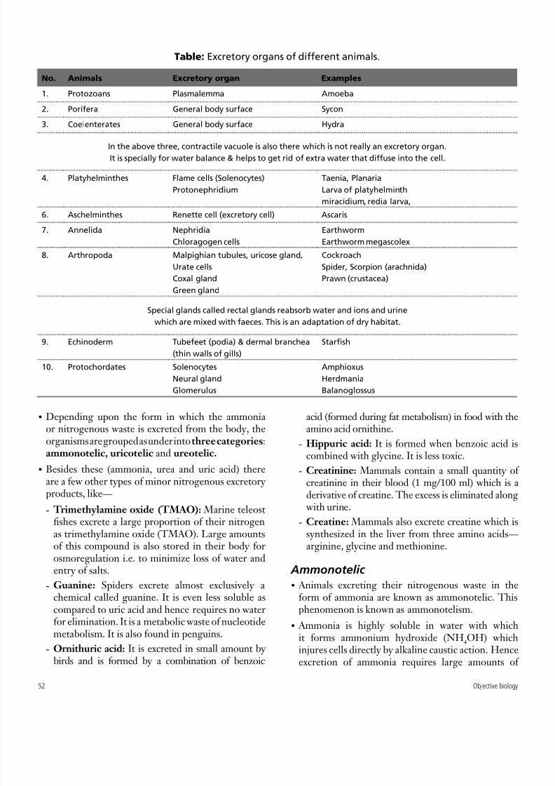
Table: Excretory organs of different animals.
No. Animals Excretory organ Examples
1. Protozoans Plasmalemma Amoeba
2. Porifera General body surface Sycon
3. Coelenterates General body surface Hydra
In the above three, contractile vacuole is also there which is not really an excretory organ.
It is specially for water balance & helps to get rid of extra water that diffuse into the cell.
4. Platyhelminthes Flame cells (Solenocytes)
Protonephridium
Taenia, Planaria
Larva of platyhelminth
miracidium, redia larva,
6. Aschelminthes Renette cell (excretory cell) Ascaris
7. Annelida Nephridia
Chloragogen cells
Earthworm
Earthworm megascolex
8. Arthropoda Malpighian tubules, uricose gland,
Urate cells
Coxal gland
Green gland
Cockroach
Spider, Scorpion (arachnida)
Prawn (crustacea)
Special glands called rectal glands reabsorb water and ions and urine
which are mixed with faeces. This is an adaptation of dry habitat.
9. Echinoderm Tubefeet (podia) & dermal branchea
(thin walls of gills)
Starfish
10. Protochordates Solenocytes
Neural gland
Glomerulus
Amphioxus
Herdmania
Balanoglossus

8/16/2019 Excretion and Waste
http://slidepdf.com/reader/full/excretion-and-waste 3/15
53Objective biology
water to be lost from the body. That is why such amode is suitable for aquatic organisms which have aconstant access to water.
No energy is required to produce ammonia.
E.g. all aquatic invertebrates, bony fishes and aquaticamphibians.
Ammonia is the first metabolic waste product ofprotein metabolism.
In anurans (amphibians) the larval tadpoles excreteammonia, while the adults produce urea.
Uricotelic
Animals which excrete their nitrogenous wastemainly in the form of uric acid and urates areknown as uricotelic. The phenomenon is known asuricotelism .
All terrestrial animals like insects, reptiles, and birdsexcrete uric acid.
Uric acid (C5H
4N
4O
3) (which require more energy)
is produced by degradation of purines (e.g. guanine)in liver and kidneys to some extent. In uricotelic
animals, excess nitrogen is first used in synthesisof purines. A purine is changed to xanthine (fromhypoxanthine or guanine) which is then oxidised to
•
•
•
•
•
•
•
uric acid. Part of uric acid is oxidised further to formallantoin and allantoic acid. Teleost fish excreteallantoate or hydration product of allantoin. In mostfishes and amphibians, allantoate is hydrolysed tourea and glyoxylate. Some marine invertebrates havegone a step further by hydrolysing urea to ammoniaand carbon dioxide. Creatine is formed in liver from
amino acids. Creatinine is produced from creatine.Conversion of ammonia to uric acid and itssubsequent elimination requires lesser amount of water as it is comparatively less soluble in waterand less toxic as compared to ammonia. Hence, itis observed in terrestrial animals that do not haveconstant access to water or rather have limitedaccess to water.
Ureotelic
Animals that excrete their nitrogenous waste mainly
in the form of urea are known as ureotelic and thephenomenon is known as ureotelism.
Urea can be stored in body for considerable periodsof time, and is least toxic. It is eliminated in the formof urine.
Ureotelism is exhibited by semi-terrestrialanimals, e.g. some earthworms, adult amphibians,elasmobranch (cartilagineous fishes) and mammals.
Frog like other amphibians is ammonotelic in tadpolestate and ureotelic in mature state. Earthworm
is similarly ammonotelic when sufficient water isavailable and ureotelic when water availability isreduced.
•
•
•
•
•
Aminotelism is the excretion of amino acids
which cannot be metabolised due to their being
in excess. The animals performing aminotelism
are called aminotelic, e.g. some molluscs (Pila,
Unio, Limnaea) and some echinoderms (starfish,
Holothuria).
1. A person has taken very high protein rich diet; his urine
will eliminate more ofa. urea b. creatinine
c. glucose d. glycogen
2. Vertebrates kidney and contractile vacuole of protozoans
resemble as both excrete
a. minerals b. water
c. glucose d. nitrogenous wastes
3. The passing out of almost solid excretory wastes in reptiles
is an indication of
a. xeric habits b. excretion through skin
c. excretion through alimentary canal
d. reutilizing water of urine 4. Most terrestrial insects get rid of bulk of their nitrogenous
wastes as
a. amino acids b. urea
c. uric acid d. ammonia
5. The flame cell system of helminthes work
a. to remove ammonium ions and uric acids
b. to remove uric acid
c. to regulate pH
d. for osmoregulation
Check your GRASP

8/16/2019 Excretion and Waste
http://slidepdf.com/reader/full/excretion-and-waste 4/15
54 Objective biology
6. Renal gland is the excretory organ of
a. annelida b. echinodermata
c. crustaceans d. molluscs
7. Deamination is first step in urea formation which means
a. reduction of ammonia
b. oxidation of ammonia
c. addition of amino group to organic molecule
d. removal of amino group from amino acid
8. Antennary glands are excretory organs of
a. spiders b. crustaceans
c. mollusca d. echinodermata
9. Protonephridia are present in platyhelminthes and
metanephridia in
a. nematodes b. arthropoda
c. annelids d. platyhelminthes only
10. Excretory organs of protochordate Amphioxus are
a. malpighian tubules b. protonephridia
c. kidney d. none of these.
11. Major source of ammonia produced by kidney comesfrom
a. leucine b. glycine
c. glutamine d. alanine
12. Uric acid is the end product of metabolism of
a. proteins
b. glomerular acids
c. fats
d. lipids
13. Trimethylamine is excreted by
a. marine teleosts
b. mollusca
c. fresh water fish
d. amphibians
14. The kidneys resembles contractile vacuoles of protozoans in
a. expelling of excess water
b. expelling out salts
c. expelling out excess glucose
d. expelling out urea and uric acid
15. In one of the following groups the toxic substance benzoic
acid after combining with amino acids glycine forms hippuric
acid as material in
a. fish b. reptiles
c. amphibians d. mammals16. Which is most toxic excretory product and require 600 ml
of water for throwing one gm nitrogen ?
a. uric acid b. urea
c. ammonia d. hippuric acid
17. Which one of these animal excretes amino acid without
deamination ?
a. rat b. earthworm
c. unio d. fly
18. Excess bile pigments in urine indicate
a. anaemia b. diabetes insipidus
c. jaundice d. all of these.
19. Ammonia is the end product of
a. glucose breakdown
b. fatty acid metabolism
c. protein metabolism
d. breakdown of biogenic amines
20. Pterydines are the excretory products of some
a. crustaceans b. molluscs
c. insects d. fish
21. The liver of which one of the following is richest source
of arginase enzyme ?
a. bony fish b. cartilaginous fish
c. frog d. rabbit
22. Excretion is required for maintaining homeostasis of body
fluids through regulation of their
a. volume, composition, pH and osmotic potential
b. volume
c. composition and pH
d. osmotic potential
23. Uric acid is excreted in
a. frog b. rabbit
c. man d. pigeon/crow
24. In human beings, gout is caused by
a. deficiency of iodine
b. excessive secretion of thyroid
c. excessive liberation of uric acid
d. deposition of uric acid
25. Deamination is the process in which
a. poisonous urea is removed from the blood and it
occurs in kidneys
b. amino acids are absorbed from digested food, and
it occurs in intestinal villi
c. amino acids are broken down to produce urea and
it occurs in liver
d. amino acids are synthesised and it occurs in
ribosomes
26. In aquatic organisms the waste end product of nitrogen
metabolism is
a. urea b. nitrogen
c. ammonia d. allantois27. Excretory products of mammalian embryo are eliminated by
a. placenta
b. amniotic fluid
c. allantois
d. ureters
28. The snakes living in deserts are mainly
a. ammonotelic b. aminotelic
c. ureotelic d. uricotelic

8/16/2019 Excretion and Waste
http://slidepdf.com/reader/full/excretion-and-waste 5/15
55Objective biology
29. The excretory product of excess metabolism of creatine
and guanine is
a. creatinine b. Urea
c. uric acid d. oxalic acid
30. In Rabbit and other terrestial mammals, the main nitrogenous
waste material is
a. uric acid b. ammonia
c. urea d. urea and uric acid
31. Nitrogenous waste products are eliminated mainly as
a. urea in tadpole and ammonia in adult frog
b. ammonia in tadpole and urea in adult frog
c. urea in both tadpole and adult frog
d. urea in tadpole and uric acid in adult frog
32. In Prawn, excretion is carried out by
a. nephrons b. malpighian tubules
c. flame cells d. reptiles
33. Excretion is
a. removal of substances not required by body
b. removal of useless substances and substances present
in excess
c. formation of substances having some role in body
d. all of the above
34. The animal which retain urea for hypertonicity is
a. man b. bird
c. elasmobranch d. amphibian
35. Aquatic animals are mostly ammonotelic because
a. ammonia helps in checking inflow of water into body
b. excretion of ammonia requires large amount of water
which is available to these animals
c. water contains less nitrogen
d. these get less light
36. Excretion of nitrogenous waste product mainly as uric acid
by birds is helpful in
a. conserving body heat
b. conserving water
c. eliminating excess water
d. eliminating excess body heat
37. Which one is the most soluble in water ?
a. uric acid b. urea
c. fatty acids d. casein
38. Excretory product of birds and reptiles is
a. urea
b. uric acid
c. ammonia
d. TMO
39. Sea Gulls excrete salts through
a. liver b. lungs
c. urine d. nasal gland
40. In Amoeba, NH3 is excreted through
a. food vacuole
b. plasma membrane
c. contractile vacuole
d. all of these.
41. Malphigian tubule are the excretory organs in
a. cockroach
b. platyhelminthes
c. Ascaris
d. Pila
42. Ammonia is changed to uric acid in the liver of
a. ammonotelic animals
b. uricotelic animals
c. ureotelic animals
d. ornithotelic animals
43. Which one is false ?
a. nephrons perform excretion through filtration,
reabsorption and secretion
b. nephridia are accessory excretory organs in Prawn
c. tapeworm have excretory flame cells
d. nephrons begin with Bowman’s capsule having
glomerulus
44. Which of the following is likely to accumulate in dangerousproportion in the blood of a person whose kidney is not
working properly ?
a. lysine b. urea
c. ammonia d. sodium chloride
45. Bony fishes are
a. ureotelic
b. uricotelic
c. aminotelic
d. ammonotelic
Answers
1. a. 2. a. 3. b. 4. c. 5. d. 6. d. 7. d. 8. b. 9. c. 10. b.
11. c. 12. b. 13. a. 14. a. 15. a. 16. c. 17. c. 18. c. 19. c. 20. c.
21. b. 22. a. 23. d. 24. d. 25. c. 26. c. 27. a. 28. d. 29. c. 30. b.
31.b. 32. d. 33. b. 34. c. 35. b. 36. b. 37. b. 38. b. 39. d. 40. a.
41.a. 42. b. 43. b. 44. b. 45. d.
8/16/2019 Excretion and Waste
http://slidepdf.com/reader/full/excretion-and-waste 6/15
56 Objective biology
Human beings are ureotelic i.e. excreting theirnitrogenous waste as urea.
Kidney
Kidney are mesodermal in origin and developedfrom nephrostomes of early embryo.
Kidney are excretory and homeostatic organ .
Man possesses a pair of dark red, bean shaped,metanephric kidneys located in the peritoneal cavityoutside the coelom (retroperitoneal).
Kidney is covered by peritoneum on the ventralside.
As the primary organs of excretion, the kidneys
perform two major functions. They filter the blood,removing water, ions, and nitrogenous wastes andforming urine and in turn regulate the volume, osmoticpressure, and pH of the blood and body fluids.
The body is very sensitive to its pH level. Outside therange of pH that is compatible with life, proteins aredenatured and digested, enzymes lose their ability tofunction, and the body is unable to sustain itself.
The kidneys maintain acid-base homeostasis byregulating the pH of the blood plasma. Gains andlosses of acid and base must be balanced.
Sources of acid gain : carbon dioxide (since CO2 andH
2O form H
2CO
3, carbonic acid, in the presence
of carbonic anhydrase); production of nonvolatileacids from the metabolism of proteins and otherorganic molecules; and loss of bicarbonate in faecesor urine.
Sources of acid loss: use of hydrogen ions in themetabolism of various organic anions; and loss ofacid in the vomitus or urine
•
•
•
•
•
•
•
•
-
-
When acid loss exceeds acid gain , alkalosis occurs. When gain exceeds loss acidosis occurs. There are various renal responses to acidosis and alkalosis.
Responses to acidosis:
Bicarbonate is added to the blood plasma by tubularcells.
Tubular cells reabsorb more bicarbonate fromthe tubular fluid.
Collecting duct cells secrete more hydrogen andgenerate more bicarbonate.
Ammoniagenesis leads to increased buffer formation(in the form of NH
3)
Responses to alkalosis:
Excretion of bicarbonate in urine.
This is caused by lowered rate of hydrogen ionsecretion from the tubular epithelial cells.
This is also caused by lowered rates of glutaminemetabolism and ammonia excretion.
Kidney also secretes hormones like – erythropoietin(which regulates red blood cell production in thebone marrow); renin (which is a key part of the renin-angiotensin-aldosterone system) and active form of vitamin D, calcitriol, and prostaglandins.
The kidneys are situated below the diaphragm on theleft and right sides, and are partially protected by theeleventh and twelfth pairs of ribs.
The right kidney is lower than the left because the
liver takes up much space on that side.Each kidney is surrounded by layers of fibrousconnective tissue and a mass of adipose (fat) tissue which cushions it and holds it in place.
The kidney consists of an outer cortex and an innermedulla. They both contains the uriniferous tubulesor nephrons (the structural and functional unit ofkidney).
The renal cortex is granular in appearance becausethe tubules here are much convoluted (proximal and
distal convoluted tubules) and contain malphigiancorpuscles.
The cortex overlies the bases of medullary pyramidsand dips down between them. These displacedportions of cortex which invade the medullaryterritory are called the renal columns of Bertini.
Cortex is sub-divided into alternating radial tractsknown as rays and labyrinths. The rays are radiallystraight since they contain the straight parts of
•
•
-
-
•
-
•
•
•
•
•
•
•
•
Item
Item
Item
Item
Item
Item
Fig. Excretory system.
8/16/2019 Excretion and Waste
http://slidepdf.com/reader/full/excretion-and-waste 7/15
57Objective biology
proximal tubules and the collecting ducts. Theyare continuous with striated medulla, hence calledmedullary rays. The labyrinths have a granularappearance because the convoluted tubules, whichcompose them, are cut irregularly.
Renal medulla mainly contains loop of Henle,collecting tubules and ducts of Bellini.
The medulla appears striated since the tubules runthrough it in a straight course radiating towards thepelvis.
The medulla consists of 10 to 15 multilobular conicalmasses, the renal pyramids, whose bases are adjacentto the cortex and whose apices form the papillae. These papillae project into cup shaped channelscalled calyces.
The spaces between calyces are part of the renal sinusand are filled with adipose tissue, vessels and nerves.
The calyces join to form the pelvis which is theexpanded beginning of the ureter.
The medial concave border of a kidney contains anotch known as hilus through which the renal arteryenters and the renal vein and ureter leave the kidney.
The structural and functional unit of a kidney isthe nephron or the uriniferous tubule.
•
•
•
•
•
•
•
Nephrons are connected to collecting ducts whichpass through the renal pyramids and open into therenal pelvis.
Urine formed in the nephrons and collecting ductsempties into the renal pelvis and is carried away fromthe kidney by the ureter.
Types of Kidney
Archinephros
It is the basic and ancestral form. Such kidney is foundtoday in larvae of certain cyclostomes (Myxine), butdo not occur in any adult vertebrate.
Glomeruili are only present in some of the posteriortubules.
Pronephros
Pronephros the most primitive excretory organs that develop in vertebrate, corresponding to the firststage of kidney development.
The pronephros develops in the anterior nephrotomesof all vertebrates.
It is a paired organ, consisting of a series of nephronsfiltering urine from both the pericardium fluids via
•
•
•
•
•
•
•
The kidney cells that make EPO are specialized and are
sensitive to low oxygen levels in the blood coming into the
kidney. These cells release erythropoietin when the oxy-
gen level is low in the kidney. Erythropoietin stimulates
the bone marrow to produce more red blood cells which in
turn increases the oxygen-carrying capacity of the blood.
Erythropoietin or EPO is a glycoprotein hormone that is a
cytokine for erythrocyte (red blood cell) precursors in the
bone marrow. Erythropoietin is available as a therapeutic
agent produced by recombinant DNA technology in mam-
malian cell culture. It is used in treating anemia resulting
from chronic renal failure or from cancer chemotherapy.
Its use is also believed to be common as a blood doping
agent in endurance sports such as bicycle racing, triathlons
and marathon running.
EPO is produced not only in the kidney but also, to a less-
er extent, in the liver. The EPO gene has been found onhuman chromosome 7 (in band 7q21). Different DNA se-
quences flanking the EPO gene act to control liver versus
kidney production of EPO.
The measurement of EPO in the blood can indicate bone
marrow disorders or kidney disease. Normal levels of EPO
are 0 to 19 mU/ml (milliunits per millilitre). Elevated levels
can be seen in polycythemia rubra vera, a disorder charac-
terized by an excess of red blood cells. Lower than normal
values of EPO are seen in chronic renal failure.
Using recombinant DNA technology, EPO has been syn-
thetically produced for use in persons with certain types
of anaemia: anemia due to kidney failure, anaemia sec-
ondary to AZT treatment of AIDS, and anaemia associated
with cancer.
EPO has been much misused as a performance-enhanc-
ing drug in endurance athletes such as reportedly cyclists,
long-distance runners, speed skaters, and Nordic (cross-
country) skiers. When misused in such situations, EPO is
thought to be especially dangerous (perhaps because de-
hydration can further increase the viscosity of the blood,increasing the risk for heart attacks and strokes. EPO has
been banned by the Tour, the Olympics, and other sports
organizations.
Erythropoietin
8/16/2019 Excretion and Waste
http://slidepdf.com/reader/full/excretion-and-waste 8/15
58 Objective biology
openings called nephrostomes and blood from theglomerulus.
The organ is active in adult forms of some primitivefish, like lampreys or hagfish. It is present at theembryo of more advanced fish and at the larval stageof amphibians.
In human beings, it is rudimentary and replaced by
mesonephros after 3.5 weeks.
Mesonephros
The mesonephros (Latin for “middle kidney ”) servesas the main excretory organ of aquatic vertebratesand as a temporary kidney in higher vertebrates.
The mesonephros is also called the Wolffian body afterCaspar Friedrich Wolff who described it in 1759.
The mesonephros is composed of the mesonephricduct (also called the Wolffian duct), mesonephric
tubules, and associated capillary tufts. A single tubuleand its associated capillary tuft is called a mesonephricexcretory unit; these units are similar in structure andfunction to nephrons of the adult kidney.
The mesonephros is derived from intermediate
Mesoderm in the vertebrate embryo.
In human males, the mesonephros gives rise to theefferent ductules of the testis, the epididymis, vasdeferens, seminal vesicle, and vestigial structuressuch as the appendix testis, appendix epididymis, andparadidymis.
The mesonephros largely regresses in human females,though vestigial structures such as Gartner’s cysts, theepoophoron, and paroophoron are common.
E.g. Fish and frog but in reptiles, birds and mammalsit is functional in embryo.
Metanephric kidney
Also called posterior kidney.
Most advanced kidney in which loop of henle ispresent.
E.g. Reptiles, birds & mammals.
Ureters, Urinary bladder and Urethra
Ureters are thin muscular tubes emerge out fromthe hilum of kidneys. Urine enters the ureter fromthe renal pelvis and is conducted along the ureter byperistaltic waves on its walls.
Ureter develops in the region of calyces.
•
•
•
•
•
•
•
•
•
•
•
•
•
•
•
Urinary bladder is sac like structure which storesurine temporarily.
Bladder has three parts– apex, fundus (or body)and neck.
Body has triangular area called trigone. Neck regionspossesses two sphincters– involuntary internalsphincter and voluntary external sphincter.
Ureters and urinary bladder are lined by flexibletransitional epithelium .
Urethra is a membranous tube, which conduct urineto the exterior.
The urethral sphincters keep the urethra closedexcept during voiding of urine.
Urethra is concerned with the release of urine as wellas semen (sperms + glandular secretion) through anaperture called urinogenital aperture at the tip of thepenis.
In the male, urethra has 4 parts–
Urinary urethra (preprostatic urethra). It is 1.0–1.5 cm long part which lies between urinary bladderand point of union with ejaculatory ducts.
Prostatic urethra. It is about 2.5 cm long and issurrounded by prostate gland.
Membranous urethra. It is about 2.5 cm long, without any covering.
Penile urethra. It is approximately 15cm longpresent inside the copulatory organ penis.
STRUCTURE OF NEPHRON ORURINIFEROUS TUBULE
Nephrons or uriniferous tubule are morphologicaland physiological unit of kidneys.
Nephrons eliminate wastes from the body, regulateblood volume and pressure, control levels ofelectrolytes and metabolites, and regulate blood pH.Its functions are vital to life and are regulatedby the endocrine system by hormones such asantidiuretic hormone, aldosterone, and parathyroidhormone.
Two types of nephrons present in kidney are:cortical and juxtamedullary nephrons.
Cortical nephrons (70–80%) close to kidneysurface, have a shorter loop of Henle and peritubularcapillary network.
Juxtamedullary nephrons (20–30%) at the junction
•
•
•
•
•
•
•
•
-
-
-
-
•
-
•
•
•
8/16/2019 Excretion and Waste
http://slidepdf.com/reader/full/excretion-and-waste 9/15
59Objective biology
of renal cortex and medulla, have a longer loop ofHenle and vasa recta.
A nephron consists of two parts– an initial filteringcomponent (the renal corpuscle) and a long tubule(renal tubule) – both made of simple cuboidalepithelium.
Renal corpuscle The renal corpuscle filters out large solutes fromthe blood, delivering water and small solutes tothe renal tubule for modification .
The renal corpuscle (or Malphigian corpuscle) iscomposed of a glomerulus and Bowman’s capsule. Where blood is filtered to begin the process of urineformation.
The malpighian corpuscle named after Marcello Malpighi (1628 - 1694), an Italian physician and
biologist. The nephron begins as a double-walled blind cupcalled Bowman’s capsule (lined by squamousepithelium) which surrounds a network of capillariesknown as glomerulus.
Glomerulus is a capillary (fenestrated) tuft thatreceives its blood supply from an afferent arterioleof the renal circulation .
Blood enters glomerular capillaries through afferentarteriole and leaves through efferent arteriole.
The diameter of afferent arteriole is much morethan that of efferent arteriole.
Bowman’s capsule is composed of visceral (simplesquamous epithelial cells) (inner) and parietal(simple squamous epithelial cells) (outer) layers.
Bowman’s capsule is named after Sir William Bowman (1816–1892), a British surgeon and anatomest.
The visceral layer lies just beneath the thickenedglomerular basement membrane and is made ofpodocytes which send foot processes over the lengthof the glomerulus.
Foot processes interdigitate with one anotherforming filtration slits that, in contrast to thosein the glomeruluar endothelium, are spanned bydiaphragms. The size of the filtration slits restrictsthe passage of large molecules (eg, albumin) and cells(e.g., red blood cells and platelets). As a result, thefiltrate leaving the Bowman’s capsule is very similarto blood plasma in composition as it passes into theproximal convoluted tubule.
•
•
•
•
•
•
•
•
•
•
•
•
In addition, foot processes have a negatively-charged coat (glycocalyx) that limits the filtration ofnegatively-charged molecules, such as albumin.
The parietal layer of Bowman’s capsule is lined by asingle layer of squamous epithelium.
Between the visceral and parietal layers is Bowman’sspace, into which the filtrate enters after passing
through the podocytes’ filtration slits.
Unlike the visceral layer, the parietal layer does notfunction in filtration. Rather, the filtration barrieris formed by three components: the diaphragms ofthe filtration slits, the thick glomerular basementmembrane, and the glycocalyx secreted bypodocytes.
Podocytes are special, less flattened cells which linethe concavity of Bowman’s capsule.
Podocytes prevents filteration of large macromolecules
that might pass through basement membrane andendothelium.
Renal tubule
Attached to each Bowman’s capsule is a long, thintubule (which functions as dialysis unit) with threedistinct regions– proximal convoluted tubule,loop of Henle and distal convoluted tubule.
The first region is called the proximal convolutedtubule. ‘Proximal’ means that it is near Bowman’scapsule, and ‘convoluted’ describes its coiled and
looped shape.
Proximal convoluted tubules (PCT) or parsconvoluta is about 14 mm long and lined by a singlelayer of cubical cells.
Cells of the proximal convoluted tubule havenumerous microvilli and mitochondria whichprovide surface area and energy and closeness ofblood capillaries.
The proximal convoluted tubule connects to thesecond region, the loop of Henle.
The loop of Henle is a U-shaped or hair pin tube thatdips deeply into the medulla within a renal pyramidand then loops back towards the cortex.
Its primary role is to concentrate the salt in theinterstitium ; the tissue surrounding the loop.
The loop of Henle is described as having adescendinglimb and an ascending limb. These limbs havedifferent properties and play different roles in urineformation.
•
•
•
•
•
•
•
•
•
•
•
•
•
•

8/16/2019 Excretion and Waste
http://slidepdf.com/reader/full/excretion-and-waste 10/15
60 Objective biology
Descending limb is permeable to water butcompletely impermeable to salt, and thus onlyindirectly contributes to the concentration of theinterstitium. As the filtrate descends deeper into thehypertonic interstitium of the renal medulla, waterflows freely out of the descending limb by osmosisuntil the tonicity of the filtrate and interstitium
equilibrate.Longer descending limbs allow more time for waterto flow out of the filtrate, so longer limbs make thefiltrate more hypertonic than shorter limbs.
Unlike the descending limb, the ascending limb of Henle’s loop is impermeable to water , a criticalfeature of the counter-current exchange mechanismemployed by the loop. The ascending limb activelypumps sodium out of the filtrate, generatingthe hypertonic interstitium that drives counter-current exchange. In passing through the ascending
limb, the filtrate grows hypotonic since it has lostmuch of its sodium content. This hypotonic filtrateis passed to the distal convoluted tubule in the renalcortex.
The thick ascending limb of the loop of Henle reachesthe glomerulus of the nephron from which the tubulearises and passes close to its afferent arteriole andefferent arteriole.
These limbs operates counter current mechanisms tomade urine hypertonic.
Counter-current multiplier system is an activeprocess which is responsible for the production ofconcentrated urine in the collecting ducts of thenephrons. Sodium and chloride ions are activelypumped from the ascending limb of the loop but wateris retained, since the ascending limb is impermeableto water. This creates a concentration gradient in themedulla in which the concentration of sodium andchloride is greatest in the region of the bend of theloop. Fluid passing from the loop of Henle to thedistal tubule is less concentrated than that enteringthe loop, but because of the high osmotic pressure in
the medulla water diffuses out of the collecting ducts,producing a concentrated urine.
Peritubular capillaries called vasa rectae are presentaround loop of Henle. They help to retain reabsorbedions and urea in the interstitial fluid, and help inmaintaining high osmotic pressure in the medulla.
The walls of the afferent arterioles contain the renin-secreting juxtaglomerular cells. At this point, thetubular epithelium is modified histologically to form
•
•
•
•
•
•
•
•
the macula densa. The juxtaglomerular cells, themacula densa and the lacis cells (specialised glandularcells present at the vascular angle formed by theafferent and efferent arterioles whose significanceat this location is unknown) are collectively knownas the juxtaglomerular apparatus. Juxtaglomerularapparatus is one component of tubuloglomerular
feedback mechanism that regulates renal blood flowand glomerular filteration rate.
Juxtaglomerular cells are the site of renin synthesisand secretion and thus plays a critical role in reninangiotensin system .
The third region of the nephron tubule is calledthe distal convoluted tubule. ‘Distal’ means thatit is farther from Bowman’s capsule than the otherregions.
The distal convoluted tubule is similar to the proximalconvoluted tubule in structure and function. Cells
lining the tubule have numerous mitochondria,enabling active transport to take place by the energysupplied by ATP.
Distal convoluted tubules from many nephrons allconnect to a common tube, the collecting duct which empties into the renal pelvis.
The collecting duct has important functions inregulating the composition of urine, as water, ions,and nutrients are reabsorbed from the filtrate in thenephron tubules and collecting ducts.
This reabsorption prevents the loss of useful nutrients,ions, and water, and provides an opportunity fortubule cells to regulate the composition of blood andthe body fluids.
The collecting duct system begins in the renal cortexand extends deep into the medulla. As the urinetravels down the collecting duct system, it passes bythe medullary interstitium which has a high sodiumconcentration as a result of the loop of Henle’scounter-current multiplier system.
Though the collecting duct is normally impermeable
to water, it becomes permeable in the presence ofantidiuretic hormone (ADH).
The collecting ducts unite with each other in themedulla to form still larger ducts of Bellini. Theducts of Bellini open into renal pelvis.
Nephron’s Blood Supply
There is an intimate association between theblood vessels and the nephrons of the kidney. This
•
•
•
•
•
•
•
•
•
•

8/16/2019 Excretion and Waste
http://slidepdf.com/reader/full/excretion-and-waste 11/15
61Objective biology
association permits both extensive filtration fromthe blood and selective reabsorption back into theblood.
After entering each kidney, therenal artery branchesrepeatedly, forming smaller and smaller arteries, untiltiny arterioles reach each of the 1 million nephrons.
Anafferent arteriole delivers blood to the glomerulus
capillaries for filtration, an efferent arterioles drainsfiltered blood away from the same glomerulus.
The efferent arteriole connects to a second networkof capillaries, the peritubular capillaries, whichare closely associated with the nephron tubule. Itis into these peritubular capillaries that water, ionsand nutrients are reabsorbed from the filtrate inthe nephron tubule. After leaving the vicinity of thenephron, blood flows through progressively larger veins until reaches the renal vein , which leaves thekidney and returns blood to the inferior vena cava.
Process of Urine Formation
The formation of urine is the result of the followingprocess–
ultra filtration or glomerular filteration ofthe blood plasma by the glomeruli;
selective reabsorption by the tubules (usefulsubstances such as sugar, salts, water are selectivelyreabsorbed from the glomerular filtrate to maintainthe internal environment); and
secretion by the tubules (the tubules secretecertain substances like urea, uric acid, anions etc.from the blood into the tubular lumen for excretioninto the urine).
Glomerular Filtration of Blood
Glomerular filtration is the first of the three processesthat form urine.
Urine formation begins with filtration of bloodthrough the epithelial walls of the glomerulus andBowman’s capsule.
The fluid portion of the blood, which consists of water, urea, ions, nutrients and small proteins, isable to move across the capillary wall. Blood cellsand larger proteins, however, cannot cross and areretained in the blood.
The molecules that leave the blood and enter theglomerular capsule are called the glomerular filtrate.It is also termed as primary urine.
•
•
•
•
-
-
-
•
•
•
•
Both kidneys produce glomerular filtrate at the rateof about 125 ml/min or 180 lt/day, equivalent to tentimes the blood volume daily.
Measuring the GFR (glomerular filtration rate) is adiagnostic test of kidney function. A decrease GFRmay be a sign of renal failure.
Water and dissolved substances are present in the
filtrate at about the same concentrations as they arein the blood.
If the glomerular filtrate were excreted from the bodyunchanged, persons would be in constant dangerof both dehydration and starvation. Persons wouldneed to spend most of their life drinking and eatingto compensate for water and nutrient losses.
Fortunately, humans do not excrete the glomerularfiltrate. Water and other useful materials arereabsorbed from the filtrate, and only a small volume
of concentrated urine is actually formed.Glomerular filtration occurs because the pressureof the blood flowing in the glomerular capillaries ishigher than the pressure of the filtrate in Bowman’scapsule. In other words, blood pressure drivesglomerular filtration, and because the process takesadvantage of a pressure gradient, glomerular filtrationdoes not require the expenditure of energy by kidneycells.
To prevent the rate of glomerular filtration fromchanging when blood pressure is altered as a result
of exercise or other conditions in the body, a certaindegree of self-regulation over filtration occurs in thekidney.
Specialized cells in the nephron wall sense changesin blood pressure and rectrete chemicals that changethe diameter of the arterioles connected to theglomerular capillaries. Changing the size of these vessels alters the amount of blood flowing throughthe glomerulus, maintaining a relatively stable rate ofglomerular filtration and urine formation.
GFR is auto-regulated by myogenic mechanism
(increase in blood pressure tends to stretch theafferent arteriole which responds by contraction;reducing the diameter & hence blood flow) and juxtaglomerular apparatus (which responds to lowblood pressure by secreting renin).
Effective Filteration Pressure (EFP) is determinedby 3 pressure -
GHP (Glomerular Hydrostatic Pressure) - It isblood pressure in a glomerular capillaries due
•
•
•
•
•
•
•
•
•
•
-

8/16/2019 Excretion and Waste
http://slidepdf.com/reader/full/excretion-and-waste 12/15
62 Objective biology
to narrower efferent arteriole and is the chiefdeterminant of EFR. Its value is 75 mmHg.
BCOP (Blood Colloid Osmotic Pressure) - It isosmotic pressure created in the blood of glomerularcapillaries due to plasma proteins. Its value is 30mmHg.
CHP (Capsular Hydrostatic Pressure) - It is caused
by fluid occupying the Bowman’s capsule and resistsfilteration. Its value is 20 mmHg.
Hence, EFP = GHP - (BCOP + CHP)
= 75 - (30 + 20) = 25 mm Hg.
About 1250 ml of blood circulates per minute throughthe two kidneys, and out of it 650-700 ml is plasma. This is called Renal Plasma Flow (RPF).
Filtration Fraction (FF) is the fraction of the plasma.
passing through kidneys which is filtered at theglomerulus, i.e., the ratio of GFR to renal plasma flow
(RPF).
Therefore FF = = = 0.17
Selective Tubular Reabsorption
Tubular reabsorption is the second process in theformation of urine from filtrate. As a result of tubularreabsorption, much of the filtrate passes out of thenephron tubule and returns to the blood through theperitubular capillaries.
As much as 99 per cent of the material in the filtrate
is reabsorbed, preventing the loss of water, nutrients,and ions from the body. As a consequence of tubularreabsorption urine contains mostly waste materialsand excess water.
Reabsorption occurs within cell, three regions of thenephron and in the collecting duct, but most of ittakes place within the proximal convoluted tubule.
The epithelial cells of the proximal convoluted tubulehave numerous microvilli which increase the surfacearea available for reabsorption.
During reabsorption, molecules move out of thelumen of the tubule and enter the tubule’s epithelialcells. They then pass out of the epithelial cells, crossinto the peritubular capillaries, and enter the blood.
Depending on the type of molecule being reabsorbed,movement into and out of epithelial cells occurs bypassive transport or active transport.
Water and urea, for example, are reabsorbedby passive transport, by which they move fromregions of higher concentration to regions of lower
-
-
•
•
•
•
•
•
•
•
•
•
•
•
•
concentration (water is reabsorbed by osmosis andurea by simple diffusion).
Water is reabsorbed in all parts of the tubules exceptthe ascending loop of Henle.
Glucose and amino acids are reabsorbed by activetransport.
The reabsorption of Na+ occurs by both passive andactive transport. Na+ moves passively by diffusionfrom the filtrate into tubule cells but is activelytransported out of the tubule cells on its way to theperitubular capillaries.
Renal threshold of a substance is its highestconcentration in the blood upto which it is totallyreabsorbed from the glomerular filtrate.
High threshold substances are almost completelyabsorbed from nephric filtrate, e.g., glucose, aminoacids, vitamin C, Na+, water. Glucose has a threshold
value of 180 mg/100 ml.Low threshold substances (in which only very smallreabsorption occurs) are urea, uric acid, xanthin,phosphate and non-threshold substances are not alall reabsorbed, e.g., creatinine, hippuric acid
Tubular Secretion
Certain chemicals in the blood that are not removedby filtration from the glomerular capillaries areremoved by a third process of urine formation calledtubular secretion.
These chemicals are removed from the blood in the
peritubular capillaries to the nephron tubule by bothpassive and active transport.
After entering the proximal or distal convolutedtubules, the chemicals are mixed with the glomerularfiltrate and eliminated from the body with the urine.
The chemicals moved by tubular secretion includethose foreign body and the ions and molecules thatare toxic at elevated levels.
Ions removed from the blood by tubular secretioninclude potassium (K+), hydrogen (H+) andammonium (NH4+).
The secretion of H+ is an important way in whichkidneys help control blood pH.
In addition to ions, other molecules, such as foodpreservatives, medicines such as penicillin andmetabolic by– products such as creatine, are removedfrom the blood by tubular secretion.
•
•
•
•
•
•
•
•
•
•
•
•
•
•
8/16/2019 Excretion and Waste
http://slidepdf.com/reader/full/excretion-and-waste 13/15
63Objective biology
The kidneys also rid the body of harmful drugssuch as marijuana, cocaine and heroin by tubularsecretion.
Route of urine flow
Role of hormones in urine formation
Certain hormones and hormone like substances areintimately related to renal function.
Hormones are important signalling moleculescontrolling the kidneys in the regulatory processes.
Four major hormones help to maintain homeostasisby regulating the concentration and amount of urineexcreted. They are: antidiuretichormone (ADH),aldosterone, angiotensin II, atrial natriuretic peptide(ANP).
Water reabsorption is controlled by antidiuretic
hormone (ADH) in negative feedback and aldosteroneregulates the transfer of sodium from the nephron tothe blood.
ADH
ADH increases the reabsorption of water by the distaltubule and collecting duct.
ADH or vasopressin (synthesized in hypothalamusand secreted by posterior pituitary gland) areproduced outside the kidney and travel to the kidney via the blood as chemical messengers.
ADH regulates water excretion by increasing thepermeability of the collecting ducts to water andsalt and by accelerating water and ion transfer in adirection determined by the osmotic gradient.
The receptors at the base of the brain form part ofthe feedback mechanism that (1) stimulates ADHoutput if the osmotic concentration of extracellularfluid (ECF) is high, so as to concentrate the urine,and (2) reduces ADH output and so dilutes the urineif osmotic concentration of ECF (extra cellular fluid)
and of plasma falls. Alcohol inhibits the release of ADH and caffeineinterferes with ADH action and sodium reabsorptionthus both artificially dilute urine to be produced.
Drugs (called diuretics) increase the production ofdilute urine and prevent the excessive water retentionand tissue swelling (edema) that may accompanycongestive heart failure, high blood pressure and otherconditions.
•
•
•
•
•
•
•
•
•
•
•
When water content in the body is low, the posteriorpituitary secretes ADH, which makes the wallsof DCT, collecting tubules and collecting ductspermeable to water. Water is reabsorbed in thesurrounding tissue having hypertonic fluid andhypertonic urine is eliminated.
Under the deficiency of ADH, a disease called diabetes
insipidus is caused in which the output of urine mayreach 3-–40 litre/day in place of normal 1.2–1.8litre/day. Frequent urination (due to inadequatelyreabsorbed water from the collecting duct) and thrustis the symptoms of the disease.
Aldosterone
Aldosterone is a hormone secreted by the outer layerof the adrenal gland (cortex part), a gland which sitslike a cap above the kidney.
Decreased blood volume and interstitial fluid level,
resulting in decreased blood pressure, triggeraldosterone secretion.
When aldosterone is present in the blood, all the Na+in the filtrate is reabsorbed by the epithelial cells ofthe collecting duct.
When aldosterone is absent, some Na+ remains inthe filtrate and is excreted with the urine.
The release of aldosterone is controlled by negativefeedback.
Retaining Na+, raises the osmotic pressure of the
blood and reduces water loss from the body.
Angiotensin-II
As blood pressure decreases, the cells of the juxtaglomerular apparatus release the enzyme reninand activate the renin-angiotensin-aldosteronepathway (RAAS). The juxtaglomerular apparatus isa small group of cells situated in the area where therenal tubule links up with the afferent and efferentarterioles. Renin converts angiotensinogen intoangiotensin I. Angiotensin converting enzyme (ACE)
then converts angiotensin I into angiotensin II, apeptide hormone that is the active form. AngiotensinII has the following effects:
Increases the synthesis and release of aldosterone
Raises blood pressure directly by constricting blood vessels
Stimulates sodium reabsorption by the proximalconvoluted tubules
•
•
•
•
•
•
•
•
•
-
-
-

8/16/2019 Excretion and Waste
http://slidepdf.com/reader/full/excretion-and-waste 14/15
64 Objective biology
1. Which of the following statements about metanephric
kidneys is true ?
a. they become functional at the end of the eighthweek
b. they are active throughout fetal development
c. they are the third pair of kidneys to develop
d. all of the above are true
2. Which of the following statements about the renal pyramids
is false ?
a. they are located in the renal medulla
b. they contain glomeruli
c. they contain collecting ducts
d. they empty urine into the calyces
3. Sodium reabsorption from the distal tubule will be increasedif there is an increase in
a. plasma potassium b. plasma volume
c. urine flow rate d. plasma osmolality
4. Antidiuretic hormone promotes the retention of water by
stimulating
a. the active transport of water
b. the active transport of chloride
c. the active transport of sodium
d. the permeability of the collecting duct
to water
5. Aldosterone stimulates sodium reabsorption and potassium
secretion in
a. the proximal convoluted tubule
b. the descending limb of the nephron loop
c. the ascending limb of the nephron loop
d. the cortical collecting duct
6. Diuretic drugs that act in the nephron loop
a. inhibit active sodium transport and promote the
excretion of salt and water
b. cause an increased flow of filtrate to the distal
convoluted tubule
c. cause an increased secretion of potassium into the
renal tubuled. all of the above
7. Reabsorption of water through the tubules occurs by
a. osmosis b. active transport
c. facilitated diffusion d. all of these
8. The countercurrent exchange in the vasa recta
a. removes Na+ from the extracellular fluid
b. maintains high concentrations of NaCl in the
extracellular fluid
c. raises the concentration of Na– in the blood leaving
the kidneys
d. all of the above
9. Refer the figure and identify the marked alphabet which
represents the sites at which tubular fluid osmolality exceeds
that of plasma by greatest amount
a. A b. B
c. D d. C & E
10. The kidneys help maintain acid-base balance by
a. the secretion of H– in the distal regions of the nephron
b. the action of carbonic anhydrase within the apical cell
membranes and the cytoplasm of the tubule cells
c. the buffering action of phosphates and ammonia inthe urine
d. all of the above.
11. The detrusor muscle is located in
a. the kidneys b. the ureters
c. the urinary bladder d. the urethra
12. Glucose reabsorption occurs in the
a. proximal tubule b. loop of Henle
c. distal tubule d. medullary collecting duct
13. Ammonia is an effective and important urinary buffer for
which of the following reasons ?
a. its production in the kidney decreases during chronicacidosis
b. only the afferent arteriole is constricted
c. only the efferent arteriole is constricted
d. the efferent and afferent arterioles are both
constricted
14. Sodium reabsorption from the distal tubule will be increased
if there is an increase in
a. plasma potassium b. plasma volume
c. urine flow rate d. plasma osmolality
Test Your LEVEL
8/16/2019 Excretion and Waste
http://slidepdf.com/reader/full/excretion-and-waste 15/15
CBSE
1. If excess water passes out from the tissue without being
restored by the kidneys, the cells would
a. burst open and die
b. take water from the plasma
c. not be affected at all
d. shriver and die
(Year 1994)
2. Which one of the four parts mentioned below does not
constitute a part of single uriniferous tubule?
a. distal convoluted tubule b. collecting duct
c. Bowman’s capsule
d. loop of Henle
(Year 1994)3. Two examples in which the nitrogenous waste are excreted
from body in the form of uric acid are
a. birds and lizards b. frogs and bony fishes
c. insects and bony fishes d. mammals and molluscs
(Year 1994, 2004)
4. The ornithine cycle removes two waste products from the
blood in liver. These products are
a. CO2 and ammonia b. ammonia and uric acid
c. CO2 and urea d. ammonia and urea.
(Year 1996, 2005)
5. In ureotelic animals, urea is formed by
a. Kreb’s cycle b. EM pathway
c. ornithine cycle d. Cori cycle.
(Year 1997)
6. The basic functional unit of human kidney is
a. nephridia b. Henle’s loop
c. nephron d. pyramid.
(Year 1997)
7. A condition of failure of kidney to form urine is called
a. anuria b. deamination
c. entropy d. none of these.
(Year 1998)
8. Solenocytes are the main excretory structure in
a. echenodermalia b. platyhelminthes
c. annelids d. mollusc
AIIMS
17. Where the conversion of harmful prussic acid into potassium
sulphocyanide takes place ?
a. bone marrow b. spleen
c. lymph glands d. liver
(Year 1996)
18. Assertion: The final reabsorption of water from urine into
blood occurs through the collecting duct of a mammalian
nephron, resulting in the production of hyperosmoticurine.
Reason: The loop of Henle is responsible for the formation
of a sodium gradient across the depth of the cortical
intersitium of a mammalian kidney.
a. If both Assertion and Reason are true and the Reason
is a correct explanation of the Assertion.
b. If both Assertion and Reason are true but Reason is
not a correct explanation of the Assertion.
c. If Assertion is true but the Reason is false.
d. If both Assertion and Reason are false.
(Year 1996)
19. Waste product of adenine and guanine metabolism are
excreted by man as
a. uric acid b. urea
c. allantoin d. ammonia.
(Year 1997)
20. Assertion: In the descending limb of loops of Henle, the
urine is hypertonic, while in ascending limb of loops of
Henle, the urine is hypotonic.
Reason: Descending limb is impermeable to Na+, while
ascending limb is impermeable to H2O.
a. If both Assertion and Reason are true and the Reason
is a correct explanation of the Assertion.b. If both Assertion and Reason are true but Reason is
not a correct explanation of the Assertion.
c. If Assertion is true but the Reason is false.
d. If both Assertion and Reason are false.
(Year 1997)
21. The cells named podocytes occur in
a. glomerulus of kidney b. wall of capillaries
c. neck region of nephrons
d. large intestine.
(Year 1998)
22. The function of renin is
a. stimulation of corpus luteum
b. vasodilation
c. to reduce blood pressure
d. degradation of angiotensinogen.
(Year 1999)
23. The end-product of ornithine cycle is
a. carbon dioxide b. uric acid
c. urea d. ammonia.
(Year 1999)
MCQ’s From Competitive Exams