Excitation-contraction coupling from the 1950s into the...
13
Proceedings of the Australian Physiological Society (2006) 37: 1-13 http://www.aups.org.au/Proceedings/37/1-13 ©A.F. Dulhunty 2006 Excitation-contraction coupling from the 1950s into the new millennium A.F. Dulhunty Division of Molecular Bioscience, John Curtin School of Medical Research, Building 54, Off Mills Rd, Australian National University, ACT 0200, Australia Summary 1. Excitation-contraction coupling is broadly defined as the process linking the action potential to contraction in striated muscle, or more narrowly, as the process coupling surface membrane depolarisation to Ca 2+ release from the sarcoplasmic reticulum. 2. We now know that excitation-contraction coupling depends on a macromolecular protein complex or “calcium release unit”. The complex extends the extracellular space within the transverse tubule invaginations of the surface membrane, across the transverse tubule membrane into the cytoplasm and then across the sarcoplasmic reticulum membrane and into the lumen of the sarcoplasmic reticulum. 3. The central element of the macromolecular complex is the ryanodine receptor calcium release channel in the sarcoplasmic reticulum membrane. The ryanodine receptor has recruited a surface membrane L-type calcium channel as a “voltage sensor” to detect the action potential and a calcium binding protein “calsequestrin” to detect in the environment within the sarcoplasmic reticulum. Consequently the calcium release channel is able to respond to surface depolarisation in a manner that depends on the Ca 2+ load within the calcium store. 4. The molecular components of the “calcium release unit” are the same in skeletal and cardiac muscle. The mechanism of excitation-contraction coupling is however different. The signal from the voltage sensor to ryanodine receptor is chemical in the heart, depending on an influx of external Ca 2+ through the surface calcium channel. In contrast conformational coupling links the voltage sensor and the ryanodine receptor in skeletal muscle. 5. Our current understanding of this amazingly efficient molecular signal transduction machine has evolved over the past 50 years. None of the proteins had been identified in the 1950s – indeed there was debate about whether the molecules involved were in fact protein. Nevertheless a multitude of questions about the molecular interactions and structures of the proteins and their interaction sites remain to be answered and provide a challenge for the next 50 years. Introduction The past 40 years have seen an explosion of information about the molecular components of many cell processes including excitation-contraction (EC) coupling which controls Ca 2+ release and triggers contraction in muscle. In the 1960s it was understood that “a switch” allowed the action potential that travelled along the transverse (t-) tubular invaginations of the surface membrane to release Ca 2+ from the sarcoplasmic reticulum (SR). Nothing was known of the molecules or signalling systems involved. It was established during the 1970s that EC coupling differed in the heart and in skeletal muscle and that the switch in cardiac muscle was simply the entry of external Ca 2+ . There was a hot debate however about the nature of the switch in skeletal muscle, whether it was chemical, mechanical or electrical. The 1970s also saw the discovery of a tiny electrical “charge movement” which reflected the movement of a dipole in the t-tubule membrane that was linked to, and preceded, Ca 2+ release. The charge movement was likened to a lever that pulled a plug from the terminal cisternae to dump Ca 2+ into the myoplasm. The molecule that generated the charge movement was thought to be the dihydropyridine receptor (DHPR) L-type Ca 2+ channel. The >2 million dalton ryanodine receptor (RyR) Ca 2+ release channel was identified in the 1980s. Expression of recombinant proteins in DHPR- or RyR-null cells in the late 1980s and 1990s confirmed that the α 1 subunit of DHPR and the RyR were essential for EC coupling. In the following decade, several interactions between the proteins have been defined and the very important role of associated proteins recognised. It is no longer thought that the DHPR and RyR transiently connect after an action potential. Rather, a tightly coupled macromolecular complex is thought to respond to changes in surface membrane potential in a manner that is highly regulated by cytoplasmic factors and by the Ca 2+ load in the SR. The molecular complex extends from the extracellular space into the lumen of the SR, spanning the t-tubule and SR membranes and the junctional gap between them. The RyR is coupled to the II-III and III-IV cytoplasmic linker loops and C-terminal tail of the α 1S subunit of the DHPR and to the soluble β 1 subunit. Among the many proteins that associate with the cytoplasmic domain of the RyR and regulate its activity are the critically important FK506 binding proteins, anchored kinases, calmodulin, Homer and members of the glutathione transferase (GST) structural family. Glycolytic enzymes are abundant around the complex. Within the SR lumen, the RyR communicates with the calcium binding protein, calsequestrin (CSQ), with the CSQ anchoring proteins triadin and junctin, with a histidine rich protein and with GSTs. The activity of the channel is modulated by phosphorylation and oxidation. The complex not only regulates Ca 2+ release from the SR, Proceedings of the Australian Physiological Society (2006) 37 1
Transcript of Excitation-contraction coupling from the 1950s into the...
Proceedings of the Australian Physiological Society (2006)37: 1-13 http://www.aups.org.au/Proceedings/37/1-13
©A.F. Dulhunty 2006
Excitation-contraction coupling from the 1950s into the new millennium
A.F. Dulhunty
Division of Molecular Bioscience, John Curtin School of Medical Research,Building 54, Off Mills Rd, Australian National University,
ACT 0200, Australia
Summary
1. Excitation-contraction coupling is broadly definedas the process linking the action potential to contraction instriated muscle, or more narrowly, as the process couplingsurface membrane depolarisation to Ca2+ release from thesarcoplasmic reticulum.
2. We now know that excitation-contraction couplingdepends on a macromolecular protein complex or “calciumrelease unit”. The complex extends the extracellular spacewithin the transverse tubule invaginations of the surfacemembrane, across the transverse tubule membrane into thecytoplasm and then across the sarcoplasmic reticulummembrane and into the lumen of the sarcoplasmicreticulum.
3. The central element of the macromolecularcomplex is the ryanodine receptor calcium release channelin the sarcoplasmic reticulum membrane. The ryanodinereceptor has recruited a surface membrane L-type calciumchannel as a “voltage sensor” to detect the action potentialand a calcium binding protein “calsequestrin” to detect inthe environment within the sarcoplasmic reticulum.Consequently the calcium release channel is able to respondto surface depolarisation in a manner that depends on theCa2+ load within the calcium store.
4. The molecular components of the “calcium releaseunit” are the same in skeletal and cardiac muscle. Themechanism of excitation-contraction coupling is howeverdifferent. The signal from the voltage sensor to ryanodinereceptor is chemical in the heart, depending on an influx ofexternal Ca2+ through the surface calcium channel.Incontrast conformational coupling links the voltage sensorand the ryanodine receptor in skeletal muscle.
5. Our current understanding of this amazinglyefficient molecular signal transduction machine has evolvedover the past 50 years.None of the proteins had beenidentified in the 1950s – indeed there was debate aboutwhether the molecules involved were in fact protein.Nevertheless a multitude of questions about the molecularinteractions and structures of the proteins and theirinteraction sites remain to be answered and provide achallenge for the next 50 years.
Introduction
The past 40 years have seen an explosion ofinformation about the molecular components of many cellprocesses including excitation-contraction (EC) couplingwhich controls Ca2+ release and triggers contraction in
muscle. In the 1960s it was understood that “a switch”allowed the action potential that travelled along thetransverse (t-) tubular invaginations of the surfacemembrane to release Ca2+ from the sarcoplasmic reticulum(SR). Nothing was known of the molecules or signallingsystems involved. It was established during the 1970s thatEC coupling differed in the heart and in skeletal muscle andthat the switch in cardiac muscle was simply the entry ofexternal Ca2+. There was a hot debate however about thenature of the switch in skeletal muscle, whether it waschemical, mechanical or electrical.The 1970s also saw thediscovery of a tiny electrical “charge movement” whichreflected the movement of a dipole in the t-tubulemembrane that was linked to, and preceded, Ca2+ release.The charge movement was likened to a lever that pulled aplug from the terminal cisternae to dump Ca2+ into themyoplasm. The molecule that generated the chargemovement was thought to be the dihydropyridine receptor(DHPR) L-type Ca2+ channel. The >2 million daltonryanodine receptor (RyR) Ca2+ release channel wasidentified in the 1980s. Expression of recombinant proteinsin DHPR- or RyR-null cells in the late 1980s and 1990sconfirmed that theα1 subunit of DHPR and the RyR wereessential for EC coupling. In the following decade, severalinteractions between the proteins have been defined and thevery important role of associated proteins recognised. It isno longer thought that the DHPR and RyR transientlyconnect after an action potential. Rather, a tightly coupledmacromolecular complex is thought to respond to changesin surface membrane potential in a manner that is highlyregulated by cytoplasmic factors and by the Ca2+ load in theSR. The molecular complex extends from the extracellularspace into the lumen of the SR, spanning the t-tubule andSR membranes and the junctional gap between them. TheRyR is coupled to the II-III and III-IV cytoplasmic linkerloops and C-terminal tail of theα1S subunit of the DHPRand to the solubleβ1 subunit. Among the many proteins thatassociate with the cytoplasmic domain of the RyR andregulate its activity are the critically important FK506binding proteins, anchored kinases, calmodulin, Homer andmembers of the glutathione transferase (GST) structuralfamily. Glycolytic enzymes are abundant around thecomplex. Within the SR lumen, the RyR communicateswith the calcium binding protein, calsequestrin (CSQ), withthe CSQ anchoring proteins triadin and junctin, with ahistidine rich protein and with GSTs. The activity of thechannel is modulated by phosphorylation and oxidation.The complex not only regulates Ca2+ release from the SR,
Proceedings of the Australian Physiological Society (2006)37 1
Excitation-contraction coupling
but also Ca2+ influx from the extracellular environmentthrough the DHPR and store-operated calcium entry(SOCE)-like channels.
Excitation-contraction coupling
In the “big picture”, EC coupling refers to the processor processes that couple an action potential (excitation) withcross bridge cycling and contraction (shortening or forcedevelopment) of the striated muscle fibre. In skeletalmuscle, the action potential is initiated in the brain and istransmittedvia sequential electrical and chemical eventsthrough the spinal cord, motor nerve and neuromuscularjunction to the muscle fibre.In the heart, the actionpotential is initiated in local pacemaker tissue andpropagates throughout the myocardium by electricaltransmission through gap junctions. The term EC couplinghas evolved narrowly within the muscle field to encapsulatethe processes that intervene between depolarization of thesurface/transverse (t-tubule) membrane and calcium releasefrom the internal calcium store, the SR.
In the 1960s
Early in the 1960s, the most pressing question washow an action potential on the surface could initiatecontraction within only 1 to 2ms in the centre of fibres thatwere 50 to 100µm in diameter. Diffusion of an activatingfactor (Ca2+) from the surface to the centre of the fibrewould take hundreds of ms! The answer to this basicquestion had been indicated by the pioneering work ofAndrew Huxley and co-workers which showed that therewere discrete “hot spots” on the surface of the muscle fibrewhich had a lower stimulus threshold for contraction thanother areas.1,2 They concluded that the “hot spots” wereassociated with some component of the “triads” describedin the SR3 that might conduct membrane depolarisationinwards. Thesehot spots were later shown to be theentrance to the fine network of transverse (t-) tubulesradiating from the fibre surface.4 The t-tubules ensured thatno part of the fibre interior was more than 1µM from amembrane that was continuous with the surface membrane.It was soon shown that the t-tubule membrane waselectrically continuous with the fibre surface and itsdetailed electrical characteristics defined.5-7
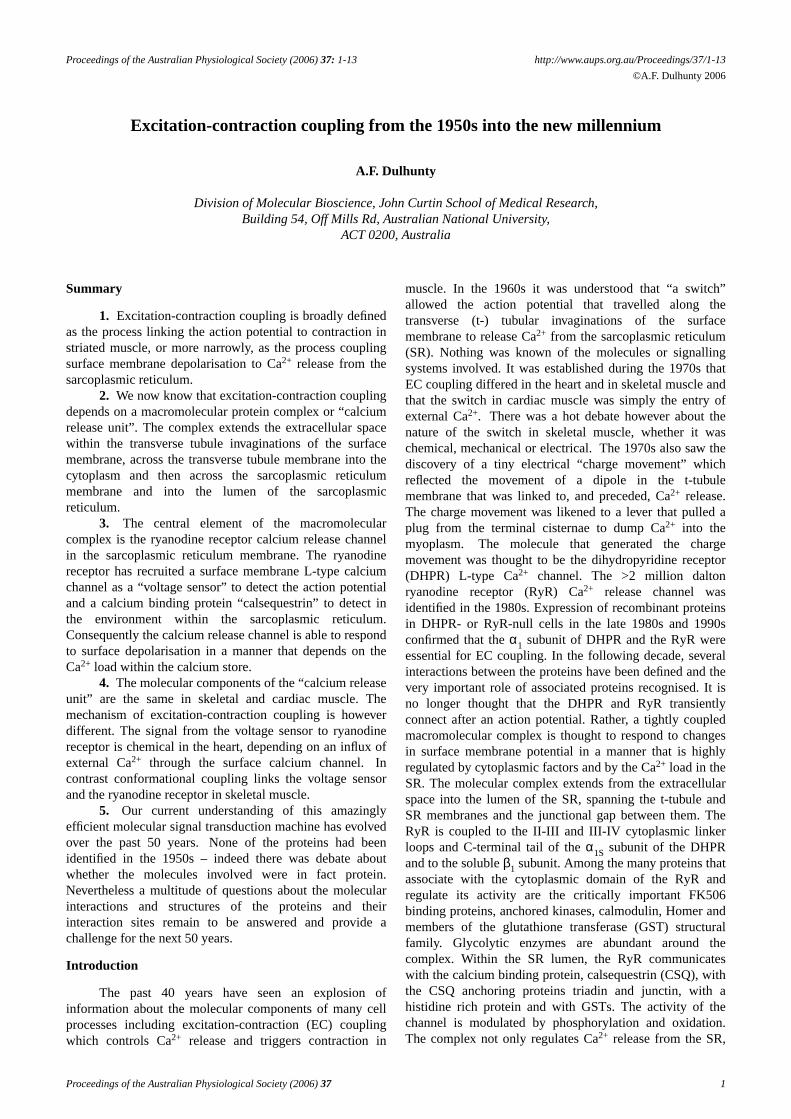
Electron microscopy also revealed a tight connectionbetween the t-tubule system and the expanded terminalcisternae sacs of the SR.Since there were 3 elements, a t-tubule flanked on either side by a terminal cisternum(Figure 1), this junction between the external and internalmembrane systems was aptly named the “triad junction”.8
A remarkable feature of the triad junction was periodicelectron densities spanning the 10nm junctional gapbetween the cytoplasmic leaflets of the t-tubule and SRmembranes. Thetriad junction and the “junctional feet”were thought to facilitate EC coupling because of theirstrategic position and the close proximity of the internal andexternal membranes. A novel technique, “glyceroltreatment”, osmotically “severed” the t-tubule system fromthe surface membrane, abolished EC coupling and
confirmed thatt-tubule continuity was essential for theaction potential to initiate contraction.6,9
Figure 1. An electron micrograph of a section through atriad junction of a frog tonic fibre, showing a central t-tubu-lar element flanked on either side by a terminal cisternaeelement of the sarcoplasmic reticulum. Thearrows point toelectron dense junctional feet spanning the junctional gapon either side of the t-tubule between the t-tubule and termi-nal cisternae. Note the rope-like structures within the termi-nal cisternae – later identified as the calcium binding pro-tein, calsequestrin. Micrograph kindly provided by ClaraFr anzini-Armstrong, modified from.10
Progress during the 1970s
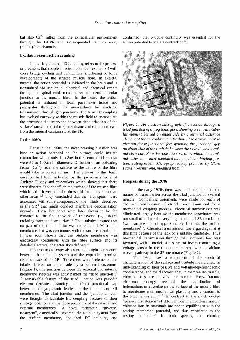
In the early 1970s there was much debate about thenature of transmission across the triad junction in skeletalmuscle. Compellingarguments were made for each ofchemical transmission, electrical transmission and for amechanical coupling process. Electrical transmission waseliminated largely because the membrane capacitance wastoo small to include the very large amount of SR membrane(with surface area of approximately 10 times the surfacemembrane11). Chemicaltransmission was argued against atthis time because of the lack of a suitable candidate.Thusmechanical transmission through the junctional feet wasfavoured, with a model of a series of levers connecting avoltage sensor in the t-tubule membrane with a calciumrelease pathway in the SR membrane (Figure 2).
The 1970s saw a refinement of the electricalcharacterisation of the surface and t-tubule membranes, anunderstanding of their passive and voltage-dependent ionicconductances and the discovery that, in mammalian muscle,chloride ions are actively transported. Freeze-fractureelectron-microscopy rev ealed the contribution ofindentations or caveolae on the surface of the muscle fibreto membrane area, mechanical plasticity and a conduit tothe t-tubule system.12,13 In contrast to the much quoted“passive distribution” of chloride ions in amphibian muscle,chloride ions in mammals are not in equilibrium with theresting membrane potential, and thus contribute to theresting potential.14 In both species, the chloride
2 Proceedings of the Australian Physiological Society (2006)37
A.F. Dulhunty
Ca2+
Actin/myosin
surface membrane
Ca2+
transverse-tubule
at rest
Ca2+
relaxed muscle
terminal cisternae
action potential
Sarcoplasmic reticulum
after action potential
Ca2+
Figure 2. A model for the mechanical mechanism of EC coupling in skeletal muscle developed in the 1970s. The position ofa lever that extended from the t-tubule to the terminal cisternae of the SR, is altered by an action potential in such a waythat a plug is removed from a pore in the terminal cisternae membrane and Ca2+ ions flow out to activate the contractileproteins.
conductance was found to be located mainly in the t-tubulemembrane.15
During this period, the skeletal muscle t-tubulemembrane was identified as a rich source of L-type calciumchannel protein, later known as the dihydropyridinereceptor (DHPR). The T-tubule membrane was used as asource of this protein by biochemists investigating voltage-dependent Ca2+ channels. Itsignificance for EC couplingwas overlooked.
In 1973, a novel and tiny electrical signal wasdiscovered in squid giant axon membranes.16,17 The signalpreceded the voltage-dependent Na+ current that underliesthe action potential in most neurones. Its activation andinactivation characteristics suggested that it reflected anev ent within the membrane, that gated the Na+ channel andhad been predicted more than two decade before byHodgkin & Huxley.18 The “asymmetric capacitive current”or “charge movement” was also named a “gating current”.The very small size of the signal (pA) and its complexelectrical behaviour provided a major technical challengefor researchers who, at that time, designed and built theirown electronic equipment.Some of the first laboratorycomputers were used to generate the complex trains ofpulses, signal subtraction and signal averaging required todissect the capacitive gating current from other ionic an
capacitive currents generated by the voltage pulses.The discovery of gating currents provided a
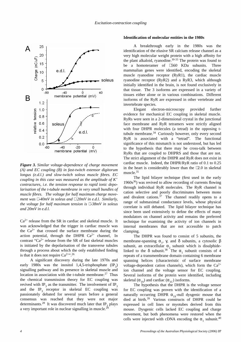
conceptual leap for EC coupling. The current in skeletalmuscle fibres was too slow to gate the voltage-dependentNa+ channel. Itpreceded Ca2+ release from the SR, itsvoltage-dependence was compatible with EC coupling andit was abolished by glycerol-treatment.19,20 Furtherevidence for its role in EC coupling was provided by thefact that differences between the voltage dependence of ECcoupling in fast- and slow-twitch fibres was reflected incharge movement (Figure 3).21 The gating current wasthought to be the movement of charged residues within thet-tubule membrane and to reflect a voltage-sensor responseto depolarisation that initiated EC coupling.The gatingcurrent was the “engine” driving a mechanical EC couplingmechanism.
At the same time as the gating current work, it wasdiscovered that EC coupling was sensitive todihydropyridine based compounds that were also agonistsor antagonists of the DHPR Ca2+ channel.22,23 This andadditional evidence that the gating current arose from theDHPR24,25 led to the hypothesis that the DHPR was thevoltage sensor for EC coupling.
Another fact that became established during the1970s was that two basically different processes triggered
Proceedings of the Australian Physiological Society (2006)37 3
Excitation-contraction coupling
e.d.l.
e.d.l.
soleus
soleus
B
A
Figure 3. Similar voltage-dependence of charge movement(A) and EC coupling (B) in fast-twitch extensor digitorumlongus (e.d.l.) and slow-twitch soleus muscle fibres. ECcoupling in this case was measured as the amplitude of K+
contractures, i.e. the tension response to rapid ionic depo-larisation of the t-tubule membrane in very small bundles ofmuscle fibres. Thevoltage for half maximum charge move-ment was∼ -40mV in soleus and∼ 20mV in e.d.l. Similarly,the voltage for half maximum tension is∼ -38mV in soleusand 20mV in e.d.l.
Ca2+ release from the SR in cardiac and skeletal muscle.Itwas acknowledged that the trigger in cardiac muscle wasthe Ca2+ that crossed the surface membrane during theaction potential, through the DHPR Ca2+ channel, Incontrast “Ca2+ release from the SR of fast skeletal musclesis initiated by the depolarisation of the transverse tubulesthrough a process about which the only established findingis that it does not require Ca2+”.26
A significant discovery during the late 1970s andearly 1980s was the inositol 1,4,5-trisphosphate (IP3)signalling pathway and its presence in skeletal muscle andlocation in association with the t-tubule membrane.27 Thusthe chemical transmission theory for EC coupling wasrevived with IP3 as the transmitter. The involvement of IP3and the IP3 receptor in skeletal EC coupling waspassionately debated for several years before a generalconsensus was reached that they were not majordeterminants.28 It was discovered much later that IP3 playsa very important role in nuclear signalling in muscle.29
Identification of molecular entities in the 1980s
A breakthrough early in the 1980s was theidentification of the elusive SR calcium release channel as avery high molecular weight protein with a high affinity forthe plant alkaloid, ryanodine.30-33 The protein was found tobe a homoteramer of∼ 560 KDa subunits. Threemammalian genes were identified, encoding the skeletalmuscle ryanodine receptor (RyR1), the cardiac muscleryanodine receptor (RyR2) and a RyR3, which althoughinitially identified in the brain, is not found exclusively inthat tissue. The 3 isoforms are expressed in a variety oftissues either alone or in various combinations.Differentisoforms of the RyR are expressed in other vertebrate andinvertebrate species.
Elegant electron-microscopy provided furtherevidence for mechanical EC coupling in skeletal muscle.RyRs were seen in a 2-dimensional crystal in the junctionalface membrane and RyR tetramers were strictly alignedwith four DHPR molecules (a tetrad) in the opposing t-tubule membrane.34 Curiously however, only every secondRyR is associated with a “tetrad”. The functionalsignificance of this mismatch is not understood, but has ledto the hypothesis that there may be cross-talk betweenRyRs that are coupled to DHPRS and those that are not.The strict alignment of the DHPR and RyR does not exist incardiac muscle.Indeed, the DHPR/RyR ratio of 0.1 to 0.25in the heart is considerably lower than the∼ 2.0 in skeletalmuscle.35
The lipid bilayer technique (first used in the early1960s36) was revived to allow recording of currents flowingthrough individual RyR molecules.The RyR channel iscation selective and poorly discriminates between monoand divalent cations.37 The channel readily opens to arange of submaximal conductance levels, whose physicalcorrelate is still debated. The lipid bilayer technique hassince been used extensively to define the effects of manymodulators on channel activity and remains the preferredtechnique for examining the activity of ion channels ininternal membranes that are not accessible to patchclamping.
The DHPR was found to consist of 5 subunits, themembrane-spanningα1, γ, and δ subunits, a cytosolic βsubunit, an extracellular α2 subunit which is disulphide-linked to theδ subunit.38 The α1 subunit consists of 4repeats of a transmembrane domain containing 6 membranespanning helices (characteristic of surface membranevoltage-dependent cation channels), which form the Ca2+
ion channel and the voltage sensor for EC coupling.Several isoforms of the protein were identified, includingskeletal (α1S) and cardiac (α1C) isoforms.
The hypothesis that the DHPR is the voltage sensorfor EC coupling was proven with the identification of anaturally occurring DHPRα1S-null dysgenic mouse thatdied at birth.39 Various constructs of DHPR could beexpressed in cell lines or myotubes derived from thismouse. Dysgeniccells lacked EC coupling and chargemovement, but both phenomena were restored when thecells were injected with cDNA encoding theα1 subunit.40
4 Proceedings of the Australian Physiological Society (2006)37
A.F. Dulhunty
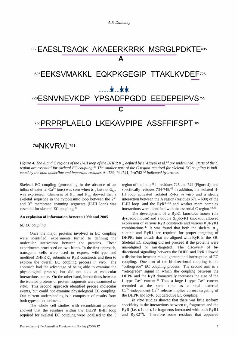
Figure 4. The A and C regions of the II-III loop of the DHPRα1Sdefined by el-Hayeket al.43 are underlined. Parts of the Cregion are essential for skeletal EC coupling.46 The smaller part of the C region required for skeletal EC coupling is indi-cated by the bold underline and important residuesAla739, Phe741, Pro74242 indicated by arrows.
Skeletal EC coupling (proceeding in the absence of aninflux of external Ca2+ ions) was seen whenα1S but not α1C,was expressed. Chimerasof α1S and α1C showed that askeletal sequence in the cytoplasmic loop between the 2nd
and 3rd membrane spanning segments (II-III loop) wasessential for skeletal EC coupling.40
An explosion of information between 1990 and 2005
(a) EC coupling
Once the major proteins involved in EC couplingwere identified, experiments turned to defining themolecular interactions between the proteins.Theseexperiments proceeded on two fronts. In the first approach,transgenic cells were used to express wild-type andmodified DHPRα1 subunits or RyR constructs and then toexplore the overall EC coupling processin vivo. Theapproach had the advantage of being able to examine thephysiological process, but did not look at molecularinteractions per se. On the other hand, interactions betweenthe isolated proteins or protein fragments were examinedinvitro. This second approach identified precise molecularev ents, but could not examine physiological EC coupling.Our current understanding is a composite of results fromboth types of experiment.
The whole cell studies with recombinant proteinsshowed that the residues within the DHPR II-III looprequired for skeletal EC coupling were localised to the C
region of the loop,41 in residues 725 and 742 (Figure 4), andspecifically residues 734-748.42 In addition, the isolated II-III loop activated isolated RyRsin vitro and a stronginteraction between the A region (residues 671 – 690) of theII-III loop and the RyR43,44 and weaker more complexinteractions were identified with the essential C region.43,45
The development of a RyR1 knockout mouse (thedyspedic mouse) and a doubleα1S/RyR1 knockout allowedexpression of various RyR constructs and variousα1/RyR1combinations.47 It was found that both the skeletal α1Ssubunit and RyR1 are required for proper targeting ofDHPRs into tetrads that are aligned with RyR in the SR.Skeletal EC coupling did not proceed if the proteins weremis-aligned or mis-targeted. The discovery of bi-directional signalling between the DHPR and RyR alloweda distinction between mis-alignment and interruption of ECcoupling. Onearm of the bi-directional coupling is the“orthograde” EC coupling process. The second arm is a“retrograde” signal in which the coupling between theDHPR and the RyR dramatically increases the size of theL-type Ca2+ current.48 Thus a large L-type Ca2+ currentrecorded at the same time as a small externalCa2+-independent Ca2+ release implies correct targeting ofthe DHPR and RyR, but defective EC coupling.
In vitro studies showed that there was little isoformspecificity in the interactions betweenα1 fragments and theRyR (i.e.α1s orα1c fragments interacted with both RyR1and RyR249). Therefore some residues that appeared
Proceedings of the Australian Physiological Society (2006)37 5
Excitation-contraction coupling
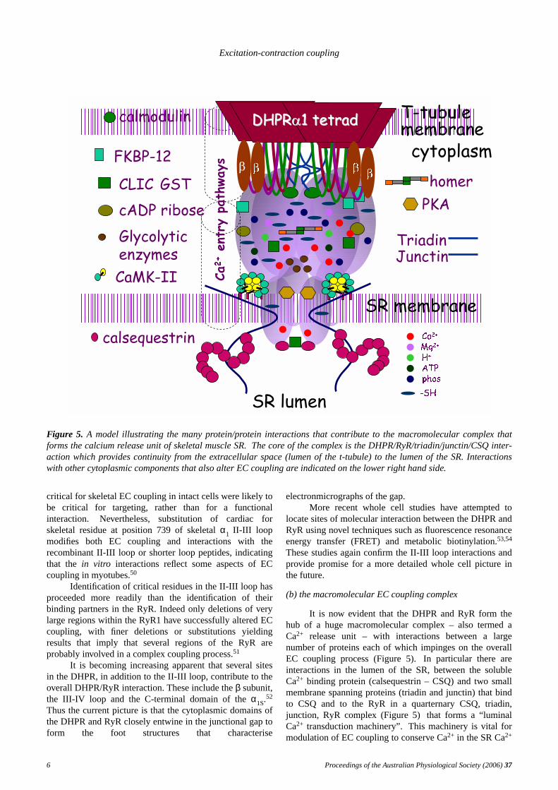
Figure 5. A model illustrating the many protein/protein interactions that contribute to the macromolecular complex thatforms the calcium release unit of skeletal muscle SR. The core of the complex is the DHPR/RyR/triadin/junctin/CSQ inter-action which pro vides continuity from the extracellular space (lumen of the t-tubule) to the lumen of the SR. Interactionswith other cytoplasmic components that also alter EC coupling are indicated on the lower right hand side.
critical for skeletal EC coupling in intact cells were likely tobe critical for targeting, rather than for a functionalinteraction. Nevertheless, substitution of cardiac forskeletal residue at position 739 of skeletal α1 II-III loopmodifies both EC coupling and interactions with therecombinant II-III loop or shorter loop peptides, indicatingthat the in vitro interactions reflect some aspects of ECcoupling in myotubes.50
Identification of critical residues in the II-III loop hasproceeded more readily than the identification of theirbinding partners in the RyR. Indeed only deletions of verylarge regions within the RyR1 have successfully altered ECcoupling, with finer deletions or substitutions yieldingresults that imply that several regions of the RyR areprobably involved in a complex coupling process.51
It is becoming increasing apparent that several sitesin the DHPR, in addition to the II-III loop, contribute to theoverall DHPR/RyR interaction. These include theβ subunit,the III-IV loop and the C-terminal domain of theα1S.
52
Thus the current picture is that the cytoplasmic domains ofthe DHPR and RyR closely entwine in the junctional gap toform the foot structures that characterise
electronmicrographs of the gap.More recent whole cell studies have attempted to
locate sites of molecular interaction between the DHPR andRyR using novel techniques such as fluorescence resonanceenergy transfer (FRET) and metabolic biotinylation.53,54
These studies again confirm the II-III loop interactions andprovide promise for a more detailed whole cell picture inthe future.
(b) the macromolecular EC coupling complex
It is now evident that the DHPR and RyR form thehub of a huge macromolecular complex – also termed aCa2+ release unit – with interactions between a largenumber of proteins each of which impinges on the overallEC coupling process (Figure 5).In particular there areinteractions in the lumen of the SR, between the solubleCa2+ binding protein (calsequestrin – CSQ) and two smallmembrane spanning proteins (triadin and junctin) that bindto CSQ and to the RyR in a quarternary CSQ, triadin,junction, RyR complex (Figure 5) that forms a “luminalCa2+ transduction machinery”.This machinery is vital formodulation of EC coupling to conserve Ca2+ in the SR Ca2+
6 Proceedings of the Australian Physiological Society (2006)37
A.F. Dulhunty
Coupled gating
AO3
O2
O1
C3s 10pA
A
B5pA
250m
control
CLIC-2
Substate opening
c
o1
C
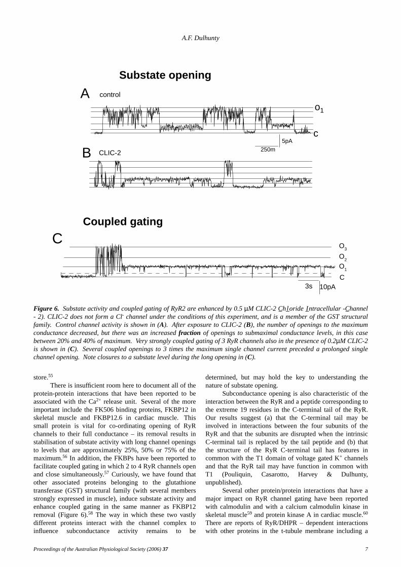
Figure 6. Substate activity and coupled gating of RyR2 are enhanced by 0.5µM CLIC-2 C_h l_oride I_ntracellular -C_hannel- 2). CLIC-2 does not form a Cl- channel under the conditions of this experiment, and is a member of the GST structuralfamily. Control channel activity is shown in (A). After exposure to CLIC-2 (B), the number of openings to the maximumconductance decreased, but there was an increasedfraction of openings to submaximal conductance levels, in this casebetween 20% and 40% of maximum.Very strongly coupled gating of 3 RyR channels also in the presence of 0.2µM CLIC-2is shown in (C). Several coupled openings to 3 times the maximum single channel current preceded a prolonged singlechannel opening. Note closures to a substate level during the long opening in (C).
store.55
There is insufficient room here to document all of theprotein-protein interactions that have been reported to beassociated with the Ca2+ release unit.Several of the moreimportant include the FK506 binding proteins, FKBP12 inskeletal muscle and FKBP12.6 in cardiac muscle.Thissmall protein is vital for co-ordinating opening of RyRchannels to their full conductance – its removal results instabilisation of substate activity with long channel openingsto levels that are approximately 25%, 50% or 75% of themaximum.56 In addition, the FKBPs have been reported tofacilitate coupled gating in which 2 to 4 RyR channels openand close simultaneously.57 Curiously, we hav e found thatother associated proteins belonging to the glutathionetransferase (GST) structural family (with several membersstrongly expressed in muscle), induce substate activity andenhance coupled gating in the same manner as FKBP12removal (Figure 6).58 The way in which these two vastlydifferent proteins interact with the channel complex toinfluence subconductance activity remains to be
determined, but may hold the key to understanding thenature of substate opening.
Subconductance opening is also characteristic of theinteraction between the RyR and a peptide corresponding tothe extreme 19 residues in the C-terminal tail of the RyR.Our results suggest (a) that the C-terminal tail may beinvolved in interactions between the four subunits of theRyR and that the subunits are disrupted when the intrinsicC-terminal tail is replaced by the tail peptide and (b) thatthe structure of the RyR C-terminal tail has features incommon with the T1 domain of voltage gated K+ channelsand that the RyR tail may have function in common withT1 (Pouliquin, Casarotto, Harvey & Dulhunty,unpublished).
Several other protein/protein interactions that have amajor impact on RyR channel gating have been reportedwith calmodulin and with a calcium calmodulin kinase inskeletal muscle59 and protein kinase A in cardiac muscle.60
There are reports of RyR/DHPR – dependent interactionswith other proteins in the t-tubule membrane including a
Proceedings of the Australian Physiological Society (2006)37 7
Excitation-contraction coupling
excitation-coupled Ca2+ entry pathway61 and a storeoperated Ca2+ channel.62
(c) RyR and DHPR structure
Like most ion channels, both the DHPR and the RyRhave eluded crystallization and high resolution X-rayanalysis. Elegant cryoelectron microscopic studies withparticle analysis have rev ealed the overall profiles of theDHPR and the RyR.The structure of the RyR, solved at a∼ 30Ao resolution in 2000, showed the four subunits, a largecytoplasmic foot and a narrower transmembraneassembly.63,64 Several sub-domains were identified in thecytoplasmic parts of each subunit. One region (domain 6)that extends furthest towards the t-tubule membrane is alikely candidate for a part of the DHPR/RyR interaction.More recently structures have been obtained with higherresolution that provide much greater detail65,66 andsecondary structural elements within the pore.67 A recentX-ray analysis of a 2-dimensional RyR crystal provided astructure with a resolution of about 30Ao that basicallyagreed with the cryoelectron microscopic images andshowed clear interactions between domain 6 of adjacentRyR tetramers, suggesting that this domain may not onlyinteract with the DHPR but also subserve coupled gatingbetween RyR channels.68 Homology modelling hasprovided an indication of secondary and tertiary structuresof some of some of the RyR domains.66
Binding sites on the 3 dimensional profile have beenidentified for several compounds including FKBP12,calmodulin, imperatoxin A69 as well as the location ofregions of major divergence between the three mammalianRyRs.70 These binding sites allowed mapping of regions inthe linear sequence to domains in the 3 dimensional profile,since binding sequences have been identified in separatestudies.
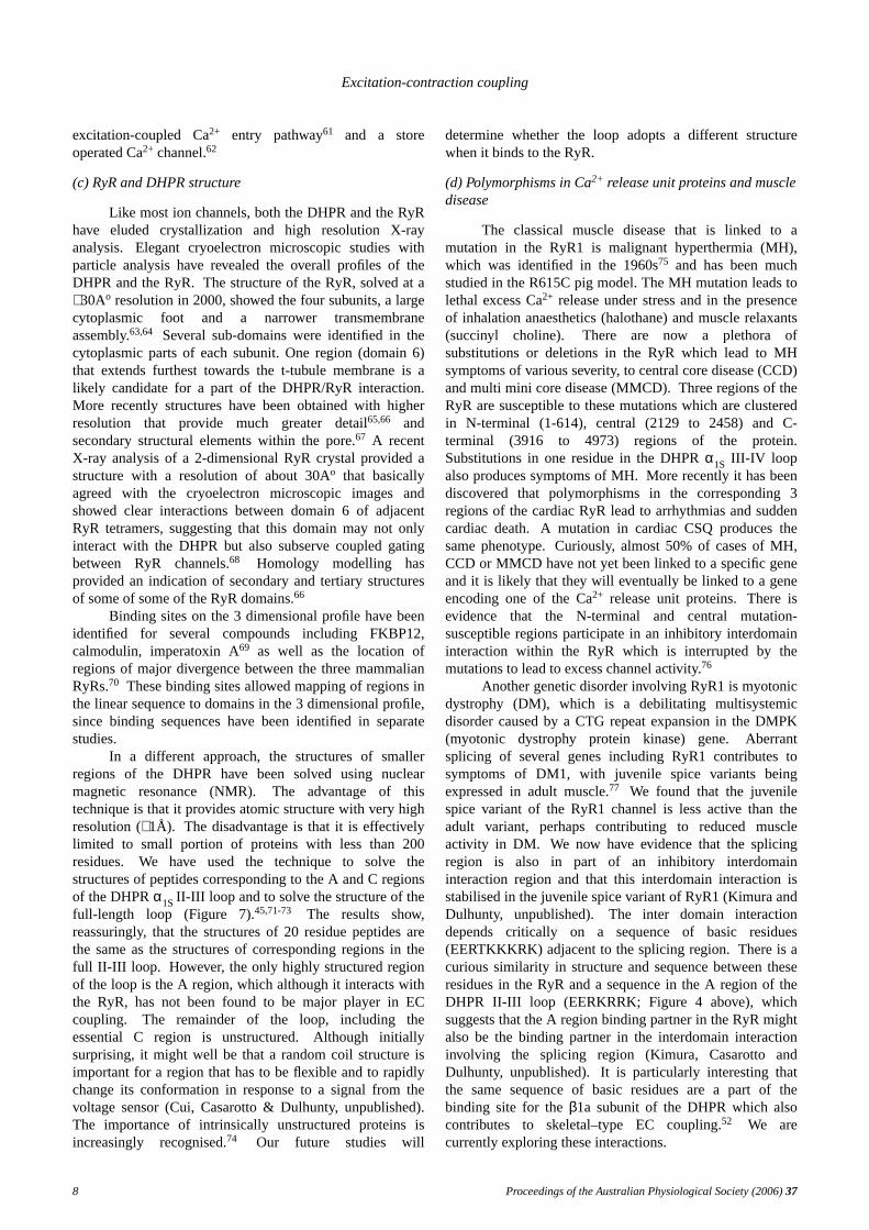
In a different approach, the structures of smallerregions of the DHPR have been solved using nuclearmagnetic resonance (NMR). The advantage of thistechnique is that it provides atomic structure with very highresolution (∼ 1Å). Thedisadvantage is that it is effectivelylimited to small portion of proteins with less than 200residues. We hav e used the technique to solve thestructures of peptides corresponding to the A and C regionsof the DHPRα1SII-III loop and to solve the structure of thefull-length loop (Figure 7).45,71-73 The results show,reassuringly, that the structures of 20 residue peptides arethe same as the structures of corresponding regions in thefull II-III loop. However, the only highly structured regionof the loop is the A region, which although it interacts withthe RyR, has not been found to be major player in ECcoupling. The remainder of the loop, including theessential C region is unstructured. Although initiallysurprising, it might well be that a random coil structure isimportant for a region that has to be flexible and to rapidlychange its conformation in response to a signal from thevoltage sensor (Cui, Casarotto & Dulhunty, unpublished).The importance of intrinsically unstructured proteins isincreasingly recognised.74 Our future studies will
determine whether the loop adopts a different structurewhen it binds to the RyR.
(d) Polymorphisms in Ca2+ release unit proteins and muscledisease
The classical muscle disease that is linked to amutation in the RyR1 is malignant hyperthermia (MH),which was identified in the 1960s75 and has been muchstudied in the R615C pig model. The MH mutation leads tolethal excess Ca2+ release under stress and in the presenceof inhalation anaesthetics (halothane) and muscle relaxants(succinyl choline). There are now a plethora ofsubstitutions or deletions in the RyR which lead to MHsymptoms of various severity, to central core disease (CCD)and multi mini core disease (MMCD). Three regions of theRyR are susceptible to these mutations which are clusteredin N-terminal (1-614), central (2129 to 2458) and C-terminal (3916 to 4973) regions of the protein.Substitutions in one residue in the DHPRα1S III-IV loopalso produces symptoms of MH. More recently it has beendiscovered that polymorphisms in the corresponding 3regions of the cardiac RyR lead to arrhythmias and suddencardiac death.A mutation in cardiac CSQ produces thesame phenotype.Curiously, almost 50% of cases of MH,CCD or MMCD have not yet been linked to a specific geneand it is likely that they will eventually be linked to a geneencoding one of the Ca2+ release unit proteins. There isevidence that the N-terminal and central mutation-susceptible regions participate in an inhibitory interdomaininteraction within the RyR which is interrupted by themutations to lead to excess channel activity.76
Another genetic disorder involving RyR1 is myotonicdystrophy (DM), which is a debilitating multisystemicdisorder caused by a CTG repeat expansion in the DMPK(myotonic dystrophy protein kinase) gene. Aberrantsplicing of several genes including RyR1 contributes tosymptoms of DM1, with juvenile spice variants beingexpressed in adult muscle.77 We found that the juvenilespice variant of the RyR1 channel is less active than theadult variant, perhaps contributing to reduced muscleactivity in DM. We now hav e evidence that the splicingregion is also in part of an inhibitory interdomaininteraction region and that this interdomain interaction isstabilised in the juvenile spice variant of RyR1 (Kimura andDulhunty, unpublished). Theinter domain interactiondepends critically on a sequence of basic residues(EERTKKKRK) adjacent to the splicing region. Thereis acurious similarity in structure and sequence between theseresidues in the RyR and a sequence in the A region of theDHPR II-III loop (EERKRRK; Figure 4 above), whichsuggests that the A region binding partner in the RyR mightalso be the binding partner in the interdomain interactioninvolving the splicing region (Kimura, Casarotto andDulhunty, unpublished). Itis particularly interesting thatthe same sequence of basic residues are a part of thebinding site for theβ1a subunit of the DHPR which alsocontributes to skeletal–type EC coupling.52 We arecurrently exploring these interactions.
8 Proceedings of the Australian Physiological Society (2006)37
A.F. Dulhunty
Figure 7. NMR solution structure of the DHPRα1S II-III loop. The A, B, C and D regions correspond to regions given inFigure 4 above. The essential central residues (734-748) are in the central part of the C region. Theonly strongly struc-tured part of the loop is the N-terminal A region. Theremainder of the loop is random coil (Cui Y, Karunasekara Y, HarveyPJ, Board PG, Dulhunty AF, Casarotto MG, unpublished data).
(e) Proteins in EC coupling as a therapeutic target
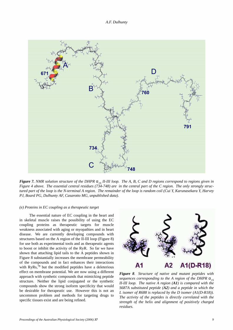
The essential nature of EC coupling in the heart andin skeletal muscle raises the possibility of using the ECcoupling proteins as therapeutic targets for muscleweakness associated with aging or myopathies and in heartdisease. We are currently developing compounds withstructures based on the A region of the II-III loop (Figure 8)for use both as experimental tools and as therapeutic agentsto boost or inhibit the activity of the RyR. So far we haveshown that attaching lipid tails to the A peptides shown inFigure 8 substantially increases the membrane permeabilityof the compounds and in fact enhances their interactionswith RyRs,78 but the modified peptides have a deleteriouseffect on membrane potential. We are now using a differentapproach with synthetic compounds that mimicking peptidestructure. Neitherthe lipid conjugated or the syntheticcompounds show the strong isoform specificity that wouldbe desirable for therapeutic use.However this is not anuncommon problem and methods for targeting drugs tospecific tissues exist and are being refined.
Figure 8. Structure of native and mutant peptides withsequences corresponding to the A region of the DHPRα1SII-III loop. The native A region (A1) is compared with theS687A substituted peptide (A2) and a peptide in which theL isomer of R688 is replaced by the D isomer (A1(D-R18)).The activity of the peptides is directly correlated with thestrength of the helix and alignment of positively chargedresidues.
Proceedings of the Australian Physiological Society (2006)37 9
Excitation-contraction coupling
Concluding comments
Despite the huge advances in our understanding ofEC coupling since the 1960s, the molecular mechanisms ofthe process are still largely unknown. We remain ignorantabout the atomic structures of the major players, the DHPRand RyR. High resolution structural determinations awaitfurther refinements of particle analysis and/or theproduction of crystals and X-ray analysis of these large andcomplex membrane spanning proteins.We do notunderstand the molecular nature of the interactions betweenthe proteins and indeed do not know which residues areinvolved in most of the interactions. This information willbe provided by many of the current studies but will alsodepend on the development of more sophisticatedtechniques such as FRET and other fluorescent probetechniques.79 Such techniques may also reveal the nature ofthe inter-domain interactions that we think are modified inthe MH-like polymorphisms and in DM.Finally, althoughall evidence strongly supports the mechanical couplingmechanism for EC coupling, mechanical coupling remainsa hypothesis. Theultimate experiment will be visualisationof the conformational changes that in the DHPR and in theRyR during EC coupling.
References
1. Huxley, AF, Straub RW. Local activation of interfibrillarstructures in striated muscle.J. Physiol. 1958;143:40-41P.
2. Huxley, AF, Taylor RE. Local activation of striatedmuscle fibres.J. Physiol.1958;144:426-41.
3. Porter, KR, Palade GE. Studies on the endoplasmicreticulum. III. Its form and distribution in striatedmuscle cells.J. Biophys. Biochem. Cytol.1957; 3:269-300.
4. Franzini-Armstrong,C. Fine structure of sarcoplasmicreticulum and tranverse tubular system in musclefibers.Fed. Proc.1964;23: 887-95.
5. Adrian, RH, Costantin LL, Peachey LD. Radial spreadof contraction in frog muscle fibres.J. Physiol.1969;204:231-257.
6. Eisenberg, RS, Gage PW. Ionic conductances of thesurface and transverse tubular membranes of frogsartorius fibers.J. Gen. Physiol.1969;53: 279-297.
7. Dulhunty, AF, Gage PW. Electrical properties of toadskeletal muscle fibres in summer and winter. J.Physiol.1973;230:619-41.
8. Franzini-Armstrong,C. Studies of the triad. I. Structureof the junction in frog twitch fibers.J. Cell Biol.1970;47: 488-498.
9. Gage, PW, Eisenberg RS. Action potentials,afterpotentials, and excitation-contraction couplingin frog sartorius fibers without transverse tubules.J.Gen. Physiol.1969;53: 298-310.
10. Franzini-Armstrong,C. Studies of the triad. IV.Structure of the junction in frog slow fibers.J. CellBiol. 1973;56: 120-8.
11. Eisenberg, BR, Kuda AM, Peter JB. Stereologicalanalysis of mammalian skeletal muscle.J. Cell Biol.
1974;60: 732-754.12. Dulhunty, AF, Franzini-Armstrong C. The passive
electrical properties of frog skeletal muscle fibres atdifferent sarcomere lengths.J. Physiol. 1977; 266:687-711.
13. Dulhunty, AF, Franzini-Armstrong C. The relativecontributions of the folds and caveolae to the surfacemembrane of frog skeletal muscle fibres at differentsarcomere lengths.J. Physiol.1975;250:513-39.
14. Dulhunty, AF. The dependence of membrane potentialon extracellular chloride concentration inmammalian skeletal muscle fibres.J. Physiol.1978;276:67-82.
15. Dulhunty, AF. Distribution of potassium and chloridepermeability over the surface and T-tubulemembranes of mammalian skeletal muscle. J.Membrane Biol.1979;45: 293-310.
16. Armstrong, CM, Bezanilla F. Currents related tomovement of the gating particles of the sodiumchannels.Nature1973;242:459-61.
17. Armstrong, CM, Bezanilla F. Charge movementassociated with the opening and closing of theactivation gates of the Na channels.J. Gen. Physiol.1974;63: 533-52.
18. Hodgkin,AL, Huxley AF. A quantitative description ofmembrane current and its application to conductionand excitation in nerve. J. Physiol. 1952; 117:500-44.
19. Schneider, MF, Chandler WK. Voltage dependentcharge movement of skeletal muscle: a possible stepin excitation-contraction coupling.Nature 1973;242:244-6.
20. Chandler, WK, Rakowski RF, Schneider MF. Effects ofglycerol treatment and maintained depolarization oncharge movement in skeletal muscle.J. Physiol.1976;254:285-316.
21. Dulhunty, AF, Gage PW. Asymmetrical chargemovement in slow- and fast-twitch mammalianmuscle fibres in normal and paraplegic rats.J.Physiol.1983;341:213-31.
22. Eisenberg, RS, McCarthy RT, Milton RL. Paralysis offrog skeletal muscle fibres by the calcium antagonistD-600.J. Physiol.1983;341:495-505.
23. Dulhunty, AF, Gage PW. Effects of extracellularcalcium concentration and dihydropyridines oncontraction in mammalian skeletal muscle. J.Physiol.1988;399:63-80.
24. Lamb,GD. Asymmetric charge movement in polarizedand depolarized muscle fibres of the rabbit.J.Physiol.1987;383:349-67.
25. Rios, E, Brum G. Involvement of dihydropyridinereceptors in excitation-contraction coupling inskeletal muscle.Nature1987;325:717-20.
26. Fabiato, A, Fabiato F. Calcium release from thesarcoplasmic reticulum.Circ. Res.1977;40: 119-29.
27. Vergara, J, Tsien RY, Delay M. Inositol1,4,5-trisphosphate: a possible chemical link inexcitation-contraction coupling in muscle.Proc.Natl. Acad. Sci. U.S.A.1985;82: 6352-6.
10 Proceedings of the Australian Physiological Society (2006)37
A.F. Dulhunty
28. Hidalgo, C, Jaimovich E. Inositol trisphosphate andexcitation-contraction coupling in skeletal muscle.J.Bioenerg. Biomembr.1989;21: 267-81.
29. Jaimovich, E, Carrasco MA. IP3 dependent Ca2+
signals in muscle cells are involved in regulation ofgene expression.Biol. Res.2002;35: 195-202.
30. Meissner, G. Ryanodine activation and inhibition of theCa2+ release channel of sarcoplasmic reticulum.J.Biol. Chem.1986;261:6300-6.
31. Smith, JS, Coronado R, Meissner G. Sarcoplasmicreticulum contains adenine nucleotide-activatedcalcium channels.Nature1985;316:446-9.
32. Fleischer, S, Ogunbunmi EM, Dixon MC, Fleer EA.Localization of Ca2+ release channels with ryanodinein junctional terminal cisternae of sarcoplasmicreticulum of fast skeletal muscle.Proc. Natl. Acad.Sci. U.S.A.1985;82: 7256-9.
33. Inui, M, Saito A, Fleischer S. Purification of theryanodine receptor and identity with feet structuresof junctional terminal cisternae of sarcoplasmicreticulum from fast skeletal muscle.J. Biol. Chem.1987;262:1740-7.
34. Block, BA, Imagaw a T, Campbell KP, Franzini-Armstrong C. Structural evidence for directinteraction between the molecular components of thetransverse tubule/sarcoplasmic reticulum junction inskeletal muscle.J. Cell Biol. 1988;107: 2587-2600.
35. Bers, DM, Stiffel VM. Ratio of ryanodine todihydropyridine receptors in cardiac and skeletalmuscle and implications for E-C coupling.Am. J.Physiol.1993;264:C1587-C1593.
36. Mueller, P, Rudin DO, Tien HT, Wescott WC.Reconstitution of cell membrane structure in vitroand its transformation into an excitable system.Nature1962;194:979-80.
37. Meissner, G. Ryanodine receptor/Ca++ release channelsand their regulation by endogenous effectors.Ann.Rev. Physiol.1994;56: 485-508.
38. Ahlijanian, MK, Westenbroek RE, Catterall WA.Subunit structure and localization ofdihydropyridine-sensitive calcium channels inmammalian brain, spinal cord, and retina.Neuron1990;4: 819-32.
39. Beam,KG, Knudson CM, Powell JA. A lethal mutationin mice eliminates the slow calcium current inskeletal muscle cells.Nature1986;320:168-170.
40. Tanabe, T, Beam KG, Adams BA, Niidome T, Numa S.Regions of the skeletal muscle dihydropyridinereceptor critical for excitation-contraction coupling.Nature1990;346:567-568.
41. Nakai,J, Tanabe T, Konno T, Adams B, Beam KG.Localization in the II-III loop of the dihydropyridinereceptor of a sequence critical for excitation-contraction coupling.J. Biol. Chem. 1998; 273:24983-6.
42. Kugler, G, Weiss RG, Flucher BE, Grabner M.Structural requirements of the dihydropyridinereceptor alpha1S II-III loop for skeletal-typeexcitation-contraction coupling.J. Biol. Chem.2004;
279:4721-8.43. el-Hayek, R, Antoniu B, Wang J, Hamilton SL,
Ikemoto N. Identification of calcium release-triggering and blocking regions of the II-III loop ofthe skeletal muscle dihydropyridine receptor. J.Biol. Chem.1995;270:22116-22118.
44. Dulhunty, AF, Lav er DR, Gallant EM, Casarotto MG,Pace SM, Curtis S. Activation and inhibition ofskeletal RyR channels by a part of the skeletalDHPR II-III loop: Effects of DHPR Ser687 andFKBP12. Biophys. J.1999;77: 189-203.
45. Haarmann,CS, Green D, Casarotto MG, Laver DR,Dulhunty AF. The random-coil ’C’ fragment of thedihydropyridine receptor II-III loop can activate orinhibit native skeletal ryanodine receptors.Biochem.J. 2003;372:305-16.
46. Nakai,J, Tanabe T, Konno T, Adams B, Beam KG.Localization in the II-III loop of the dihydropyridinereceptor of a sequence critical for excitation-contraction coupling.J. Biol. Chem. 1998; 273:24983-24986.
47. Nakai,J, Ogura T, Protasi F, Franzini-Armstrong C,Allen PD, Beam KG. Functional nonequality of thecardiac and skeletal ryanodine receptors.Proc. Natl.Acad. Sci. U.S.A.1997;94: 1019-22.
48. Nakai,J, Dirksen RT, Nguyen HT, Pessah IN, BeamKG, Allen PD. Enhanced dihydropyridine receptorchannel activity in the presence of ryanodinereceptor.Nature1996;380:72-5.
49. Dulhunty, AF, Curtis SM, Cengia L, Sakowska M,Casarotto MG. Peptide fragments of thedihydropyridine receptor can modulate cardiacryanodine receptor channel activity andsarcoplasmic reticulum Ca2+ release.Biochem. J.2004;379:161-72.
50. Dulhunty, AF, Karunasekara Y, Curtis SM, Harvey PJ,Board PG, Casarotto MG. Role of someunconserved residues in the "C" region of theskeletal DHPR II-III loop.Fr ont. Biosci.2005; 10:1368-81.
51. Protasi,F, Paolini C, Nakai J, Beam KG, Franzini-Armstrong C, Allen PD. Multiple regions of RyR1mediate functional and structural interactions withalpha(1S)-dihydropyridine receptors in skeletalmuscle.Biophys. J.2002;83: 3230-44.
52. Cheng, W, Altafaj X, Ronjat M, Coronado R.Interaction between the dihydropyridine receptorCa2+ channel {beta}-subunit and ryanodine receptortype 1 strengthens excitation-contraction coupling.Proc. Natl. Acad. Sci. U.S.A.2005;102:19225-30.
53. Papadopoulos, S, Leuranguer V, Bannister RA, BeamKG. Mapping sites of potential proximity betweenthe dihydropyridine receptor and RyR1 in muscleusing a cyan fluorescent protein-yellow fluorescentprotein tandem as a fluorescence resonance energytransfer probe.J. Biol. Chem.2004;279: 44046-56.
54. Lorenzon, NM, Haarmann CS, Norris EE,Papadopoulos S, Beam KG.Metabolic biotinylationas a probe of supramolecular structure of the triad
Proceedings of the Australian Physiological Society (2006)37 11
Excitation-contraction coupling
junction in skeletal muscle.J. Biol. Chem. 2004;279:44057-64.
55. Beard,NA, Lav er DR, Dulhunty AF. Calsequestrin andthe calcium release channel of skeletal and cardiacmuscle.Prog. Biophys. Mol.Biol. 2004;85: 33-69.
56. Ahern, GP, Junankar PR, Dulhunty AF.Subconductance states in single channel activity ofskeletal muscle ryanodine receptors after removal ofFKBP12.Biophys. J.1996;72: 146-62.
57. Lehnart, SE, Huang F, Marx SO, Marks AR.Immunophilins and coupled gating of ryanodinereceptors.Curr. Top. Med. Chem.2003;3: 1383-91.
58. Dulhunty, AF, Pouliquin P, Coggan M, Gage PW,Board PG. A recently identified member of theglutathione transferase structural family modifiescardiac RyR2 substate activity, coupled gating andactivation by Ca2+ and ATP. Biochem. J. 2005;390:333-43.
59. Dulhunty, AF, Lav er D, Curtis SM, Pace S, HaarmannC, Gallant EM. Characteristics of irreversible ATPactivation suggest that native skeletal ryanodinereceptors can be phosphorylated via an endogenousCaMKII. Biophys. J.2001;81: 3240-52.
60. Reiken, S, Gaburjakova M, Guatimosim S, GomezAM, D’Armiento J, Burkhoff D, Wang J, Vassort G,Lederer WJ, Marks AR. Protein kinase Aphosphorylation of the cardiac calcium releasechannel (ryanodine receptor) in normal and failinghearts. Role of phosphatases and response toisoproterenol.J. Biol. Chem.2003;278:444-53.
61. Hurne,AM, O’Brien JJ, Wingrove D, CherednichenkoG, Allen PD, Beam KG, Pessah IN. Ryanodinereceptor type 1 (RyR1) mutations C4958S andC4961S reveal excitation-coupled calcium entry(ECCE) is independent of sarcoplasmic reticulumstore depletion. J. Biol. Chem. 2005; 280:36994-7004.
62. Ma,J, Pan Z. Junctional membrane structure and storeoperated calcium entry in muscle cells.Fr ont.Biosci.2003;8: d242-55.
63. Radermacher, M, Wagenknecht T, Grassucci R, FrankJ, Inui M, Chadwick C, Fleischer S. Cryo-EM of thenative structure of the calcium releasechannel/ryanodine receptor from sarcoplasmicreticulum.Biophys. J.1992;61: 936-40.
64. Serysheva, II, Orlova EV, Chiu W, Sherman MB,Hamilton SL, van Heel M. Electron cryomicroscopyand angular reconstitution used to visualize theskeletal muscle calcium release channel.Nat.Struct. Biol.1995;2: 18-24.
65. Samso, M, Wagenknecht T, Allen PD. Internalstructure and visualization of transmembranedomains of the RyR1 calcium release channel bycryo-EM.Nat. Struct. Mol. Biol.2005;12: 539-44.
66. Serysheva, II, Hamilton SL, Chiu W, Ludtke SJ.Structure of Ca2+ release channel at 14 A resolution.J. Mol. Biol. 2005;345:427-31.
67. Ludtke, SJ, Serysheva, II, Hamilton SL, Chiu W. Thepore structure of the closed RyR1 channel.Structure
(Camb)2005;13: 1203-11.68. Yin, CC, Blayney LM, Lai FA. Physical coupling
between ryanodine receptor-calcium releasechannels.J. Mol. Biol. 2005;349:538-46.
69. Wagenknecht, T, Samso M. Three-dimensionalreconstruction of ryanodine receptors.Fr ont. Biosci.2002;7: d1464-74.
70. Liu, Z, Zhang J, Wang R, Wayne Chen SR,Wagenknecht T. Location of divergent region 2 onthe three-dimensional structure of cardiac muscleryanodine receptor/calcium release channel.J. Mol.Biol. 2004;338:533-45.
71. Casarotto,MG, Gibson F, Pace SM, Curtis SM,Mulcair M, Dulhunty AF. A structural requirementfor activation of skeletal ryanodine receptors bypeptides of the dihydropyridine receptor II-III loop.J. Biol. Chem.2000;275:11631-7.
72. Casarotto,MG, Green D, Pace S, Young J, DulhuntyAF. Activating the ryanodine receptor withdihydropyridine receptor II-III loop segments: sizeand charge do matter. Fr ont. Biosci. 2004; 9:2860-72.
73. Cui, Y, Karunasekara Y, Harvey PJ, Board PG,Dulhunty AF, Casarotto MG. 1H, 13C and 15Nassignments for the II-III loop region of the skeletaldyhydropyridine receptor. J. Biomol. NMR2005;32:89-90.
74. Tompa, P. The interplay between structure and functionin intrinsically unstructured proteins.FEBS Lett.2005;579:3346-54.
75. Denborough,MA, Forster JF, Hudson MC, Carter NG,Zapf P. Biochemical changes in malignanthyperpyrexia.Lancet1970;1: 1137-8.
76. Ikemoto, N, Yamamoto T. Regulation of calciumrelease by interdomain interaction within ryanodinereceptors.Fr ont. Biosci.2002;7: d671-83.
77. Kimura,T, Nakamori M, Lueck JD, Pouliquin P, AoikeF, Fujimura H, Dirksen RT, Takahashi MP, DulhuntyAF, Sakoda S. Altered mRNA splicing of theskeletal muscle ryanodine receptor andsarcoplasmic/endoplasmic reticulum Ca2+-ATPase inmyotonic dystrophy type 1.Hum. Mol. Genet.2005;14: 2189-200.
78. Dulhunty, AF, Cengia L, Young J, Pace SM, Harvey PJ,Lamb GD, Chan YN, Wimmer N, Toth I, CasarottoMG. Functional implications of modifying RyR-activating peptides for membrane permeability. Br.J. Pharmacol.2005;144:743-54.
79. Bannister, JP, Chanda B, Bezanilla F, Papazian DM.Optical detection of rate-determining ion-modulatedconformational changes of the ether-a-go-go K+
channel voltage sensor. Proc. Natl. Acad. Sci. U.S.A.2005;102:18718-23.
Received 16 January 2006, in revised form 24 February2006. Accepted26 February 2006.© A.F. Dulhunty 2006.
12 Proceedings of the Australian Physiological Society (2006)37
A.F. Dulhunty
Author for correspondence:Prof. A.F. DulhuntyDivision of Molecular Bioscience,John Curtin School of Medical Research,Building 54, Off Mills Rd,Australian National University,ACT 0200, Australia
Tel: +61 2 6125 4491Fax: +61 2 6125 4761E-mail: [email protected]
Proceedings of the Australian Physiological Society (2006)37 13

![A mathematical model for active contraction in healthy and failing ...xl/PLOS-myocytes.pdf · important ion regulating myocardial excitation-contraction coupling [16]. Continuous](https://static.fdocuments.net/doc/165x107/5f8068044621df3bd9599405/a-mathematical-model-for-active-contraction-in-healthy-and-failing-xlplos-.jpg)