EXCEPTS FROM LATEST ACR BI- RADS ATLAS (5TH · PDF fileEXCEPTS FROM LATEST ACR BI-RADS ATLAS...
52
EXCEPTS FROM LATEST ACR BI- RADS ATLAS (5 TH EDITION)… Smriti Hari Additional Professor, Radiology All India Institute of Medical Sciences New Delhi
Transcript of EXCEPTS FROM LATEST ACR BI- RADS ATLAS (5TH · PDF fileEXCEPTS FROM LATEST ACR BI-RADS ATLAS...

EXCEPTS FROM LATEST ACR BI-RADS ATLAS (5TH EDITION)…
Smriti Hari Additional Professor, Radiology All India Institute of Medical Sciences New Delhi

Breast imaging in the 1980s
Poor Quality
Poor Reporting
Vague terms
Vague recommendations
Need to standardise, measure and improve quality of radiology communication

BI-RADS--- Important Components
Lexicon: dictionary of terms
Atlas to demonstrate the terms
Report organization: Comprehensive multimodality reporting
Clear final assessment categories
Recommendation of the most appropriate management
Data mangement and outcome audit

Report Structure… Mammography
Indication for examination Description of the overall breast composition
Clear description of any important findings
Comparison to previous examination(s) Assessment Management

Breast Composition Categories a. The breasts are almost entirely fatty
b.There are scattered areas of fibroglandular density
c.The breasts are heterogeneously dense, which may obscure small masses
d.The breasts are extremely dense, which lowers the sensitivity of mammography

a.Entirely Fatty

b. Scattered Areas of Fibroglandular Density

c.Heterogeneously Dense

d. Extremely Dense

Lexicon
Only standard terminology
No embellishments
No ambiguity; AVOID appears to show, likely,
noted, poorlymarginated
Do not repeat the complete description in the
final impression

Location and Labelling

Pictorial Depiction of finding is higly appreciated by the Surgeon

45 Y lady with palpable lump in Left breast UOQ
Mass Architectural distortion Asymmetry Calcification
Shape Round Oval Irregular
Margins Circumscribed Obscured Microlobulated Indistinct Spiculated
Density High Low Isodense
BI-RADS 1 BI-RADS 2 BI-RADS 3 BI-RADS 4 BI-RADS 5 BI-RADS 6

ACR-BIRADS Assessment Categories
Category Description Likelihood of malignancy Recommendation
0 Incomplete Unknown Special views, US,
MRI; comparison
with old studies
1 Negative No evidence of malignancy Routine screening
2 Benign finding No evidence of malignancy Routine screening
3 Probably benign
finding
Less than 2 % chance of
malignancy
Follow-up imaging
4 Suspicious
abnormality
2 to 95 %chance of malignancy
Biopsy
5 Highly suggestive
of malignancy Greater than 95%chance of
malignancy
Biopsy
6 Known malignancy 100 % malignant Definitive treatment

BI-RADS 4 — Big inhomogeneous Category
BIRADS-4
BIRADS-4A
Low Suspicion
(2-10%)
BIRADS-4B
Moderate Suspicion
(10-50%)
BIRADS-4C
High Suspicion
(50-95%)

Why 4A, 4B, 4C?
BIRADS 4 has a wide range(2%-95%) of probability of malignancy. Good to stratify into 4a, 4b and 4c
BIRADS 4A, 4B----awaited results is benign
BIRADS 4C--------- awaited result is malignant
Establish Imaging-histology concordance to minimize false negatives due to sampling error
If the Bx result is nonconcordant--- Further action warranted (Repeat biopsy(VAB)/ Surgical excision)

BI-RADS 0 Category; Incomplete assessment
Further imaging,prior films or additional
information required to complete assessment
Should include specific suggestions for the next course of action (spot-compression magnification views, US, etc)
Category 0 should not be used for diagnostic breast
imaging findings that warrant further evaluation with MRI. Final assessment should be assigned in a report that is made before the MRI examination is per-formed

BI-RADS 1 Category; Negative for cancer
Benign findings are described in the report
BI-RADS 2 Category; Negative for cancer
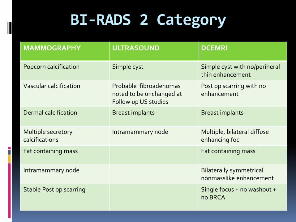
BI-RADS 2 Category
MAMMOGRAPHY ULTRASOUND DCEMRI
Popcorn calcification Simple cyst Simple cyst with no/periheral thin enhancement
Vascular calcification Probable fibroadenomas noted to be unchanged at Follow up US studies
Post op scarring with no enhancement
Dermal calcification Breast implants Breast implants
Multiple secretory calcifications
Intramammary node
Multiple, bilateral diffuse enhancing foci
Fat containing mass Fat containing mass
Intramammary node
Bilaterally symmetrical nonmasslike enhancement
Stable Post op scarring Single focus + no washout + no BRCA

BI-RADS 3 Category; Probabily benign
MAMMOGRAPHY ULTRASOUND DCEMRI
Circumscribed mass on a baseline mammogram
Complicated cyst
Abnormal focal enhancement likely due to hormonal influence
Focal asymmetry Breast abscess
Solitary group of punctate calcifications
Solid mass with circumscribed margins, an oval shape, and parallel orientation

Management of BIRADS 3 lesions Short interval follow-up imaging every 6 months for 2 years (watchful waiting)
95%, no growth, bx is avoided
2-3 %, a benign process grows, and requires bx to establish
its nature
1-2 % are cancer, growth is seen on follow-up, and bx initiates treatment. Although some time is lost in the process, the cancers found at follow-up are usually still small and curable.
Biopsy may be performed in selected cases (patient preference or overriding clinical concern)

BI-RADS 3… Palpable vs nonpalpable masses
Graf O, Helbich TH, Fuchsjaeger MH, et al.
Follow-up of palpable circumscribed noncalcified solid breast masses at mammography and US: can biopsy be averted? Radiology 2004; 233(3):850–856.
Harvey JA, Nicholson BT, LoRusso AP, et al.
Short-term follow-up of palpable breast lesions with benign imaging features: evaluation of 375 lesions in 320 women. AJR 2009; 193(3):1723–1730.
Palpable noncalcified solid breast masses with benign morphology at mammography and US can be managed similarly to nonpalpable BI-
RADS category 3 lesions, with short-term follow-up (6-month intervals for 2 years)

BI-RADS 4 Category; Suspicious for malignancy
BI-RADS 4A BI-RADS 4B BI-RADS 4C
Solid mass partially circumscribed with sonographic features suggestive of a fibroadenoma
Complex cystic and solid masses
Solid, irregular masses with illdefined margins
Complicated cyst or a breast abscess
Intraductal masses
Recent cluster of microcalcifications
Radio-pathological correlations should be precise: a partially circumscribed mass with ill-defined margins with
a fibroadenoma result is acceptable, but in case of a diagnosis of papillary lesion, surgery should be proposed.
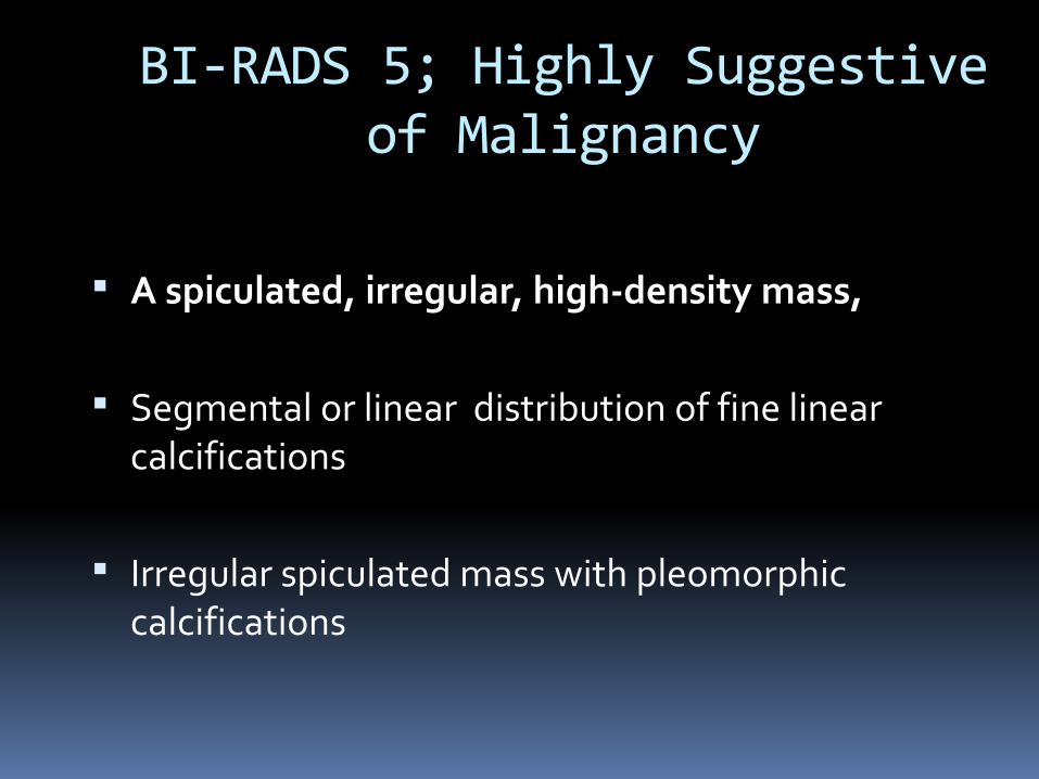
A spiculated, irregular, high-density mass,
Segmental or linear distribution of fine linear calcifications
Irregular spiculated mass with pleomorphic calcifications
BI-RADS 5; Highly Suggestive of Malignancy

BI-RADS Category 6; Known Biopsy-Proven Malignancy
Biopsy proven malignancy (imaging performed after percutaneous biopsy but prior to complete surgical excision), in which there are no mammographic abnormalities other than the known cancer that might need additional evaluation.

Microcalcifications
Perception of microcal is easy but…..
Characterization is difficult
Trick is to establish the location › Ducts----- Mostly malignant
› Lobule-----Mostly benign
› Outside the TDLU-------Definitely Benign
Morphology and distribution provide clues to the location of microcal

Microcalcifications Decoded
Descriptor PPV for Malignancy
Appropriate BI-RADS Assessment
Category
Coarse heterogeneous
7-50 % 4A
Amorphous 13- 26% 4B
Fine pleomorphic 28- 40% 4B
Fine Linear branching
53- 80% 4C

How to Categorize Cysts?
High Resolution US should be performed

43 y/o with palpable lump
BIRADS 0

Local tenderness, malaise on questioning
Thick walled Complicated cyst
BIRADS 3
What BIRADS 3 or 4?

45y/o with lump in the left breast
BIRADS 0

BIRADS 4c

Simple vs Complicated vs Complex cyst
Cyst should be judged with the worst features Thick internal septations
Mural solid nodules
Microlobulated margins
Fibrovascular stalk
20% of complex cysts are malignant
BIRADS 4b/ 4c
VAB or Surgical excision more appropriate

Scenario 1
Mammography; Incomplete” (BI-RADS® category 0) assessment due to an asymmetry recommending additional US examination
US examination then is performed showing no abnormal findings
Is it appropriate to also assess the US examination as BI-RADS® category 0, recommend additional MRI
examination?

Scenario 1
If diagnostic mammography is performed concurrently with US, an overall BI-RADS® assessment category should be given
BI-RADS® category 1/ BI-RADS® category 2/ BI-RADS® category 3/ BI-RADS® category 4
• BI-RADS® category 0 should not be used for diagnostic breast imaging findings that warrant further evaluation with MRI
• Incorporate this recommendation into the patient management recommendations in the combined mammography/US report.

Scenario 2
Axillary adenopathy is seen at screening mammography with no suspicious findings in the breasts.
What should the BI-RADS® final assessment be?

Scenario 2
In the absence of a known infectious or inflammatory cause, isolated unilateral axillary adenopathy should receive a suspicious (BI-RADS® category 4) assessment
If a benign cause is elucidated, a benign (BI-RADS® category 2) assessment would be appropriate.
Bilateral axillary adenopathy would be assessed as benign (BI-RADS® category 2) in some situations and as suspicious (BI-RADS® category 4) in others.

Scenario 3
A woman in her 20s discovered a palpable breast mass. Imaging shows probable fibroadenoma.
What should the assessment be? Is biopsy always necessary?

Scenario 3
Probably benign (BI-RADS category 3)
Recommend follow up imaging, unless the woman prefers biopsy or even excision if the mass is cyclically painful
Even if biopsy is done for this category 3 lesion, the probably benign assessment should not change.

Report Structure… Ultrasound Indication for examination
Statement of scope and technique of breast US examination
Succinct description of the overall breast composition (screening only)
Clear description of any important findings
Comparison to previous examination(s), including correlation with physical, mammography, or MRI findings
Composite report
Assessment
Management

Tissue Composition (Screening)
Homogeneous background echotexture-fat
Homogeneous background echotexture-fibroglandular
Heterogeneous background echotexture

Labeling and Measurement
The longest horizontal dimension followed by the vertical measurement, and the anteroposterior last (ML x SI x AP)

oThe longest horizontal dimension followed by the vertical measurement, and the anteroposterior last (ML x SI x AP)
oClock face location and distance from the nipple.
oDistance from skin and chest wall
o 17x26x12mm (ML x SI x AP)
o9’0 C , 2cm from the nipple
o 11 mm from skin and 18 mm from chest wall
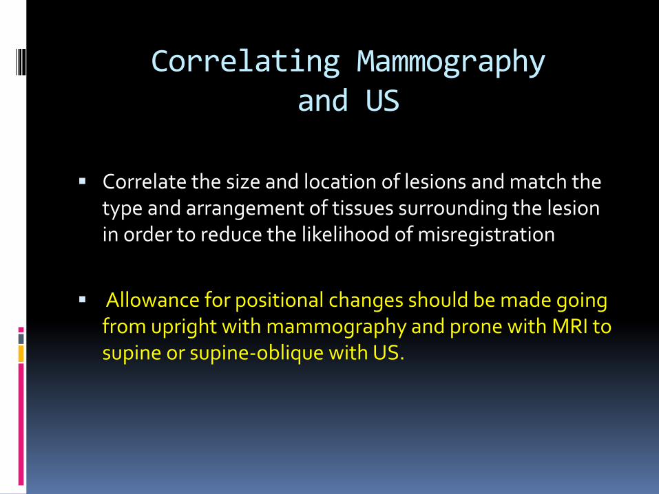
Correlating Mammography and US
Correlate the size and location of lesions and match the type and arrangement of tissues surrounding the lesion in order to reduce the likelihood of misregistration
Allowance for positional changes should be made going from upright with mammography and prone with MRI to supine or supine-oblique with US.

If a sonographic finding corresponds to a palpable abnormality, or to a mammographic or MRI finding, this should be stated explicitly in the US report.
If the US finding is new or has no correlate, this should also be stated in the report.
In a follow up US, the current report should describe any changes. An increase of 20% or more in the longest dimension of a probably benign solid mass within 6 months may prompt biopsy.
An increase of only 1–2 mm in lesion size may be related to differences in scanning technique or patient
positioning.

Exceptions to this rule occur when the characteristically benign features on one examination supersede the less specific benign features on the other examination. Eg. partially circumscribed, noncalcified mass at mammography, superseded by simple cyst at US.
When more than one type of examination is performed the examinations be reported together with an overall assessment and management recommendations
The overall assessment (and concordant management recommendations) should reflect the more abnormal of the individual assessments
Composite Reports

Scenario 4
A woman undergoes breast US examination to evaluate spontaneous bloody nipple discharge, and I see a mass within a duct.
How do I describe this using the BI-RADS® lexicon?

Scenario 4
•Intraductal location •Size •Presence of vascularity •Clock face position •Distance from the nipple •Length of the duct segment that contains the mass

Scenario 5
A 32-year-old woman presents with a large, painful breast mass. Her US shows an abscess. Aspiration is performed for culture sensitivity and relief of symptoms.
What assessment and management recommendations should be provided in the breast imaging report?
Benign (category 2) assessment
A management recommendation of aspiration should be made

Scenario 6
A circumscribed mass in a 42-year-old woman recorded as right breast, 10 o’clock, 5 cm posterior to the nipple. She returned for a 6-month follow-up US, and the mass was now seen located at 11:00 in the right breast 6 cm posterior to the nipple.
How should lesion location be reported on the follow-up US?

Scenario 6
Due to minor differences in both patient positioning and angles of insonation difficult to precisely duplicate the scanning conditions of a previous examination
Ensure that the mass depicted on both examinations is one and the same.
Images labeled either precisely as on the previous examination or as actually located on the current examination.
If the current actual location is used in labeling, and if there is a slight difference the report could state, “The right breast mass seen previously at 10:00 position, 5 cm posterior to the nipple is the same mass seen on today’s exam in the right breast at 11:00 position, 6 cm posterior to the nipple, the minor difference being due to variability in patient positioning.”

Scenario 7
For bilateral screening US performed with no abnormality identified, what images should I
record?
Record one image in one plane (ordinarily radial) for each quadrant, and record one image of the retroareo-lar region just behind the nipple.