EvEnt-RElatEd BRain PotEntials (ERP): an ovERviEw
20
24 Volume 7 Número 2 Dezembro 2011 Neurociência da Linguagem EVENT-RELATED BRAIN POTENTIALS (ERP): AN OVERVIEW Aline da Rocha Gesualdi (CEFET-RJ) e Aniela Improta França (UFRJ) ABSTRACT This text offers a practical overview of biological and technological aspects of an electromagnetic cognitive assessment tool named Event-Related Brain Potential (ERP), focusing on its application to language research. Because of its high temporal resolution (milliseconds), the ERP is considered an accurate measure for cognitive research, and has been widely used in neuroscience of language since the 1980’s. The text intends to raise the most relevant questions about the technique, while discussing the major interdisciplinary issues that arise with its use. KEY WORDS: EEG-ERP technique, cognitive research, neuroscience of language 1. INTRODUCTION: FROM THE WORLD EVENTS TO THE ELECTRICAL SIGNALS AND THEIR TRANSDUCTION IN AND OUT OF THE BRAIN Over the last 40 years, the field of neuroscience of language has been making clear progress with the introduction of non-invasive brain research techniques. Among them, the Event-Related Brain Potential (ERP) stands out as a very precise cognitive measurement that is low-cost and relatively easy to handle. This measurement usually entails the formation of a multidisciplinary team of researchers, including linguists, signal processing engineers, speech therapists, psychologists, neuroscientists, among others. The ERP provides a continuous sampling of the brain’s electrical activity. This sampling can be related to different sorts of linguistic phenomena produced in the cortex of volunteers performing linguistic tasks, while monitored by the electroencephalogram – EEG (Osterhout, Holcomb, 1995). The ERP involves a signal amplification that adds up and averages specifically time-locked epochs 1 that may present a lower signal-to-noise ratio (SN) 2 than those of the original waveforms (Figure 1). Figure 1: The averaging of several EEG segments into the averaged ERP waveform 1. Replications of a stimulus. 2. A signal is information that persists in response to a stimulus. Noise may be randomly generated. In signal processing the aim is to eliminate all the noise that comes mixed with the signal so that only the signal persists. Thus, the signal to noise ratio is a quality measure to sinal processing. It is defined as the ratio of signal power to the noise power. A ratio higher than 1:1 indicates more signal than noise. GESUALDI, Aline da Rocha; FRANÇA, Aniela Improta. Event-related brain potentials (ERP): an overview. Revista LinguíStica / Revista do Programa de Pós-Graduação em Linguística da Universidade Federal do Rio de Janeiro. Volume 7, número 2, dezembro de 2011. ISSN 1808-835X 1. [http://www.letras.ufrj.br/poslinguistica/revistalinguistica]
Transcript of EvEnt-RElatEd BRain PotEntials (ERP): an ovERviEw

24Volume 7 Número 2 Dezembro 2011Neurociência da Linguagem
EvEnt-RElatEd BRain PotEntials (ERP): an ovERviEw
Aline da Rocha Gesualdi (CEFET-RJ) e Aniela Improta França (UFRJ)
AbstrActThis text offers a practical overview of biological and technological aspects of an electromagnetic cognitive assessment tool named Event-Related Brain Potential (ERP), focusing on its application to language research. Because of its high temporal resolution (milliseconds), the ERP is considered an accurate measure for cognitive research, and has been widely used in neuroscience of language since the 1980’s. The text intends to raise the most relevant questions about the technique, while discussing the major interdisciplinary issues that arise with its use.
Key words: EEG-ERP technique, cognitive research, neuroscience of language
1. intRoduction: fRom thE woRld EvEnts to thE ElEctRical signals and thEiR tRansduction in and out of thE BRainOver the last 40 years, the field of neuroscience of language has been making clear progress with the introduction of non-invasive brain research techniques. Among them, the Event-Related Brain Potential (ERP) stands out as a very precise cognitive measurement that is low-cost and relatively easy to handle. This measurement usually entails the formation of a multidisciplinary team of researchers, including linguists, signal processing engineers, speech therapists, psychologists, neuroscientists, among others. The ERP provides a continuous sampling of the brain’s electrical activity. This sampling can be related to different sorts of linguistic phenomena produced in the cortex of volunteers performing linguistic tasks, while monitored by the electroencephalogram – EEG (Osterhout, Holcomb, 1995). The ERP involves a signal amplification that adds up and averages specifically time-locked epochs1 that may present a lower signal-to-noise ratio (SN)2 than those of the original waveforms (Figure 1).
Figure 1: The averaging of several EEG segments into the averaged ERP waveform
1. Replications of a stimulus. 2. A signal is information that persists in response to a stimulus. Noise may be randomly generated. In signal processing the aim is to eliminate all the noise that comes mixed with the signal so that only the signal persists. Thus, the signal to noise ratio is a quality measure to sinal processing. It is defined as the ratio of signal power to the noise power. A ratio higher than 1:1 indicates more signal than noise.
GESUALDI, Aline da Rocha; FRANÇA, Aniela Improta. Event-related brain potentials (ERP): an overview. Revista LinguíStica / Revista do Programa de Pós-Graduação em Linguística da Universidade Federal do Rio de Janeiro. Volume 7, número 2, dezembro de 2011. ISSN 1808-835X 1. [http://www.letras.ufrj.br/poslinguistica/revistalinguistica]

25Volume 7 Número 2 Dezembro 2011Neurociência da Linguagem
The ERP takes advantage from the fact that electromagnetic activity can be sensed at the scalp by the EEG (electroencephalographer), developed by a German Psychiatrist, Hans Berger (1873 — 1941; Figure 2) and put to use in humans since 1929.
Figure 2: German Psychiatrist Hans Berger
While providing poor spatial resolution, the EEG has outstanding temporal precision, to the order of the millisecond time-scale. Thus, the ERP inherits this favorable time resolution from the EEG and is considered an on-line measure of microscopic cognitive activity that takes place in the cortex. Nevertheless, the EEG and the ERP are not a direct measure of the world events in the brain. As the acronym reveals, ERPs are just event-related. One thing is the event out there in the world, emitting an infinite wealth of analog signals3; another, is how the human brain represents and processes this event within its cognitive limitations. In order to process stimulation from world events, the human brain uses an electrical propagation system that is based on the neuron, which is an excitable cell (Figure 3).
Figure 3: The neuron: an electrically excitable cell being monitored by a recording electrode that plots the flow of electrical current across the cell membrane over time. Notice that electrical current can be incremented ( examples A
and B) or inhibited (example C), and when it goes above threshold, it fires.
The analog events captured by the sensory nerves are readily transformed into digital signals4 that travel from one neuron to the next. This analog to digital transformation implies in a format change which promotes a huge information reduction, from the infinite possible value of the analog system
3. An analog system is one that encodes information as a continuum. There is no inherent limit to values that can be assigned at any point. In fact, information in the analog system is infinite. 4. A digital system is one in which information has discrete values. For instance, in Figure 2, there is a threshold line. Any stimulus below threshold does not generate an action potential. But any stimulus above threshold generates the same action potential. So there are two discrete possibilities only, one below and the other above the threshold.

26Volume 7 Número 2 Dezembro 2011Neurociência da Linguagem
to the two possible values of the digital system: zeros and ones. This means that when our ears, eyes, skin or tongue capture the physical energy of a world event, this event is not interpreted on the basis of this physical energy, be it acoustic waves, light intensity, degrees of pressure and temperature or the kinds of ingested element, such as minerals, food etc. The different kinds of stimuli the brain can capture, go through a stimulus transduction process5 that unifies a wide range of sensory information into a unique binary neural code in the brain.
When a neuron fires, it conducts a signal, named action potential, from the cell body down its axon. No matter the strength of the signal, if it exceeds the threshold, the neuron fires and the value of its action potential is 1. With any signal below threshold, the neuron rests - doesn’t fire - and its value is zero. Thus, it is a digital, all-or-none process.
This transduction process converts two aspects of the infinite information carried in an analog wave, its amplitude and its frequency (Figure 4).
Figure 4: Two aspects of the analog data: (i) in the top graph, its vertical dimension that is amplitude, projected on the y-axis; and (ii) in the bottom graph, the number of cycles in a given time frame, frequency, projected on the x-axis.
The amplitude is converted on the y-axis and the process is named discretization (Figure 5; Cf. 3.0).
Figure 5: Example of signal discretization. On the top graph we have the analog signal. The middle graph is a train of impulses distant T seconds from each other, and the bottom graph is the discretized signal which is the multiplication of
the top and middle signals.
5. Two examples of stimulus transduction: (i) transduction of auditory stimuli takes place at the cochlear hair cells, which are the recep-tor cells for hearing. The analog vibration of the basilar membrane corresponding to the mechanical air rarefaction that enter the ear is transformed into spike trains: hair cells on the basal part of the membrane are stimulated by high frequencies, while hair cells on the apical part of the membrane are stimulated by low frequencies; (ii) transduction in the visual system takes place at the retina which has sensory cells that convert the intensity of signals into signals that also travel through the axons in digital spike trains.
27Volume 7 Número 2 Dezembro 2011Neurociência da Linguagem
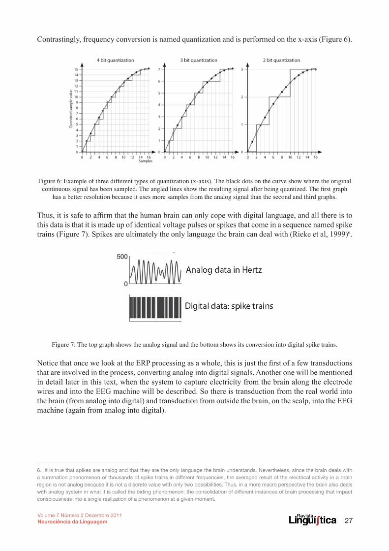
Contrastingly, frequency conversion is named quantization and is performed on the x-axis (Figure 6).
Figure 6: Example of three different types of quantization (x-axis). The black dots on the curve show where the original continuous signal has been sampled. The angled lines show the resulting signal after being quantized. The first graph
has a better resolution because it uses more samples from the analog signal than the second and third graphs.
Thus, it is safe to affirm that the human brain can only cope with digital language, and all there is to this data is that it is made up of identical voltage pulses or spikes that come in a sequence named spike trains (Figure 7). Spikes are ultimately the only language the brain can deal with (Rieke et al, 1999)6.
Figure 7: The top graph shows the analog signal and the bottom shows its conversion into digital spike trains.
Notice that once we look at the ERP processing as a whole, this is just the first of a few transductions that are involved in the process, converting analog into digital signals. Another one will be mentioned in detail later in this text, when the system to capture electricity from the brain along the electrode wires and into the EEG machine will be described. So there is transduction from the real world into the brain (from analog into digital) and transduction from outside the brain, on the scalp, into the EEG machine (again from analog into digital).
6. It is true that spikes are analog and that they are the only language the brain understands. Nevertheless, since the brain deals with a summation phenomenon of thousands of spike trains in different frequencies, the averaged result of the electrical activity in a brain region is not analog because it is not a discrete value with only two possibilities. Thus, in a more macro perspective the brain also deals with analog system in what it is called the biding phenomenon: the consolidation of different instances of brain processing that impact consciousness into a single realization of a phenomenon at a given moment.

28Volume 7 Número 2 Dezembro 2011Neurociência da Linguagem
2. Biological undERPinings: micRo PERsPEctivEsIn order to understand the micro components of these highly complex operations it is important to be aware of some basic biological foundations. On the microscopic level, the brain is constituted by two cell types: neuroglia (glia, the Greek for glue) and neurons. Neuroglia are about fifty times more numerous than neurons in the brain. Neuroglial cells, or simply glial cells, are peculiarly branched cells arranged in a fine web of tissue. Traditionally, they were only regarded as the supporting structure of nervous tissue. However, neuroglia are now known to be involved in neuronal growth and migration. There are three types of glial cells in the brain: astrocytes, oligodendrocytes and microcytes (Figure 8).
Figure 8: The three types of glial cells: astrocyte, microglia, oligodendrocytes
Astrocytes are star-shaped cells that, besides a major supportive role in the central nervous system, are also recruited in the formation of the network on which neurons grow. Filling in the space within neurons, astrocytes hold neurotransmitters, that are released by neurons, and provide part of the fundamental control of chemical concentrations in extracellular space. Astrocytes also clean up brain waste and digest dead neurons. More recently, these cells have been connected with the controversial issue of adult neurogenesis, that is, the growth of new neural tissue. Oligodendrocytes (astroglia and oligodendroglia) are involved in myelin formation, and this functions as insulation to the nerve fibers. It also provides transport of material to neurons and the control of the ionic environment of neurons. Microcytes (microglia) are the smallest of the glial cells. They phagocytize waste products of nerve and play an important role in the immune system of the brain by protecting it from invading microorganisms.
The other type of cell in the brain is its primary functional unit: the neuron. Until recently, the official estimate was that the brain held 100 billion neurons. This figure was challenged by research with more advanced cell count techniques that estimated the cell count of 100 billion just for the cerebellum (Andersen, Korbo and Pakkenberg, 1992). Neurons operate in large sets forming neuronal circuits or neural nets. The most striking feature of these cells is that they produce electrical signals that operate as information bits. All data reaching or leaving the body is transformed into electrical signals that are transported and processed by neurons. Each neuron can be divided into three parts: dendrites, cell body or soma, and axons (Figure 9).

29Volume 7 Número 2 Dezembro 2011Neurociência da Linguagem
Figure 9: The neuron
The cell body contains the nucleus and other cell organelles such as mitochondrion, rough and smooth endoplasmatic reticula, lysosomes, ribosomes and Golgi complex, that are responsible for cell metabolic maintenance. Departing from the cell body are dendrites and axons: cell extensions, also named processes. Dendrites are tree-like structures that collect electrical signals from other nerve cells. Again, neurons are electrically excitable and are known to be able to transmit this excitation. So, an electrical signal reaches a dendrite, goes through the cell body, which generates outgoing signal down the axon in the form of the action potential mentioned above.
3. Biological undERPinings: macRo PERsPEctivEsFrom micro to macro, in the cranium vault the neuron and glial cells bundled together form two layers of brain tissue that fill the cerebral cavities. The outermost layer is the gray matter. Under it lies an inner core of white matter, a much thicker layer.
The gray matter is a vital 2 to 6 mm layer of tissue known as the cerebral cortex, the Latin word for bark (Figure 10). It is structured by nerve cells and fibers of different sizes and shapes, but it is specially rich in nerve cell bodies that are gray to the naked eye. Of all the cerebral cortex, 90% is neocortex, involved in higher functions in humans, such as sensory perception, generation of motor commands, spatial reasoning, conscious thought and language.
Figure 10: The gray and white matters

30Volume 7 Número 2 Dezembro 2011Neurociência da Linguagem
Most cognitive processing takes place in the neocortex. This tissue is accommodated within the limits of cranium vault through intense folding, forming bulges and creases, named respectively gyri and sulci, which are the plural for gyrus and sulcus (Figure 11).
Figure 11: The cortex, an enlarged detail from the left parietal lobe
The convoluted surface of the gyri and sulci enlarges the extension of the sheet of gray matter available for all this processing. Resulting from this is the possibility for the human being to compute incredibly large quantities of information. Other animal species do not have the same amount of folding as we do and, therefore, have more limited processing space.
The white matter composes the deeper parts of the brain. Despite the fact that it is also formed by nerve cells, it is essentially different from the cortex, because it is richer in nerve cell axons, arranged in bundles or fibers. These fibers are sheathed with myelin, an insulating layer made up of glial cells, rich in protein and fatty substances that are white in color to the naked eye. The purpose of the myelin sheath is to allow rapid and efficient transmission of impulses along the nerve cells. There are three types of fibers in the white matter: (i) association fibers, that interconnect structures within the same hemisphere; (ii) projection fibers, that unite the hemispheres to the lower parts of the brain; and (iii) commissural fibers, that connect the two hemispheres. While the white matter takes over information flow, the gray matter is mostly connected with information processing.
4. communication within thE BRainResorting to a between-neuron transmission system, named synapse, the action potential can travel at high speed along an incredible network of neurons. And there are two kinds of synapses: electrical and chemical. The electrical synapse is the direct connection between two neurons that are very close to each other, so there can be direct transfer of the electrical signal. In fact, in the narrow gap between the two cells, there is a junction of protein that allows ions to go from the cytoplasm of one cell to that of the next. Although the electrical synapse is much less common in mammals, they can be found in human fetuses, in early embryonic neuronal development, and in adults, between non-neural cells and in cardiac, glandular and liver cells.
Chemical synapses are highly complex information systems about which whole books have been written and a lot is still unknown. But, in essence, the chemical mechanism that was first described in

31Volume 7 Número 2 Dezembro 2011Neurociência da Linguagem
the early 1900’s, by the Austrian neuroscientist Otto Loewi, consists of a contact zone between two neurons: action potential to the axonal buttons triggers the release of molecules of neurotransmitters that are kept in synaptic vesicles. These molecules reach the synaptic cleft, that is, the contact zone with the next neuron, and are absorbed by receptors, ion channels, placed at the postsynaptic membrane of the dendrites of the next cell, that open for the active synapse and close for the resting synapse (Figure 12).
Figure 12: The chemical synapse
Depending on the neurotransmitter released, the action potential that will flow to the next neuron may be propagated, blocked or modified. This means that the neurotransmitters modulate the electrical bits being transmitted along the neuron.
Taking a closer look at ion channels, it is important to point out that they are protein tunnels located on the lipid bilayer of the neuron plasmatic membrane. They make a fundamental cell system responsible for several biological processes, from excitation to restriction, signaling to deletion and absorption to excretion. These channels go through instant conformational changes that make them turn into gate-like structures that can open and close allowing up to 10 million ions to flow into or out of the cell at each second. By opening and closing their gates, ion channels get to be pretty selective as they act according to the type of ion (sodium, potassium, calcium) they allow to pass, and in which direction. (Figure 13).

32Volume 7 Número 2 Dezembro 2011Neurociência da Linguagem
Figure 13: Ion channels selectivity
Action potentials are ultimately generated by these special channels. When the channels are closed, the membrane potential is near its resting potential (Figure 14).
Figure 14: The cell in its resting potential, when the inside contains a positive charge in relation to the outside.
When the membrane potential increases to a precisely defined threshold value as high as –50 mV (from the resting potential of –70 mV), the gates open rapidly, allowing an inflow of sodium ions. As gates start opening, they trigger a gate-opening trend, which depolarizes the membrane bit by bit all along the cell. The process continues up to an apex when all ion channels are open. This causes a large upswing in the membrane potential (depolarization) which underlies the action potential (Figure 15).
Figure 15: The stimulated cell. When the action potential travels along the axon, the inside of the cell contains a more positive charge in relation to the outside.

33Volume 7 Número 2 Dezembro 2011Neurociência da Linguagem
After depolarization reaches its peak, a process of repolarization starts, in which sodium and potassium channels close, blocking positively charged ions from entering the cell. At the same time, negatively charged ions return to the cell. This causes a transient negative shift called the refractory period that prevents an action potential from traveling back the way it just came.
Figure 16: the refractory period
Since action potentials are all-or-nothing, they don’t vary in size, only in how often they occur. Thus, the only way that information such as the strength or duration of a stimulus can be encoded is by variation of the frequency of action potentials. Some axons can conduct action potentials at frequencies up to 1000 times per second.
After the action potential leaves the cell body, it is transmitted along the axon in a special way. Because the axon is coated by a myelin sheath (Figure 12), the action potential moves fast toward the end of the axon, that branches out. But to allow for even faster propagation, instead of allowing a steady electric flow of the action potentials, the sheath is interrupted in several locations exposing the neuron plasmatic membrane. These uninsulated locations are named nodes of Ranvier, which are capable of allowing ionic flow and the propagation of the electrical activity (Figure 17).
Figure 17: Node of Ranvier
So the action potential moves from a node of Ranvier to the next in a jumping manner, technically named saltatory conduction. This process not only makes conduction faster, but also helps to preserve the signal established by the chemical synapse.

34Volume 7 Número 2 Dezembro 2011Neurociência da Linguagem
Synapses are very productive processes. After one firing takes place, it takes one-thousandth of a second for the neuron to be ready to transmit another action potential. While it is ready to fire, the cell membrane is in resting potential (Gazzaniga ,1998).
Since all this is a fleeting process that can be reiterated indefinitely, and since the number and frequency of firings varies in relation to external stimulation, the action potentials function as digital codes that can be deciphered by cortical processing. So this is how the mind–brain relationship is established for cognition to take place.
5. monitoRing thE BRain with an EEgEEG signals are obtained through electrodes placed on the scalp over multiple areas of the brain and connected on one tip to the EEG amplifier and on the other to a computer screen that shows the electrical activity at each electrode point or derivation, as it is technically called (Figure 18).
Figure 18: Running an EEG.
Nevertheless, bioelectricity in the cortex is propagated not only through neurons but also throughout the brain. Before it reaches the more external parts of the brain where the electrodes may sense it, it encounters propagation barriers, such as non-neuronal tissues (meninges and bone) and the cerebral spinal fluid, which absorb electricity, diminish the strength and deviate the signal. Thus, when the spike reaches the cranium vault, the waves that can be sensed by the electrodes on the scalp are attenuated and have to be amplified in order to be studied. Another negative point about the propagation barriers is that the waves sensed at the scalp have poor spatial resolution, in the order of centimeters, so it is impossible to correlate the position of the electrode on the scalp with the precise spot in the brain, where the electrical signal was originated. Thus, although there is a very homogeneous distribution of electrodes, space precision cannot be reached.

35Volume 7 Número 2 Dezembro 2011Neurociência da Linguagem
6. distRiBution of thE ElEctRodEsThe electrical information captured and stored by the electroencephalogram (EEG) comes from various points at the scalp. These points are technically called derivations. Although there are several ways to distribute the electrodes at the scalp, the most common system is the International 10-207 (Figure 19)
Figure 19: EEG electrode positions
Since the electrical measurement does not have an absolute value, and depends on certain characteristics of the metal used in the electrode, it is essential that the activity at each electrode be measured in reference to a point in which the electricity is known to be inactive or almost inactive. This way it is possible to compare electrical measures taken by different EEGs.
One earlobe or both earlobes connected are commonly used as the reference channel to each derivation because they are close to the where the other electrodes will be placed and they are electrically inactive (Luck, 2005; Figure 20).
Figure 20: The earlobe electrode as a common reference to every derivation.
7. This system is based on the relationship between the location of an electrode and the underlying area of cerebral cortex. The “10” and “20” refer to the fact that the actual distances between adjacent electrodes are either 10% or 20% of the total front-back or right-left distance of the skull.

36Volume 7 Número 2 Dezembro 2011Neurociência da Linguagem
The electrical signal the EEG captures comes from the brain, and, as stated elsewhere, travels through the brain in a digital fashion. However on the outside of the brain, at the scalp, where the electrode is placed, the signal arrives as an analog value. This can be explained because the electrical activity that can be sensed by the electrode tip at the scalp does not come from one single neuron. Rather, it is the average activity of aggregates of millions of neurons. In fact, the EEG performs the recording of the temporal and spatial summation of all excitatory and inhibitory post-synaptic potentials in the brain along the time (Lopes da Silva, 1999).
7. thE EEg’s tissuE-ElEctRodE intERfacEThe brain is immersed in cerebro-spinal fluid and encased in a bone structure, and it thus separated separated from the scalp. So the electrode’s pick up point at the scalp cannot be accurately connected to a point at the cortex, but rather to a small area where there is summation of electrical activity from thousands of neurons. Thus, this value out of the brain is necessarily analog again. This moment in which digital information in the brain is transformed into analogical data traveling along the electrode wire is the second transduction mentioned in 2.0. This transfer from the ionic conduction to electronic conduction, that is signal discretization - takes place at the tissue-electrode interface (Strong, 1970).
Working with this interface is not trivial. Firstly, it is necessary to create a chemical biding condition between the skin and the electrode, and this is achieved through the use of a saline electrode cream or gel (electrolyte) that should be applied at each derivation. Before applying the gel, it is also advisable to scrape the skin lightly with sandpaper or with a pad soaked in acetone in order to remove the first three skin sub-layers which constitute the epidermis. The epidermis is a barrier that holds and attenuates the signal and provokes high impedance8 at high frequencies. Note that the procedure of removing the three layers is not invasive, because the epidermis gets constant renovation.
Secondly, it is necessary to diminish the electrode motion which may create a mechanical disturbance in the ionic distribution resulting in artifact9. Again the gel is a good solution to stabilize the electrode at the scalp. Whenever it is too dry or the gel layer is too thin, there are more losses and distortions in the eletrode- electrolyte relationship, specially concerning the low-frequency waves, which are exactly the waves of interest to the language researcher (Neuman, 1998)
Granted that the skin-electrode interface is well maintained, the EEG captures an averaged analog value referenced to the common electrode (earlobe) and amplifies it so that it can be processed more easily (Figure 21).
Figure 21: Unipolar EEG recording configuration
8. It is a measure of the overall opposition of a circuit to current, in other words: how much the circuit impedes the flow of current.9. Undesired signal distortion

37Volume 7 Número 2 Dezembro 2011Neurociência da Linguagem
The raw data amplified also goes through two filters to define the bottom and upper limits of the region of interest. For the bottom limit, a low-pass filter is used to eliminate unstable signals under 0.1 Hz (Luck, 2004). Sometimes this activity results from unwanted cross-talking among electrodes. For the upper limit, a high-pass filter is used at 100Hz. Another filter – notch – comes built-in to eliminate all waves exactly at 60Hz which coincides with the electrical net frequency. In sum, these are the three filters applied during signal acquisition, right before the signal is discretized and quantized (Figure 22).
Figure 22: Ideal representation of the three filters applied to the signal
8. using thE ERP to monitoR cognitivE activity Throughout its history, the EEG has been more commonly used in the diagnosis of diseases such as seizure disorders, mental confusion, and in the evaluation of head injuries, tumors, infections, degenerative diseases, and metabolic disturbances that affect the brain. It is also used to evaluate sleep disorders and to investigate periods of unconsciousness or to confirm brain death in a comatose patient. In fact, conditions such as epilepsy, attention deficit disorder, dyslexia, sleep disorders and many others can be accurately characterized in terms of the bioelectric wave patterns displayed by the EEG (Bear, Connors, Paradiso, 2001).
Differently from the raw EEG data, collected mainly from the brain at rest or during sleep, ERP data does not come spontaneously. It can be extracted if it is time-locked to stimuli that will be related to the resulting wave. ERPs are the summation of synchronized postsynaptic activity of a large population of neurons recorded at the scalp as small voltage fluctuations in the EEG are time-locked to sensory or motor stimuli (Donchin & Coles, 1991).
But not every the electrical activity from a neuron is perceived by the electrodes. The ERP is a resulting wave mainly from the summation of the activity of pyramidal cells, the largest and more numerous neocortical cell type. Pyramidal neurons account for up to 3/4 of all neocortical cells.
In more detail, ERPs can be extracted from certain neocortical regions, where excitatory input to pyramidal neurons result in a net negativity in the region of the apical dendrite and a positivity in the area of the soma (Figure 23). The voltage contrast between these two regions of the pyramidal neuron will turn it into tiny dipole. The ERP will be constructed from the summation of thousands of dipoles, which carry similar cortical orientation and synchronous activity (Luck, 2005).

38Volume 7 Número 2 Dezembro 2011Neurociência da Linguagem
Figure 23: The pyramidal cell
9. ERP ExPERimEnts in languagE REsEaRch: PREsEnting stimuli, tRiggERing, avERaging and gRand-avERaging REsPonsEsThe ERP technique implies in adding multiple occurrences of the signal (electrical data) in relation to a target phenomenon. For instance, taking ERP turned to language research, let’s suppose a volunteer is visually stimulated by 80 sentences with the same syntactic structure: a transitive verb with its object and subject. Each sentence has a noun phrase as subject, a verb phrase and a determiner phrase, appearing on the computer screen, phrase by phrase, for 200 ms each. In 50% of the tokens, these sentences will yield an incongruous reading exactly at the transition between the verb and the determiner (verb-complement merge). The remaining 50% of sentences will yield congruous readings, as can be seen below in Table (1).
Table 1: Experiment stimuli in groups: congruous Vs incongruous
The presentation of the stimuli in this experiment (Figure 24) previews a fixation cross appearing on the screen for 2000ms, then the three phrases will appear for 200 ms each. Finally, there will be a 1000 ms window for the volunteer’s response, probably the touch of a certain button if the sentence is congruous and another button if it is incongruous.
Figure 24: Presentation scheme
All 40 waves, picked by each electrode placed on the scalp, resulting from the verb-complement merge of the congruous sentences will be averaged, and so will those of the incongruous sentences. Since all 80 sentences have exactly the same structure and imagining that the frequency of the words used to form the stimulus will also be equivalent, then if the resulting averaged wave forms for the

39Volume 7 Número 2 Dezembro 2011Neurociência da Linguagem
congruous condition compared to that for the incongruous conditions, are significantly different from each other, then we can say that verb-complement incongruity causes a recognizable electrical effect which can be characterized by a certain wave, with a positive or negative polarity, with a given latency and amplitude.
Also since the stimuli were carefully manipulated to have only one contrasting aspect, in this case, the selection of the object by the verb, it is possible to identify exactly a region of interest in the EEG continuous sampling. This is done by placing a mark, named trigger, at a specific moment in time that will be exactly the same for every stimulus, marking the beginning of the region of interest. So we can program the epochs to contain EEG stretches that go from the trigger to for instance, 800ms. This way we only have to process the waves within that time window. Usually, triggers are square waves that are sent from the computer to the EEG apparatus, via the parallel port. So if the researcher assigns a trigger with a certain width to the congruous stimuli, and another width to the incongruous stimuli, it is possible to separate the regions of interest from the rest of the EEG continuous sampling and it is also possible to separate the conditions: large width to the congruous condition and narrow width to the incongruous, for instance. It is necessary to reserve one channel of the EEG to receive this information from the computer (Figure 25).
Figure 25: The raw EEG sampling per derivation and the triggers sent in the bottom channel.
Averaging the waves is a wise procedure beyond contrasting sets of stimuli: it also helps eliminate an unavoidable experimental problem. If each and every cognitive task the human being performs produces an electric response, then if the volunteer scratches the hand, adjusts to the chair, yawns or blinks during the EEG recording, exactly at the same time that she is processing the verb-complement merge, there will be an electric component resulting from this motor activity that will be contaminating the recording of the electrical activity related to the target stimulation. But since this intervening component, known as artifact, appears at random, not repeatedly associated with all the tokens of the target event, it simply gets cancelled by the linear summation and averaging of the multiple signals. So the averaged ERP wave is free from artifacts and can be analyzed in terms of its main parameters: a specific latency (the onset time of the peak), amplitude (how high the peak is) and topographic distribution (regions on the scalp where it is elicited).
40Volume 7 Número 2 Dezembro 2011Neurociência da Linguagem
Nevertheless, looking into the averaged data from one volunteer, that is, the summation of 40 repetitions of a phenomenon, at each electrode point of one volunteer, might still not result in a signal to noise ratio that can discriminate subtle linguistic phenomena the researcher wants to study. Thus, there is a supplementary way to amplify these differences and this is the grand-averaging of all responses. This means getting all 40 responses of one subject at one electrode point and averaging with those of each volunteer in your experiment, from 15 to 20 people, in most cases. The differences between conditions, if they exist at all, tend to surface more vividly after the grand-averaging. The reasoning behind this practice is that since these are fleeting phenomena, there would be very little difference between individuals in a control group (healthy individuals).
However, grand-averaging brings in some degree of controversy. Some researchers believe that there can be many individual differences among the electrical activities. They believe these differences are so big that grand-averaging should be avoided because it could distort the characteristics of the real potentials. Nevertheless, to be on the safe side and still use this technique, researchers might examine the standard deviation among volunteers. If it is irrelevant, then use grand-averaging as a resource to make ERP differences more vivid. This procedure will be discussed in more detail in the next chapter.
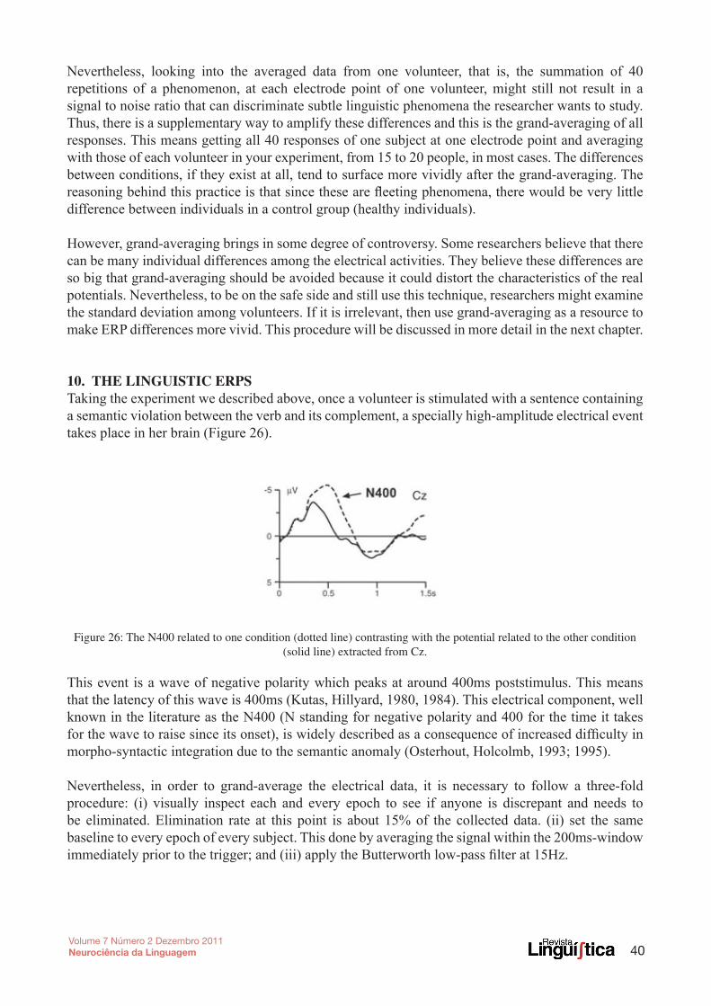
10. thE linguistic ERPsTaking the experiment we described above, once a volunteer is stimulated with a sentence containing a semantic violation between the verb and its complement, a specially high-amplitude electrical event takes place in her brain (Figure 26).
Figure 26: The N400 related to one condition (dotted line) contrasting with the potential related to the other condition (solid line) extracted from Cz.
This event is a wave of negative polarity which peaks at around 400ms poststimulus. This means that the latency of this wave is 400ms (Kutas, Hillyard, 1980, 1984). This electrical component, well known in the literature as the N400 (N standing for negative polarity and 400 for the time it takes for the wave to raise since its onset), is widely described as a consequence of increased difficulty in morpho-syntactic integration due to the semantic anomaly (Osterhout, Holcolmb, 1993; 1995).
Nevertheless, in order to grand-average the electrical data, it is necessary to follow a three-fold procedure: (i) visually inspect each and every epoch to see if anyone is discrepant and needs to be eliminated. Elimination rate at this point is about 15% of the collected data. (ii) set the same baseline to every epoch of every subject. This done by averaging the signal within the 200ms-window immediately prior to the trigger; and (iii) apply the Butterworth low-pass filter at 15Hz.

41Volume 7 Número 2 Dezembro 2011Neurociência da Linguagem
Note that these procedures may differ according to the cognitive task being analyzed. For language research the presented parameters are the most commonly used. The resulting ERP potential can finally be related to the cognitive activity.
Besides the N400, there are other well-known ERPs related to linguistic conditions: (i) ELAN (early left anterior negativity), that relates to the early (between 125 to 180 ms) perception that there is something wrong with word class choice, for instance John bought smiled; (ii) P600, that is connected with anomalous syntactic structure (Cf Gouvea’s How to examine the P600 using language theory: what are the syntactic processes reflected in this component in this volume, for an extensive review); and (iii) LPC (late positive component), a late positivity (between 500 and 800 ms) that relates to the irregular word formation of morphologically irregular words (for instance, fastly, cutted).
11. conclusion:For the electrical engineer, signal processing offers the building blocks to a number of research and multidisciplinary applications. Its main objective is to enhance the signal and downplay the noise in a way that the raw signal is minimally disturbed or modulated. In order to do that it is essential to distinguish between signal and noise by examining and processing the raw data.
Despite the fact that, technically, it does not seem to matter the kind of signal one wishes to detect - whether it comes from the motion of nano gold particles inserted in the bloodstream of patients to help uncover cancerous cells; or a pattern generated by a passive sonar set out to detect navy vessels; or the patterns generated at the collision site in a particle accelerator – be it any kind of signal, the interface between the professional performing the signal processing and the one providing expertise from the basic area that studies the signal must be clear and fertile. Otherwise interdisciplinary efforts might reduce science into technological redtape, which inevitably amplifies noise in detriment of signal.
Working with cortical signals related with language processing has proven to be a challenging mission for the signal processing professional. The signal is very much contaminated by several types of noise. Besides, the linguistic ERPs are the result of the summation of non-linear signals and this makes acquisition an exacting task. But for the engineer, the perspective of shedding light onto the human beings’ exclusive characteristic and the building blocks of thought structured by intricate syntax algorithms in the brain seems worth circumventing difficulties along the way.
On the other hand, for the traditional linguist, it might seem too dry and even useless to bother meddling with this level of technology presented here, even if this was nothing but a short and practical overview of the main aspects of Event-Related Brain Potentials. Mastering these techniques might also seem an achievement beyond a linguist’s grasp and a complete waste of time in view of so much linguistic literature to catch up with. After all, linguistics is rather complex on its own right. Nevertheless, how can one make the point that language is in the brain, taking the material aspects of this statement for granted?
If it is not for the sake of one’s investigative spirit, then for the mere need to understand what those machines are doing and how neuroscientists are doing things that get more and more frequently published in most linguistic magazines (even in those that are not known for their experimental facet), it is highly important to be technologically educated. And at the end of the day one might discover pleasure and light in the findings that this interdisciplinary practice might bring.

42Volume 7 Número 2 Dezembro 2011Neurociência da Linguagem
PotenciAis cerebrAis relAcionAdos Ao evento (erP): umA visão gerAl
REsumoEste texto oferece uma visão prática dos aspectos biológicos e tecnológicos da ferramenta eletromagnética de avaliação cognitiva chamada potencial relacionado a evento (ERP), com foco na sua aplicação na pesquisa de linguagem. Por causa de sua alta resolução temporal (milissegundos), o ERP é considerado um medida exata para a pesquisa cognitiva, e tem sido amplamente utilizada na neurociência da linguagem desde 1980. O texto tem a intenção de levantar as questões mais relevantes sobre a técnica, ao discutir os problemas interdisciplinares que possam surgir com a sua utilização.
PalavRas-chavE: a técnica EEG-ERP; pesquisa cognitiva; neurociência da linguagem
REfEREncEs:Andersen, B. B.; Korbo, l. ; Pakkenberg, B. (1992). A quantitative study of the human cerebellum with unbiased stereological techniques. Journal of Comparative Neurology 326(4): 549–60.
Bear, Mark; Barry Connors and Michael Paradiso (2001). Neuroscience: exploring the brain. Baltimore: Lippencott Williams & Wilkins. 666 pp.
Donchin, E., & Coles, M. G. H. (1991). While an undergraduate waits. Neuropsychologia, 29(6), 557-569.
Gazzaniga, Michael S.; Richard B. Ivry and George R. Mangun (1998). Language and the brain. In Cognitive neuroscience: the biology of the mind, New York: W. W. Norton, pp. 289–321.
Kutas, Marta and Steven A. Hillyard (1980). Reading senseless: brain potentials reflect semantic incongruity. Science, 207: 203–5.
Kutas, Marta and Steven A. Hillyard (1984). Brain potentials during reading reflect word expectancy and semantic association. Nature, 307 (5947): 161–163.
Lopes da Silva, Fernando H. and Van Rotterdam A. (1999) Biophysical Aspects of EEG and MEG Generation. In Niedermeyer, Ernst and Fernando H. Lopes da Silva (eds.) Electroencephalography, basic principles, clinical applications, and related fields. Baltimore-Munich: Urban & Schwarzenberg, 4ed., pp. 15-26.
Luck, S. J. (2004) Ten Simple Rules for Designing and Interpreting ERP Experiments. [In:] Event-Related Potentials: A Methods Handbook, Handy, T.C. (Ed.)
Luck, S. J. (2005). An introduction to the event-related potential technique. Cambridge, MA. MIT Press.
Neuman, MR. Biopotential amplifiers. In Medical Instrumntation: Application and design. J.G. Webster, Ed. 3rd Ed. New York, NY, Wyley, 1998, pp 183-232.
Osterhout, L; Holcomb, P. J. (1995) Event related potentials and language comprehension. Rugg, Michael D (Ed); Coles, Michael G. H (Ed). (1995). Electrophysiology of mind: Event-related brain potentials and cognition. (pp. 171-215). xii, 220 pp. New York, NY, US: Oxford University Press.

43Volume 7 Número 2 Dezembro 2011Neurociência da Linguagem
Osterhout L.; and Phillip J. Holcomb P. J (1993). Event-related potentials and syntactic anomaly: evidence of anomaly detection during the perception of continuous speech. Language and Cognitive Processes, 8(4): 413–37.
Rieke, F., Warland, D. van Stevenick, R., Bialek, W. (1999). Spikes: Exploring the neural code. A Bradford Book , MIT Press, Cambridge, Massachusetts, 395p.
Strong, P. (1970). Biophysical Measurements. Tektronix, Inc., Beaverton, Oregon.