Evaluationof Plasmodiumvivax ...
14
ORIGINAL PAPER Evaluation of Plasmodium vivax isolates in Thailand using polymorphic markers Plasmodium merozoite surface protein (PvMSP) 1 and PvMSP3 Nutnicha Suphakhonchuwong 1 & Wanna Chaijaroenkul 2 & Kanchana Rungsihirunrat 3 & Kesara Na-Bangchang 2 & Jiraporn Kuesap 1 Received: 14 June 2018 /Accepted: 1 October 2018 /Published online: 10 October 2018 # Springer-Verlag GmbH Germany, part of Springer Nature 2018 Abstract Malaria is a significant public health problem in several tropical countries including Thailand. The prevalence of Plasmodium vivax infection has been increasing in the past decades. Plasmodium vivax merozoite surface protein (PvMSP) gene encodes a malaria vaccine candidate antigen. Its polymorphic nature leads to antigenic variation, the barrier for vaccine development, drug resistance, and potential for multiple-clone infections within the malaria patients. The objective of this study was to investigate the genetic diversity of PvMSP1 and PvMSP3 gene in P. vivax populations in Thailand. A total of 100 P. vivax isolates collected from the western (Kanchanaburi and Tak Provinces) and southern (Ranong Provinces) regions along the Thai-Myanmar border were analyzed using polymerase chain reaction and restriction fragment length polymorphism (PCR-RFLP). Analysis of the F1, F2, and F3 regions of PvMSP1 revealed 5, 2, and 3 allelic variants, respectively. Three major types of PvMSP3-α and two major types of PvMSP3-β were identified based on the PCR product sizes. After digestion with restriction enzymes, 29, 25, 26, and 18 patterns were distinguished by RFLP for PvMSP1 (F2, Alu I), PvMSP1 (F2, Mnl I), PvMSP3-α, and PvMSP3-β, respectively. Combination of each family variant (PvMSP1 and PvMSP3) resulted in high genetic polymorphism of P. vivax population. Additionally, using PvMSP1 polymorphic marker revealed a significant association between multiple-genotype infections and P. vivax parasitemia. The results strongly supported that P. vivax populations in the endemic areas along the Thai-Myanmar border are highly diverse. Keywords Plasmodium vivax . Merozoite surface protein 1 (MSP1) . Merozoite surface protein 3 (MSP3) . Polymorphism Introduction Plasmodium vivax has replaced Plasmodium falciparum as the primary human malaria species in Thailand since 2008 (WHO 2016). In Thailand, the high prevalence of malaria is found near the international borders of Thai-Myanmar, Thai- Cambodian, Thai-Malaysian, and Thai-Laos. The control of P. vivax infection is limited by the relapsing nature of this malaria species due to its hypnozoite stage in the liver. Although resistance of P.vivax to the first-line drug chloro- quine has been reported in several parts of the world particu- larly South East Asia (Marlar et al. 1995; Thanh et al. 2015), there has been no confirmed case of chloroquine-resistant P. vivax in Thailand (Phyo et al. 2011). This may suggest the revolution of P. vivax in adapting its biology to overcome antimalarial action. Genetic analysis of P. vivax essential pro- teins is important as background information to guide emer- gence and development of drug resistance. Studies on the genetic diversity of P. vivax have been lim- ited due to the lack of optimal procedure for continuous culture Section Editor: Kevin S. W. Tan * Jiraporn Kuesap [email protected] Nutnicha Suphakhonchuwong [email protected] Wanna Chaijaroenkul [email protected] Kanchana Rungsihirunrat [email protected] Kesara Na-Bangchang [email protected] 1 Faculty of Allied Health Sciences, Thammasat University, Pathumthani, Thailand 2 Chulabhorn International College of Medicine, Thammasat University, Pathumthani, Thailand 3 College of Public Health Sciences, Chulalongkorn University, Bangkok, Thailand Parasitology Research (2018) 117:3965–3978 https://doi.org/10.1007/s00436-018-6106-1
Transcript of Evaluationof Plasmodiumvivax ...
ORIGINAL PAPER
Evaluation of Plasmodium vivax isolates in Thailand using polymorphicmarkers Plasmodium merozoite surface protein (PvMSP) 1 and PvMSP3
Nutnicha Suphakhonchuwong1& Wanna Chaijaroenkul2 & Kanchana Rungsihirunrat3 & Kesara Na-Bangchang2
&
Jiraporn Kuesap1
Received: 14 June 2018 /Accepted: 1 October 2018 /Published online: 10 October 2018# Springer-Verlag GmbH Germany, part of Springer Nature 2018
AbstractMalaria is a significant public health problem in several tropical countries including Thailand. The prevalence of Plasmodiumvivax infection has been increasing in the past decades. Plasmodium vivax merozoite surface protein (PvMSP) gene encodes amalaria vaccine candidate antigen. Its polymorphic nature leads to antigenic variation, the barrier for vaccine development, drugresistance, and potential for multiple-clone infections within the malaria patients. The objective of this study was to investigate thegenetic diversity of PvMSP1 and PvMSP3 gene in P. vivax populations in Thailand. A total of 100 P. vivax isolates collected fromthe western (Kanchanaburi and Tak Provinces) and southern (Ranong Provinces) regions along the Thai-Myanmar border wereanalyzed using polymerase chain reaction and restriction fragment length polymorphism (PCR-RFLP). Analysis of the F1, F2, andF3 regions of PvMSP1 revealed 5, 2, and 3 allelic variants, respectively. Three major types of PvMSP3-α and two major types ofPvMSP3-β were identified based on the PCR product sizes. After digestion with restriction enzymes, 29, 25, 26, and 18 patternswere distinguished by RFLP for PvMSP1 (F2,Alu I), PvMSP1 (F2,Mnl I), PvMSP3-α, andPvMSP3-β, respectively. Combinationof each family variant (PvMSP1 and PvMSP3) resulted in high genetic polymorphism of P. vivax population. Additionally, usingPvMSP1 polymorphic marker revealed a significant association between multiple-genotype infections and P. vivax parasitemia.The results strongly supported that P. vivax populations in the endemic areas along the Thai-Myanmar border are highly diverse.
Keywords Plasmodium vivax . Merozoite surface protein 1 (MSP1) .Merozoite surface protein 3 (MSP3) . Polymorphism
Introduction
Plasmodium vivax has replaced Plasmodium falciparum asthe primary human malaria species in Thailand since 2008(WHO 2016). In Thailand, the high prevalence of malaria isfound near the international borders of Thai-Myanmar, Thai-Cambodian, Thai-Malaysian, and Thai-Laos. The control ofP. vivax infection is limited by the relapsing nature of thismalaria species due to its hypnozoite stage in the liver.Although resistance of P.vivax to the first-line drug chloro-quine has been reported in several parts of the world particu-larly South East Asia (Marlar et al. 1995; Thanh et al. 2015),there has been no confirmed case of chloroquine-resistantP. vivax in Thailand (Phyo et al. 2011). This may suggest therevolution of P. vivax in adapting its biology to overcomeantimalarial action. Genetic analysis of P. vivax essential pro-teins is important as background information to guide emer-gence and development of drug resistance.
Studies on the genetic diversity of P. vivax have been lim-ited due to the lack of optimal procedure for continuous culture
Section Editor: Kevin S. W. Tan
* Jiraporn [email protected]
Nutnicha [email protected]
Wanna [email protected]
Kanchana [email protected]
Kesara [email protected]
1 Faculty of Allied Health Sciences, Thammasat University,Pathumthani, Thailand
2 Chulabhorn International College of Medicine, ThammasatUniversity, Pathumthani, Thailand
3 College of Public Health Sciences, Chulalongkorn University,Bangkok, Thailand
Parasitology Research (2018) 117:3965–3978https://doi.org/10.1007/s00436-018-6106-1
of the clinical isolates (Panichakul et al. 2007). Analysis ofgenetic markers is, therefore, a preferable approach to deter-mine antimalarial drug resistance, as well as to distinguishreinfection, relapse, and multiple infections (Kim et al. 2006;Kirchgatter and del Portillo 1998; Veron et al. 2009). Besides,these genetic analyses constitute the essential elements fordrug and vaccine development for malaria control. A numberof malaria genetic markers have recently been identified in-cluding apical membrane antigen 1 (AMA1), which encodes aprotein of all malaria species for red cell invasion (Kim et al.2006; Triglia et al. 2000), and Duffy binding protein (DBP)which encodes a protein on human red cells carrying Duffy-negative blood group (Hamblin and Di Rienzo 2000).However, both genes are not highly polymorphic and, thus,are not suitable for use as genetic markers for parasite diver-sity. The circumsporozoite surface protein (CSP) which en-codes a protein located on the surface of malaria in sporozoitestage (Imwong et al. 2005) is also not suitable to use as agenetic marker due to high variation in the immunodominantT cell epitopes of CSP (Imwong et al. 2005; Souza-Neiraset al. 2010).
Several merozoite surface proteins (MSPs) which encodeproteins located on the surface of P. vivax merozoites havebeen identified including PvMSP1, PvMSP3, PvMSP4,PvMSP5, PvMSP7, and PvMSP10 (Garzon-Ospina et al.2011; Gomez et al. 2006; Imwong et al. 2005; Putaporntipet al. 2009; Rungsihirunrat et al. 2011). Among these,PvMSP1 and PvMSP3 are highly polymorphic and may bereliably genetic markers for P. vivax diversity (Imwong et al.2005; Rungsihirunrat et al. 2011). The protein is essential forparasite invasion into human red blood cells. The PvMSP1gene is divided into 13 blocks; the highly variable blocks arelocated in the three regions, i.e., block 2 (F1 region), block 6–8 (F2 region), and block 10 (F3 region) (Imwong et al. 2005).It has been reported that antibody against PvMSP1 can inhibitP. vivax invasion to red cells, and this gene is proposed as apotential target for vaccine candidate (Ak et al. 1998;Espinosa et al. 2003). The PvMSP3 is also crucial for parasitered cell invasion; however, the inhibitory effect on red cellinvasion was not found using an antibody against PvMSP3.Ten ofPvMSP3multigene families have been identified (Jianget al. 2013); among these, three original members of PvMSP3have been demonstrated, i.e., PvMSP3-α, PvMSP3-β, andPvMSP3-γ (Galinski et al. 2001). Genetic analysis ofPvMSP3-α and PvMSP3-β has been performed in differentP. vivax endemic areas such as Bangladesh, China,Colombia, Korea, India, Pakistan, and Thailand (Bruce et al.1999; Imwong et al. 2005; Kang et al. 2014; Kibria et al. 2015;Kim et al. 2006; Li et al. 2015; Moon et al. 2009; Raza et al.2013; Rungsihirunrat et al. 2011; Yang et al. 2006). Bothgenes serve as reliable genetic tools to distinguish diverseP. vivax isolates and study population dynamics. The presentstudy aimed to investigate the genetic diversity of PvMSP1
and PvMSP3 in P. vivax isolates in malaria-endemic areas ofThailand using PCR-RFLP.
Material and methods
Sample collection
One hundred blood samples were collected during 2014–2015from patients with P. vivax infection (finger prick) whoattended malaria clinics in the western (Kanchanaburi andTak Provinces) and southern (Ranong Province) regions alongthe Thai-Myanmar border. Blood samples were spotted ontoglass slides for blood smear and filter papers for DNA extrac-tion. Thin and thick blood smears were prepared and exam-ined microscopically for the presence of P. vivax.Approval ofthe study protocol was obtained from the Human EthicalReview Board of Thammasat University (no. 110/2556).
DNA extraction
The genomic DNA of P. vivax was extracted from driedblood-spotted filter paper using DNA Geneius™ MicrogDNA Extraction Kit (Geneaid Biotech Ltd., New TaipeiCity, Taiwan) and stored at −20 °C until use.
Amplification of PvMSP1 polymorphisms
The three polymorphic regions of PvMSP1 (PvMSP1-F1,PvMSP1-F2, and PvMSP1-F3) were analyzed according tothe previously described method with modification (Imwonget al. 2005). The PvMSP1-F1 polymorphism was performedusing nested polymerase chain reaction (nested-PCR). Briefly,the reaction was performed in a total volume of 10 μlconsisting of 1× Taq buffered with KCl, 2 mM of MgCl2,0.125 mM of dNTP, 0.5 μM of each oligonucleotide primer,and 0.125 unit of Taq DNA polymerase (Thermo Scientific,MA, USA). The gene was amplified using 0.5 μl of the ex-tracted genomic DNA template. First amplified product wasused as a template for the nested reaction with the same com-ponents as first amplification. The PvMSP1-F2 gene was am-plified using the nested PCR-RFLP. The reaction (10 μl)consisted of 1× Taq buffered with KCl, 1 mM of MgCl2,0.125 mM of dNTP, 0.25 μM of each oligonucleotide primer,and 0.125 unit of Taq DNA polymerase (Thermo Scientific,MA, USA). The amplified DNA product was digested (at37 °C for 16 h) with the restriction enzymes Alu I and Mnl I(New England Biolabs Inc., Hertfordshire, UK). ForPvMSP1-F3 amplification, conventional PCR was performedusing similar reaction condition as that was described forPvMSP1-F2 except the primers used. The PCR and RFLPproducts were separated on 1.2 and 1.8% agarose gel,
3966 Parasitol Res (2018) 117:3965–3978
respectively, and visualized under UV illumination after stain-ing with ethidium bromide.
Amplification of PvMSP3 polymorphisms
The PvMSP3 polymorphisms were analyzed using the nestedpolymerase chain reaction-restriction fragment length poly-morphism (nested PCR-RFLP). The primers and reaction con-ditions used for amplification of the two polymorphicPvMSP3 regions (PvMSP3-α and PvMSP3-β)were accordingto the previously described method with modification(Rungsihirunrat et al. 2011). Briefly, the total volume of10 μl reaction consisted of 1× Taq buffer with KCl, 2.5 mMof MgCl2, 0.2 mM of dNTP, 0.2 mM of each oligonucleotideprimer, and 0.05 unit of Taq DNA polymerase (ThermoScientific, MA, USA). The extracted genomic DNA (1 μl)was used as the template for amplification, and the first PCRproduct was used as the template for the nested amplification.The PCR products were separated on 1.5% agarose gel andvisualized under UV illumination after staining with ethidiumbromide. After individual digestion with restriction enzymes,Hha I (for PvMSP3-α) and Pst I (for PvMSP3-β) (NewEngland Biolabs Inc., Hertfordshire, UK) in total volume of20μl (37 °C for 16 h), the digested products were separated on1.8% agarose gel electrophoresis and visualized under UVillumination after staining with ethidium bromide.
Purification of PCR products and DNA sequencing
The selected PCR amplification products were purified usingQIAquick PCR extraction kits (QIAGEN, Germany) and se-quenced by ABI 3730XL DNA Analyzer (Thermo scientific,MA, USA). The DNA sequences were translated to proteinsequences by using the translated online program, the ExpertProtein Analysis System (ExPASy) biology server (https://web.expasy.org) (SIB, Geneva, Switzerland), and sequencealignment were performed by Clustal X (EBI, UK).
Analysis of allele frequencies and multiple infections
The allele frequency was analyzed according to the PCR pat-terns. Size of the PCR products and restriction fragments wereestimated based on their mobility compared to the standardDNA ladder marker (Fermentas, Thermo Scientific,MA, USA).
The multiple infections were initially considered when morethan one PCR products with different sizes were detected in asample, or when the summed size of the restriction fragmentsexceeded the size of the PCR products. The multiplicity of in-fection (MOI)was applied for the determination of the frequencyof multiple infections. The MOI is implying the average of thenumber of different parasite genotype coinfecting in the samplethat was calculated by dividing the total number of clones by thenumber of PCR positive samples (Koepfli et al. 2009).
Determination of parasitemia
Parasite parasitemia was determined according to the WHOguideline (WHO2014) of which hyperparasitemia is indicatedif parasite density is > 100,000 parasites/μl. In present study,parasitemia ranged from 355 to 74,667 parasites/μl and, there-fore, was classified as low (< 4286 parasites/μl) and moderate(4286–100,000 parasites/μl) parasitemia.
Statistical analysis
Data analysis was performed using SPSS software version21.0 (IBM Corporation, NY, USA). The difference in the fre-quencies of the polymorphic gene alleles and association be-tween parasite density and multiple genotype infections (mul-tiple clone infection) were determined using chi-square andFisher’s exact test. The statistical significance level was set atα = 0.05 for all tests.
Results
Out of 100 samples, 74 samples were obtained from the west-ern and 26 samples from the southern regions of Thailand.Mono-P. vivax infection was found in all patients.
Genetic diversity of PvMSP1
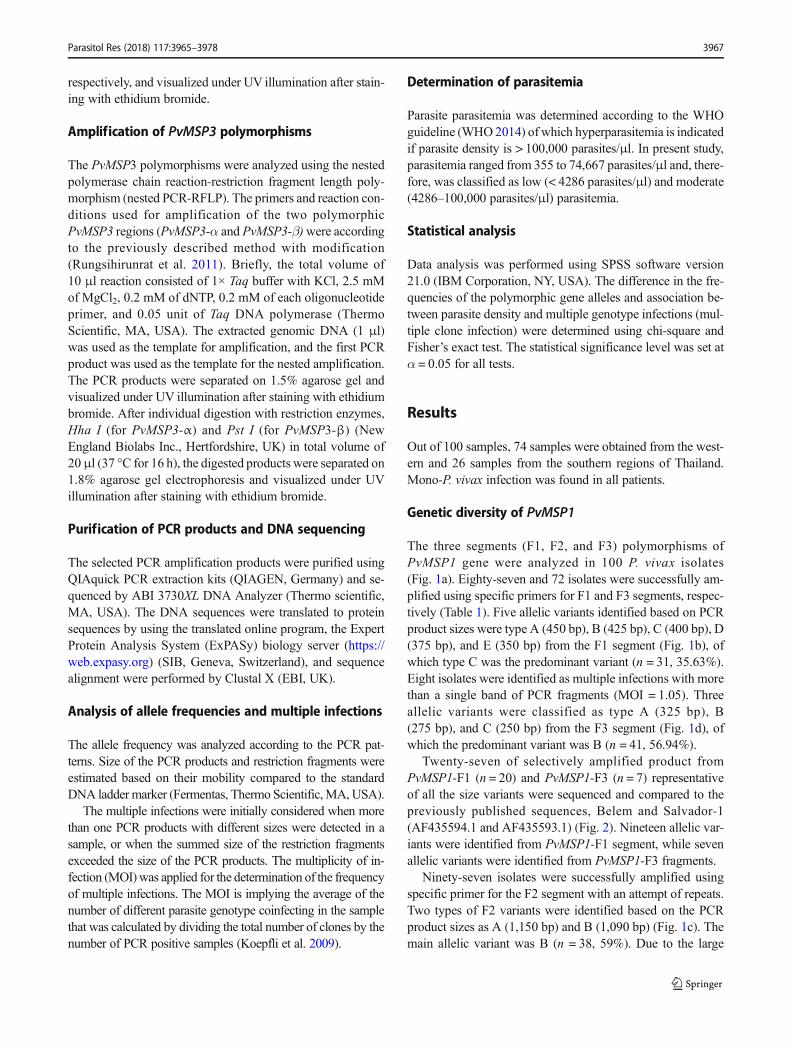
The three segments (F1, F2, and F3) polymorphisms ofPvMSP1 gene were analyzed in 100 P. vivax isolates(Fig. 1a). Eighty-seven and 72 isolates were successfully am-plified using specific primers for F1 and F3 segments, respec-tively (Table 1). Five allelic variants identified based on PCRproduct sizes were type A (450 bp), B (425 bp), C (400 bp), D(375 bp), and E (350 bp) from the F1 segment (Fig. 1b), ofwhich type C was the predominant variant (n = 31, 35.63%).Eight isolates were identified as multiple infections with morethan a single band of PCR fragments (MOI = 1.05). Threeallelic variants were classified as type A (325 bp), B(275 bp), and C (250 bp) from the F3 segment (Fig. 1d), ofwhich the predominant variant was B (n = 41, 56.94%).
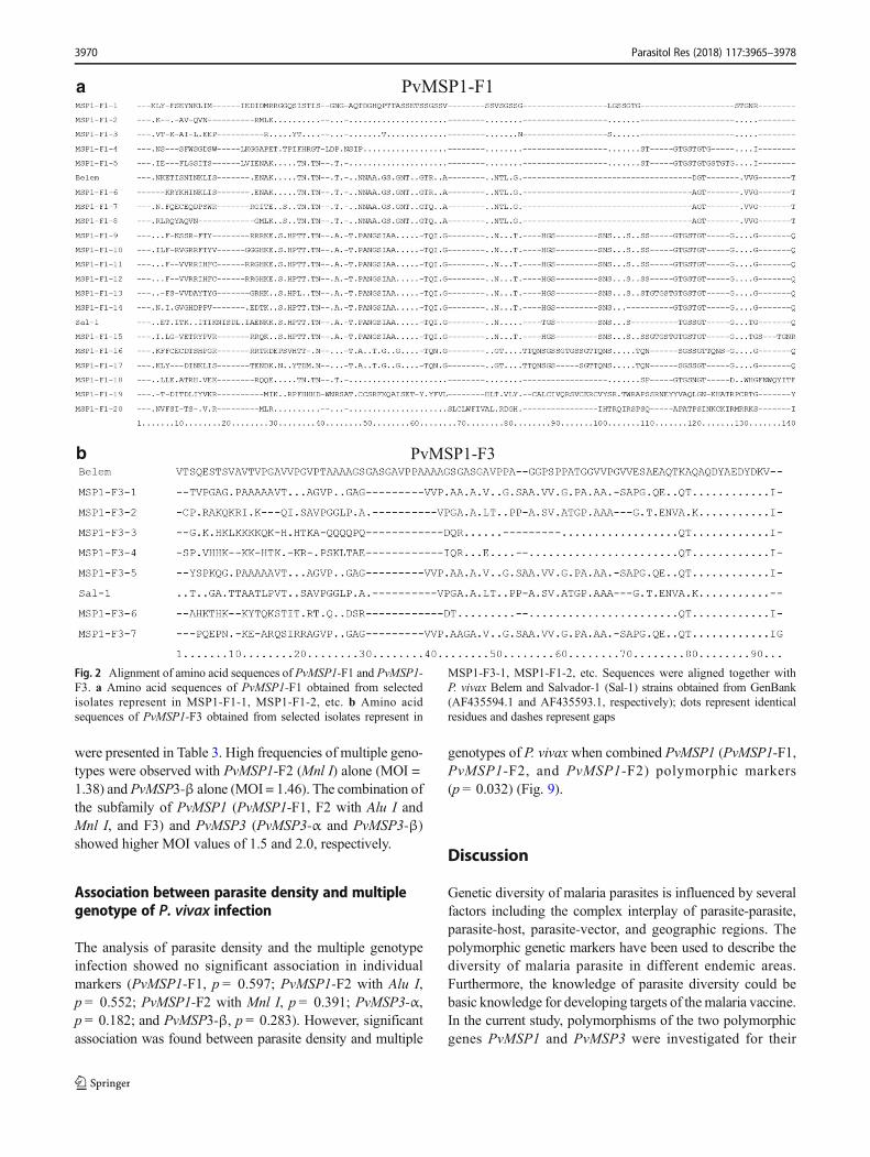
Twenty-seven of selectively amplified product fromPvMSP1-F1 (n = 20) and PvMSP1-F3 (n = 7) representativeof all the size variants were sequenced and compared to thepreviously published sequences, Belem and Salvador-1(AF435594.1 and AF435593.1) (Fig. 2). Nineteen allelic var-iants were identified from PvMSP1-F1 segment, while sevenallelic variants were identified from PvMSP1-F3 fragments.
Ninety-seven isolates were successfully amplified usingspecific primer for the F2 segment with an attempt of repeats.Two types of F2 variants were identified based on the PCRproduct sizes as A (1,150 bp) and B (1,090 bp) (Fig. 1c). Themain allelic variant was B (n = 38, 59%). Due to the large
Parasitol Res (2018) 117:3965–3978 3967
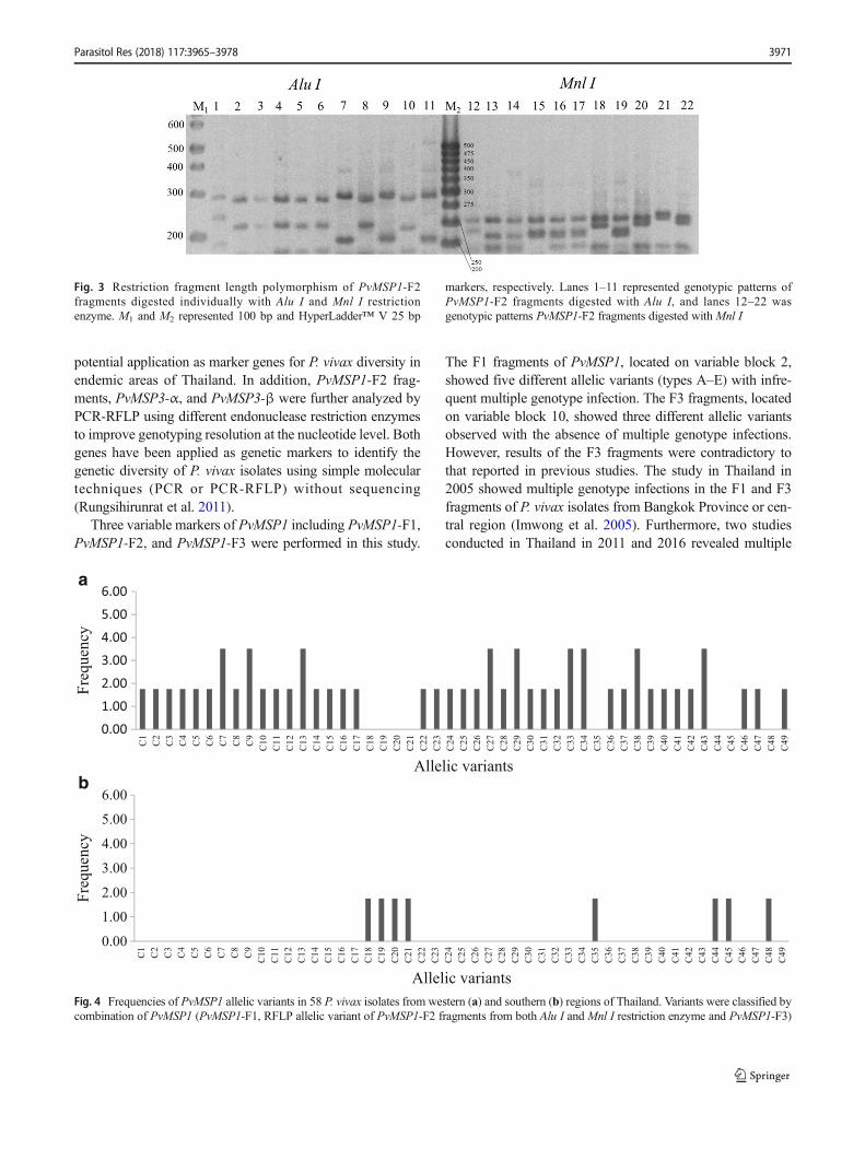
fragment size of PCR product with more than 1 kb and lowdiversity of this variant, RFLP was used to improve genotyp-ing resolution at nucleotide level by digesting PCR productsof the F2 variants with Alu I and Mnl I restriction enzymes(Imwong et al. 2005). Twenty-nine and 25 RFLP patternswere obtained after digestion with Alu I andMnl I, respective-ly (Fig. 3), of which 26 and four RFLP patterns identified fromthe western sample after digested with Alu I andMnl I, respec-tively. Twenty-three and three RFLP patterns of Alu I andMnlIwere identified from the samples collected from the southernregion, respectively. Twenty-nine and 17 isolates were identi-fied as multiple infections after digested with Alu I andMnl I,respectively (Table 1). The distribution of PvMSP1-F1 andPvMSP1-F2 digested with Alu I and Mnl I were significantlydifferent between the two endemic areas (p = 0.02, p < 0.001,and p < 0.001, respectively) (Table 1).
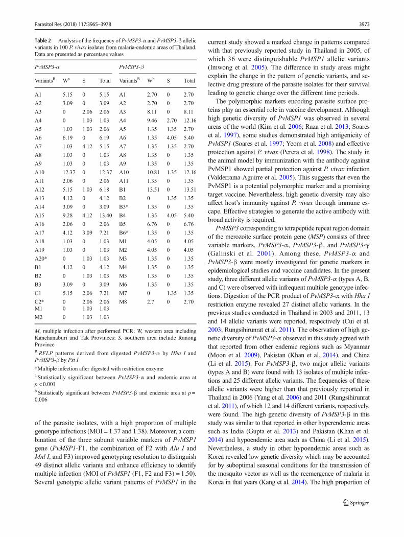
When combining all the three variables markers ofPvMSP1 (F1, F2, and F3), 49 genotypic patterns were found(Fig. 4), 41 and eight genotypic patterns from western andsouthern isolates, respectively.
Genetic diversity of PvMSP3
Analysis of the polymorphisms of the two segments(PvMSP3-α and PvMSP3-β) of the PvMSP3 regions wasperformed in 100 P. vivax isolates (Fig. 5a). Ninety-sevenisolates were successfully amplified using specific primerfor PvMSP3-α. Three allelic variants were identified as typeA (1.9 kb), B (1.5 kb), and type C (1.2 kb) with correspond-ing frequencies of 80.41%, 8.25% and 9.28%, respectively(Fig. 5b). Two isolates (2.6%) were identified as multipleinfections with more than a single band of PCR fragments.For PvMSP3-β, 75 isolates were successfully amplified
using the specific primer. Two allelic variants were identi-fied as type A (1.7–2.2 kb) and type B (1.4–1.5 kb) withcorresponding frequencies of 52.70 and 29.73%, respec-tively (Fig. 5c). Thirteen isolates (17.57%) were identifiedas multiple infections. Type C (0.65 kb) variant was notfound as a single genotype but in combination with othergenotypes. RFLP was further performed withHha I enzymedigestion for PvMSP3-α and Pst I for PvMSP3-β after PCRamplification to improve efficiency in the differentiation ofPvMSP3 variants at the nucleotide level (Fig. 6). Twenty-seven different genotypic patterns of the PvMSP3-α RFLPfragment were detected with a clear common fragment sizeof 1000 bp in all isolates, together with distinct smallerfragments ranging from 100 to 500 bp (20 and 13 genotypicpatterns from western and southern isolates, respectively).Twenty-five distinct genotypic patterns of the PvMSP3-βRFLP fragment were detected with smaller fragments rang-ing from 200 to 1500 bp (23 and 8 genotypic patterns fromwestern and southern isolates, respectively) (Table 2).Multiple P. vivax genotype infections with PvMSP3-α andPvMSP3-β were identified in five and 15 isolates from thewest and southern regions, respectively. The frequency ofPvMSP3-α and PvMSP3-β was significantly different intwo endemic areas (p < 0.001 and p = 0.006) (Table 2).
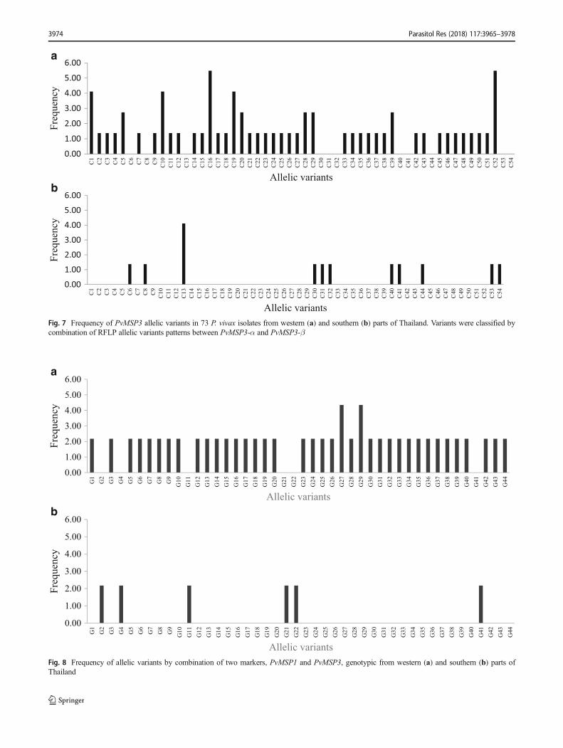
When combining both marker genes (PvMSP3-α andPvMSP3-β), 54 genotypic patterns were found (Fig. 7), 43and 11 genotypic patterns from western and southern isolates,respectively.
Genetic diversity of PvMSP1-PvMSP3 combination
The polymorphic patterns of PvMSP1 (F1, F2, and F3) andPvMSP3 (PvMSP3-α and PvMSP3-β) were successfully
Fig. 1 a Plasmodium vivax merozoite surface protein 1 gene (PvMSP1)consisting of seven conserved region (black) and six variable regions(white) (Imwong et al. 2005). b allelic variants of PvMSP1-F1 (350–450 bp): lanes 1–5 represent type C (400 bp), E (350 bp), A (450 bp),D (375 bp), and B (425 bp), respectively. cAllelic variants ofPvMSP1-F2
(1090-1150 bp): lanes 1 and 4 = type B (1090 bp), lanes 2 and 3 = type A(1150 bp). d Allelic variants of PvMSP1-F3 (250–325 bp): lanes 1 and 2= type C (250 bp), lanes 3 and 4 = type A (325 bp) and B (275 bp),respectively. M = 100 bp markers
3968 Parasitol Res (2018) 117:3965–3978
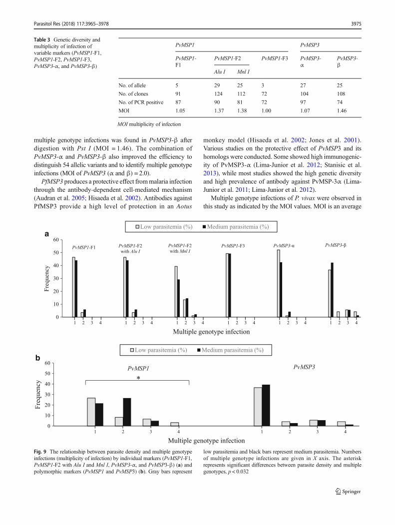
analyzed in 46 P. vivax isolates. The combined genotypicanalysis of both genes improved efficiency to distinguish ge-netic variants of P. vivax isolates in Thailand. Results revealedthe high genetic diversity of the combined markers with 44genotypic patterns (95.65%); of these, 38 and six genotypicpatterns were identified from western and southern isolates,respectively (Fig. 8).
Multiplicity of infection
Overall, the majority of P. vivax in this study had a single-clone infection (43%), followed by two clones (34%), threeclones (17%), and four clones (6%), respectively. Results ofthe multiplicity of infection (MOI) of each marker (PvMSP1-F1, PvMSP1-F2, PvMSP1-F3, PvMSP3-α, and PvMSP3-β)
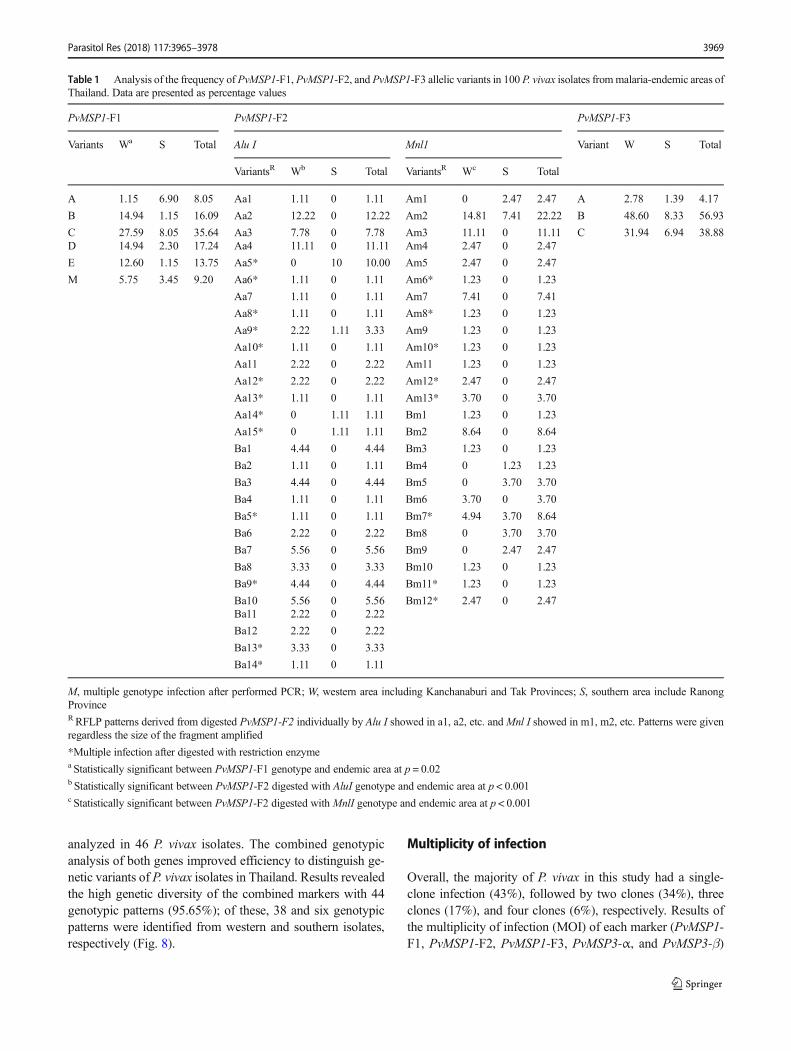
Table 1 Analysis of the frequency of PvMSP1-F1, PvMSP1-F2, and PvMSP1-F3 allelic variants in 100 P. vivax isolates frommalaria-endemic areas ofThailand. Data are presented as percentage values
PvMSP1-F1 PvMSP1-F2 PvMSP1-F3
Variants Wa S Total Alu I Mnl1 Variant W S Total
VariantsR Wb S Total VariantsR Wc S Total
A 1.15 6.90 8.05 Aa1 1.11 0 1.11 Am1 0 2.47 2.47 A 2.78 1.39 4.17
B 14.94 1.15 16.09 Aa2 12.22 0 12.22 Am2 14.81 7.41 22.22 B 48.60 8.33 56.93
C 27.59 8.05 35.64 Aa3 7.78 0 7.78 Am3 11.11 0 11.11 C 31.94 6.94 38.88D 14.94 2.30 17.24 Aa4 11.11 0 11.11 Am4 2.47 0 2.47
E 12.60 1.15 13.75 Aa5* 0 10 10.00 Am5 2.47 0 2.47
M 5.75 3.45 9.20 Aa6* 1.11 0 1.11 Am6* 1.23 0 1.23
Aa7 1.11 0 1.11 Am7 7.41 0 7.41
Aa8* 1.11 0 1.11 Am8* 1.23 0 1.23
Aa9* 2.22 1.11 3.33 Am9 1.23 0 1.23
Aa10* 1.11 0 1.11 Am10* 1.23 0 1.23
Aa11 2.22 0 2.22 Am11 1.23 0 1.23
Aa12* 2.22 0 2.22 Am12* 2.47 0 2.47
Aa13* 1.11 0 1.11 Am13* 3.70 0 3.70
Aa14* 0 1.11 1.11 Bm1 1.23 0 1.23
Aa15* 0 1.11 1.11 Bm2 8.64 0 8.64
Ba1 4.44 0 4.44 Bm3 1.23 0 1.23
Ba2 1.11 0 1.11 Bm4 0 1.23 1.23
Ba3 4.44 0 4.44 Bm5 0 3.70 3.70
Ba4 1.11 0 1.11 Bm6 3.70 0 3.70
Ba5* 1.11 0 1.11 Bm7* 4.94 3.70 8.64
Ba6 2.22 0 2.22 Bm8 0 3.70 3.70
Ba7 5.56 0 5.56 Bm9 0 2.47 2.47
Ba8 3.33 0 3.33 Bm10 1.23 0 1.23
Ba9* 4.44 0 4.44 Bm11* 1.23 0 1.23
Ba10 5.56 0 5.56 Bm12* 2.47 0 2.47Ba11 2.22 0 2.22
Ba12 2.22 0 2.22
Ba13* 3.33 0 3.33
Ba14* 1.11 0 1.11
M, multiple genotype infection after performed PCR; W, western area including Kanchanaburi and Tak Provinces; S, southern area include RanongProvinceRRFLP patterns derived from digested PvMSP1-F2 individually by Alu I showed in a1, a2, etc. andMnl I showed in m1, m2, etc. Patterns were givenregardless the size of the fragment amplified
*Multiple infection after digested with restriction enzymea Statistically significant between PvMSP1-F1 genotype and endemic area at p = 0.02b Statistically significant between PvMSP1-F2 digested with AluI genotype and endemic area at p < 0.001c Statistically significant between PvMSP1-F2 digested with MnlI genotype and endemic area at p < 0.001
Parasitol Res (2018) 117:3965–3978 3969
were presented in Table 3. High frequencies of multiple geno-types were observed with PvMSP1-F2 (Mnl I) alone (MOI =1.38) and PvMSP3-β alone (MOI = 1.46). The combination ofthe subfamily of PvMSP1 (PvMSP1-F1, F2 with Alu I andMnl I, and F3) and PvMSP3 (PvMSP3-α and PvMSP3-β)showed higher MOI values of 1.5 and 2.0, respectively.
Association between parasite density and multiplegenotype of P. vivax infection
The analysis of parasite density and the multiple genotypeinfection showed no significant association in individualmarkers (PvMSP1-F1, p = 0.597; PvMSP1-F2 with Alu I,p = 0.552; PvMSP1-F2 with Mnl I, p = 0.391; PvMSP3-α,p = 0.182; and PvMSP3-β, p = 0.283). However, significantassociation was found between parasite density and multiple
genotypes of P. vivax when combined PvMSP1 (PvMSP1-F1,PvMSP1-F2, and PvMSP1-F2) polymorphic markers(p = 0.032) (Fig. 9).
Discussion
Genetic diversity of malaria parasites is influenced by severalfactors including the complex interplay of parasite-parasite,parasite-host, parasite-vector, and geographic regions. Thepolymorphic genetic markers have been used to describe thediversity of malaria parasite in different endemic areas.Furthermore, the knowledge of parasite diversity could bebasic knowledge for developing targets of themalaria vaccine.In the current study, polymorphisms of the two polymorphicgenes PvMSP1 and PvMSP3 were investigated for their
PvMSP1-F1
PvMSP1-F3
a
b
Fig. 2 Alignment of amino acid sequences of PvMSP1-F1 and PvMSP1-F3. a Amino acid sequences of PvMSP1-F1 obtained from selectedisolates represent in MSP1-F1-1, MSP1-F1-2, etc. b Amino acidsequences of PvMSP1-F3 obtained from selected isolates represent in
MSP1-F3-1, MSP1-F1-2, etc. Sequences were aligned together withP. vivax Belem and Salvador-1 (Sal-1) strains obtained from GenBank(AF435594.1 and AF435593.1, respectively); dots represent identicalresidues and dashes represent gaps
3970 Parasitol Res (2018) 117:3965–3978
potential application as marker genes for P. vivax diversity inendemic areas of Thailand. In addition, PvMSP1-F2 frag-ments, PvMSP3-α, and PvMSP3-β were further analyzed byPCR-RFLP using different endonuclease restriction enzymesto improve genotyping resolution at the nucleotide level. Bothgenes have been applied as genetic markers to identify thegenetic diversity of P. vivax isolates using simple moleculartechniques (PCR or PCR-RFLP) without sequencing(Rungsihirunrat et al. 2011).
Three variable markers of PvMSP1 including PvMSP1-F1,PvMSP1-F2, and PvMSP1-F3 were performed in this study.
The F1 fragments of PvMSP1, located on variable block 2,showed five different allelic variants (types A–E) with infre-quent multiple genotype infection. The F3 fragments, locatedon variable block 10, showed three different allelic variantsobserved with the absence of multiple genotype infections.However, results of the F3 fragments were contradictory tothat reported in previous studies. The study in Thailand in2005 showed multiple genotype infections in the F1 and F3fragments of P. vivax isolates from Bangkok Province or cen-tral region (Imwong et al. 2005). Furthermore, two studiesconducted in Thailand in 2011 and 2016 revealed multiple
0.001.002.003.004.005.006.00
C1
C2
C3
C4
C5
C6
C7
C8
C9
C1
0
C1
1
C1
2
C1
3
C1
4
C1
5
C1
6
C1
7
C1
8
C1
9
C2
0
C2
1
C2
2
C2
3
C2
4
C2
5
C2
6
C2
7
C2
8
C2
9
C3
0
C3
1
C3
2
C3
3
C3
4
C3
5
C3
6
C3
7
C3
8
C3
9
C4
0
C4
1
C4
2
C4
3
C4
4
C4
5
C4
6
C4
7
C4
8
C4
9
Fre
qu
ency
Allelic variants
0.00
1.00
2.00
3.00
4.00
5.00
6.00
C1
C2
C3
C4
C5
C6
C7
C8
C9
C1
0
C1
1
C1
2
C1
3
C1
4
C1
5
C1
6
C1
7
C1
8
C1
9
C2
0
C2
1
C2
2
C2
3
C2
4
C2
5
C2
6
C2
7
C2
8
C2
9
C3
0
C3
1
C3
2
C3
3
C3
4
C3
5
C3
6
C3
7
C3
8
C3
9
C4
0
C4
1
C4
2
C4
3
C4
4
C4
5
C4
6
C4
7
C4
8
C4
9
Fre
qu
ency
Allelic variants
a
b
Fig. 4 Frequencies of PvMSP1 allelic variants in 58 P. vivax isolates from western (a) and southern (b) regions of Thailand. Variants were classified bycombination of PvMSP1 (PvMSP1-F1, RFLP allelic variant of PvMSP1-F2 fragments from both Alu I and Mnl I restriction enzyme and PvMSP1-F3)
Fig. 3 Restriction fragment length polymorphism of PvMSP1-F2fragments digested individually with Alu I and Mnl I restrictionenzyme. M1 and M2 represented 100 bp and HyperLadder™ V 25 bp
markers, respectively. Lanes 1–11 represented genotypic patterns ofPvMSP1-F2 fragments digested with Alu I, and lanes 12–22 wasgenotypic patterns PvMSP1-F2 fragments digested with Mnl I
Parasitol Res (2018) 117:3965–3978 3971

genotype infections of F3 fragments in isolates from TakProvince, the west region of Thailand (Kosaisavee et al.2011; Maneerattanasak et al. 2016). These results suggestthe parasite genetic diversity in different time periods andendemic areas even in the same region. Furthermore, the ge-netic polymorphism of PvMSP1-F1 and PvMSP1-F3 investi-gated in many regions around the world, particularly in hyper-endemic areas such as India (Kim et al. 2006), southernPakistan (Raza et al. 2013), and Bangladesh (Kibria et al.2015) demonstrated higher genetic diversity than that ob-served in this study. According to moderate allelic size varia-tion of PvMSP1-F1 and PvMSP1-F3, sequencing of the am-plified fragments was performed, and a higher number ofP. vivax with different allelic variants was demonstrated
compared with the results obtained from gel electrophoresis.This suggests that sequencingmethod and alignment of aminoacid sequences provide high efficiency to distinguish variousallelic variants, although the specific equipment is requiredwith high-cost reagents. On the other hand, the conventionalPCR with gel electrophoresis could be a convenient methodfor general laboratory and research unit. The method is rela-tively cost-effective and straightforward without the require-ment of specific equipment.
The PvMSP1-F2 is located on variable blocks 6–8. Thenested PCR analysis of the PvMSP1-F2 revealed two majorallelic variants (types A and B) with the large fragment andlow genetic diversity. Further analysis using Alu I and Mnl Irestriction enzymes revealed 29 and 25 distinct allelic variants
Fig. 6 Restriction fragment length polymorphism of Plasmodium vivaxmerozoite surface protein 3 includes PvMSP3-α (a) and PvMSP3-β (b).M represented 100-bp marker. a The different allelic variation of
PvMSP3-α after PCR-RFLP using Hha I enzyme. b The different allelicvariation of PvMSP3-β after PCR-RFLP using Pst I enzyme
Fig. 5 a Plasmodium vivax merozoite surface protein 3 gene (PvMSP3)(Jiang et al. 2013). Ten of PvMSP3 genes labeled as 3.1, 3.2, etc. arerepresented with black boxes. Areas of two amplified segments includingPvMSP3-α (3.10) and PvMSP3-β (3.5) are present in horizontal boldlines. b Allelic variants of PvMSP3-α fragments (1.2–1.9 k bp): lanes
1–2 = type A (1.9 kb), lane 3 = type B (1.5 k bp), and lane 4 = type C(1.2 k bp), respectively. c Allelic variants of PvMSP3-β (0.65–2.2 k bp):lane 1 = type A (1.7–2.2 k bp), lane 2 = type B (1.4–1.5 kb). Multipleinfections of type A (1.7–2.2 k bp) and type C (0.65 k bp) isolates aredemonstrated in lanes 3 and 4. M = 100 bp marker
3972 Parasitol Res (2018) 117:3965–3978
of the parasite isolates, with a high proportion of multiplegenotype infections (MOI = 1.37 and 1.38). Moreover, a com-bination of the three subunit variable markers of PvMSP1gene (PvMSP1-F1, the combination of F2 with Alu I andMnl I, and F3) improved genotyping resolution to distinguish49 distinct allelic variants and enhance efficiency to identifymultiple infection (MOI of PvMSP1 (F1, F2 and F3) = 1.50).Several genotypic allelic variant patterns of PvMSP1 in the
current study showed a marked change in patterns comparedwith that previously reported study in Thailand in 2005, ofwhich 36 were distinguishable PvMSP1 allelic variants(Imwong et al. 2005). The difference in study areas mightexplain the change in the pattern of genetic variants, and se-lective drug pressure of the parasite isolates for their survivalleading to genetic change over the different time periods.
The polymorphic markers encoding parasite surface pro-teins play an essential role in vaccine development. Althoughhigh genetic diversity of PvMSP1 was observed in severalareas of the world (Kim et al. 2006; Raza et al. 2013; Soareset al. 1997), some studies demonstrated high antigenicity ofPvMSP1 (Soares et al. 1997; Yeom et al. 2008) and effectiveprotection against P. vivax (Perera et al. 1998). The study inthe animal model by immunization with the antibody againstPvMSP1 showed partial protection against P. vivax infection(Valderrama-Aguirre et al. 2005). This suggests that even thePvMSP1 is a potential polymorphic marker and a promisingtarget vaccine. Nevertheless, high genetic diversity may alsoaffect host’s immunity against P. vivax through immune es-cape. Effective strategies to generate the active antibody withbroad activity is required.
PvMSP3 corresponding to tetrapeptide repeat region domainof the merozoite surface protein gene (MSP) consists of threevariable markers, PvMSP3-α, PvMSP3-β, and PvMSP3-γ(Galinski et al. 2001). Among these, PvMSP3-α andPvMSP3-β were mostly investigated for genetic markers inepidemiological studies and vaccine candidates. In the presentstudy, three different allelic variants of PvMSP3-α (types A, B,and C) were observed with infrequent multiple genotype infec-tions. Digestion of the PCR product of PvMSP3-α with Hha Irestriction enzyme revealed 27 distinct allelic variants. In theprevious studies conducted in Thailand in 2003 and 2011, 13and 14 allelic variants were reported, respectively (Cui et al.2003; Rungsihirunrat et al. 2011). The observation of high ge-netic diversity of PvMSP3-α observed in this study agreed withthat reported from other endemic regions such as Myanmar(Moon et al. 2009), Pakistan (Khan et al. 2014), and China(Li et al. 2015). For PvMSP3-β, two major allelic variants(types A and B) were found with 13 isolates of multiple infec-tions and 25 different allelic variants. The frequencies of theseallelic variants were higher than that previously reported inThailand in 2006 (Yang et al. 2006) and 2011 (Rungsihirunratet al. 2011), of which 12 and 14 different variants, respectively,were found. The high genetic diversity of PvMSP3-β in thisstudy was similar to that reported in other hyperendemic areassuch as India (Gupta et al. 2013) and Pakistan (Khan et al.2014) and hypoendemic area such as China (Li et al. 2015).Nevertheless, a study in other hypoendemic areas such asKorea revealed low genetic diversity which may be accountedfor by suboptimal seasonal conditions for the transmission ofthe mosquito vector as well as the reemergence of malaria inKorea in that years (Kang et al. 2014). The high proportion of
Table 2 Analysis of the frequency of PvMSP3-α and PvMSP3-β allelicvariants in 100 P. vivax isolates from malaria-endemic areas of Thailand.Data are presented as percentage values
PvMSP3-α PvMSP3-β
VariantsR Wa S Total VariantsR Wb S Total
A1 5.15 0 5.15 A1 2.70 0 2.70
A2 3.09 0 3.09 A2 2.70 0 2.70
A3 0 2.06 2.06 A3 8.11 0 8.11
A4 0 1.03 1.03 A4 9.46 2.70 12.16
A5 1.03 1.03 2.06 A5 1.35 1.35 2.70
A6 6.19 0 6.19 A6 1.35 4.05 5.40
A7 1.03 4.12 5.15 A7 1.35 1.35 2.70
A8 1.03 0 1.03 A8 1.35 0 1.35
A9 1.03 0 1.03 A9 1.35 0 1.35
A10 12.37 0 12.37 A10 10.81 1.35 12.16
A11 2.06 0 2.06 A11 1.35 0 1.35
A12 5.15 1.03 6.18 B1 13.51 0 13.51
A13 4.12 0 4.12 B2 0 1.35 1.35
A14 3.09 0 3.09 B3* 1.35 0 1.35
A15 9.28 4.12 13.40 B4 1.35 4.05 5.40
A16 2.06 0 2.06 B5 6.76 0 6.76
A17 4.12 3.09 7.21 B6* 1.35 0 1.35
A18 1.03 0 1.03 M1 4.05 0 4.05
A19 1.03 0 1.03 M2 4.05 0 4.05
A20* 0 1.03 1.03 M3 1.35 0 1.35
B1 4.12 0 4.12 M4 1.35 0 1.35
B2 0 1.03 1.03 M5 1.35 0 1.35
B3 3.09 0 3.09 M6 1.35 0 1.35
C1 5.15 2.06 7.21 M7 0 1.35 1.35
C2* 0 2.06 2.06 M8 2.7 0 2.70M1 0 1.03 1.03
M2 0 1.03 1.03
M, multiple infection after performed PCR; W, western area includingKanchanaburi and Tak Provinces; S, southern area include RanongProvinceR RFLP patterns derived from digested PvMSP3-α by Hha I andPvMSP3-β by Pst I
*Multiple infection after digested with restriction enzymea Statistically significant between PvMSP3-α and endemic area atp < 0.001b Statistically significant between PvMSP3-β and endemic area at p =0.006
Parasitol Res (2018) 117:3965–3978 3973
0.00
1.00
2.00
3.00
4.00
5.00
6.00
G1
G2
G3
G4
G5
G6
G7
G8
G9
G10
G11
G12
G13
G14
G15
G16
G17
G18
G19
G20
G21
G22
G23
G24
G25
G26
G27
G28
G29
G30
G31
G32
G33
G34
G35
G36
G37
G38
G39
G40
G41
G42
G43
G44
Fre
qu
ency
Allelic variants
0.00
1.00
2.00
3.00
4.00
5.00
6.00
G1
G2
G3
G4
G5
G6
G7
G8
G9
G10
G11
G12
G13
G14
G15
G16
G17
G18
G19
G20
G21
G22
G23
G24
G25
G26
G27
G28
G29
G30
G31
G32
G33
G34
G35
G36
G37
G38
G39
G40
G41
G42
G43
G44
Fre
qu
ency
Allelic variants
a
b
Fig. 8 Frequency of allelic variants by combination of two markers, PvMSP1 and PvMSP3, genotypic from western (a) and southern (b) parts ofThailand
0.001.002.003.004.005.006.00
C1
C2
C3
C4
C5
C6
C7
C8
C9
C1
0
C1
1
C1
2
C1
3
C1
4
C1
5
C1
6
C1
7
C1
8
C1
9
C2
0
C2
1
C2
2
C2
3
C2
4
C2
5
C2
6
C2
7
C2
8
C2
9
C3
0
C3
1
C3
2
C3
3
C3
4
C3
5
C3
6
C3
7
C3
8
C3
9
C4
0
C4
1
C4
2
C4
3
C4
4
C4
5
C4
6
C4
7
C4
8
C4
9
C5
0
C5
1
C5
2
C5
3
C5
4
Fre
qu
ency
Allelic variants
a
b
Fig. 7 Frequency of PvMSP3 allelic variants in 73 P. vivax isolates from western (a) and southern (b) parts of Thailand. Variants were classified bycombination of RFLP allelic variants patterns between PvMSP3-α and PvMSP3-β
3974 Parasitol Res (2018) 117:3965–3978
multiple genotype infections was found in PvMSP3-β afterdigestion with Pst I (MOI = 1.46). The combination ofPvMSP3-α and PvMSP3-β also improved the efficiency todistinguish 54 allelic variants and to identify multiple genotypeinfections (MOI of PvMSP3 (α and β) = 2.0).
PfMSP3 produces a protective effect frommalaria infectionthrough the antibody-dependent cell-mediated mechanism(Audran et al. 2005; Hisaeda et al. 2002). Antibodies againstPfMSP3 provide a high level of protection in an Aotus
monkey model (Hisaeda et al. 2002; Jones et al. 2001).Various studies on the protective effect of PvMSP3 and itshomologs were conducted. Some showed high immunogenic-ity of PvMSP3-α (Lima-Junior et al. 2012; Stanisic et al.2013), while most studies showed the high genetic diversityand high prevalence of antibody against PvMSP-3α (Lima-Junior et al. 2011; Lima-Junior et al. 2012).
Multiple genotype infections of P. vivax were observed inthis study as indicated by the MOI values. MOI is an average
Table 3 Genetic diversity andmultiplicity of infection ofvariable markers (PvMSP1-F1,PvMSP1-F2, PvMSP1-F3,PvMSP3-α, and PvMSP3-β)
PvMSP1 PvMSP3
PvMSP1-F1
PvMSP1-F2 PvMSP1-F3 PvMSP3-α
PvMSP3-β
Alu I Mnl I
No. of allele 5 29 25 3 27 25
No. of clones 91 124 112 72 104 108
No. of PCR positive 87 90 81 72 97 74
MOI 1.05 1.37 1.38 1.00 1.07 1.46
MOI multiplicity of infection
0
10
20
30
40
50
60
1 2 3 4 1 2 3 4 1 2 3 4 1 2 3 4 1 2 3 4 1 2 3 4
Fre
qu
ency
Multiple genotype infection
Low parasitemia (%) Medium parasitemia (%)
PvMSP1-F1 PvMSP1-F3PvMSP1-F2
with Alu IPvMSP1-F2
with Mnl IPvMSP3-α PvMSP3-β
0
10
20
30
40
50
60
43214321
Fre
qu
ency
Multiple genotype infection
Low parasitemia (%) Medium parasitemia (%)
PvMSP1 PvMSP3
*
b
a
Fig. 9 The relationship between parasite density and multiple genotypeinfections (multiplicity of infection) by individual markers (PvMSP1-F1,PvMSP1-F2 with Alu I and Mnl I, PvMSP3-α, and PvMSP3-β) (a) andpolymorphic markers (PvMSP1 and PvMSP3) (b). Gray bars represent
low parasitemia and black bars represent medium parasitemia. Numbersof multiple genotype infections are given in X axis. The asteriskrepresents significant differences between parasite density and multiplegenotypes, p < 0.032
Parasitol Res (2018) 117:3965–3978 3975
number of distinct parasite genotype coinfection in a patient(Pacheco et al. 2016) associated with the transmission inten-sity, duration of infection, and individual P. vivax number ofblood stages. Although Thailand is considered thehypoendemic area of malaria, the high level of MOI wasfound in this study. This may suggest the continued persis-tence of hypnozoites that could be reactivated any time, or thenew infection of P. vivax at the same time with the preexis-tence ofP. vivax infection (Hisaeda et al. 2002). The resistanceof P. falciparum to antimalarials is of concern.
Apart from the association betweenMOI and P. vivax trans-mission, multiple genotype infection was supposed to be as-sociated with severity of infection (de Roode et al. 2005a; deRoode et al. 2005b; Pacheco et al. 2016). Malaria parasitedensity is one of the important criteria for severe malaria,particularly that caused by P. falciparum. Although, the char-acterization of severe P. vivax patients has not been well de-fined compared with P. falciparum due to accumulation ofP. vivax-infected RBC in bone marrow (Imirzalioglu et al.2006; Ru et al. 2009) and spleen (Machado Siqueira et al.2012), correlation between peripheral vivax parasitemia andP. vivax endothelial activation factors (angiopoietin-2, andvon-Willebrand-Factor) were demonstrated (Barber et al.2015). Enhancing of these endothelial activation factors wasassociated with the severity of malaria (Barber et al. 2015;Conroy et al. 2010; de Jong et al. 2016). In the present study,no isolates were identified as hyperparasitemia. In order toinvestigate for the association between the multiple genotypesand severity of malaria infection, the severity of P. vivaxparasitemia by parasitemia was classified as low (< 4286 par-asites/μl) and moderate (4286–100,000 parasites/μl) levels. Asignificant association was found when using PvMSP1 as apolymorphic marker. The results support previous studies inthe animal model in mice that malaria severity and virulencein multiple genotype infections were higher than single geno-type infection (de Roode et al. 2005a, b). In addition, theassociation between multiple genotype infections of P. vivaxand disease severity was also reported in complicated malariapatients in Colombia (Pacheco et al. 2016).
Conclusion
The high genetic diversity and multi-clonogenicity of theP. vivax populations in Thailand were demonstrated usingPCR-RFLP. Both PvMSP1 and PvMSP3 can be used as effi-cient genetic markers to determine genetic polymorphisms ofthe P. vivax population, as well as to differentiate betweenmultiple clone infection. Results showed high efficiency ofthe molecular technique to differentiate the genetic diversityof P. vivax that could be useful for identification of relapse,reinfection, and multiple infections in P. vivax isolates inThailand. Moreover, regardless of the limited number of high
parasitemia isolates, the significant association betweenparasitemia and multiple genotype infections was which couldcontribute to disease severity in P. vivax infection was found.
Funding information The study was supported by the national researchcouncil of Thailand and Thammasat University with Contract Nos. 040/2557 and 036/2558. KN and WC were supported by the Center ofExcellence in Pharmacology and Molecular Biology of Malaria andCholangiocarcinoma of Thammasat University, the National ResearchCouncil of Thailand (NRCT), and the National Research UniversityProject of Thailand (NRU), Office of Higher Education Commission ofThailand.
Compliance with ethical standards
Ethics approval and consent to participate The procedure of this studywas approved by the Human Ethical Review Board of ThammasatUniversity (no. 110/2556). All participants wrote the informed consentsfor participating in this study, and the data was nameless with no individ-ual names of participants captured.
Conflict of interest The authors declare that they have no conflict ofinterest.
References
AkM et al (1998) Humoral immune responses against Plasmodium vivaxMSP1 in humans living in an endemic malaria area in Flores,Indonesia. Southeast Asian J Trop Med Public Health 29:685–691
Audran R et al (2005) Phase I malaria vaccine trial with a long syntheticpeptide derived from the merozoite surface protein 3 antigen. InfectImmun 73:8017–8026. https://doi.org/10.1128/iai.73.12.8017-8026.2005
Barber BE et al (2015) Parasite biomass-related inflammation, endothelialactivation, microvascular dysfunction and disease severity in vivaxmalaria. PLoS Pathog 11:e1004558. https://doi.org/10.1371/journal.ppat.1004558
Bruce MC, Galinski MR, Barnwell JW, Snounou G, Day KP (1999)Polymorphism at the merozoite surface protein-3alpha locus ofPlasmodium vivax: global and local diversity. Am J Trop MedHyg 61:518–525
Conroy AL et al (2010) Endothelium-based biomarkers are associatedwith cerebral malaria in Malawian children: a retrospective case-control study. PLoS One 5:e15291. https://doi.org/10.1371/journal.pone.0015291
Cui L et al (2003) Genetic diversity and multiple infections ofPlasmodium vivax malaria in Western Thailand. Am J Trop MedHyg 68:613–619
de Jong GM, Slager JJ, Verbon A, van Hellemond JJ, van Genderen PJJ(2016) Systematic review of the role of angiopoietin-1 andangiopoietin-2 in Plasmodium species infections: biomarkers ortherapeutic targets? Malar J 15:581. https://doi.org/10.1186/s12936-016-1624-8
de Roode JC, Helinski ME, Anwar MA, Read AF (2005a) Dynamics ofmultiple infections and within-host competition in genetically di-verse malaria infections. Am Nat 166:531–542. https://doi.org/10.1086/491659
deRoode JC et al (2005b) Virulence and competitive ability in geneticallydiverse malaria infections. Proc Natl Acad Sci U S A 102:7624–7628. https://doi.org/10.1073/pnas.0500078102
3976 Parasitol Res (2018) 117:3965–3978
Espinosa AM et al (2003) Expression, polymorphism analysis, reticulo-cyte binding and serological reactivity of two Plasmodium vivaxMSP-1 protein recombinant fragments. Vaccine 21:1033–1043
GalinskiMR, Ingravallo P, Corredor-Medina C, Al-Khedery B, PovoaM,Barnwell JW (2001) Plasmodium vivax merozoite surface proteins-3beta and-3gamma share structural similarities with P. vivax mero-zoite surface protein-3alpha and define a new gene family. MolBiochem Parasitol 115:41–53
Garzon-Ospina D, Romero-Murillo L, Tobon LF, Patarroyo MA (2011)Low genetic polymorphism of merozoite surface proteins 7 and 10in Colombian Plasmodium vivax isolates. Infect Genet Evol 11:528–531. https://doi.org/10.1016/j.meegid.2010.12.002
Gomez A, Suarez CF, Martinez P, Saravia C, Patarroyo MA (2006) Highpolymorphism in Plasmodium vivax merozoite surface protein-5(MSP5). Parasitology 133:661–672. https://doi.org/10.1017/s0031182006001168
Gupta P, Pande V, Eapen A, Singh V (2013) Genotyping of MSP3betagene in Indian Plasmodium vivax. J Vector Borne Dis 50:197–201
Hamblin MT, Di Rienzo A (2000) Detection of the signature of naturalselection in humans: evidence from the Duffy blood group locus.Am J Hum Genet 66:1669–1679. https://doi.org/10.1086/302879
Hisaeda H, Saul A, Reece JJ, Kennedy MC, Long CA, Miller LH,Stowers AW (2002) Merozoite surface protein 3 and protectionagainst malaria in Aotus nancymai monkeys. J Infect Dis 185:657–664. https://doi.org/10.1086/339187
Imirzalioglu C, Soydan N, Schaller M, Bretzel RG, Chakraborty T,Domann E (2006) Diagnosis of mixed Plasmodium malariae andP. vivax infection in a development aid volunteer by examination ofbone-marrow specimens by real-time PCR. J Clin Microbiol 44(6):2307–2310
Imwong M et al (2005) Practical PCR genotyping protocols forPlasmodium vivax using Pvcs and Pvmsp1. Malar J 4:20. https://doi.org/10.1186/1475-2875-4-20
Jiang J, Barnwell J, Meyer E, Galinski M (2013) Plasmodium vivaxmerozoite surface protein-3 (PvMSP3): expression of an 11 membermultigene family in blood-stage parasites. PLoS One 8:e63888.https://doi.org/10.1371/journal.pone.0063888
Jones TR et al (2001) Protection of Aotus monkeys by Plasmodiumfalciparum EBA-175 region II DNA prime-protein boost immuni-zation regimen. J Infect Dis 183:303–312. https://doi.org/10.1086/317933
Kang JM et al (2014) Polymorphic patterns of the merozoite surfaceprotein-3beta in Korean isolates of Plasmodium vivax. Malar J 13:104. https://doi.org/10.1186/1475-2875-13-104
Khan SN et al (2014) PCR/RFLP-based analysis of genetically distinctPlasmodium vivax population of Pvmsp-3alpha and Pvmsp-3betagenes in Pakistan. Malar J 13:355. https://doi.org/10.1186/1475-2875-13-355
Kibria MG, Elahi R, Mohon AN, Khan WA, Haque R, Alam MS (2015)Genetic diversity of Plasmodium vivax in clinical isolates fromBangladesh. Malar J 14:267. https://doi.org/10.1186/s12936-015-0790-4
Kim JR et al (2006) Genetic diversity of Plasmodium vivax in Kolkata,India. Malar J 5:71. https://doi.org/10.1186/1475-2875-5-71
Kirchgatter K, del Portillo HA (1998) Molecular analysis of Plasmodiumvivax relapses using theMSP1molecule as a genetic marker. J InfectDis 177:511–515
Koepfli C et al (2009) Evaluation of Plasmodium vivax genotypingmarkers for molecular monitoring in clinical trials. J Infect Dis199:1074–1080. https://doi.org/10.1086/597303
Kosaisavee V, Hastings I, Craig A, Lek-Uthai U (2011) The geneticpolymorphism of Plasmodium vivax genes in endemic regions ofThailand. Asian Pac J Trop Med 4:931–936. https://doi.org/10.1016/s1995-7645(11)60221-6
Li YC, Wang GZ, Meng F, Zeng W, He CH, Hu XM, Wang SQ (2015)Genetic diversity of Plasmodium vivax population before
elimination of malaria in Hainan Province, China. Malar J 14:78.https://doi.org/10.1186/s12936-015-0545-2
Lima-Junior JC et al (2011) B cell epitope mapping and characterizationof naturally acquired antibodies to the Plasmodium vivax merozoitesurface protein-3alpha (PvMSP-3alpha) in malaria-exposed individ-uals from Brazilian Amazon. Vaccine 29:1801–1811. https://doi.org/10.1016/j.vaccine.2010.12.099
Lima-Junior JC et al (2012) Influence of HLA-DRB1 and HLA-DQB1alleles on IgG antibody response to theP. vivaxMSP-1,MSP-3alphaandMSP-9 in individuals from Brazilian endemic area. PloS One 7:e36419. https://doi.org/10.1371/journal.pone.0036419
Machado Siqueira A et al (2012) Spleen rupture in a case of untreatedPlasmodium vivax infection. PLoS Negl Trop Dis 6(12):e1934
Maneerattanasak S, Gosi P, Krudsood S, Tongshoob J, Lanteri CA,Snounou G, Khusmith S (2016) Genetic diversity amongPlasmodium vivax isolates along the Thai–Myanmar border ofThailand. Malar J 15:75. https://doi.org/10.1186/s12936-016-1136-6
Marlar T, Myat Phone K, Aye Yu S, Khaing Khaing G, Ma S, Myint O(1995) Development of resistance to chloroquine by Plasmodiumvivax in Myanmar. Trans R Soc Trop Med Hyg 89:307–308
Moon SU et al (2009) High frequency of genetic diversity of Plasmodiumvivax field isolates in Myanmar. Acta Trop 109:30–36. https://doi.org/10.1016/j.actatropica.2008.09.006
PachecoMA, Lopez-PerezM, Vallejo AF, Herrera S, Arévalo-HerreraM,Escalante AA (2016) Multiplicity of infection and disease severityin Plasmodium vivax. PLoS Negl Trop Dis 10:e0004355. https://doi.org/10.1371/journal.pntd.0004355
Panichakul T, Sattabongkot J, Chotivanich K, Sirichaisinthop J, Cui L,Udomsangpetch R (2007) Production of erythropoietic cells in vitrofor continuous culture of Plasmodium vivax. Int J Parasit 37:1551–1557. https://doi.org/10.1016/j.ijpara.2007.05.009
Perera KL, Handunnetti SM, Holm I, Longacre S, Mendis K (1998)Baculovirus merozoite surface protein 1 C-terminal recombinantantigens are highly protective in a natural primate model for humanPlasmodium vivax malaria. Infect Immun 66:1500–1506
Phyo AP et al (2011) Dihydroartemisinin-piperaquine versus chloroquinein the treatment of Plasmodium vivax malaria in Thailand: a ran-domized controlled trial. Clin Infect Dis 53:977–984. https://doi.org/10.1093/cid/cir631
Putaporntip C, Jongwutiwes S, FerreiraMU, Kanbara H, UdomsangpetchR, Cui L (2009) Limited global diversity of the Plasmodium vivaxmerozoite surface protein 4 gene. Infect Genet Evol 9:821–826.https://doi.org/10.1016/j.meegid.2009.04.017
Raza A, Ghanchi NK, Thaver AM, Jafri S, Beg MA (2013) Geneticdiversity of Plasmodium vivax clinical isolates from southernPakistan using pvcsp and pvmsp1 genetic markers. Malar J 12:16.https://doi.org/10.1186/1475-2875-12-16
Ru YX, Mao BY, Zhang FK, Pang TX, Zhao SX, Liu JH,Wickramasinghe SN (2009) Invasion of erythroblasts by : a newmechanism contributing to malarial anemia. UltrastructPathol 33(5):236–242
Rungsihirunrat K, Chaijaroenkul W, Siripoon N, Seugorn A, Na-Bangchang K (2011) Genotyping of polymorphic marker(MSP3alpha and MSP3beta) genes of Plasmodium vivax field iso-lates from malaria-endemic of Thailand. Trop Med Int Health 16:794–801. https://doi.org/10.1111/j.1365-3156.2011.02771.x
Soares IS, Levitus G, Souza JM, Del Portillo HA, Rodrigues MM (1997)Acquired immune responses to the N- and C-terminal regions ofPlasmodium vivax merozoite surface protein 1 in individuals ex-posed to malaria. Infect Immun 65:1606–1614
Souza-Neiras WC et al (2010) Plasmodium vivax circumsporozoitegenotypes: a limited variation or new subspecies with majorbiological consequences? Malar J 9:178. https://doi.org/10.1186/1475-2875-9-178
Stanisic DI et al (2013) Naturally acquired immune responses to P. vivaxmerozoite surface protein 3α, and merozoite surface protein 9 are
Parasitol Res (2018) 117:3965–3978 3977
associated with reduced risk ofP. vivaxmalaria in young PapuaNewGuinean children. PLoS Negl Trop Dis 7:e2498. https://doi.org/10.1371/journal.pntd.0002498
Thanh PV et al (2015) Confirmed Plasmodium vivax resistance to chlo-roquine in Central Vietnam. Antimicrob Agents Chemother 59:7411–7419. https://doi.org/10.1128/aac.00791-15
Triglia T, Healer J, Caruana SR, Hodder AN, Anders RF, Crabb BS,Cowman AF (2000) Apical membrane antigen 1 plays a central rolein erythrocyte invasion by Plasmodium species. Mol Microbiol 38:706–718
Valderrama-Aguirre A et al (2005) Antigenicity, immunogenicity,and protective efficacy of Plasmodium vivax MSP1 PV200l: apotential malaria vaccine subunit. Am jJ Trop Med Hyg 73:16–24
Veron V, Legrand E, Yrinesi J, Volney B, Simon S, Carme B (2009)Genetic diversity of msp3alpha and msp1_b5 markers of
Plasmodium vivax in French Guiana. Malar J 8:40. https://doi.org/10.1186/1475-2875-8-40
WHO (2014) Severe malaria. Trop Med Int Health 19(Suppl 1):7–131.https://doi.org/10.1111/tmi.12313_2
WHO (2016) World malaria report 2016. WHO Global malariaprogramme
Yang Z et al (2006) Genetic structures of geographically distinctPlasmodium vivax populations assessed by PCR/RFLP anal-ysis of the merozoite surface protein 3beta gene. Acta Trop100:205–212. https://doi.org/10.1016/j.actatropica.2006.10.011
Yeom JS et al (2008) Naturally acquired IgM antibody responseto the C-terminal region of the merozoite surface protein 1 ofPlasmodium vivax in Korea: use for serodiagnosis of vivaxmalaria. J Parasitol 94:1410–1414. https://doi.org/10.1645/ge-1484.1
3978 Parasitol Res (2018) 117:3965–3978