Evaluation and Management of Primary Amenorrhea Libby Crockett, MD Department of Obstetrics and...
63
Evaluation and Management of Primary Amenorrhea Libby Crockett, MD Department of Obstetrics and Gynecology University of Nebraska Medical Center
-
Upload
miles-lawrence -
Category
Documents
-
view
213 -
download
1
Transcript of Evaluation and Management of Primary Amenorrhea Libby Crockett, MD Department of Obstetrics and...

Evaluation and Management of Primary AmenorrheaLibby Crockett, MDDepartment of Obstetrics and GynecologyUniversity of Nebraska Medical Center

Disclosure….• I have no financial conflicts of interest.

Objectives
• 1. Understand the causes of primary amenorrhea
• 2. Understand how to elicit a pertinent history and perform a focused physical exam to evaluate primary amenorrhea
• 3. Understand how to perform and interpret selected diagnostic tests and imaging to evaluate primary amenorrhea

Puberty—The Mechanism
• Initiated by release of pulsatile GnRH (hypothalamus)• Specifically see increased pulsatile patterns of
FSH & LH (these start during sleep and eventually go throughout the day)• With pulses of GnRH, peaks of estradiol result
and eventually menarche appears• By late puberty, the mature HPO axis is intact
and ovulation occurs
Timing of Puberty
• Major determinant—GENETICS• Also affected by geographic location, exposure to
light, general health and nutrition and psychological factors• Recent studies have demonstrated a decline in the
age of menarche• Critical Body Weight?• Studies have shown 47.8 kg in general• Shift in body composition to more fat 16-23.5%• Has been linked to protein Leptin

Stages of Pubertal Development
• Stages• 1. Accelerated Growth• 2. Breast Development (Thelarche)• 3. Pubarche• 4. Menarche
• Generally takes 4.5 years • Differs culturally between Ethnic Groups

Puberty and EthnicityEthnic Group Mean Age of
ThelarcheMean Age Adrenarche
Mean Age Menarche
African American Girls
9.5 years 9.5 years 12.1 years
Mexican-American Girls
9.8 years 10.3 years 12.2 years
Caucasian Girls 10.5 years 10.5 years 12.7 years
Wu T, Mendola P, Buck GM. Ethnic differences in the presence of secondary sex characteristics and menarche among US girls: the Third National Health and Nutrition Examination Survey. 1988-1994. Pediatrics. 110: 752, 2002.

Assessing Pubertal Development
• Tanner Staging• Developed by James Tanner and originally published in 1968 as
an objective way to assess pubertal development.
Breast Pubic Hair
Prepubertal (Stage 1)
Elevation of the papilla only No pubic hair
Stage 2 Elevation of the breast and papilla as a small mound, areola diameter enlarged
Sparse, long, pigmented hair chiefly along labia majora
Stage 3 Further enlargement without separation of the breast and areola
Dark, coarse, curled hair sparsely spread over mons
Stage 4 Secondary mound of areola and papilla above the breast
Adult-type hair, abundant but limited to mons
Stage 5 Recession of areola to contour of breast
Adult-type spread in quantity and distribution
Speroff and Fritz. Abnormal Puberty and Growth Problems. Clinical Gynecological Endocrinology and Infertility: Seventh Edition. 2005. pg 365-392

Tanner Staging


Causes of Primary Amenorrhea
• American Society of Reproductive Medicine classifies causes of primary amenorrhea into three distinct groups
• Primary Amenorrhea with:• Breast Development (30%)• No breast development AND high FSH (40%)• No breast development AND low FSH. (30%)
ASRM Practice Committee. Amenorrhea. Fertil Steril 2008.

Causes of Primary AmenorrheaCategory Approximate Frequency (%)
+ Breast Development 30%
Mullerian agenesis 10% Androgen insensitivity 9% Vaginal Septum 2% Imperforate hymen 1% Constitutional Delay 8%No breast development & HIGH FSH 40%
No breast development & LOW FSH 30%
ASRM Practice Committee. Amenorrhea. Fertil Steril 2008.

Mullerian Agenesis• Mayer-Rokitansky-Kuster-Hauser (MRKH) syndrome• Complete absence of uterus, cervix and the upper
2/3 of the vagina• Incidence 1/5000 (1/4000-1/10,000 female newborns)• Normal XX Karyotype• Normal ovarian function• Otherwise normal pubertal development
• Causes• Mutations in Antimullerian Hormone or
Antimullerian Hormone receptor• Association with Wnt gene
Deligeoroglou et. Al 2010 & ASRM Practice Committee. Amenorrhea. Fertil Steril 2008.

Uterine Development Video• Hill, M.A. (2013) Uterus Development Movie. Retrieved August
5, 2013, from http://php.med.unsw.edu.au/embryology/index.php?title=Uterus_Development_Movie

Mullerian Agenesis
• Evaluation: Normal breast development, normal secondary sexual characteristics• Laboratory: Normal XX karyotype, normal
LH, FSH• Pelvic Exam: • Normal external genitalia• absence of internal
midline structures• + vaginal dimple

Diagnosis
Left: MRI showing absence of uterus and vagina
Mullerian Agenesis
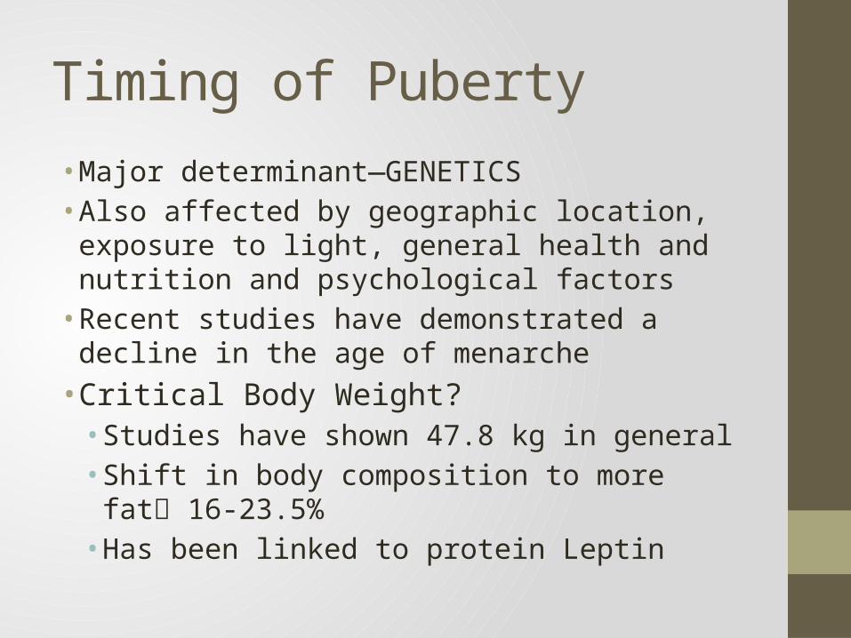
• Associated Conditions• 1/3 concurrent urinary tract anomalies• Ex: Ectopic kidney, renal agenesis, horseshoe
kidney• 12% associated skeletal anomalies • Ex: spinal anomalies, absent digits, webbed
fingers, toes• Part of work up needs to include an abdominal
CT to evaluate for renal anomalies

Mullerian Agenesis: Treatment• Dilators (Frank and Ingram)• Dilate at a 15 degree angle daily after warm bath for 20
minutes. • Progressively work up to larger dilators• Success defined as non-painful intercourse or vaginal
length of 7cm• Studies demonstrate up to an 88% success rate at 19
months of use.

McIndoe Neovagina
• Use a skin graft or artificial skin placed over a mold forming a tube with one closed end• Incision made in the vaginal dimple and cavity
dissected to level of peritoneum. • Labia majora are sewn together. • Bed rest for 7 days and then mold removed.

McIndoe Neovagina
Other Forms of Surgical Management
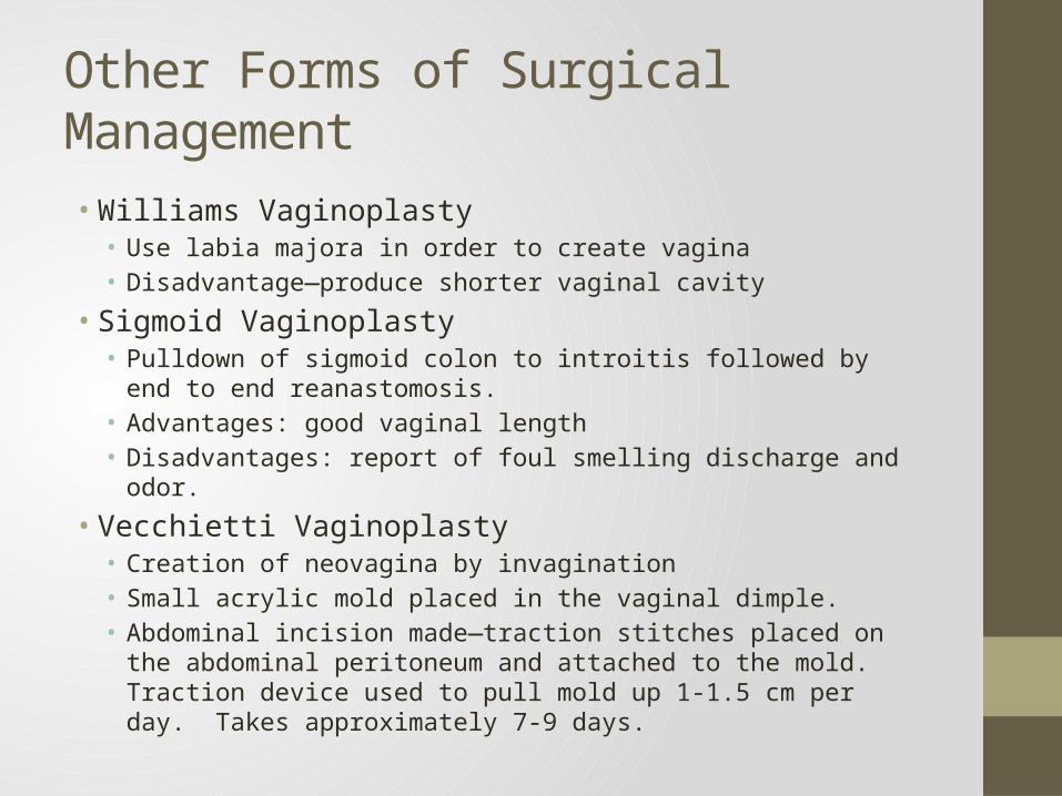
• Williams Vaginoplasty• Use labia majora in order to create vagina• Disadvantage—produce shorter vaginal cavity
• Sigmoid Vaginoplasty• Pulldown of sigmoid colon to introitis followed by end to end
reanastomosis. • Advantages: good vaginal length • Disadvantages: report of foul smelling discharge and odor.
• Vecchietti Vaginoplasty• Creation of neovagina by invagination• Small acrylic mold placed in the vaginal dimple. • Abdominal incision made—traction stitches placed on the abdominal
peritoneum and attached to the mold. Traction device used to pull mold up 1-1.5 cm per day. Takes approximately 7-9 days.

Transverse Vaginal Septum
• Failure of canalization of distal third of the vagina• Most common in upper and middle third of vagina
• Diagnosis• Usually present after puberty with amenorrhea and
pelvic pain• Can present with hematocolpos, hematometra• Does not bulge with valsalva maneuver• MRI helps with diagnosis
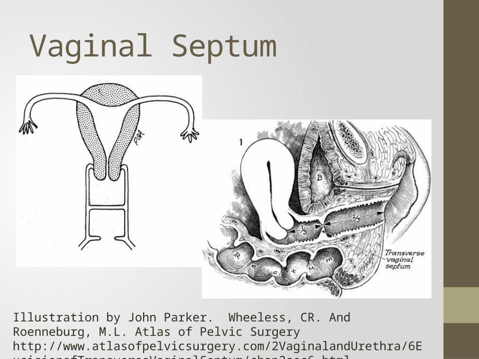
Vaginal Septum
Illustration by John Parker. Wheeless, CR. And Roenneburg, M.L. Atlas of Pelvic Surgery http://www.atlasofpelvicsurgery.com/2VaginalandUrethra/6ExcisionofTransverseVaginalSeptum/chap2sec6.html

Imperforate Hymen• Most common obstructive lesion
of the female genital tract• 1/1000 female births
• Classic appearance of bulging, blue-domed, translucent membrane
• Can present with hematocolpos or urinary retention
• Differs from vaginal septum in that an imperforate hymen bulges with valsalva
• Treatment: Surgical Resection• Hymenectomy versus
hymenotomy

Androgen Insensitivity• Incidence 1/60,000 • although 9% of causes of primary amenorrhea
• Genetics: X-linked recessive• Phenotype: Female; Genotype: XY• Female external genitalia with small vaginal dimple• Absent uterus and cervix. • Cryptorchidic gonads• Absent axillary and pubic hair• +Breast development
• Cause: Mutations in the androgen receptor
Deligeoroglou et al 2010

Androgen Insensitivity
• Physical Exam• Slim and taller than average female• Large breasts with juvenile nipples• Absent pubic/axillary hair, no acne or other signs of
androgen action• May have inguinal hernia• Normal external genitalia, blind vaginal pouch and
absence of midline structures• Laboratory: Testosterone in the normal to high male
range
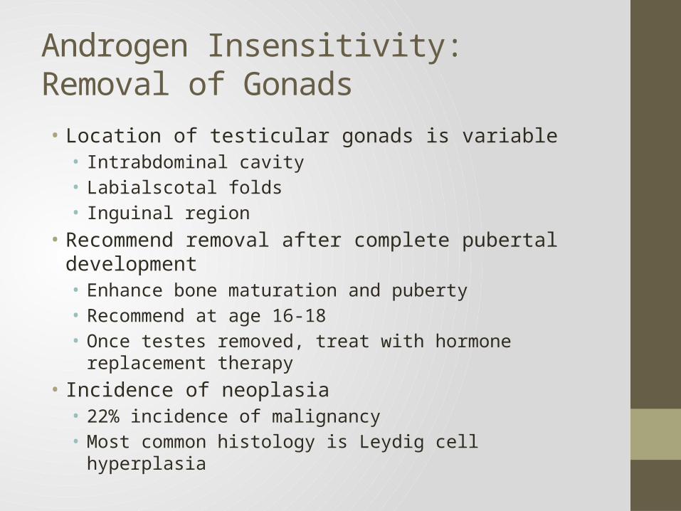
Androgen Insensitivity: Removal of Gonads• Location of testicular gonads is variable• Intrabdominal cavity• Labialscotal folds• Inguinal region
• Recommend removal after complete pubertal development • Enhance bone maturation and puberty• Recommend at age 16-18• Once testes removed, treat with hormone replacement therapy
• Incidence of neoplasia• 22% incidence of malignancy • Most common histology is Leydig cell hyperplasia


Causes of Primary AmenorrheaCategory Approximate Frequency
+ Breast Development 30%
No Breast Development: HIGH FSH 40% 46XX 15% 46XY 5% 45XO 20%No Breast Development: LOW FSH 30%
Current Evaluation of Amenorrhea. Fertility and Sterility 2008 80(3): S219-S225.
Hypergonadotrophic HypogonadismGonadal dysgenesis

Turner’s Syndrome
• Classically 45 XO or mosaic• Incidence 2,500-10,000 liveborns• 99% of pregnancies affected end in SAB
• Cause: Absence of ovarian determinant genes result in premature loss of germ cells• Fetuses with Turner’s have the same amount of
germ cells at midgestation as do 46, XX• As gestation continues, accelerated loss of germ cells
occurs• Many XO individuals lose all germ cells prior to birth;
less than 15% have enough germ cells to start pubertal process by adolescence

Turner’s Syndrome

Turner’s Syndrome• Associated Abnormalities• Cardiac Anomalies• Coartation of aorta in 30% of patients, also bicuspid aortic
valve, mitral valve prolapse• Recommend echocardiography be performed every 3-5 years
• Renal Anomalies• Horseshoe Kidney• Need retroperitoneal ultrasound once diagnosed
• Hypothyroidism • 10% of patients with Turner’s Syndrome• Recommend yearly screening of T4/TSH and antibodies
• Deafness (audiometry)

Gonadal Dysgenesis: 46XX• Refers to a number of conditions in which abnormal
development leads to streak gonads• Incidence: <1/10,000 in women less than 30• Inherited• Familial inheritance 7-30%• Premutations in the FMR1 gene (Fragile X Syndrome)
• 15% of carriers have POF• Associated with autoimmune diseases (18-30%)
• Hashimoto’s Thyroiditis, Addison’s disease, hypoparathyroidism, vitiligo
• Acquired• Radiation, chemotherapy• Environmental
• Childhood viruses

Gonadal Dysgenesis: 46XX• Diagnosis:• >3 months of amenorrhea + FSH in the menopausal range • Ultrasound; >60% of patients have undetectable ovaries by
ultrasound. Majority show no follicular growth• DEXA scan in addition to screening for autoimmune diseases
• Hormone Replacement• Low-dose estradiol(1/2 mg/day and step up) for 12-18 months
before addition of progestogenic agent• Add progesterone in order for regular menstruation.
• Fertility• 5-10% of spontaneous pregnancy as patients with gonadal
dysgenesis will cycle inconsistently. • Recommend OCPs in adolescent population to prevent unwanted
pregnancy.

Gonadal Dysgenesis: 46 XY Swyer Syndrome• Cause: Associated with mutations in the SRY gene. • Streak gonads present; No testes formation• Therefore Anti-Mullerian hormone and testosterone
are not produced thus• Normal uterus and fallopian tubes, female external
genitalia• Estrogen also not produced from streak gonads
therefore breast development does not occur• Elevated FSH/LH
• Streak Gonads need removal as they are at increased risk (25%) for germ cell tumors: most common gonadoblastoma.

Swyer Syndrome
Dysgerminoma in an adolescent patient with Swyer Syndrome


Causes of Primary AmenorrheaCategory Approximate Frequency (%)
+ Breast Development 30
No breast development: HIGH FSH 40
No breast development: low FSH 30
Constitutional Delay 10
Prolactinomas 5
Kallman Syndrome 2
Other CNS 3
Stress, weight loss, anorexia 3
PCOS 3
Other 4
Current Evaluation of Amenorrhea. Fertility and Sterility 2008 80(3): S219-S225
Hypogonadotropic Hypogonadism

Prolactinoma• Most common cause of pituitary related amenorrhea• Mechanism• Elevated PRL levels can suppress hypothalamic GnRH secretion• Higher the PRL level, the greater disruption of the menstrual cycle• Rule out hypothyroidism!• Medications?
• Imaging• MRI of pituitary fossa if PRL is >100ng/mL OR if visual symptoms
• Treatment• Bromocriptine/Cabergoline• 80-90% of hyperprolactinemia will resolve and 80% of
microadenomas will shrink• Resort to transsphenoidal surgery if medical therapy fails.

Prolactinoma
Fig. 2. a MRI: microadenoma 8 mm (arrow) in a 14-year-old female. b MRI: macroadenoma in a 15-year-old male (pretreatment). c MRI: empty sella post-treatment in the same patient

Other Pituitary Causes of Amenorrhea
Tumors Space-occupying Lesions
Pituitary Necrosis Systemic Inflammatory Disease
Prolactinoma Empty Sella Syndrome
Sheehan’s Syndrome Sarcoidosis
Nonfunctional Tumors (craniopharyngioma)
Arterial Aneurysm
Panhypopituitarism Hemochromatosis
Metastatic Tumors
Deligeoroglou et al. Evaluation and management of adolescent amenorrhea. Annals of the New York Academy of Sciences. 2010; pg 23-32

Hypothalamic Amenorrhea• Functional hypothalamic amenorrhea• Stress, nutrition, and exercise related• Alterations in normal pulsatile release of GnRH.
• Mechanism• Complex interplay between neuropeptides• Leptin• Lower levels of leptin (malnourished, anorexia) seem to
decrease amount of leptin and cause amenorrhea.• Cortisol• Stress related levels CRH interfere and inhibit GnRH
• Exercise• Chronic imbalance between calorie intake and consumption
lead to hypothalamic dysfunction
Hypothalamic Amenorrhea
• Primary Amenorrhea and Eating Disorders• Despite treatment, adolescents with
eating disorders and primary amenorrhea progress through puberty at a slowed rate• Estimate weight at which menarche will
resume by prepubertal weight

Female Athlete Triad
• Established as a diagnosis in 1992• Amenorrhea, osteoporosis, and eating disorder
among female athletes• Most common: gymnastics, ballet, and long-
distance running• Bone Mineral Density evaluation• Athletes general have higher BMD• A Z-score less than -1.0 requires evaluation

Estrogen and the Bone

Female Athlete Triad• Counseling about Eating Disorders• Screening tests:• Eating Attitudes Test• SCOFF questionnaires
• Dietician• Minimal goal of 30 kcal/kg of lean body mass• Dairy, iron, and protein rich foods
• Referral to psychologist• Role of Oral Contraceptives• Some improvement in BMD but does not restore bone
mass to age –matched controls• Need to address underlying pathology—focused
counseling with regard to nutrition and psychology
Kallman Syndrome• Cause• X-linked recessive mutation in the KAL gene• Codes of an adhesion moleculeresults in lack of migration of
GnRH neurons from the olfactory placcode.• Characteristics• Hypogonaotrophic hypogonadism• Anosmia• Midline facial defects• Occasional renal agenesis• See absence of pubertal development and primary amenorrhea
• Treatment• Hormone replacement therapy to promote sexual maturation• Fertility is possible using IM gonadotropins

Kallman Syndrome

Polycystic Ovarian Syndrome• 5-10% of adult women and increasing in prevalence in the
adolescent population• Diagnosis• At least 2 of the following:• Chronic Anovulation• Clinical or biochemical evidence of excess androgen• Polycystic ovaries on ultrasound
• Typically present with secondary amenorrhea/oligomenorrhea but represent 3% of diagnoses of primary amenorrhea
• Important to diagnose given metabolic abnormalities• Recent study of adolescent population showed 62% had already
developed insulin resistance• Dyslipidemia• Obesity

Polycystic Ovarian Syndrome

Congenital Adrenal Hyperplasia
• Enzyme defect leading to excessive androgen production• Milder form of disease diagnosed later in life (late onset)• May present with primary amenorrhea but even more
classical: hirsutism, virilization, anovulation• Most commonly a defect in 21-hydroxylase leading to an
accumulation of its substrate 17-hydroxyprogesterone• Diagnosis:• Fasting 17-OHP• If >300ng/dLACTH stim test• Levels >1000 ng/dL are indicative of late-onset CAH

Constitutional Delay
• Puberty occurs at a time greater than 2.5 standard deviations from the mean• Family history of delayed puberty• Characteristics: • Significantly shorter• Bone age lags behind age matched controls• Often present at early Tanner stage 2• Low gonadotropin levels • Diagnosis of exclusion—exclude other
reproductive disorders

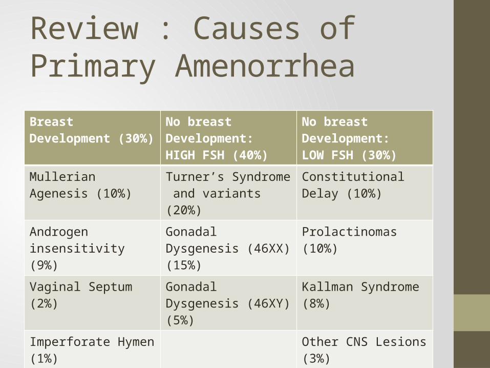
Review : Causes of Primary Amenorrhea
Breast Development (30%)
No breast Development:HIGH FSH (40%)
No breast Development:LOW FSH (30%)
Mullerian Agenesis (10%) Turner’s Syndrome and variants (20%)
Constitutional Delay (10%)
Androgen insensitivity (9%)
Gonadal Dysgenesis (46XX) (15%)
Prolactinomas (10%)
Vaginal Septum (2%) Gonadal Dysgenesis (46XY) (5%)
Kallman Syndrome (8%)
Imperforate Hymen (1%) Other CNS Lesions (3%)
Constitutional Delay (8%) Stress, weight loss, anorexia (3%)PCOS (3%)
Other (4%)

Review: Evaluation of Amenorrhea• Patient History• OB/GYN: Pubertal development, premenstrual
symptoms, dysmenorrhea/cyclic abdominal pain• Past Medical History: chronic illness, exposure to
radiation, current medications• Social History: exercise, weight loss, illicit drug use• Family History: history of pubertal delay, infertility, • Review of Systems: anosmia, galactorrhea,
headaches, visual changes, hirsutism or acne, s/sx of thyroid disease, vasomotor symptoms

Review: Evaluation of Amenorrhea• Physical Exam• Growth chart/BMI• Secondary sexual characteristics: Tanner staging,
breast development, pubic hair• Dysmorphic features: webbed neck, short stature,
widely spaced nipples• Hirsute features, Acne• Thyroid exam• Pelvic exam: rudimentary or absent uterus, transverse
vaginal septum, imperforate hymen, virilization, clitiromegaly

Physician 2006


Case• 16 year old African American Female presenting to her
primary care physician with absence of menarche• On exam• Minimal axillary hair tanner stage II• Tanner stage II breast development• Pelvic exam not performed
• Laboratory Values• FSH >20 and LH >40 • TSH, PRL normal
• Ultrasound Performed• Absence of reproductive organs

Work-up• What do you want to do now?• Karyotype: 46XY• Diagnosis: Swyer Syndrome
• Now what?• Patient taken to the operating room to for bilateral gonadectomy• Operative findings: rudimentary uterus, streak gonads, and a 1 cm
nodule along the area of the left gonad.• Histopathology revealed a seminoma of the left gonad
• Follow up• Patient referred to gynecological oncology
• Negative AFP, B-hcg, LDH, CMP, CXR, Abdominopelvis CT • Surveillance
• On hormone replacement therapy• Regular follow up with GYN ONC and REI

Questions??

Resources• Alvarez NR et al. Complete Androgen Insensitivity Syndrome: The Role of the Endocrine Surgeon. The American Surgeon.
2005: 71; 241-243• Bekx et al. Characteristics of Adolescents Presenting to a Multidisciplinary Clinic for Polycystic Ovarian Syndrome. J Pediatric
Adolesc Gynecol. 2010. 23:7-10• Butler WJ, Price TM. Sexual Development and puberty. Precis: an update in obstetrics and gynecology. Reproductive
endocrinology, 3rd ed. 2007.• Cameron et al. Non-Chromosomal, Non-Iatrogenic Premature Ovarian Failure in an Adolescent Population: A Case Series.
Pediatric Adolescent Gynecology. 2009: 21(3) pg 3-8.• Current Evaluation of Amenorrhea. Fertility and Sterility 2008 80(3): S219-S225.• Deligeoroglou et al. Evaluation and management of adolescent amenorrhea. Annals of the New York Academy of Sciences.
2010; pg 23-32• Fideleff HL, et al. Prolactinoma in Children and Adolescents. Horm Res. 2009; 72:192-205• Garcia-Mayor , RV, Andrade MA, Rios M, Lage M, Diquex C, Casanueva FF, Serum Leptin levels in normal children: relationship
to age, gender, body mass index, pituitary-gonadal hormones, and pubertal stage. J of Clinical Endocrinology and Metabolism. 82: 8249, 1997.
• Kataoka, ML. Togashi K, Yamaoka T, Koyama T, Ueda H, Kobayashi H, Rahman M, Higuchi T, Fujii S. Posterior cul-de-sac obliteration associated with endometriosis: MR Imaging Evaluation. Radiology. March 2005. (234) 815-823
• Lindenman et al. Mullerian Agenesis: An Update. Obstetrics and Gynecology. 1997. 90(2): 307-312. • Massin et al. Idiopathic Premature Ovarian Failure in 63 Young Women. Horm Res. 2006; 65:89-95.• Master-Hunter T and Heiman D. Amenorrhea: Evaluation and Treatment. American Family Physician. 2006. 73 (8): 1374-1382.• Speroff and Fritz. Clinical Gynecological Endocrinology and Infertility: Seventh Edition. 2005. • Sultan et al. Disorders of Androgen Action. Seminars in Reproductive Medicine. 2002: 20(3); 217-227• Fideleff HL, et al. Prolactinoma in Children and Adolescents. Horm Res. 2009; 72:192-205• Timmreck LS and Reindollar RH. Contemporary issues in primary amenorrhea. Obstetrics and Gynecology Cinics of North
America. 2003: (30); 287-302• Witkop et al. Understanding the Spectrum of the Female Athlete Triad. Obstetrics and Gynecology. 2010; 116(6): 1444-1448• Wu T, Mendola P, Buck GM. Ethnic differences in the presence of secondary sex characteristics and menarche among US
girls: the Third National Health and Nutrition Examination Survey. 1988- 1994. Pediatrics. 110: 752, 2002.