ESTHETIC AND FUNCTIONAL REHABILITATION OF MAXILLARY...
4
Sarswat A et al. Richmond Crown case report 68 HECS International Journal of Community Health and Medical Research |Vol. 5|Issue 3| July – September 2019 Harsukh Educational Charitable Society International Journal of Community Health and Medical Research Journal home page:www.ijchmr.com doi: 10.21276/ijchmr ISSN E: 2457-0117 ISSN P: 2581-5040 Index Copernicus ICV 2018=62.61 Case Report ESTHETIC AND FUNCTIONAL REHABILITATION OF MAXILLARY ANTERIOR TEETH BY RICHMOND CROWN: A CASE REPORT 1 Anika Saraswat, 2 Ajay Nagpal , 3 Rohit Paul, 4 Raghav Pratap, 1,4 Post graduate student, 2 Reader, 3 Professor and Head Department of Conservative Dentistry and Endodontics, K. D Dental College and Hospital. Mathura- 281006. India ABSTRACT When endodontically treated teeth with the loss of coronal tooth structure are left untreated for a long period they may undergo supra- eruption, drifting, tipping, and rotation of adjacent and opposing teeth. This may be challenging to the clinician, when fabricating a crown because of inadequate interocclusal space. Patients with reduced interocclusal clearance and having very steep incisal guidance are most difficult to manage. Richmond crown is a feasible approach for such cases that can be performed with very less incisal clearance to accommodate post, core and crown thickness. KEYWORDS: Interocclusal, Incisal guidance, Richmond crown, Post, Core Corresponding author: Dr.Anika Saraswat; Post graduate student, Department of Conservative Dentistry and Endodontics, K. D Dental College and Hospital. Mathura- 281006. Uttar Pradesh, India This article may be cited as: Saraswat A, Nagpal A, Paul R, Pratap R . Esthetic And Functional Rehabilitation Of Maxillary Anterior Teeth By Richmond Crown: A Case Report HECS Int J Comm Health Med Res 2019; 5(3):68-71. NTRODUCTION To restore badly damaged endodontically treated teeth is a common problem faced by many restorative dentists. The broken teeth often require extra support from the root canal for the additional retention of the restoration. 1 In cases where the remaining crown structure is not sufficient to retain full-coverage crown, post and core is a treatment option to increase the retention and resistance form of tooth. 2 The major concern with the post and core procedure is fracture of post or root, dislodgement of post–core assembly, loss of the restorative seal, and injury to the periodontium. 3,4 The situation may be further worsening in patient with deep bite, which leads to maximum oblique forces. In such cases, there should be adequate core reduction, so that the required thickness for metal ceramic crown can be obtained for better esthetics. In 1878, the Richmond crown was introduced with a threaded tube incorporated into the root canal with a screw-retained crown. The Richmond crown was indicated for grossly decayed single tooth with very much reduced crown height and with increased deep bite and decreased overjet. 5 This case report illustrates the management of grossly decayed central and lateral incisor with reduced interocclusal space with Richmond crown by an unexacting and minimally invasive technique. CASE REPORT A 24-year-old male patient reported to Department of Conservative Dentistry and Endodontics with a chief complaint of fractured upper right front teeth and wanted it to be replaced with artificial teeth. On taking a detailed history, it was revealed that the patient underwent trauma 1 year back and his maxillary right central and lateral incisor got fractured. Clinical examination showed Ellis Class-III fracture and discoloration along with pain and tenderness irt #11,12.[Figure1] Radiographic evaluation revealed radiolucency involving pulp and periapical area irt #11,12 [Figure 2]. The diagnostic impressions were made, and model analysis was done to assess the amount of space available for restoration. It was found that there was increased overbite and decreased overjet to restore the tooth to normal function and esthetics, so it was planned to give a Richmond crown irt #11,12 to the patient. The entire treatment plan was explained to the patient and consent was obtained from the patient. I
Transcript of ESTHETIC AND FUNCTIONAL REHABILITATION OF MAXILLARY...
Sarswat A et al. Richmond Crown case report
68 HECS International Journal of Community Health and Medical Research |Vol. 5|Issue 3| July – September 2019
Harsukh Educational Charitable Society International Journal of Community Health and Medical Research
Journal home page:www.ijchmr.com doi: 10.21276/ijchmr
ISSN E: 2457-0117 ISSN P: 2581-5040 Index Copernicus ICV 2018=62.61
Case Report ESTHETIC AND FUNCTIONAL REHABILITATION OF MAXILLARY ANTERIOR TEETH BY RICHMOND CROWN: A CASE REPORT 1Anika Saraswat, 2Ajay Nagpal , 3Rohit Paul, 4 Raghav Pratap,
1,4Post graduate student, 2 Reader, 3Professor and Head Department of Conservative Dentistry and Endodontics, K. D Dental College and Hospital. Mathura- 281006. India
ABSTRACT When endodontically treated teeth with the loss of coronal tooth structure are left untreated for a long period they may undergo supra-eruption, drifting, tipping, and rotation of adjacent and opposing teeth. This may be challenging to the clinician, when fabricating a crown because of inadequate interocclusal space. Patients with reduced interocclusal clearance and having very steep incisal guidance are most difficult to manage. Richmond crown is a feasible approach for such cases that can be performed with very less incisal clearance to accommodate post, core and crown thickness.
KEYWORDS: Interocclusal, Incisal guidance, Richmond crown, Post, Core
Corresponding author: Dr.Anika Saraswat; Post graduate student, Department of Conservative Dentistry and Endodontics, K. D Dental College and Hospital. Mathura- 281006. Uttar Pradesh, India
This article may be cited as: Saraswat A, Nagpal A, Paul R, Pratap R . Esthetic And Functional Rehabilitation Of Maxillary Anterior Teeth By Richmond Crown: A Case Report HECS Int J Comm Health Med Res 2019; 5(3):68-71.
NTRODUCTION To restore badly damaged endodontically treated teeth is a common problem faced by many restorative dentists. The broken teeth often require extra support from the root canal for the additional retention of the restoration.1
In cases where the remaining crown structure is not sufficient to retain full-coverage crown, post and core is a treatment option to increase the retention and resistance form of tooth.2 The major concern with the post and core procedure is fracture of post or root, dislodgement of post–core assembly, loss of the restorative seal, and injury to the periodontium.3,4 The situation may be further worsening in patient with deep bite, which leads to maximum oblique forces. In such cases, there should be adequate core reduction, so that the required thickness for metal ceramic crown can be obtained for better esthetics.
In 1878, the Richmond crown was introduced with a threaded tube incorporated into the root canal with a screw-retained crown. The Richmond crown was indicated for grossly decayed single tooth with very much reduced crown height and with increased deep bite and decreased overjet.5
This case report illustrates the management of grossly decayed central and lateral incisor with reduced interocclusal space with Richmond crown by an unexacting and minimally invasive technique.
CASE REPORT
A 24-year-old male patient reported to Department of Conservative Dentistry and Endodontics with a chief complaint of fractured upper right front teeth and wanted it to be replaced with artificial teeth. On taking a detailed history, it was revealed that the patient underwent trauma 1 year back and his maxillary right central and lateral incisor got fractured. Clinical examination showed Ellis Class-III fracture and discoloration along with pain and tenderness irt #11,12.[Figure1] Radiographic evaluation revealed radiolucency involving pulp and periapical area irt #11,12 [Figure 2]. The diagnostic impressions were made, and model analysis was done to assess the amount of space available for restoration. It was found that there was increased overbite and decreased overjet to restore the tooth to normal function and esthetics, so it was planned to give a Richmond crown irt #11,12 to the patient. The entire treatment plan was explained to the patient and consent was obtained from the patient.
I
Sarswat A et al. Richmond Crown case report
69 HECS International Journal of Community Health and Medical Research |Vol. 5|Issue 3| July – September 2019
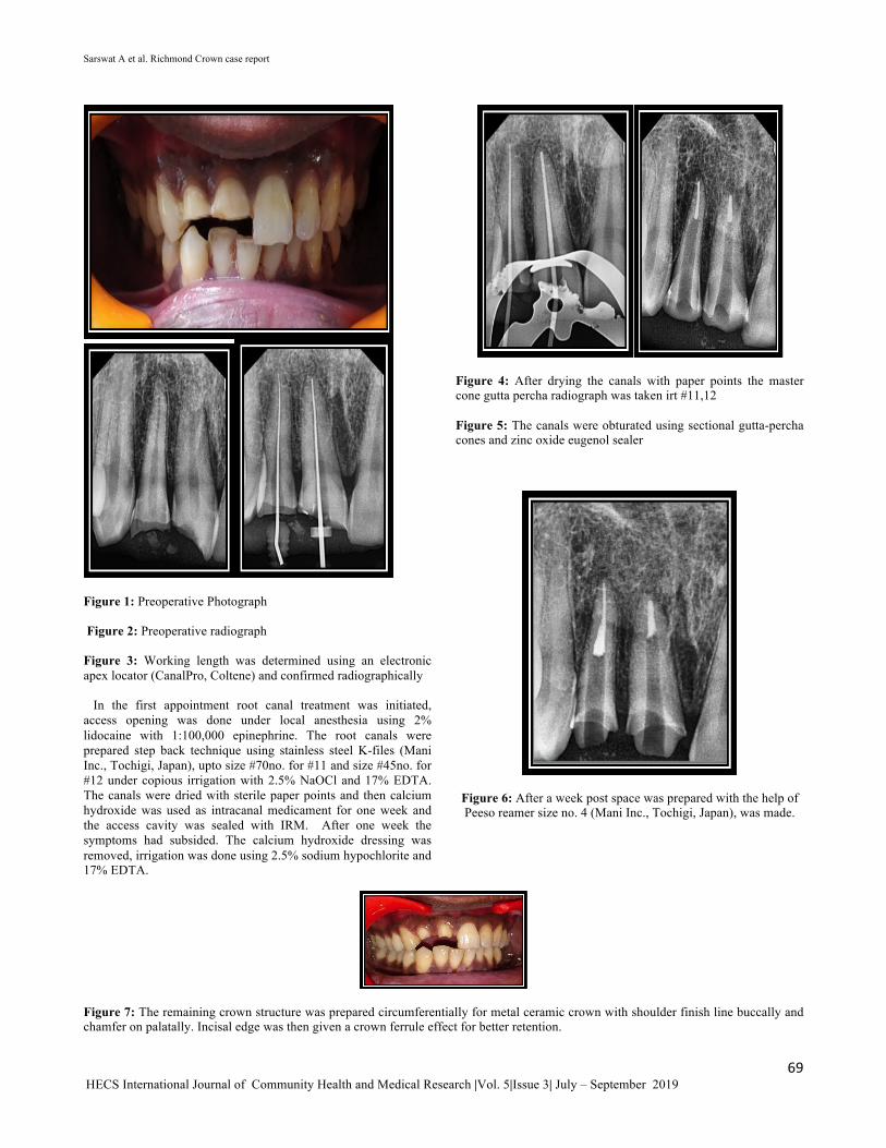
Figure 1: Preoperative Photograph
Figure 2: Preoperative radiograph
Figure 3: Working length was determined using an electronic apex locator (CanalPro, Coltene) and confirmed radiographically
In the first appointment root canal treatment was initiated, access opening was done under local anesthesia using 2% lidocaine with 1:100,000 epinephrine. The root canals were prepared step back technique using stainless steel K-files (Mani Inc., Tochigi, Japan), upto size #70no. for #11 and size #45no. for #12 under copious irrigation with 2.5% NaOCl and 17% EDTA. The canals were dried with sterile paper points and then calcium hydroxide was used as intracanal medicament for one week and the access cavity was sealed with IRM. After one week the symptoms had subsided. The calcium hydroxide dressing was removed, irrigation was done using 2.5% sodium hypochlorite and 17% EDTA.
Figure 4: After drying the canals with paper points the master cone gutta percha radiograph was taken irt #11,12
Figure 5: The canals were obturated using sectional gutta-percha cones and zinc oxide eugenol sealer
Figure 6: After a week post space was prepared with the help of Peeso reamer size no. 4 (Mani Inc., Tochigi, Japan), was made.
Figure 7: The remaining crown structure was prepared circumferentially for metal ceramic crown with shoulder finish line buccally and chamfer on palatally. Incisal edge was then given a crown ferrule effect for better retention.
Sarswat A et al. Richmond Crown case report
70 HECS International Journal of Community Health and Medical Research |Vol. 5|Issue 3| July – September 2019
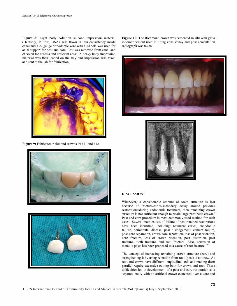
Figure 8: Light body Addition silicone impression material (Dentsply, Milford, USA), was flown in thin consistency inside canal and a 22 gauge orthodontic wire with a J-hook was used for axial support for post and core. Post was removed from canal and checked for defects and deficient areas. A heavy body impression material was then loaded on the tray and impression was taken and sent to the lab for fabrication.
Figure 9: Fabricated richmond crowns irt #11 and #12
Figure 10: The Richmond crown was cemented in situ with glass ionomer cement used in luting consistency and post cementation radiograph was taken
DISCUSSION
Whenever, a considerable amount of tooth structure is lost because of fracture/caries/secondary decay around previous restorations/during endodontic treatment, then remaining crown structure is not sufficient enough to retain large prosthetic crown.6 Post and core procedure is most commonly used method for such cases.7 Several main causes of failure of post retained restorations have been identified, including: recurrent caries, endodontic failure, periodontal disease, post dislodgement, cement failure, post-core separation, crown core separation, loss of post retention, core fracture, loss of crown retention, post distortion, post fracture, tooth fracture, and root fracture. Also, corrosion of metallic posts has been proposed as a cause of root fracture.8,9
The concept of increasing remaining crown structure (core) and strengthening it by using retention from root (post) is not new. As root and crown have different longitudinal axis and making them parallel require excessive cutting both for crown and root. These difficulties led to development of a post and core restoration as a separate entity with an artificial crown cemented over a core and

Sarswat A et al. Richmond Crown case report
71 HECS International Journal of Community Health and Medical Research |Vol. 5|Issue 3| July – September 2019
remaining tooth structure.10 This two-step technique improved marginal adaptation and allowed for a variation in the path of insertion of the crown.9 In coarse of time till today, different designs/techniques/materials have been evolved;6 however, no single system provides the perfect restorative solution for every clinical circumstance, and each situation requires an individual evaluation Although in present time the simplified “one-visit” prefabricated post are most commonly used; yet custom posts have their own advantages and indications so are still in use.7 Richmond crown 6,7,10,11 is not post and core system but it is customized, castable post and crown system as both are single unit and casted together. Design include casting of post and crown coping as single unit over which ceramic is fired and cemented onside canal and over prepared crown structure having same path of insertion. Ferrule collar is incorporated to increase mechanical resistance, retention apart from providing antirotational effect. Major technical drawback of this design is excessive cutting in making two different axis parallel which results in weakening of tooth and also this design increases stresses at post apex causing root fracture. Few indications for Richmond crown are grossly decayed or badly broken single tooth where remaining crown height is very less and incases with steep incisal guidance (deep bite and very less overjet). As less cervical tooth structure subjected to flexion forces under function and this design provides more cervical stiffening than other post system and is needed to protect the crown margins and to resist leakage. Case selection is very important here. The bulk of the remaining tooth above the restorative margin should be at least 1.5mm to 2mm to achieve resistance form. Even cases with steep incisal guidance are also subjected to more flexion forces along with very limited space for restoration. Such tooth if given with post and core first over which crown is cemented, needs adequate thickness which is a limitation here. To compensate this inadequacy if core is made thin then it is weak and also presents sharp margins and edges acting as stress points for overlying crown. Metal free crowns are predisposed to fracture whereas metal ceramic crowns tends to be a bulky crown in giving required thickness for metal coping and ceramic over it resulting in compromised esthetics. Richmond crown is best possibility in both these conditions as less crown cutting is required to make two axis parallel in grossly decayed tooth and also it require less thickness for best esthetic results. The advantages of this design are custom fitting to the root configuration, little or no stress at cervical margin, high strength, availability of considerable space for ceramic firing and incisal clearance, eliminate cement layer between core and crown so reduces chances of cement failure. Although certain disadvantages are time consuming, more appointments for patient, high cost, high modulus of elasticity than dentine (10 times greater than natural dentin), less retentive than parallel-sided posts, and acts as a wedge during occlusal load transfer. If ceramic fractures then it is difficult to retrieve and can lead to tooth fracture. The clinician must judge every situation on its individual merits and select a procedure that fulfills the needs of the case while maximizing retention and minimizing stress. Although in a clinical situation, success is dictated by the remaining tooth structure available after endodontic therapy.12,13,14
CONCLUSION
This clinical report describes the rehabilitation of a male patient with fractured maxillary central and lateral incisor with the Richmond crowns to improve the function and esthetics with a
minimally invasive procedure. There are many post-and-core materials/ techniques available to the clinician for a variety of clinical procedures and thus each clinical situation should be evaluated on an individual basis. Richmond crown is very much indicated in situations with very less incisal clearance to accommodate core + cement + crown thickness.
REFERENCES
1. Chakravarthy Y, Chamarthy S. Richmond crown for restoration of badly mutilated posterior teeth: A case report. J Evid Based Med Healthc 2015;2:4500-7.
2. Dausage P, Mallikarjuna K, Gupta S, Marya J. Richmond crown esthestics importance for lost tooth structure. Univ J Dent Sci 2015;3:60-3.
3. Zuckerman GR. Practical considerations and technical procedures for post-retained restorations. J Prosthet Dent 1996;75:135-9.
4. Sirimai S, Riis DN, Morgano SM. An in vitro study of the fracture resistance and the incidence of vertical root fracture of pulpless teeth restored with six post-and-coresystems. J Prosthet Dent 1999;81:262-9.
5. Mishra P, Mantri SS, Deogade S, Gupta P. Richmond crown: A lost state of art. Int J Dent Health Sci 2015;2:448-53.
6. Hudis SI, Goldstein GR. Restoration of endodontically treated teeth: a review of the literature. J Prosthet Dent. 1986;55:33-38.
7. Fernandes AS, Dessai GS. Factors affecting the fracture resistance of post-core reconstructed teeth: a review. Int J Prosthodont. 2001 Jul-Aug;14(4):355-63.
8. Purton DG, Payne JA. Comparison of carbon fiber and stainless steel root canal posts. Quintessence Int. 1996;27:93-97.
9. Smith CT, Schuman NJ, Wasson W. Biomechanical criteria for evaluating prefabricated post-and-core systems: a guide for the restorative dentist. Quintessence Int. 1998;29:305-312.
10. Smith CT, Schuman N. Prefabricated post-and-core systems: an overview. Compend Contin Educ Dent. 1998;19:1013-1020.
11. Rupika G, Jagadish S, Shashikala K, and Keshava PS. Restoration of badly broken, endodontically treated posterior teeth. J Conserv Dent. 2009 Jul-Sep; 12(3): 123–128.
12. Khan F, Mishra SK. Restoration of anterior esthetics with Richmond crown. BLDE Univ J Health Sci 2017;2:109-11.
13. Priyanka Dausage et al Richmond Crown Esthestics Importance For Lost Tooth Structure. University JDent Scie 2015;3:1
14. Antariksha Dod et al. Managing fractured central incisor with Richmond Crown –A Case report Journal of Applied Dental and Medical Sciences 2016;2:1