PROKARYOTES, BACTERIA, & VIRUSES By carter reid. Eukaryotes v. Prokaryotes.
Upload
teresa-stevensonCategory
view
217download
0
Essential IdeaEssential Idea
Eukaryotes have a much more complex cell structure than prokaryotes.

All cells share certain characteristics♦ Cells tend to be microscopic.
♦ All cells are enclosed by a membrane.
♦ All cells are filled with cytoplasm.
♦ Plasma membranePlasma membrane
♦ Chromosomes (Chromosomes (carry genes) carry genes)
♦ Ribosomes Ribosomes (make proteins)(make proteins)

nucleus
cell membrane
organelles
There are two cell types:
1. Prokaryotes
2. Eukaryotes
ProkaryotesProkaryotes
Assessment StatementAssessment Statement
Draw and label a diagram of the ultrastructure of Escherichia coli (E. coli) as an example of a prokaryote. The diagram should show the cell wall, plasma membrane, cytoplasm, pili, flagella, ribosomes and nucleoid (region containing naked DNA).

Draw and label a prokaryoteDraw and label a prokaryote
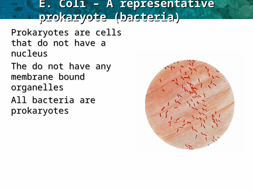
E. Coli – A representative prokaryote E. Coli – A representative prokaryote (bacteria)(bacteria)
Prokaryotes are cells that Prokaryotes are cells that do not have a nucleusdo not have a nucleus
The do not have any The do not have any membrane bound membrane bound organellesorganelles
All bacteria are prokaryotesAll bacteria are prokaryotes

Diagram of E. ColiDiagram of E. Coli

ProkaryotesProkaryotes
The general size of a prokaryotic The general size of a prokaryotic cell is about 1-2 um.cell is about 1-2 um.Note the absence of membrane Note the absence of membrane bound organellesbound organellesThere is There is no true nucleus no true nucleus with a with a nuclear membranenuclear membraneTheThe ribosome's are smaller ribosome's are smaller than than eukaryotic cellseukaryotic cellsThe slime capsule is used as a The slime capsule is used as a means of attachment to a surfacemeans of attachment to a surfaceOnly flagellate bacteria have the Only flagellate bacteria have the flagellumflagellumPlasmids are very small circular Plasmids are very small circular pieces of DNA that maybe pieces of DNA that maybe transferred from one bacteria to transferred from one bacteria to another.another.

ProkaryotesProkaryotes
• Example Eshericha coli (E. coli)
• In a prokaryotic cell, DNA is In a prokaryotic cell, DNA is circular and in the cytoplasm circular and in the cytoplasm called the nucleoidcalled the nucleoid

Prokaryote cellular structure functionProkaryote cellular structure function
Assessment StatementAssessment StatementAnnotate the E. coli diagram with the functions of each
named structure.

Annotate: Label & FunctionAnnotate: Label & Function

Prokaryote cellular structure functionProkaryote cellular structure function
Cell Wall:Cell Wall:
Made of a murein (not cellulose), which is a glycoprotein or peptidoglycan (i.e. a Made of a murein (not cellulose), which is a glycoprotein or peptidoglycan (i.e. a protein/carbohydrate complex). There are two kinds of bacterial cell wall, which are protein/carbohydrate complex). There are two kinds of bacterial cell wall, which are identified by the Gram Stain technique when observed under the microscope. Gram identified by the Gram Stain technique when observed under the microscope. Gram positive bacteria stain purple, while Gram negative bacteria stain pink. The technique is positive bacteria stain purple, while Gram negative bacteria stain pink. The technique is still used today to identify and classify bacteria. We now know that the different staining still used today to identify and classify bacteria. We now know that the different staining is due to two types of cell wallis due to two types of cell wall
Plasma membrane:Plasma membrane:
Controls the entry and exit of substances, pumping some of them in by active transport.Controls the entry and exit of substances, pumping some of them in by active transport.
Cytoplasm:Cytoplasm:
Contains all the enzymes needed for all metabolic reactions, since there are no organelles.Contains all the enzymes needed for all metabolic reactions, since there are no organelles.
Ribosome:Ribosome:
The smaller (70 S) type are all free in the cytoplasm, not attached to membranes (like RER). The smaller (70 S) type are all free in the cytoplasm, not attached to membranes (like RER). They are used in protein synthesis which is part of gene expression.They are used in protein synthesis which is part of gene expression.
Nucleoid:Nucleoid:
Is the region of the cytoplasm that contains DNA. It is not surrounded by a nuclear Is the region of the cytoplasm that contains DNA. It is not surrounded by a nuclear membrane. DNA is always a closed loop (i.e. a circular), and not associated with any membrane. DNA is always a closed loop (i.e. a circular), and not associated with any proteins to form chromatin.proteins to form chromatin.

Prokaryote cellular structure functionProkaryote cellular structure function
Flagella:Flagella:
These long thread like attachments are generally considered to be for movement. They have These long thread like attachments are generally considered to be for movement. They have an internal protein structure that allows the flagella to be actively moved as a form of an internal protein structure that allows the flagella to be actively moved as a form of propulsion. The presence of flagella tends to be associated with the pathogenicity of the propulsion. The presence of flagella tends to be associated with the pathogenicity of the bacterium. The flagella is about 20nm in diameter. This structure should not be confused bacterium. The flagella is about 20nm in diameter. This structure should not be confused with the eUkaryotic flagella seen in protoctista.with the eUkaryotic flagella seen in protoctista.
Pilli:Pilli:
These thread like projections are usually more numerous than the flagella. They are These thread like projections are usually more numerous than the flagella. They are associated with different types of attachment. In some cases they are involved in the associated with different types of attachment. In some cases they are involved in the transfer of DNA in a process called conjugation or alternatively as a means of preventing transfer of DNA in a process called conjugation or alternatively as a means of preventing phagocytosis.phagocytosis.
Slime Capsule:Slime Capsule:
A thick polysaccharide layer outside of the cell wall, like the glycocalyx of eukaryotes. Used A thick polysaccharide layer outside of the cell wall, like the glycocalyx of eukaryotes. Used for sticking cells together, as a food reserve, as protection against desiccation and for sticking cells together, as a food reserve, as protection against desiccation and chemicals, and as protection against phagocytosis. In some species the capsules of chemicals, and as protection against phagocytosis. In some species the capsules of many cells in a colony fuse together forming a mass of sticky cells called a biofilm. many cells in a colony fuse together forming a mass of sticky cells called a biofilm. Dental plaque is an example of a biofilm.Dental plaque is an example of a biofilm.

Prokaryote cellular structure functionProkaryote cellular structure function
Plasmids:Plasmids:
Extra-nucleoid DNA of up to 400 kilobase pairs. Plasmids can self-replicate particularly Extra-nucleoid DNA of up to 400 kilobase pairs. Plasmids can self-replicate particularly before binary fission. before binary fission.
They are associated with conjunction which is horizontal gene transfer. They are associated with conjunction which is horizontal gene transfer.
It is normal to find at least one anti-biotic resistance gene within a plasmid. This should not It is normal to find at least one anti-biotic resistance gene within a plasmid. This should not be confused with medical phenomena but rather is an ecological response to other be confused with medical phenomena but rather is an ecological response to other antibacterial compounds produced by other microbes. Commonly fungi will produce anti-antibacterial compounds produced by other microbes. Commonly fungi will produce anti-bacterial compounds which will prevent the bacteria replicating and competing with the bacterial compounds which will prevent the bacteria replicating and competing with the bacteria for a resource. bacteria for a resource.
conjugationconjugation
Direct contact between bacterial cells in which plasmid DNA is transferred between a donor Direct contact between bacterial cells in which plasmid DNA is transferred between a donor cell and a recipient cell.cell and a recipient cell.
There is no equal contribution to this process, no fertilisation and no zygote formation. It There is no equal contribution to this process, no fertilisation and no zygote formation. It cannot therefore be regarded as sexual reproduction.cannot therefore be regarded as sexual reproduction.

Binary Fission – Asexual Reproduction in ProkaryotesBinary Fission – Asexual Reproduction in Prokaryotes
Prokaryotes reproduce by Prokaryotes reproduce by binary fissionbinary fission
They copy their They copy their circular circular chromosomechromosome
The cell grows longer with The cell grows longer with the two chromosomes the two chromosomes attached to the inside attached to the inside membranemembrane
The membrane The membrane pinches pinches together in the centretogether in the centre
Two daughter cells are Two daughter cells are formedformed

Prokaryote Binary FissionProkaryote Binary Fission(a)(a). Reproduction signa. Reproduction signal: The cell receives a signal, of l: The cell receives a signal, of
internal or external origin that initiates the cell internal or external origin that initiates the cell division. E.coli replicates about once every 40 division. E.coli replicates about once every 40 minutes when incubated at 37minutes when incubated at 37oo C. If however we C. If however we increase the concentration of carbohydrate nutrients increase the concentration of carbohydrate nutrients that the cell is supplied with then the division time that the cell is supplied with then the division time can be reduced to 20 minutes. There is a suggestion can be reduced to 20 minutes. There is a suggestion here that an external signal (nutrient concentration) here that an external signal (nutrient concentration) is acting as the reproductive signal.is acting as the reproductive signal.
(b). Replication of DNA(b). Replication of DNA: bacterial cells have a single : bacterial cells have a single condensed loop of DNA. This is copied by a process condensed loop of DNA. This is copied by a process known as semi-conservative replication to produce known as semi-conservative replication to produce two copies of the DNA molecule one for each of the two copies of the DNA molecule one for each of the daughter cells. The replication begins at a single daughter cells. The replication begins at a single point (point (oriori)on the loop of DNA. The process proceeds )on the loop of DNA. The process proceeds around the loop until two loop have been produced, around the loop until two loop have been produced, each a copy of the original. The process finishes at each a copy of the original. The process finishes at a single point on the loop of DNA called the a single point on the loop of DNA called the terter position.position.

Prokaryote Binary FissionProkaryote Binary Fission
((c). Segregation of DNA:c). Segregation of DNA: One DNA loop will be One DNA loop will be provided for each of the daughter cells.provided for each of the daughter cells.
As the new loops form the As the new loops form the ori ori site becomes site becomes attached to some contractile proteins that pull the attached to some contractile proteins that pull the two ori sites, and therefore the loops, to opposite two ori sites, and therefore the loops, to opposite ends of the cell. This is an active process that ends of the cell. This is an active process that requires the bacteria to use energy for the requires the bacteria to use energy for the segregation.segregation.
(d). Cytokinesis: (d). Cytokinesis: Cell separation.Cell separation.
This occurs once the DNA loop replication and This occurs once the DNA loop replication and segregation is complete. The DNA completes a segregation is complete. The DNA completes a process of condensing whilst the plasma process of condensing whilst the plasma membrane begins to form a 'waist' or constriction membrane begins to form a 'waist' or constriction in the middle of the cell. As the plasma membrane in the middle of the cell. As the plasma membrane begins to pinch and constrict the membrane fuses begins to pinch and constrict the membrane fuses and seals with additional new membrane also and seals with additional new membrane also being formed.being formed.


Prokaryote cellProkaryote cell
A typicalrod-shapedbacterium
A thin section through thebacterium Bacilluscoagulans (TEM)
Pili
Nucleoid
Ribosomes
Plasmamembrane
Cell wall
Capsule
Flagella
Bacterialchromosome
0.5 µm

Binary Fission AnimationBinary Fission Animation
http://www.classzone.com/books/hs/ca/sc/http://www.classzone.com/books/hs/ca/sc/bio_07/animated_biology/bio_07/animated_biology/bio_ch05_0149_ab_fission.htmlbio_ch05_0149_ab_fission.html

Prokaryotes AnimationProkaryotes Animation
Watch Animation of Prokaryotes Structure and Watch Animation of Prokaryotes Structure and function.function.
http://www.wiley.com/legacy/college/boyer/http://www.wiley.com/legacy/college/boyer/0470003790/animations/cell_structure/0470003790/animations/cell_structure/cell_structure.swfcell_structure.swf
http://highered.mcgraw-hill.com/olcweb/cgi/http://highered.mcgraw-hill.com/olcweb/cgi/pluginpop.cgi?it=swf::500::500::/sites/dl/pluginpop.cgi?it=swf::500::500::/sites/dl/free/0073375225/594358/free/0073375225/594358/BinaryFission.swf::BinaryFissionBinaryFission.swf::BinaryFission

Eurkaryotic Cell CharacteristicsEurkaryotic Cell Characteristics
Eukaryotic cells have a nucleus that contain its DNA
Eukaryotic cells have membrane-bound organelles. Nucleus Mitochondria Chloroplasts (plants only)
Eukaryotes are bigger than Prokaryotes
Animal and plant cells are eukaryotes

HHuman Liver Celluman Liver Cellss

Draw a Eukaryotic Liver CellDraw a Eukaryotic Liver Cell
Assessment StatementAssessment Statement
Draw and label a diagram of the ultrastructure of a liver cell as an example of an animal cell. The diagram should show free ribosomes, rough endoplasmic reticulum (rER), lysosome, Golgi apparatus, mitochondrion and nucleus. The term Golgi apparatus will be used in place of Golgi body,

Draw a Eukaryotic Liver CellDraw a Eukaryotic Liver Cell
N: NucleusN: Nucleus
PM: plasma membranePM: plasma membrane
M: mitochondriaM: mitochondria
rER: Rough endoplasmic rER: Rough endoplasmic reticulumreticulum
GA: Golgi apparatusGA: Golgi apparatus
L: LysosomeL: Lysosome
MV: MicrovilliMV: Microvilli

Annotate DiagramAnnotate Diagram
Assessment Statement
Annotate the E. coli diagram with the functions of each named structure.

Annotate: Label & FunctionAnnotate: Label & Function

NucleusNucleus
Nucleus: Nucleus: This is the largest of the This is the largest of the organelles. The nucleus contains organelles. The nucleus contains the chromosomes which during the chromosomes which during interphase are to be found the interphase are to be found the nucleolus.nucleolus.
The nucleus has a The nucleus has a double double membranemembrane with pores(NP). with pores(NP).
The The nucleus controls the cells nucleus controls the cells functions functions through the expression of through the expression of genes. genes.
Some cells are Some cells are multi nucleated multi nucleated such as the such as the muscle fibremuscle fibre

Plasma MembranePlasma Membrane
Plasma membrane: Plasma membrane: controls which controls which substances can enter and exit a cell. substances can enter and exit a cell. It is a fluid structure that can It is a fluid structure that can radically change shape.radically change shape.
The membrane is a double layer of The membrane is a double layer of water repellant molecules. water repellant molecules.
Receptors in the outer surface Receptors in the outer surface detect signals to the cell and relay detect signals to the cell and relay these to the interior.these to the interior.
The membrane has pores that run The membrane has pores that run through the water repellant layer through the water repellant layer called channel proteins.called channel proteins.

MitochondriaMitochondria:..:..
MitochondriaMitochondria: location of aerobic : location of aerobic respiration and a major synthesis of ATP respiration and a major synthesis of ATP region..region..
Double membrane Double membrane organelle.organelle.
Inner membrane has folds called cristae. Inner membrane has folds called cristae. This is the site of oxidative This is the site of oxidative phosphorylation. phosphorylation.
Centre of the structure is called the matrix Centre of the structure is called the matrix and is the location of the and is the location of the Krebs cycle. Krebs cycle.
Oxygen is consumed in the Oxygen is consumed in the synthesis of synthesis of ATP ATP on the inner membraneon the inner membrane
The The more active more active a cell the greater the a cell the greater the number of number of mitochondriamitochondria..

Rough endoplasmic reticulumRough endoplasmic reticulum (rER). (rER).
rER form a network of rER form a network of tubules with a maze like tubules with a maze like structure.structure.
In general these run away In general these run away from the nucleus from the nucleus
The 'rough' on the reticulum The 'rough' on the reticulum is caused by the presence of is caused by the presence of ribosomes.ribosomes.
Proteins made here are Proteins made here are secreted out of the cellsecreted out of the cell

Ribosomes:Ribosomes:
the free ribosome the free ribosome produces proteins for produces proteins for internal use within the internal use within the cell.cell.

Golgi apparatusGolgi apparatus..
Modification of proteins Modification of proteins prior to secretion.prior to secretion.
proteins for secretion are proteins for secretion are modified modified
possible addition of possible addition of carbohydrate or lipid carbohydrate or lipid components to proteincomponents to protein
packaged into vesicles packaged into vesicles for secretionfor secretion

Lysozyme:Lysozyme:
Vesicles in the above Vesicles in the above diagram that have diagram that have formed on the golgi formed on the golgi apparatus.apparatus.
Containing hydrolytic Containing hydrolytic enzymes.enzymes.
Functions include the Functions include the digestion of old digestion of old organelles, engulfed organelles, engulfed bacteria and viruses.bacteria and viruses.

Vocabulary Practice (12 mnutes)Vocabulary Practice (12 mnutes)
1. free ribosomes,
2. rough endoplasmic reticulum (rER)
3. lysosome,
4. Golgi apparatus,
5. mitochondrion
6. and nucleus
3 minutes 3 minutes – One student – One student gives function –the other gives function –the other identifies organelleidentifies organelle
3 minutes 3 minutes -- Switch-- Switch
3 minutes 3 minutes – One student – One student gives organelle –the gives organelle –the other describes functionother describes function
3 minutes 3 minutes -- Switch-- Switch

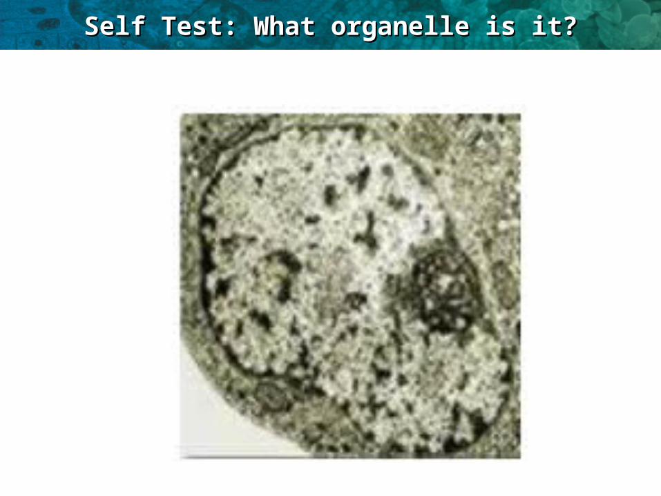
IB Assessment StatementIB Assessment Statement
Identify structures from liver in electron micrographs of liver cells.

VideosVideos
Video about Animal CellsVideo about Animal Cells
http://www.youtube.com/watch?http://www.youtube.com/watch?v=cj8dDTHGJBY&feature=BFa&list=PL3EED4C1D684D3Av=cj8dDTHGJBY&feature=BFa&list=PL3EED4C1D684D3ADF&lf=contextDF&lf=context
Video about Plant CellsVideo about Plant Cells
http://www.youtube.com/watch?http://www.youtube.com/watch?v=9UvlqAVCoqY&list=SP3EED4C1D684D3ADFv=9UvlqAVCoqY&list=SP3EED4C1D684D3ADF
Video about Cells in general Video about Cells in general
https://www.youtube.com/watch?https://www.youtube.com/watch?v=yZu6DfcPHr8&feature=player_embedded#t=6v=yZu6DfcPHr8&feature=player_embedded#t=6

Self Test: What organelle is it?Self Test: What organelle is it?

Self Test: What organelle is it?Self Test: What organelle is it?

Self Test: What organelle is it?Self Test: What organelle is it?
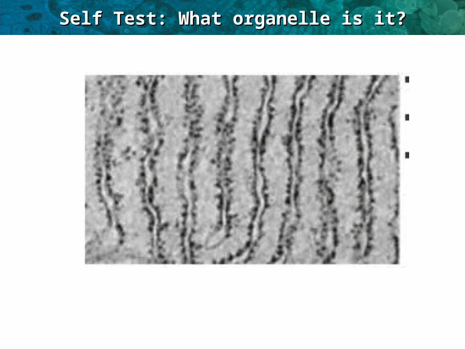
Self Test: What organelle is it?Self Test: What organelle is it?
Self Test: What organelle is it?Self Test: What organelle is it?

IB Assessment StatementIB Assessment Statement
2.3.4 Compare prokaryotic and eukaryotic cells.

Only organisms of the Only organisms of the domains Bacteria and domains Bacteria and ArchaeaArchaea consist of prokaryotic cells consist of prokaryotic cells
Protists, fungi, animals, and plantsProtists, fungi, animals, and plants all consist all consist of eukaryotic cellsof eukaryotic cells
ProkaryoteProkaryote vsvs. Eukaryote. Eukaryote

Prokaryote cellProkaryote cell
A typicalrod-shapedbacterium
A thin section through thebacterium Bacilluscoagulans (TEM)
0.5 µm
Pili
Nucleoid
Ribosomes
Plasmamembrane
Cell wall
Capsule
Flagella
Bacterialchromosome

Eukaryote Eukaryote cellscells
Flagellum
Centrosome
CYTOSKELETON
Microfilaments
Intermediate filaments
Microtubules
Peroxisome
Microvilli
ENDOPLASMIC RETICULUM (ER
Rough ER Smooth ER
MitochondrionLysosome
Golgi apparatus
Ribosomes:
Plasma membrane
Nuclear envelope
NUCLEUS
In animal cells but not plant cells: LysosomesCentriolesFlagella (in some plant sperm)
Nucleolus
Chromatin

Prokaryotes vs. EukaryotesProkaryotes vs. Eukaryotes
nucleus
cell membrane
organelles
cytoplasm
Prokaryotic vs. EukaryoticProkaryotic vs. Eukaryotic

Prokaryotic vs. EukaryoticProkaryotic vs. Eukaryotic

IB ASSESSMENT STATEMENTIB ASSESSMENT STATEMENT
State three differences between plant and animal cells.

EukaryotesEukaryotes
• All eukaryotes have the same following All eukaryotes have the same following components:components:• RibosomesRibosomes• MitochondriaMitochondria• NucleusNucleus• Endoplasmic Reticulum (ER) Endoplasmic Reticulum (ER) • Rough ERRough ER• Golgi body apparatusGolgi body apparatus

Eukaryotic: Plant vs. AnimalEukaryotic: Plant vs. Animal
PlantsPlants Cell WallCell Wall
ChloroplastsChloroplasts
General Rectangular General Rectangular ShapeShape
Large VacuolesLarge Vacuoles
Stores polysaccharide in Stores polysaccharide in the form of STARCHthe form of STARCH
AnimalsAnimals NO cell wallNO cell wall
NO ChloroplastNO Chloroplast
Irregular shapeIrregular shape
Small VacuolesSmall Vacuoles
Stores polysaccharide in Stores polysaccharide in the form of Glycogenthe form of Glycogen

LE 6-9aLE 6-9a
Flagellum
Centrosome
CYTOSKELETONMicrofilaments
Intermediate filaments
Microtubules
Peroxisome
Microvilli
ENDOPLASMIC RETICULUM (ER
Rough ER Smooth ER
Mitochondrion Lysosom
e
Golgi apparatus
Ribosomes:
Plasma membrane
Nuclear envelope
NUCLEUS
In animal cells but not plant cells: LysosomesCentriolesFlagella (in some plant sperm)
NucleolusChromatin

LE 6-9bLE 6-9b
Roughendoplasmicreticulum
In plant cells but not animal cells: ChloroplastsCentral vacuole and tonoplastCell wallPlasmodesmata
Smoothendoplasmicreticulum
Ribosomes(small brown dots)
Central vacuole
Microfilaments
Intermediatefilaments
Microtubules
CYTOSKELETON
Chloroplast
Plasmodesmata
Wall of adjacent cell
Cell wall
Nuclearenvelope
Nucleolus
Chromatin
NUCLEUS
Centrosome
Golgiapparatus
Mitochondrion
Peroxisome
Plasmamembrane

A representative plant cell and a diagram of a ‘’Typical’’ Plant A representative plant cell and a diagram of a ‘’Typical’’ Plant CellCell

Other Eukaryotes – ProtistsOther Eukaryotes – ProtistsParamecium and AmeobaParamecium and Ameoba

Plants vs. AnimalsPlants vs. Animals

Plant Vs. AnimalPlant Vs. Animal

Nature of ScienceNature of Science
Developments in scientific research follow improvements in apparatus—the invention of electron microscopes led to greater understanding of cell structure. (1.8)

http://www.history-of-the-microscope.orghttp://www.history-of-the-microscope.org
Assignment: History of the Microscope Assignment: History of the Microscope WebquestWebquest

The electron microscopeThe electron microscope
• Invented in Germany (1930’s)
• Able to view samples 200 times smaller than light microscopes
• Invention of the electron microscope revealed a whole
new level of cellular detail.

Resolution - comparisonResolution - comparison
Resolution
Millimetres (mm)
Micrometres ( m)
Nanometres (nm)
Eye 0.1 100 100,000
Light Microscope
0.0002 0.2 200
Electron microscope
0.000001 0.001 1