Microbial profile of symptomatic pericoronitis lesions: a ...
Upload
porag25Category
view
1.014download
5
Prepared by :
Dr.Afiqur Rahman SouravIntern Doctor
Department Of Paediatric Dentistry
Dhaka Dental College Hospital

• What is it?
A transitory type of gingivitis is often
observed in young children when the
primary or permanent teeth are
erupting.
Often localized
Associated with difficult eruption,
Subsides after the teeth emerge into
the oral cavity.


Name: Ubaidul
IslamAge: 6 years 2months
Address: Mirpur
Date : 5th
November’14
ERUPTION GINGIVITIS

• The greatest increase in the incidence of
gingivitis in children is often seen in the 6-
to 7-year age group when the permanent
teeth begin to erupt.
• This inflammation is most commonly
associated with the eruption of the first
and second permanent molars, and the
condition can be painful.

• This increase in gingivitis apparently occurs
because the gingival margin receives no
protection from the coronal contour of the tooth
during the early stage of active eruption, where
Food debris, materia alba, and bacterial plaque
often collect around and beneath the free tissue,
partially cover the crown of the erupting tooth,
and cause the development of an inflammatory
process.


• Pain
• Redness of the Gingiva
• Swelling of the Gingiva
• Bleeding on probing
• Anorexia & Dysphagia
• Mostly heals after complete eruption of the teeth
• In cases of Molar,if pericoronitis develops then pus
discharge sometimes associated with Lymphnode
enlargement and fever.
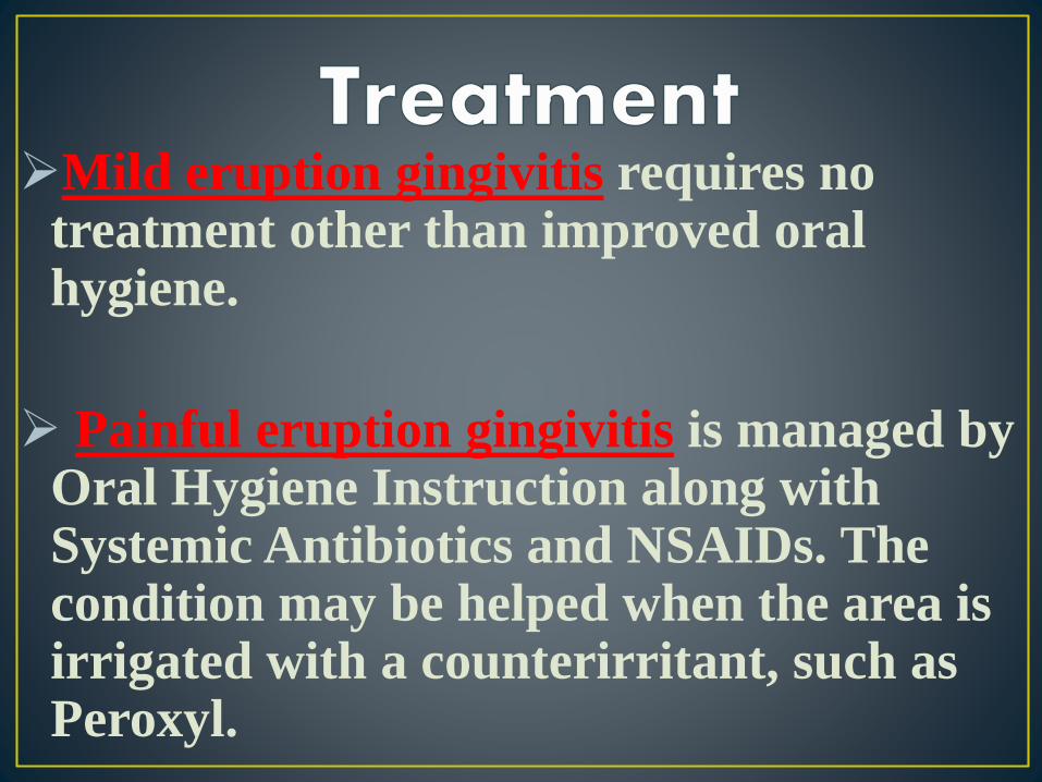
Mild eruption gingivitis requires no treatment other than improved oral hygiene.
Painful eruption gingivitis is managed by Oral Hygiene Instruction along with Systemic Antibiotics and NSAIDs. The condition may be helped when the area is irrigated with a counterirritant, such as Peroxyl.

• If it is Untreated and there is operculum
covering the erupting tooth partially,it ma
develop Pericoronitis and Pericoronal
Abscess.pericoronitis

What is it?
Pericoronitis is defined as inflammation
of the oral soft tissues surrounding the
crown of a partially erupted tooth.

• The major cause is the food and debris
impaction and microbial flora that develops
in the distally located pseudopocket.

Red, swollen, suppurating lesion that is exquisitely tender with
radiating pain to ear, throat and floor of mouth. It is usually
associated with fever and lymph node enlargement.
The diagnosis of pericoronitis is mainly clinical with three
distinct diagnostic categories recognized:
1) Acute pericoronitis,
2) Sub-acute pericoronitis, and
3) Chronic pericoronitis.

Acute Pericoronitis
Trismus, fever, pain, dysphagia, extraoral swelling,
malaise, halitosis, pus discharge, sore throat, and
anorexia. Pain may disturb sleep, lymphadenitis
involving the deep cervical lymph nodes may be
present.

Subacute Pericoronitis
Pain, dysphagia, intraoral swelling, halitosis, pus discharge,
sore throat. Associated pain is most often described as
continuous, dull, and is occasionally sharp or throbbing.
Unlike acute attacks, radiation of painful symptoms into
adjacent muscles is rare. The individual does not have
limited mouth opening. This is a distinguishing feature from
acute pericoronitis.

Chronic Pericoronitis
It is diagnosed based on a history of temporary dull
aching low grade pain that typically lasts only 1-2
days. Signs include palpable non-tender
submandibular lymph nodes and macerated buccal
tissue consistent with cheek biting.

1. Pericoronal abscess
2. It may spreads posteriorly in oropharyngeal area.
3. Dysphagia
4. Involvement of lymph nodes- posterior and deep
cervical.
5. Peritonsillar abscess.
6. Ludwig's angina

PERICORONITIS
•OPERCULECTOMY
•EXTRACTION (usually not done in children)

Operculectomy is contra-indicated in Acute
condition.So Acute condition is eliminated by-
NSAIDs to relieve pain.
-Acetaminophen, Ibuprofen etc.
Systemic Antibiotics when Fever and Lymph Node
Enlargement is present.
-Penicillin, Erythromycin, Cephalosporins etc.
Oral Hygiene Instructions.

Operculectomy is done in Three procedures:
•Scalpel
•Laser
•Electrocautary

Operculect
omy