
Equipment Available at UC Davis (underlined are...
9
Equipment Available at UC Davis (underlined are updates in summer 2015) Ferrara Lab (shared with Doug Stephens) Equipment available in the Ferrara Lab is, unless otherwise mentioned, housed in the basement, second and third floors of Genomic and Biomedical Sciences Facility and includes: • Three Verasonics Vantage 256 systems with a wide range of transducers. Two systems have been synchronized to form a 512 element system. Real-time thermometry is available. Two systems are “HIFU” capable. • Imasonics 128 element HIFU array with embedded imaging. • Image Guided Therapy system for MRgFUS (Fig. 1-2). Includes annular array capable of delivering ~100 acoustic Watts, motion control system, thermal mapping and treatment planning, pulsers, delay generation and Bruker interface. LabfUS system with 16-element annular array, lambda/256 phase accuracy and high speed motion control. Integrates in real time with MR system using Thermoguide software. • 256-element Image Guided Therapy system for MRgFUS is currently on order and expected in the fall of 2015. • Modeling software: Field II, FOCUS, Custom Rayleigh Plesset, microbubble simulation packages, ChemDraw, Matlab, AsiPRO • Custom ultrasound transducers • Quantitative real-time PCR machine • Image-guided therapy system for real-time MR-US • Luxtron temperature sensor with extensions for MRI temperature measurement • Hydrophones for low intensity and high intensity calibration • 2 Siemens Antares ultrasound systems with research interfaces and custom transducers • 1 Siemens Sequoia ultrasound system housed in the Center for Molecular and Genomic Imaging (biomedical engineering) • 1 Siemens Sequoia ultrasound system housed in the School of Veterinary Medicine Figure 1. Overview of Themoguide system. Software is embedded within the MRI Paravision console for (upper) image reconstruction and transfer. In addition, an 8-core PC runs the bulk of the Thermoguide system including treatment planning, PID temperature control, trajectory control, temperature maps and thermal dose maps. MRI console communicates with MR coils; US console communicates with US array, motor controller, water heating and degassing system, and rf pulsing and receiving. Motors allow trajectories to travel at 3 cm/s, facilitating real-time mild hyperthermia. a b c d Figure 2. a. Bruker 7T with installed insert. b. View of insert within magnet bore (coil shown in background), insert is displaced to center of coil before imaging, c. rack of degassing, temperature control, pumps, amplifiers, timing controllers for array, d. Thermoguide and MR consoles. • Aurora Radio Frequency tracking system Volume Coil MRI phased Array Tissu e Motion controller Thermoguide C++ interface Paravision interface on MRI console Volume Coil Tissu e Motion controller Thermoguide C++ interface Paravision interface on MRI console Volume Coil Tissue Motion controller Thermoguide system Control- trajectory Heating of fluid to maintain temperature Motion correction Temperature and thermal dose estimation Optional MRI reconstruction Thermoguide C++ interface Paravision interface on MRI console Transducer Array Thermoguide in Paravision Embedded recon software- to continuously recon and transfer MRI images Direct Data Transfer
Transcript of Equipment Available at UC Davis (underlined are...

Equipment Available at UC Davis (underlined are updates in summer 2015) Ferrara Lab (shared with Doug Stephens) Equipment available in the Ferrara Lab is, unless otherwise mentioned, housed in the basement, second and third floors of Genomic and Biomedical Sciences Facility and includes: • Three Verasonics Vantage 256 systems with a wide range of transducers. Two systems have been
synchronized to form a 512 element system. Real-time thermometry is available. Two systems are “HIFU” capable.
• Imasonics 128 element HIFU array with embedded imaging. • Image Guided Therapy system for MRgFUS (Fig. 1-2). Includes annular array capable of delivering ~100
acoustic Watts, motion control system, thermal mapping and treatment planning, pulsers, delay generation and Bruker interface. LabfUS system with 16-element annular array, lambda/256 phase accuracy and high speed motion control. Integrates in real time with MR system using Thermoguide software.
• 256-element Image Guided Therapy system for MRgFUS is currently on order and expected in the fall of 2015.
• Modeling software: Field II, FOCUS, Custom Rayleigh Plesset, microbubble simulation packages, ChemDraw, Matlab, AsiPRO
• Custom ultrasound transducers • Quantitative real-time PCR machine • Image-guided therapy system for real-time MR-US • Luxtron temperature sensor with extensions for MRI temperature measurement • Hydrophones for low intensity and high intensity calibration • 2 Siemens Antares ultrasound systems with research interfaces and custom transducers • 1 Siemens Sequoia ultrasound system housed in the Center for Molecular and Genomic Imaging
(biomedical engineering) • 1 Siemens Sequoia ultrasound system housed in the School of Veterinary Medicine
Figure 1. Overview of Themoguide system. Software is embedded within the MRI Paravision console for (upper) image reconstruction and transfer. In addition, an 8-core PC runs the bulk of the Thermoguide system including treatment planning, PID temperature control, trajectory control, temperature maps and thermal dose maps. MRI console communicates with MR coils; US console communicates with US array, motor controller, water heating and degassing system, and rf pulsing and receiving. Motors allow trajectories to travel at 3 cm/s, facilitating real-time mild hyperthermia.
a b c d
Figure 2. a. Bruker 7T with installed insert. b. View of insert within magnet bore (coil shown in background), insert is displaced to center of coil before imaging, c. rack of degassing, temperature control, pumps, amplifiers, timing controllers for array, d. Thermoguide and MR consoles. • Aurora Radio Frequency tracking system
Volume Coil
MRI phased Array
Tissue
Motion controller
Thermoguide C++ interface
Paravision interface on MRI console
Volume Coil
Tissue
Motion controller
Thermoguide C++ interface
Paravision interface on MRI console
Volume Coil
Tissue
Motion controller
Thermoguide systemControl- trajectoryHeating of fluid to maintain temperatureMotion correctionTemperature and thermal dose estimationOptional MRI reconstruction
Thermoguide C++ interface
Paravision interface on MRI console
Transducer Array
Thermoguide in ParavisionEmbedded reconsoftware- to continuously recon and transfer MRI images
DirectData Transfer

• 5 microscopes, three upright, two inverting, one is Zeiss 510 confocal, integrated with high speed cameras, optics, one with motorized 3D acquisition of multiple white and fluorescent channels
• bioacoustics and ultrasonics laboratory. Includes several clinical ultrasound systems with research interfaces, microscopes, discrete pulsers and receivers, transducers, custom high frequency ultrasound system with real time capabilities
• Agilent network analyzer • AccuSizer • ENI Radio Frequency generator • Mettler Toledo Excellence Plus Balance 120g/ 31g x 0.1mg/0.01mg with HAUG deionizer warning light • Vapro vaper pressure osmometer • Infinite M1000 quadruple monochromator microplate reader • Supermicro 7046GT-TRF SuperServer • Ultrasound pulsers and receivers (4 sets of receivers, pulsers & amplifiers from Ritec, Panametrics, etc.) • Oscilloscopes • Arbitrary waveform generators • Imacon 100 million frame per second camera (7 CCD’s) (for viewing US contrast agent cavitation in super
realtime) • 30 ns, class 4, kHz PRF copper ion vapor laser • 2 Fume and 1 flow hoods • Fluorimeter • Particle Sizing Systems submicron and micron particle sizers • Zeta potential analyzer • Sonicaters (2 probe and 1 bath) • Particle extruder and purification columns • -20˚ freezer, large refrigerator • Incubators for cell culture and CAM model • Full tissue culture facility, including laminar flow hood microscope • Vacuum Oven • Custom gas injection system for contrast agent preparation • Shaking incubator – 5 x 2L capacity • 3 heated water baths and 5 heat blocks • Custom Radio Frequency heating device • Gel electrophoresis apparatus • NanoDrop spectrophotometer • Varian HPLCs – with analytical and preparative capabilities and Bioscan radiation detector • Rotary evaporator • Cryopreservation tank • Centrifuges: 1 benchtop refrigerated centrifuge (large capacity, Beckman 25R), 1 refrigerated
microcentrifuge (Heraeus Fresco), 1 standard microcentrifuge (Eppendorf 5415D), 2 small benchtop centrifuges (Thermo Centra CL2)
• Dessicators • Autoclave (Department) • High field FT-NMR (Chemistry Department) • IR (Chemistry Department) • Mass spectrometers (Campus and Chemistry Department) • Cold room (Department) • Darkroom • EPR (Campus) • 2-photon microscopy and Fluorescence recovery after photobleaching (FRAP) available from the campus
Optical Core facility • Lyophilizer • Band saw • Drill press • Filtered and weight enabled glove box

• BD FacSCAN (flow cytometry) Flow cytometry • The Ferrara group has access to additional flow cytometry instruments and cell sorters through the UC
Davis flow cytometry core. Histology/Pathology equipment The Ferrara group has access to a histology lab at the Center for Comparative Medicine with: • 4 computers • 2 small screening microscopes • dissecting microscope • five headed microscope • two headed microscope equipped with a Sony digital camera and a biosafety hood • 2 fume hoods • embedding station • microtome-staining station • two headed Olympus RX40 Microscopy Pathology is also performed by the Animal Resource Service facility under the direction of Dr. Steve Griffey. This facility can perform immunohistochemistry and has a beta type system capable of automatically scanning in the full resolution histology of an entire tumor. Other equipment Equipment available in shared facilities at the UC Davis includes: Center for Nano- and Micro-Manufacturing (CNM2) The College of Engineering (COE) at UC Davis has advanced facilities geared for nanotechnology research. The microfabrication is fully equipped with photolithography, etching, mask making, metal deposition, and furnaces, vacuum systems, and assembly. Examples of equipment include Solitec Spinner, Karl Suss mask aligner, Technics PECVD, E-beam evaporator, CHA Sputtering systems, Dektak Profilometer, RIE etching systems, SEM, and wet etch benches. To support nano-scale fabrication, the microfabrication facility is equipped with nano-imprint and SEM-based nanolithography tools. A more detailed list of equipment is presented in Table 1. Table 1. CNM2 Capabilities and Selected Equipment Capability Area Detail and Comments MOS Processing Litho: 0.25 um minimum feature size
Furnaces: MRL, Thermco, Oxidation, anneal, Etch: Extensive repertory of wet and dry etch chemistries Deposition: CVD dielectric (Si3N4 and SiO2) PVD, with exceptional diverse target set
Nanofabrication Litho: 4” nanoimprint tool E-beam definition tool Deposition: CVD, Evaporator, all metals
Physical and Chemical Analysis
Physical Analysis: SEM/EDS, TEM Surface Analysis: Auger Electrical Analysis : Parametric, thermal, emission, EIS Chemical Analysis: FTIR
MEMS Processing
Litho:Thin and thick, spun-on and solid-film photopolymers Flexible Photolith w/ backside alignment Bulk Silicon Etch: Wet and Dry Miscellaneous: Wafer Bonding (various processes)
Silicon Design Full suite of Device and Process Modeling tools available.
Equipment: Furnaces: MRL Furnace LFE Barrel Asher Imperial IV Scanning Electron

Figure 3: Metalorganic Chemical Vapor Deposition System with capability to grow six, two inch wafers.
Microprocessor Oven Microscope Joel 840 Thermco Furnace LAM Research 590 E-beam writing tool (FEI) Nanometrics Jetfirst 150 RTA Technics Low Temp. Epi Solitec Resist Spinner &
Hot Plate Material Dev Corporation CV Plotter
Technics 900 PECVD Plasma-Therm 790 Series RIE
Mask Maker: Mann 3600 Pattern Generator
Zeiss Surface Inspection Microscope
Technics 800 RIE Karl Suss Mask Aligner Nikon NSR 1505G6E Prometrix FT650 CHA E-Beam Evaporator Olympus Microscope Semitool ST 2600 with
TC-20 Resistivity Monitor RCA Spin Rinser Dryer
CHA Sputtering System Nikon Micropattern Analyzer
Veeco Dektak 3030 Profilometer
All Purpose Rinser Dryer
Veeco Ion Beam Sputtering
LAMPAS M-2 Rudolph AutoEL Ellipsometer
Ball Bonder, Wire Bonder
Dicing Saw Four Point Probe UV Laser Scribe Equipment for GaN growth available at CNM2
• Metalorganic Chemical Vapor Deposition System. This MOCVD system is capable of growing six, two inch wafers (Fig. 3). It has four group III sources and ammonia. It is a shower head reactor with sources for p-and n-type growths. This system allows us to grow InN, GaN, AlN, BN, ternary and quaternary layers.
• High Resolution X-Ray Diffraction (Philips). This instrument is available for rocking curve and reciprocal strain mapping studies.
• Zeiss Ultraphot Microscope- This microscope is intended for Nomarski Interference microscopy to study surfaces of as-grown layers.
Integrated Nanodevices and Systems Laboratory - (Inano) Housed in the Kemper Hall engineering building at UC Davis, the Inano laboratory has the following equipment:
• HP/Agilent 4155 Semiconductor Parameter Analyzer • Keithley Source Measure Unit Model: 236 • Alessi Microtech on-wafer probe station • Electrical transport properties measurement setup with Cryofab cryostats • CVD Reactor for synthesis of nanowire and nanotubes (Firstnano) • Function Generator, Manufacturer: Agilent, Model: 33120A • Tunable laser, Manufacturer: HP, Model: 81689A
UC Davis Chemistry Department The Chemistry Department provides full access its facilities for Mass spectrometry(MS), nuclear magnetic resonance (NMR), infrared, and combustion analysis , which will be used for compound and protein characterization. In addition, the Chemistry department provides full access to machine, wood, electronic, and glass-blowing shops.
Center for Molecular and Genomic Imaging (CMGI) The Ferrara group has access to the CMGI, which is located in the same building- Genomic and Biomedical Sciences Facility-as the Ferrara lab.
PET imaging microPET P4: (Siemens Preclinical Solutions): Commercial animal PET scanner with 22 cm bore, 20 cm transaxial field of view, 8 cm axial field of view. Sensitivity is 2.25% at the center of the field of view with an energy window of 250-750 keV and a timing window of 10 ns (default values). With maximum a posteriori (MAP) reconstruction incorporating accurate system model (standard reconstruction algorithm that we use), image resolution is ~ 1.8 mm isotropically (6 µL volumetric resolution). Full performance details on this scanner can be found in the following publication from our group: Tai YC et al, “Performance Evaluation of the microPET P4”, Phys Med Biol 2001. This scanner has been in use at UC Davis since March 2002 and has been utilized for over 400 studies. microPET II: This system has been developed in Simon Cherry’s group with NCI and NIBIB funding over the last four years. It has 90 LSO detector modules in a 16 cm diameter ring, with each detector module containing 14 x 14 individual LSO crystals of size 0.975 x 0.975 x 12.5 mm. The scanner aperture is 15.3 cm, with an 8 cm transaxial field of view and a 4.9 cm axial field of view. Sensitivity is 2.26% at the center of the field of view with an energy window of 250-750 keV and a timing window of 10 ns (default values). With maximum a posteriori (MAP) reconstruction incorporating accurate system model (standard reconstruction algorithm that we use), image resolution is ~ 1.0 mm isotropically (1 µL volumetric resolution). Initial performance details on this scanner can be found in the following publication from our group: Tai YC et al, “MicroPET II: design, development and initial performance of an improved microPET scanner for small-animal imaging”, Phys Med Biol 2003. We have now completed optimization and characterization of the scanner, and stability testing, and are starting to use it for animal studies that require the highest spatial resolution. We have conducted around 80 animal studies on the system over the past 6 months. Focus 120 (Siemens Preclinical Solutions): Commercial animal PET scanner with 15 cm bore, 10 cm transaxial field of view, 8 cm axial field of view. Sensitivity is 6% at the center of the field of view with an energy window of 250-750 keV and a timing window of 10 ns (default values). With maximum a posteriori (MAP) reconstruction incorporating accurate system model (standard reconstruction algorithm that we use), image resolution is ~ 1.5 mm isotropically (3.4 µL volumetric resolution). Inveon DPET (Siemens Preclinical Solutions): Installed August 2010. The Inveon DPET is built upon the proven technology of LSO crystal elements for the detection of the 511keV gamma photons from the positron decay of radioactive nuclides. This is the third generation of commercial small animal PET technology using LSO as the photon detection material. The scanner has for rings of detector blocks (16 blocks per ring) with the blocks consisting of a 20x20 array of 1.59mmx1.59mmx10mm crystals. The crystal blocks have a packing fraction of 92%, which gives the scanner greater than 10% system sensitivity for photon detection. The bore size is 12cm with an active transaxial field-of-view of 10cm and an axial field-of-view of 12.7cm. This long field of view makes it an easy task to perform whole body mouse imaging and larger single field of view rat imaging. Siemens also provides two radioactive sources for transmission scanning (57Co) used for attenuation correction and Calibration source (68Ge) used for normalizing and calibrating the scanner.
Figure 4. Imaging equipment available in CMGI. From left to right: Xenogen IVIS 100 optical imaging Siemens CT/SPECT, Siemens Sequioa US, Siemens Focus 120 PET, microPET II/CT.

microSPECT/CT Imaging InveonMM SPECT/CT (Siemens Preclinical Solutions): The CT scanner for high-resolution anatomic imaging in mice is integrated with a dual-head SPECT system. The CT system comes with a variable focus x-ray source that can operate in microfocus mode (< 6 microns) providing an image resolution of 15 microns for specimen work, as well as operating with a spot size of 50 microns and at 65 watts for high-speed in vivo imaging. The x-ray detector is a 14-bit 4064x4064 pixel CCD system coupled via optical fiber taper to a GOS screen. The field of view can be as large as 100 mm x 100 mm. Respiratory gating is implemented on the system. Also includes a real time reconstruction engine to provide whole-body mouse images with 100 micron resolution in <10 minutes, and a high performance 64-bit LINUX workstation for visualization of large datasets. The microSPECT system consists of two 15 x 15 cm detector heads comprising 2x2x10 mm NaI(Tl) crystals. Six pinhole collimators (0.5, 1, 2 and 3 mm single hole apertures and 0.5 and 1.0 mm 5 pinhole apertures) and one parallel hole collimator are provided. The system can be used for imaging a range of radionuclides, including 125I, 131I, 99mTc and 111In. MR Imaging Biospec 70/30USR (Bruker BioSpin): Installed May 2010. The Bruker Biospec 7T (300 MHz) horizontal bore system is equipped for in vivo small-animal imaging and spectroscopy. The system utilizes Bruker’s Paravision imaging software, which incorporates all standard as well as recently developed imaging protocols in a user-friendly interface that also allows reasonably sophisticated image processing. The Biospec 7T has two gradient sets. The larger is 200mm i.d and the gradient strength is capable of 200mT/m at 200A. The smaller gradient set is 116 mm i.d and gradient strength is capable of 450mT/m at 200A. There are four Bruker volume proton imaging coils: a circular polarized 15.4cm coil for use with the large gradient (rabbit), a circular polarized 60mm coil for use with the small gradient (mouse/rat), a linear 72mm coil for use with the small gradient (mouse/rat) and a circular polarized 23mm coil for mouse brain imaging. There are also mouse and rat brain phased array coils, three proton surface coils (10mm, 20mm and 30mm), and 20mm surface coils for 1H/13C and 1H/31P. The system also has an electronic filter for 19F imaging. The system came with custom animal beds for positioning and incorporate heating to warm the animals. Physiologic monitoring can also be incorporated to improve image quality. Optical Imaging IVIS 100 (Xenogen Corp.): We have a commercial high-sensitivity bioluminescence imaging system for whole-body mouse imaging that consits of a Roper 1300 EB cooled CCD camera, a 50 mm f0.95 lens, a IGC-APD Crogenics CRYOTIGER® cryogenic refrigeration unit, Xenogen imaging chamber with standard components, PC running windows with high resolution monitor, control software, 2 copies of the Living Image® analysis software and a Sirius-2 Tube luminometer accessory. We have funding to upgrade this for fluorescence imaging as well. Maestro II (CRi): The CRi Maestro 2 system consists of a light-tight enclosure, a heated animal stage, a high intensity broad-band excitation source, a liquid-crystal tunable filter that allows selection of spectral information between 650 and 950 nm in the emitted fluorescence signal, and a sensitive cooled CCD camera. The excitation source emits over a broad spectrum and into the near infra-red and is thus suitable for in vivo studies with a range of fluorophores and fluorescent proteins. The excitation light passes through an excitation filter wheel and is delivered to the animal via fiberoptic light guides. The returning fluorescence from the specimen passes through a zoom lens (with motorized collar for magnification and field of view control) and an emission filter wheel with long-pass filters that block the excitation light. The signal then passes through a liquid-crystal tunable filter, unique to CRi Inc., that allows the selection of emitted fluorescence signals in narrow wavelength bands of down to 5 nm and can be switched rapidly to enable the fast acquisition of images in multiple spectral bands. The spectral range for fluorescence emission detection is 500-950 nm with an achievable spectral resolution of 5 nm. The cooled, scientific-grade CCD with 1.4 megapixels (1392 x 1040) allows for sensitive detection of the light in the narrow spectral bands. It is capable of 15 frames per second, and has 12-bit digitization. The Maestro2 system includes CRi’s proprietary software for spectral unmixing, in which the multispectral data is used to model and classify the signals emitted from the fluorophore of interest and to separate this from tissue autofluorescence. This is critically important, as tissue autofluorescence is a key limiting factor in sensitive detection at depth with fluorescence. This spectral unmixing software has also been used successfully to separate multiple fluorophores that have distinct spectral characteristics, allowing the simultaneous imaging of more than one fluorescent probe or fluorescent protein

Other CMGI equipment used by the Ferrara group Cryostat CM1850 (Leica): The CM1850 microtome allows fresh frozen tissue sectioning with thicknesses from 1 µm up to 60 µm. The thickness can be set in increments of 1 µm (0-10 µm, 2 µm (10-20 µm), and 5 µm (20-60 µm). The temperature in the chamber can be chosen between 0 °C and -35 °C. The maximum specimen size is 55 mm. Tissue samples are fixated and frozen in OCT (TissueTek, USA) by liquid nitrogen or ice spray. In addition, the cryostat has a peltier unit which cools down up to -60 °C. Protocols for histological staining (H&E), immunohistochemistry and autoradiography are set up. A vertical specimen stroke of 59 mm allows sectioning of a whole mouse (up to 6 weeks of age) for whole body autoradiography. Special specimen holders and protocols for whole-body sectioning have been set up.
Phosphor Imager STORM 860 (Amersham Biosciences): The STORM imager comprises “filmless” autoradiography with storage phosphor screens. The spatial resolution of the laser is about 100 µm. Two phosphor screens with a size of 35 cm x 43 cm and an intrinsic resolution of 50 µm are available for autoradiography. The ImageQuant software package allows qualitative and quantitative analysis of the data. Radiochemistry Lab The CMGI radiochemistry laboratory (Fig.5), located in the basement of the Genomic and Biomedical Sciences Facility, contains an RDS 111 (CTI Inc.) 11 MeV negative ion biomedical cyclotron, primarily for the production of 18F and 11C to support PET imaging studies. Radiochemistry labs have 3 VonGahlen research hot cells all with tweezer manipulators, glove ports as well as full front and rear door access, 2 hot cells with a set of CRL manipulators as well as a set of dual minicells. Each hot cell houses a shielded dose calibrator (Capintec, CRC-15). Automated modules for remote synthesis include SYNTHIA, an automated synthesis unit for C-11 chemistry, and the GE nucelophilic and electrophilic boxes for fluorination chemistry. Analytical equipment includes 2 Beckmann Gold HPLC systems with on line diode array detection (126 detector), UV detection (116 detector) and radiochemical detection (3200 flow cell, Bioscan) as well as an Agilent 6890 gas chromatography system and an AR200 thin layer chromatography scanner (Bioscan). All equipment is located in the CMGI immediately adjacent to small-animal imaging facilities. Translating Engineering Advances to Medicine Facility (TEAM) TEAM’s mission is to provide a supportive infrastructure for translational interdisciplinary research. Currently, TEAM occupies space in the Genomic and Biomedical Sciences Facility building (along with the Ferrara lab and CMGI) with over $1M of equipment, organized into three spaces (Fig. 6):
Figure 5. Radiochemistry laboratory (left) and GE TRACERLab module (top) installed at the CMGI.
Figure 6: Organization of the Translating Engineering Advances to Medicine Facility (TEAM)

1) TEAM’s Design and Prototyping Space (Fig. 7) features many of the industry’s most advanced technologies such as injection molding, printed circuit board manufacturing, laser cutting and machining, and multiple additive manufacturing (e.g., 3D printing) technologies.
2) TEAM’s Metalworking Fabrication Shop (Fig. 8) offers advanced CNC mill, lathe, and other metalworking equipment.
3) The TEAM Molecular Prototyping and BioInnovation Lab (Fig. 9) extends the prototyping capacity of the TEAM Design, Prototyping, and Fabrication Facilities to the molecular scale by facilitating the design of novel biomolecules (e.g., proteins and nucleic acids), molecular devices, and engineered cellular and cell-free systems (e.g., synthetic biology). In addition, the facility serves students as a hub for a wide variety of associated experiential learning opportunities in science, engineering, innovation, and entrepreneurship.
Figure 7. TEAM’s Design and Prototyping Space
Figure 8. TEAM’s Metalworking Fabrication Shop
Figure 9. TEAM’s Molecular Prototyping and BioInnovation Lab
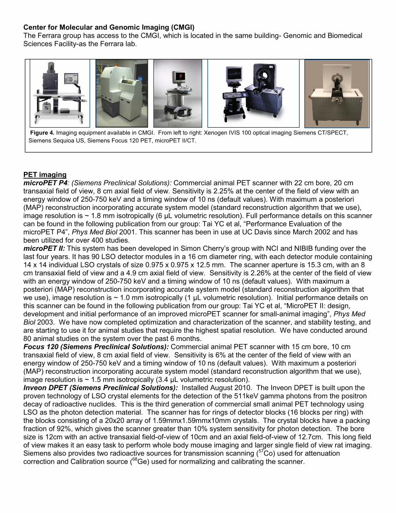
TEAM’s equipment includes Objet 3D printers (Eden and Connex), computers and CAD software, 3D Scanner with articulating scanning stage (Fig. 10), multiple extrusion-based 3D printers, Kern Laser Cutter, LPKF PCB mill, 3 axis CNC mill, metalworking lathe, Morgan press injection molding machine, SLA 3D Printer, Dual Extrusion FDM 3D Printer, among others. TEAM’s equipment is operated as instrument cores based on recharge.
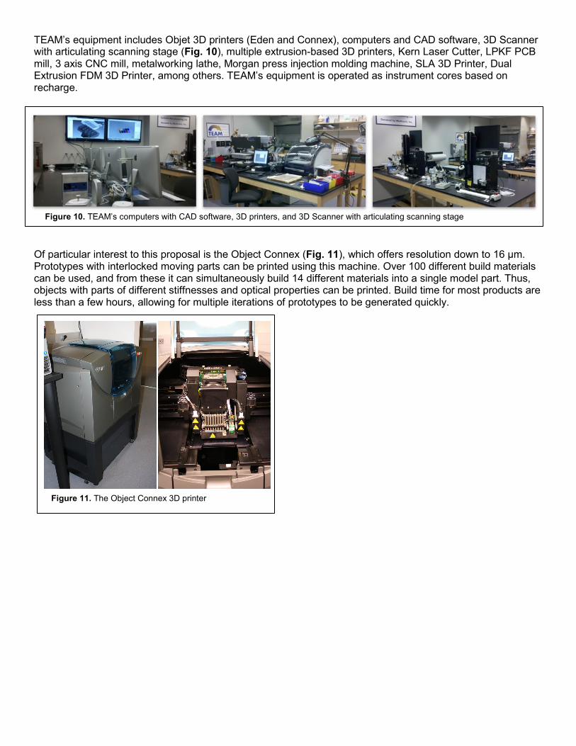
Of particular interest to this proposal is the Object Connex (Fig. 11), which offers resolution down to 16 µm. Prototypes with interlocked moving parts can be printed using this machine. Over 100 different build materials can be used, and from these it can simultaneously build 14 different materials into a single model part. Thus, objects with parts of different stiffnesses and optical properties can be printed. Build time for most products are less than a few hours, allowing for multiple iterations of prototypes to be generated quickly.
Figure 10. TEAM’s computers with CAD software, 3D printers, and 3D Scanner with articulating scanning stage
Figure 11. The Object Connex 3D printer

![[I-DO II NCTU] Community Empowerment](https://static.fdocuments.net/doc/165x107/559f6c0c1a28ab0d1e8b4850/i-do-ii-nctu-community-empowerment.jpg)


![[NCTU 2011 I-DO Camp] Facebook Class](https://static.fdocuments.net/doc/165x107/5560fff8d8b42a91388b54bf/nctu-2011-i-do-camp-facebook-class.jpg)


![[NCTU] [CCCA] vim rocks](https://static.fdocuments.net/doc/165x107/5554afa4b4c90502618b552b/nctu-ccca-vim-rocks.jpg)