Eosinophilic Cholecystitis, A Rare Case Report with a ...
3
Ann Colorectal Res 2020;8(4):206-208. Eosinophilic Cholecystitis, A Rare Case Report with a Review of the Literature Nasim Matani 1 , MD; Neda Soleimani 1 , MD; Sahand Mohammadzadeh 1 *, MD 1 Department of Pathology, Shiraz University of Medical Sciences, Shiraz, Iran Case Report Introduction: The clinical presentation of eosinophilic cholecystitis (EC) is similar to that of typical cholecystitis, commonly manifesting as right upper quadrant (RUQ) tenderness along with Murphy’s sign. In the literature, some reported cases have been associated with hyper-eosinophilic syndrome (HES), infections, drugs, and parasitosis; however, its etiology often remains unknown. Case Presentation: We report a 32-year-old woman who presented with two-day RUQ pain that radiated to the right shoulder. A clinical picture of severe acute cholecystitis was recorded, and the patient underwent open cholecystectomy. Microscopically, we observed an edematous wall with occasional acute and chronic inflammatory cells, predominantly composed of eosinophils. Due to peripheral eosinophilia, we recommended the patient to be assessed for hyper-eosinophilic syndrome, eosinophilic gastroenteritis, and parasitosis. On follow-up, parasitic gastroenteritis was diagnosed, and the patent underwent proper treatment. Conclusion: Since the EC signs are similar to those of typical cholecystitis, it should not be considered a separate entity. The significance of EC diagnosis is that it can be associated with other diseases that should also be taken into consideration. Please cite this paper as: Matani N, Soleimani N, Mohammadzadeh S. Eosinophilic Cholecystitis, A Rare Case Report with a Review of the Literature. Ann Colorectal Res. 2020;8(4):206-208. doi: 10.30476/ACRR.2020.87773.1062. *Corresponding authors: Sahand Mohammadzadeh, MD; Department of Pathology, Shiraz University of Medical Sciences, Shiraz, Iran Tel: +98-9173078147; Fax: +98-71-323017 Email: [email protected] Received: 15-08-2020 Revised: 16-09-2020 Accepted: 31-10-2020 Journal compilation © 2020 Annals of Colorectal Research, Shiraz University of Medical Sciences Keywords: Eosinophilic cholecystitis, Gallbladder disease, Cholecystectomy Abstract Introduction T he clinical manifestations of eosinophilic cholecystitis (EC) are similar to those of typical cholecystitis, with right upper quadrant (RUQ) tenderness being prominent. Some cases of EC have been associated with hyper-eosinophilic syndrome (HES), parasitosis, infections, and drugs; however, the etiology remains unknown in most cases (1). EC accounts for 0.25 to 6.4% of cholecystitis cases (2). Unfortunately, EC is clinically indistinguishable from other causes of cholecystitis. However, peripheral eosinophilia is sometimes also observed, as in parasitic disease and HES (3). In the microscopic examination, the massive transmural infiltration of eosinophils, which encompass 90% or more of the inflammatory infiltrate, is its main histologic characteristic (4). EC usually is acalculous, though calculi are found in 30% of cases (5). When its involvement is limited to the
Transcript of Eosinophilic Cholecystitis, A Rare Case Report with a ...

Ann Colorectal Res 2020;8(4):206-208.
Eosinophilic Cholecystitis, A Rare Case Report with a Review of the Literature
Nasim Matani1, MD; Neda Soleimani1, MD; Sahand Mohammadzadeh1*, MD
1Department of Pathology, Shiraz University of Medical Sciences, Shiraz, Iran
Case Report
Introduction: The clinical presentation of eosinophilic cholecystitis (EC) is similar to that of typical cholecystitis, commonly manifesting as right upper quadrant (RUQ) tenderness along with Murphy’s sign. In the literature, some reported cases have been associated with hyper-eosinophilic syndrome (HES), infections, drugs, and parasitosis; however, its etiology often remains unknown.Case Presentation: We report a 32-year-old woman who presented with two-day RUQ pain that radiated to the right shoulder. A clinical picture of severe acute cholecystitis was recorded, and the patient underwent open cholecystectomy. Microscopically, we observed an edematous wall with occasional acute and chronic inflammatory cells, predominantly composed of eosinophils. Due to peripheral eosinophilia, we recommended the patient to be assessed for hyper-eosinophilic syndrome, eosinophilic gastroenteritis, and parasitosis. On follow-up, parasitic gastroenteritis was diagnosed, and the patent underwent proper treatment.Conclusion: Since the EC signs are similar to those of typical cholecystitis, it should not be considered a separate entity. The significance of EC diagnosis is that it can be associated with other diseases that should also be taken into consideration.
Please cite this paper as:Matani N, Soleimani N, Mohammadzadeh S. Eosinophilic Cholecystitis, A Rare Case Report with a Review of the Literature. Ann Colorectal Res. 2020;8(4):206-208. doi: 10.30476/ACRR.2020.87773.1062.
*Corresponding authors: Sahand Mohammadzadeh, MD;Department of Pathology, Shiraz University of Medical Sciences, Shiraz, IranTel: +98-9173078147; Fax: +98-71-323017Email: [email protected]
Received: 15-08-2020Revised: 16-09-2020Accepted: 31-10-2020
Journal compilation © 2020 Annals of Colorectal Research, Shiraz University of Medical Sciences
Keywords: Eosinophilic cholecystitis, Gallbladder disease, Cholecystectomy
Abstract
Introduction
The clinical manifestations of eosinophilic cholecystitis (EC) are similar to those of typical
cholecystitis, with right upper quadrant (RUQ) tenderness being prominent. Some cases of EC have been associated with hyper-eosinophilic syndrome (HES), parasitosis, infections, and drugs; however, the etiology remains unknown in most cases (1). EC accounts for 0.25 to 6.4% of cholecystitis cases (2).
Unfortunately, EC is clinically indistinguishable from other causes of cholecystitis. However, peripheral eosinophilia is sometimes also observed, as in parasitic disease and HES (3). In the microscopic examination, the massive transmural infiltration of eosinophils, which encompass 90% or more of the inflammatory infiltrate, is its main histologic characteristic (4). EC usually is acalculous, though calculi are found in 30% of cases (5). When its involvement is limited to the
Eosinophilic cholecystitis, a rare case report
http://colorectalresearch.sums.ac.ir/ 207
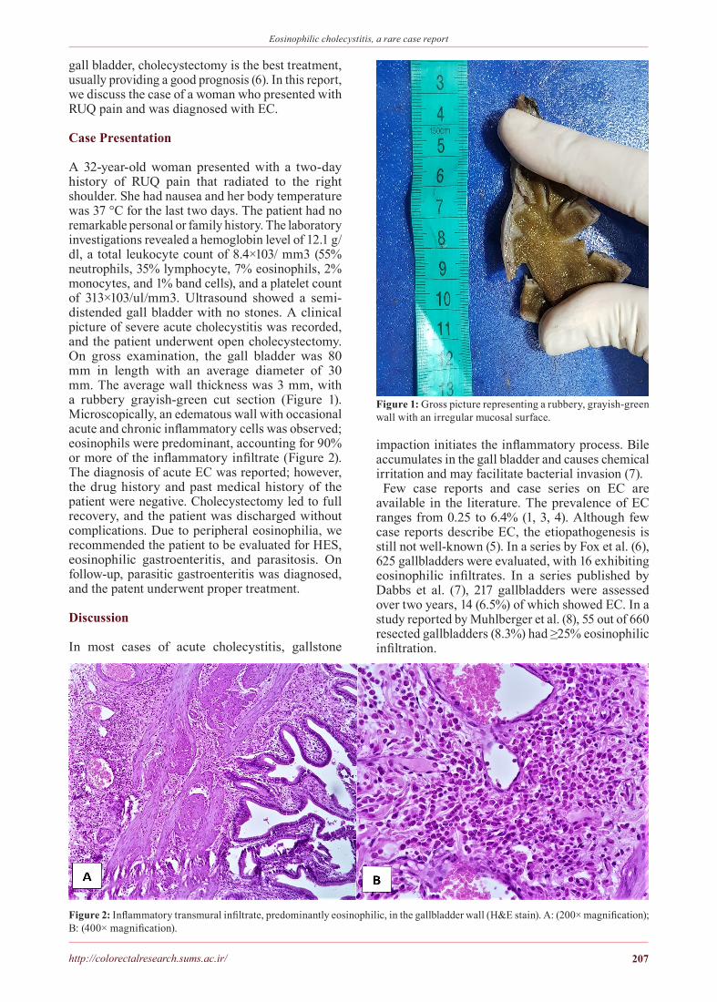
gall bladder, cholecystectomy is the best treatment, usually providing a good prognosis (6). In this report, we discuss the case of a woman who presented with RUQ pain and was diagnosed with EC.
Case Presentation
A 32-year-old woman presented with a two-day history of RUQ pain that radiated to the right shoulder. She had nausea and her body temperature was 37 °C for the last two days. The patient had no remarkable personal or family history. The laboratory investigations revealed a hemoglobin level of 12.1 g/dl, a total leukocyte count of 8.4×10³/ mm3 (55% neutrophils, 35% lymphocyte, 7% eosinophils, 2% monocytes, and 1% band cells), and a platelet count of 313×103/ul/mm3. Ultrasound showed a semi-distended gall bladder with no stones. A clinical picture of severe acute cholecystitis was recorded, and the patient underwent open cholecystectomy. On gross examination, the gall bladder was 80 mm in length with an average diameter of 30 mm. The average wall thickness was 3 mm, with a rubbery grayish-green cut section (Figure 1). Microscopically, an edematous wall with occasional acute and chronic inflammatory cells was observed; eosinophils were predominant, accounting for 90% or more of the inflammatory infiltrate (Figure 2). The diagnosis of acute EC was reported; however, the drug history and past medical history of the patient were negative. Cholecystectomy led to full recovery, and the patient was discharged without complications. Due to peripheral eosinophilia, we recommended the patient to be evaluated for HES, eosinophilic gastroenteritis, and parasitosis. On follow-up, parasitic gastroenteritis was diagnosed, and the patent underwent proper treatment.
Discussion
In most cases of acute cholecystitis, gallstone
impaction initiates the inflammatory process. Bile accumulates in the gall bladder and causes chemical irritation and may facilitate bacterial invasion (7).
Few case reports and case series on EC are available in the literature. The prevalence of EC ranges from 0.25 to 6.4% (1, 3, 4). Although few case reports describe EC, the etiopathogenesis is still not well-known (5). In a series by Fox et al. (6), 625 gallbladders were evaluated, with 16 exhibiting eosinophilic infiltrates. In a series published by Dabbs et al. (7), 217 gallbladders were assessed over two years, 14 (6.5%) of which showed EC. In a study reported by Muhlberger et al. (8), 55 out of 660 resected gallbladders (8.3%) had ≥25% eosinophilic infiltration.
Figure 1: Gross picture representing a rubbery, grayish-green wall with an irregular mucosal surface.
Figure 2: Inflammatory transmural infiltrate, predominantly eosinophilic, in the gallbladder wall (H&E stain). A: (200× magnification); B: (400× magnification).

Matani N et al.
Ann Colorectal Res 2020;8(4)208
1. Keyal NK, Adhikari P, Baskota BD, Rai U, Thakur A. Eosinophilic Cholecystitis presenting with Common Bile Duct Sludge and Cholangitis: A Case Report. Journal of the Nepal Medical Association. 2020 Mar 1;58(223).
2. Kuwahara T, Kobayashi Y, Yun Y, Kanda A, Asako M, Ueki S, Iwai H. Eosinophilic Cholecystitis Occurred in a Patient With Refractory Eosinophilic Airway Inflammation: A Case Report. Allergy & Rhinology. 2019 Aug;10:2152656719869607.
3. Hasan A, Nafie K. Eosinophilic cholecystitis: a rare cause of acute cholecystitis ‘case report’. Al-Azhar Assiut Medical Journal. 2019 Oct 1;17(4):417.
4. Caesar J, Jordan M, Hills M. Case report: A rare case of eosinophilic
cholecystitis presenting after talc pleurodesis for recurrent pneumothorax. Respiratory medicine case reports. 2017 Jan 1;20:16-8.
5. Ranaee R, Khosravi M, Vosough Z. A Rare Case of Eosinophilic Cholecystitis Presenting in a Patient with Thalassemia Intermedia; a Case Report. Journal of Advances in Medical and Biomedical Research. 2019;27(121):54-7.
6. Dwivedi T, Chavan R. Eosinophilic Cholecystitis with Lipomatosis: a rare case report and review of literature. Annals of Pathology and Laboratory Medicine. 2016 Feb 25;3(1):C23-26.
7. Khan S, Hassan MJ, JairaJPuri ZS, Jetley S, Husain M. Clinicopathological study of eosinophilic cholecystitis: five year single institution experience. Journal
of clinical and diagnostic research: JCDR. 2017 Aug;11(8):EC20.
8. Ramesh K, Harpal S, Chakma S, Kaur N, DEEPIKA D. ASCARIASIS AS A CAUSE OF ACALCULUS EOSINOPHILIC CHOLECYSTITIS-A RARE CASE REPORT. International Journal of Clinical and Biomedical Research. 2016 Jan 29:54-6.
9. Garzón GL, Jaramillo BL, Valero HJ, Quintero CE. Eosinophilic cholecystitis in children: case series. Journal of Pediatric Surgery. 2020 Jun 3.
10. Jagtap SV, Jokhi CD, Shukla D, Jagtap SS, Kulkarni SR. Idiopathic eosinophilic cholecystitis with Cholelithiasis-a rare histopathological variant. Asian Pac J Health Sci.. 2017 Mar 30;4(1):217-9.
References
A review of the literature revealed 27 individuals with acute cholecystitis confirmed as EC since 1949, the majority of whom exhibited HES, eosinophilic cholangiopathy, parasitic infestation, or acalculous cholecystitis (3, 4). When peripheral eosinophilia is observed, the diagnosis of HES should be considered (2).
Acalculous cholecystitis is another cause of EC, resulting in the invasion of the mucosal surface by eosinophils (9). Different factors cause acute acalculous cholecystitis, including critical illness, major burns, and herbal medicine (10). Patients with acalculous cholecystitis are three to four times more likely to have EC than patients with gall stones.
If no etiology is found, the case will be considered as idiopathic EC. This entity is quite challenging since laboratory tests cannot establish the cause. Considering the presence of peripheral eosinophilia in some cases, there might be a number of etiologies leading to such a disease (10). Abdominal ultrasound results may be unremarkable or may reveal signs representing typical cholecystitis such as gallbladder distension, wall thickening, or the sonographic Murphy sign (8).
In our patient, peripheral eosinophilia of about 7%
was recorded during the hospital course. The etiology for eosinophilia was unknown. Cholecystectomy led to full recovery, and the patient was discharged without complications. Considering the presence of peripheral eosinophilia, we recommended that the patient be evaluated for HES, eosinophilic gastroenteritis, and parasitosis. The prognosis of EC is desirable (6). When the disease is limited to the gall bladder, cholecystectomy, preferably performed laparoscopically, is the best option (7).
Since EC may occur under many conditions and can be associated with other diseases, the possible associated etiologies should be investigated when it is diagnosed. Pathologists should carefully differentiate EC from other causes of cholecystitis that may have a worse prognosis (9). In the follow-up of our patient, parasitic gastroenteritis was diagnosed and treated.
Ethical Approval: The research was carried out in agreement with the World Medical Association Declaration of Helsinki. The study was approved by the Ethics Committee of Shiraz University of Medical Sciences.
Conflicts of Interests: None declared.