
Endodontic periodontic interrelationship
68
ENDODONTIC-PERIODONTIC INTERRELATIONSHIP PRESENTED BY: PRABLEEN ARORA MDS STUDENT 1
-
Upload
sls-amg -
Category
Health & Medicine
-
view
227 -
download
1
Transcript of Endodontic periodontic interrelationship

ENDODONTIC-PERIODONTIC INTERRELATIONSHIP
PRESENTED BY:PRABLEEN ARORA
MDS STUDENT1
INTRODUCTION
o In 1964, Simring and Goldeberg - first described therelationship between endodontics and periodontics.
• ENDODONTIC LESION is used to denote aninflammatory process in the periodontal tissuesresulting from noxious agents present in the root canalsystem of the tooth, usually a root canal infection.
• PERIODONTAL LESION is used to denote aninflammatory process in the periodontal tissuesresulting from accumulation of dental plaque on theexternal tooth surfaces.
2
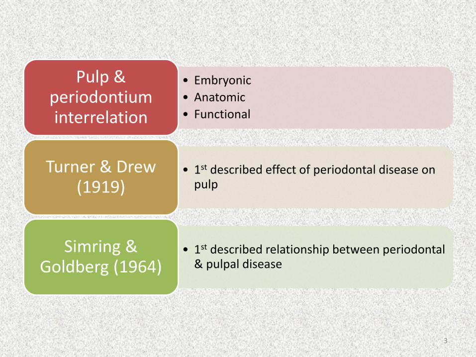
• Embryonic
• Anatomic
• Functional
Pulp & periodontiuminterrelation
• 1st described effect of periodontal disease on pulp
Turner & Drew (1919)
• 1st described relationship between periodontal & pulpal disease
Simring & Goldberg (1964)
3
HISTORY
• Cahn (1927) was one of the first investigator to state that periodontal
disease had an influence on the pulpal tissue. This influence to the close
proximity of these structures in the region of the apical foramen.
• Bender (1936), in a study reported that a large number of extracted teeth
had lateral and accessory canals. He presented evidence that pulpal
inflammation and degeneration could result from periodontal disease.
• Chacker (1974) in his studies, described vascular anastomoses through
lateral and accessory canals. He also commented on the potential for
communication through exposed dentinal tubules. 4
• Torabinejad (1985) studied 25 teeth from a single patient
and could not establish any interrelationships between
periodontal and pulpal diseases.
• Hiatt (1977) and Hemington (1979) stated that there is no
apparent relationship between periodontal and pulpal
disease.
5
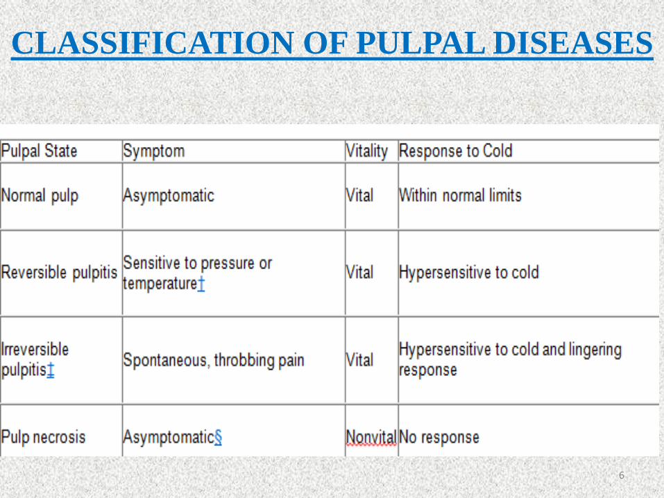
CLASSIFICATION OF PULPAL DISEASES
6

CLASSIFICATION OF PERIRADICULAR DISEASES
Periradicular
StateSymptom Pulpal Status
Percussion
Response
Palpation
Response
periapical
radiolucencySinus Tract
Normal
periapexNone Varies None None Not present Not present
Acute
periradicular
periodontitis
Painful Inflamed Painful Varies Not present Not present
Chronic
periradicular
periodontitis
None Nonvital None None Present Not present
Acute
periradicular
abscess
Painful Inflamed Painful Painful Varies Not present
Chronic
periradicular
abscess
None Nonvital None None Present Present
Condensing
osteitisNone/pain Inflamed None None Radiopaque Not present
7
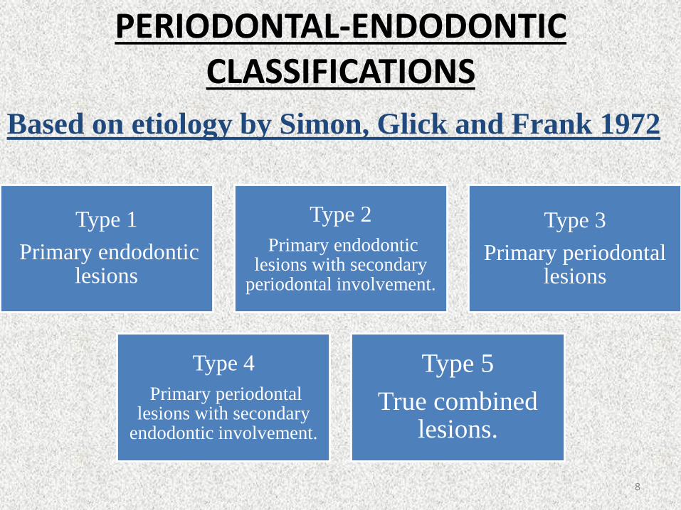
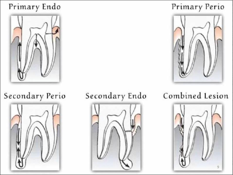
PERIODONTAL-ENDODONTIC CLASSIFICATIONS
Based on etiology by Simon, Glick and Frank 1972
Type 1
Primary endodontic lesions
Type 2
Primary endodontic lesions with secondary
periodontal involvement.
Type 3
Primary periodontal lesions
Type 4
Primary periodontal lesions with secondary
endodontic involvement.
Type 5
True combined lesions.
8
9
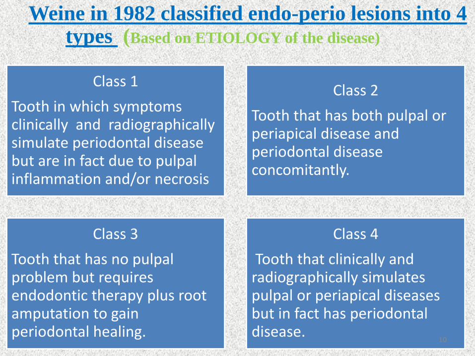
Weine in 1982 classified endo-perio lesions into 4 types (Based on ETIOLOGY of the disease)
Class 1
Tooth in which symptoms clinically and radiographicallysimulate periodontal disease but are in fact due to pulpalinflammation and/or necrosis
Class 2
Tooth that has both pulpal or periapical disease and periodontal disease concomitantly.
Class 3
Tooth that has no pulpalproblem but requires endodontic therapy plus root amputation to gain periodontal healing.
Class 4
Tooth that clinically and radiographically simulates pulpal or periapical diseases but in fact has periodontal disease.
10
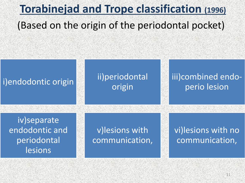
Torabinejad and Trope classification (1996)
(Based on the origin of the periodontal pocket)
i)endodontic originii)periodontal
originiii)combined endo-
perio lesion
iv)separate endodontic and
periodontal lesions
v)lesions with communication,
vi)lesions with no communication,
11
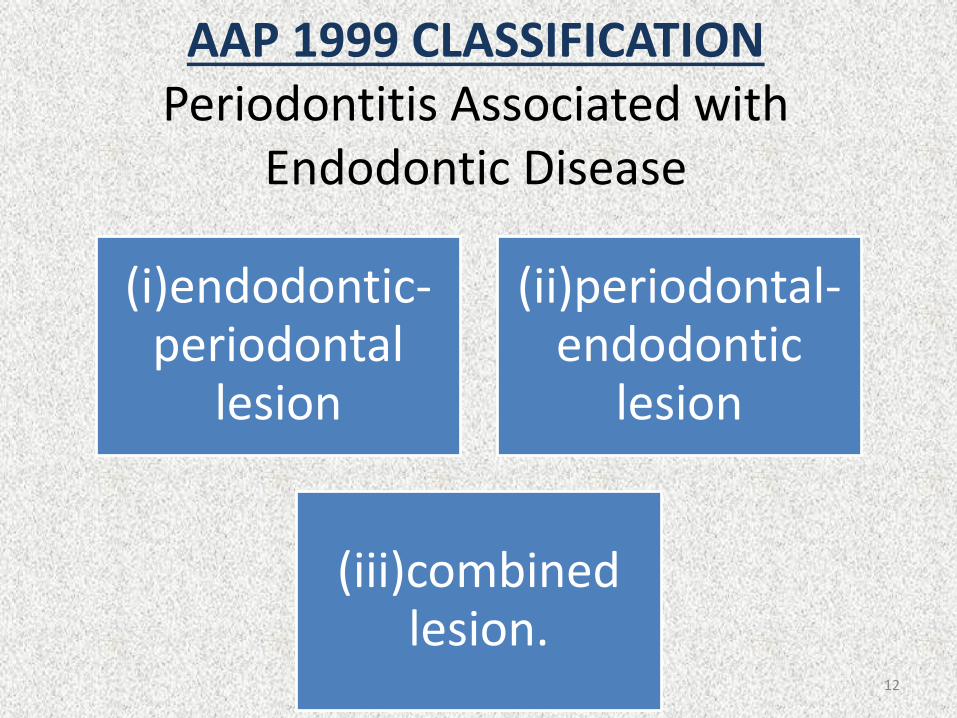
AAP 1999 CLASSIFICATIONPeriodontitis Associated with
Endodontic Disease
(i)endodontic-periodontal
lesion
(ii)periodontal-endodontic
lesion
(iii)combined lesion.
12
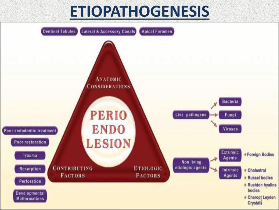
ETIOPATHOGENESIS
13

Anatomic Considerations of Pulpal and Periodontal Continuum
Apical foramen
Furcation canals
Lateral canals,
Accessory canals
14
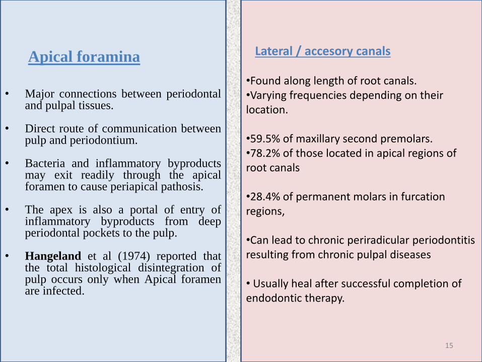
Apical foramina
• Major connections between periodontaland pulpal tissues.
• Direct route of communication betweenpulp and periodontium.
• Bacteria and inflammatory byproductsmay exit readily through the apicalforamen to cause periapical pathosis.
• The apex is also a portal of entry ofinflammatory byproducts from deepperiodontal pockets to the pulp.
• Hangeland et al (1974) reported thatthe total histological disintegration ofpulp occurs only when Apical foramenare infected.
Lateral / accesory canals
•Found along length of root canals.•Varying frequencies depending on their location.
•59.5% of maxillary second premolars.•78.2% of those located in apical regions of root canals
•28.4% of permanent molars in furcationregions,
•Can lead to chronic periradicular periodontitis resulting from chronic pulpal diseases
• Usually heal after successful completion of endodontic therapy.
15

• Kirkham reported that out of 100 permanent human teeth extracted as result of severe periodontal disease, only 2 teeth possessed accessory canals within periodontal pockets.
• Thus, likelihood that primary periodontal infections will reach dental pulp through accessory canals is rare.
16

Advanced pulpitis
Pulp necrosis
Inflammatory bone
resorption at root apex
Apical periodontitis or
an apical abscess.
Known as retrograde periodontitis
• Because it represents periodontal tissue breakdown from apical to cervical direction
Identified as a periapical
radiolucency(PARL)
17

Dentinal TubulesMaintain a tapered
structure along length from pulpodentinal
complex (PDC) to DEJ
Diameter of 2.5 μm at PDC and 0.9 μm at DEJ.
Permeable structure
Permeability Changes at different locations along root surface according to size & density of dentinal
tubules.
Bacterial colonization in tubules from
infected root canals.
Dentinal tubules may allow pulpal irritation from chronic
periodontal infections.
Presence of patent dentinal tubules, especially when cementum layer is denuded, favors the spread of microorganisms between the pulp and periodontal tissues.
18
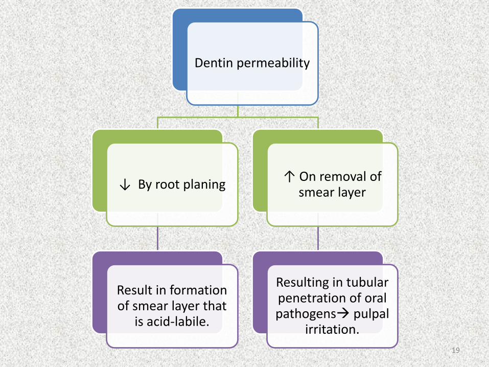
Dentin permeability
↓ By root planing
Result in formation of smear layer that
is acid-labile.
↑ On removal of smear layer
Resulting in tubular penetration of oral pathogens pulpal
irritation.
19
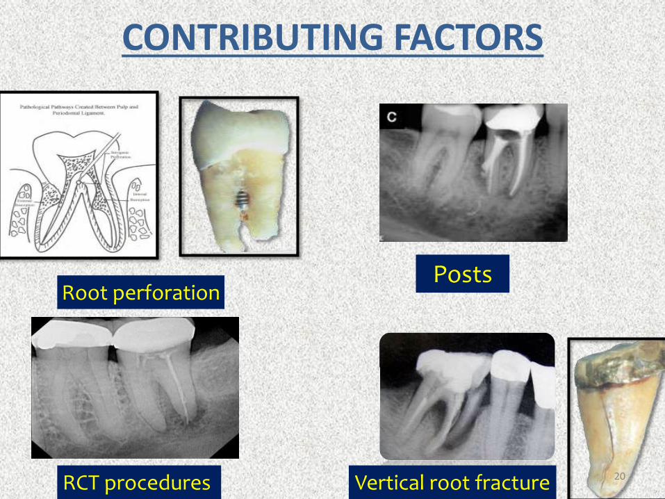
CONTRIBUTING FACTORS
Root perforation
Vertical root fractureRCT procedures
Posts
20
ETIOLOGICAL FACTORSLIVE PATHOGENS
• Encountered in a diseased pulp and in periradicular tissues may include bacteria, fungi and viruses.
BACTERIA• Bacteria. Bacteria play a crucial role in the formation and progression of
both endodontic and periodontal diseases . The periradicular tissues become involved when bacteria invade the pulp, causing either partial or total necrosis.
• Kakehashi et al. (92), in a classic study, demonstrated the relationship between the presence of bacteria and diseases in pulp and periradiculartissues.
• In their study, pulps of normal rats were exposed and left open to the oral environment. Consequently, pulp necrosis ensued, followed by inflammation of periradicular tissue and lesion formation.
• However, when the same procedure was performed on germ-free rats, not only did the pulps remain vital and relatively noninflamed, the exposure sites showed evidence of dentin repair. 21
• Moller et al. confirmed these findings in monkeys and reported that noninfected necrotic pulp tissue did not induce periradicularlesions or inflammatory reactions.
• Nonetheless, once the pulp became infected, periradicular lesions and inflammation occurred in the apical tissues.
• Proteolytic bacteria predominate in the root-canal flora, which changes, over time, to a more anaerobic microbiota.
• Rupf et al. (167) studied the profiles of periodontal pathogens in pulpal and periodontal diseases associated with the same tooth.
• Specific PCR methods were used to detect Aggregatibacteractinomycetemcomitans, Tannerella forsythia, Eikenella corrodens, Fusobacterium nucleatum, Porphyromonas gingivalis, Prevotellaintermedia and Treponema denticola.
• These pathogens were found in all endodontic samples and also in teeth with chronic apical periodontitis and chronic adult periodontitis.
• It therefore appears that periodontal pathogens.
22
Fungi (Yeasts)
• Presence and Prevalence of fungi associated with endodonticdisease is well documented.
• Fungal colonization associated with radicular pathosis has been demonstrated in untreated root caries, dentinal tubules, failing root-canal treatments , apices of teeth wit asymptomatic apical periodontitis and periradicular tissues.
• Majority of the recovered fungi were candida albicans.
• Fungi colonization - Immune compromising diseases such ascancer
- Certain intra canal mediaments,
- Local and systemic antibiotics and
- Previous unsuccessful endodontic therapy. 23

• Reduction in the numbers of specific strains of bacteria in the root canal during endodontic treatment may allow fungal overgrowth in the remaining low nutrient environment.
• Another possibility is that fungi may gain access to the root canal from the oral cavity as a result of poor asepsis during endodontic treatment or post-preparation procedures.
• In addition, it has been demonstrated that the presence of fungi in root canals is directly associated with their presence in saliva and oral tissues .
• These findings further stress the importance of using aseptic endodontic and periodontal techniques, maintaining the integrity of dental hard tissues and covering the tooth crown as soon as practical with a well-sealed permanent restoration in order to prevent reinfection.
24

Viruses
• There is increasing evidence to suggest that viruses plays an important role in both endo-perio disease.
• Herpes simplex virus is frequently detected in Gingival crevicular fluid and periodontal lesions
• Cytomegalovirus was found in about 55% of periodontalpocket (Hutter 1991).
• Epstein-Bair Virus type 1 was detected in more than 40% ofpocket (Slots et al ).
• Sabet et al suggested that human cytomegalovirus and E-Bvirus play a role in the pathogenesis of symptomatic periapicallesions.
25
Non-living etiologic agents
A. Extrinsic agents
Foreign Bodies
Dentin and cementum chips, amalgam, root canal fillingmaterials, cellulose fibers from absorbent paper points,gingival retraction cords, leguminous food and calculus likedeposits.
Response to foreign body – acute or chronic reaction –symptomatic or asymptomatic conditions.
Mechanical or surgical removal of the foreign body is thetreatment of choice.
26
Intrinsic agents.
Cholesterol
• Present in apical periodontitis ( Histopathologicfinding) - induce a typical foreign body reaction.
• Cholesterol released by disintegrating erythrocytes ofstagnant blood vessels within periapical lesion,lymphocytes, plasma cells which die in great numbersand disintegrate in chronic periapical lesions or by thecirculating plasma lipids.
• Accumulation of cholesterol crystals in inflamedperiapical tissues - causes of failure of Endo therapy
27
Russell Bodies
• Found in most inflamed tissues, accumulations of anEosinophilic substance.
• Caused by the synthesis of excessive amounts ofnormal secretary protein in certain plasma cellsengaged in active synthesis of immunoglobulins.
Rushton hyaline bodies
• Unique feature of some odontogenic cysts.
• They are keratinous in nature, of hematogenousorigin, a specialized secretory product of odontogenicepithelium.
28
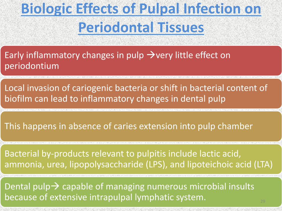
Biologic Effects of Pulpal Infection on Periodontal Tissues
Early inflammatory changes in pulp very little effect on periodontium
Local invasion of cariogenic bacteria or shift in bacterial content of biofilm can lead to inflammatory changes in dental pulp
This happens in absence of caries extension into pulp chamber
Bacterial by-products relevant to pulpitis include lactic acid, ammonia, urea, lipopolysaccharide (LPS), and lipoteichoic acid (LTA)
Dental pulp capable of managing numerous microbial insults because of extensive intrapulpal lymphatic system.
29
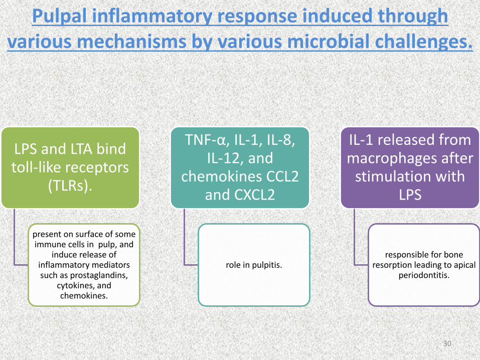
Pulpal inflammatory response induced through various mechanisms by various microbial challenges.
LPS and LTA bind toll-like receptors
(TLRs).
present on surface of some immune cells in pulp, and
induce release of inflammatory mediators such as prostaglandins,
cytokines, and chemokines.
TNF-α, IL-1, IL-8, IL-12, and
chemokines CCL2 and CXCL2
role in pulpitis.
IL-1 released from macrophages after
stimulation with LPS
responsible for bone resorption leading to apical
periodontitis.
30
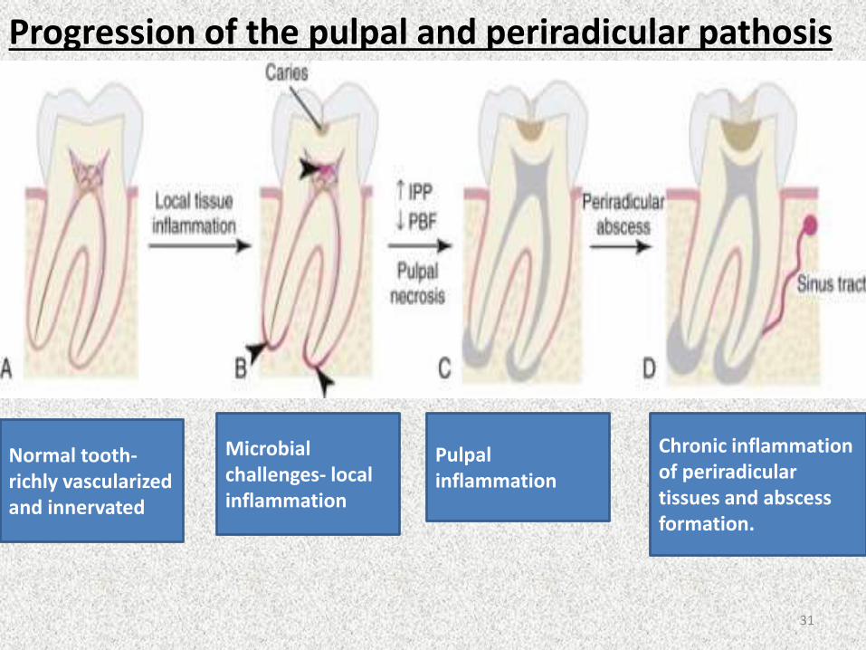
Progression of the pulpal and periradicular pathosis
Normal tooth-richly vascularizedand innervated
Microbial challenges- local inflammation
Pulpalinflammation
Chronic inflammation of periradiculartissues and abscess formation.
31
BIOLOGICAL EFFECTS OF PERIODONTAL INFECTION ON THE DENTAL PULP
• Langeland et al 1974 indicated then when pathologic changes do occur in the pulp of the tooth as a result of advanced periodontal disease
pulp does not usually undergo degenerative changes as long as the main canal has not been involved.
If the vasculature of the pulp remains vital, no inflammatory reaction occurs and there is no symptom of pulp pathosis 32
• Kobayashi et al 1990 compared the microflora from root canal and periodontal pockets of caries free teeth (necrotic and non-vital).
• Concluded that there were far fewer bacteria in root canal but both areas showed similar bacteria strains which suggested that the periodontal pocket may be the source of bacteria found in infection within the root canal system.
33
• Protection and preservation of the cementum and dentin surrounding the tooth also play important role in preserving the health of the pulp and preventing the ingress of periodontal pathogens.
• Dentin thickness also contributes to the protection of the pulp.
• Stanley 1968 stated that if a 2-mm thickness of dentin remains between the pulp and irritating stimulus, there is little chance of pulpal damage.
34
Periodontal Procedures
• Pulpal changes seen in periodontal disease were closely related to
periodontal treatment .
• Vigorous scaling and root planning removes cementum –
- expose dentinal tubules
- transport irritants
- pulpal inflammation
- necrosis of the dental pulp
• Presence of an intact cementum layer - protection of the pulp from
plaque and other periodontal pathogens that migrate along the root
surface during the development of advanced periodontal disease.
35
Precautions during periodontal therapy
• Avoid use of irritating chemicals
• Minimise use of ultrasonics & rotary scaling instruments when < 2 mm of dentin thickness remaining
• Allow minor irritation to subside before adding insult to injury
36
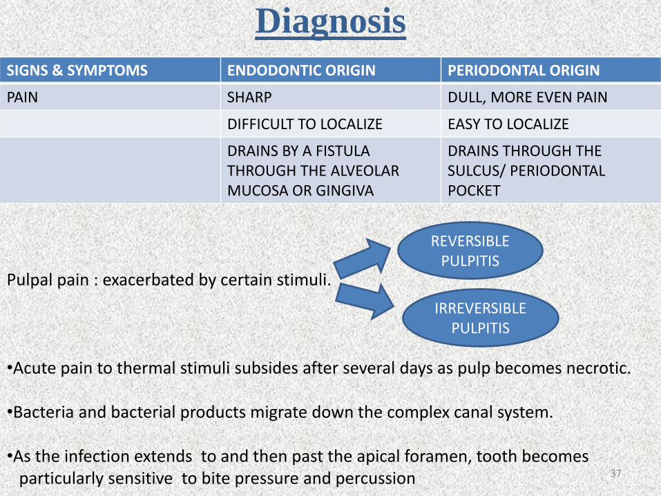
Diagnosis
SIGNS & SYMPTOMS ENDODONTIC ORIGIN PERIODONTAL ORIGIN
PAIN SHARP DULL, MORE EVEN PAIN
DIFFICULT TO LOCALIZE EASY TO LOCALIZE
DRAINS BY A FISTULA THROUGH THE ALVEOLAR MUCOSA OR GINGIVA
DRAINS THROUGH THESULCUS/ PERIODONTAL POCKET
Pulpal pain : exacerbated by certain stimuli.
•Acute pain to thermal stimuli subsides after several days as pulp becomes necrotic.
•Bacteria and bacterial products migrate down the complex canal system.
•As the infection extends to and then past the apical foramen, tooth becomes particularly sensitive to bite pressure and percussion
REVERSIBLE PULPITIS
IRREVERSIBLE PULPITIS
37

• Necrotic tooth periradicular abscess elevation
of tooth.
Patient with irreversible pulpitis or chronic pulpalinfection may be completely asymptomatic.
38
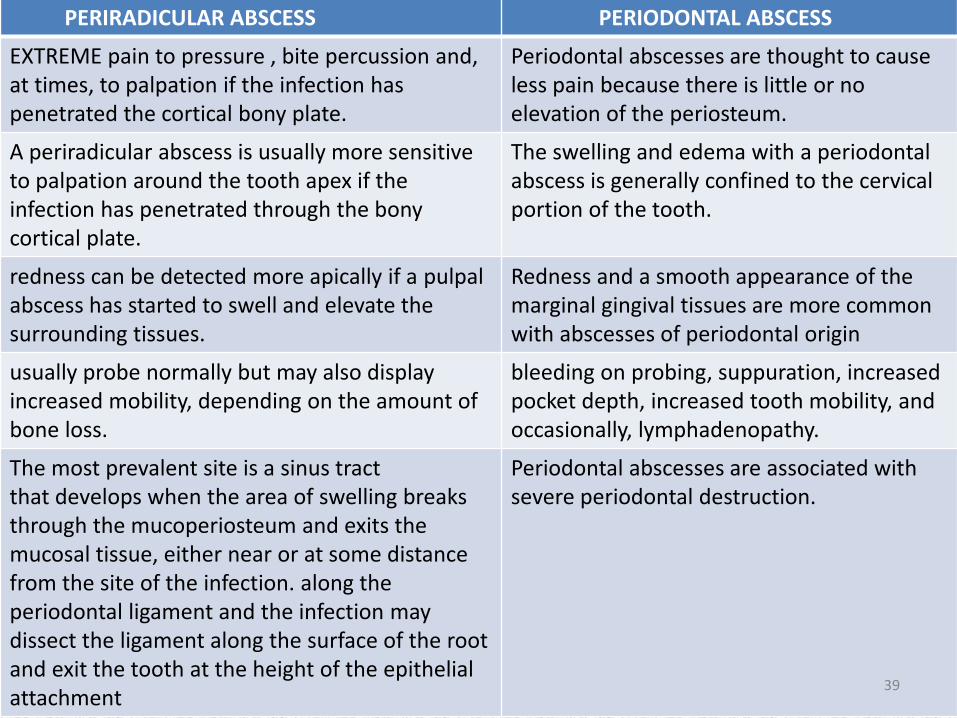
PERIRADICULAR ABSCESS PERIODONTAL ABSCESS
EXTREME pain to pressure , bite percussion and, at times, to palpation if the infection has penetrated the cortical bony plate.
Periodontal abscesses are thought to cause less pain because there is little or noelevation of the periosteum.
A periradicular abscess is usually more sensitive to palpation around the tooth apex if the infection has penetrated through the bony cortical plate.
The swelling and edema with a periodontal abscess is generally confined to the cervicalportion of the tooth.
redness can be detected more apically if a pulpalabscess has started to swell and elevate the surrounding tissues.
Redness and a smooth appearance of the marginal gingival tissues are more common with abscesses of periodontal origin
usually probe normally but may also display increased mobility, depending on the amount of bone loss.
bleeding on probing, suppuration, increased pocket depth, increased tooth mobility, and occasionally, lymphadenopathy.
The most prevalent site is a sinus tractthat develops when the area of swelling breaks through the mucoperiosteum and exits the mucosal tissue, either near or at some distance from the site of the infection. along the periodontal ligament and the infection may dissect the ligament along the surface of the root and exit the tooth at the height of the epithelial attachment
Periodontal abscesses are associated withsevere periodontal destruction.
39

ORThe path of least resistance may be along the periodontal ligament and the infection may dissect the ligament along the surface of the root and exit the tooth at the height of the epithelial attachment
This results in a periodontal defect that probes along a narrow path to the apexof the root. Both the sinus tract and narrow sulcular lesions can usually be traced to the infected tooth or offending root using a gutta percha point.
40
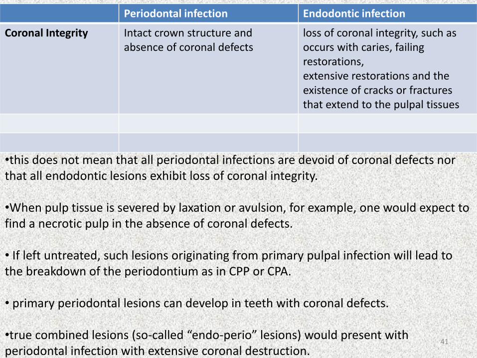
Periodontal infection Endodontic infection
Coronal Integrity Intact crown structure and absence of coronal defects
loss of coronal integrity, such as occurs with caries, failing restorations,extensive restorations and the existence of cracks or fractures that extend to the pulpal tissues
•this does not mean that all periodontal infections are devoid of coronal defects nor that all endodontic lesions exhibit loss of coronal integrity.
•When pulp tissue is severed by laxation or avulsion, for example, one would expect to find a necrotic pulp in the absence of coronal defects.
• If left untreated, such lesions originating from primary pulpal infection will lead to the breakdown of the periodontium as in CPP or CPA.
• primary periodontal lesions can develop in teeth with coronal defects.
•true combined lesions (so-called “endo-perio” lesions) would present with periodontal infection with extensive coronal destruction.
41
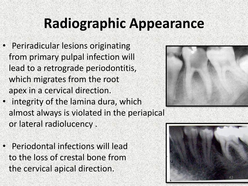
Radiographic Appearance
• Periradicular lesions originating from primary pulpal infection willlead to a retrograde periodontitis,which migrates from the root apex in a cervical direction.
• integrity of the lamina dura, which almost always is violated in the periapicalor lateral radiolucency .
• Periodontal infections will leadto the loss of crestal bone from the cervical apical direction.
42
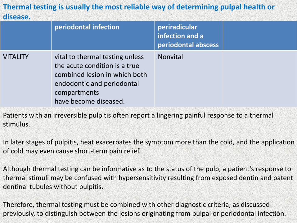
Thermal testing is usually the most reliable way of determining pulpal health or disease.
periodontal infection periradicularinfection and a periodontal abscess
VITALITY vital to thermal testing unless the acute condition is a true combined lesion in which both endodontic and periodontal compartmentshave become diseased.
Nonvital
Patients with an irreversible pulpitis often report a lingering painful response to a thermal stimulus.
In later stages of pulpitis, heat exacerbates the symptom more than the cold, and the application of cold may even cause short-term pain relief.
Although thermal testing can be informative as to the status of the pulp, a patient’s response to thermal stimuli may be confused with hypersensitivity resulting from exposed dentin and patent dentinal tubules without pulpitis.
Therefore, thermal testing must be combined with other diagnostic criteria, as discussed previously, to distinguish between the lesions originating from pulpal or periodontal infection.43
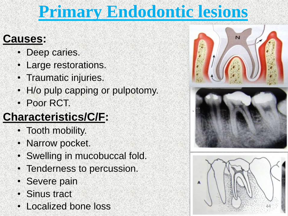
Primary Endodontic lesions
Causes: • Deep caries.
• Large restorations.
• Traumatic injuries.
• H/o pulp capping or pulpotomy.
• Poor RCT.
Characteristics/C/F:• Tooth mobility.
• Narrow pocket.
• Swelling in mucobuccal fold.
• Tenderness to percussion.
• Severe pain
• Sinus tract
• Localized bone loss 44
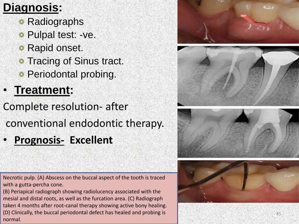
Diagnosis: Radiographs
Pulpal test: -ve.
Rapid onset.
Tracing of Sinus tract.
Periodontal probing.
• Treatment:
Complete resolution- after
conventional endodontic therapy.
• Prognosis- Excellent
Necrotic pulp. (A) Abscess on the buccal aspect of the tooth is traced with a gutta-percha cone. (B) Periapical radiograph showing radiolucency associated with the mesial and distal roots, as well as the furcation area. (C) Radiograph taken 4 months after root-canal therapy showing active bony healing. (D) Clinically, the buccal periodontal defect has healed and probing is normal.
45
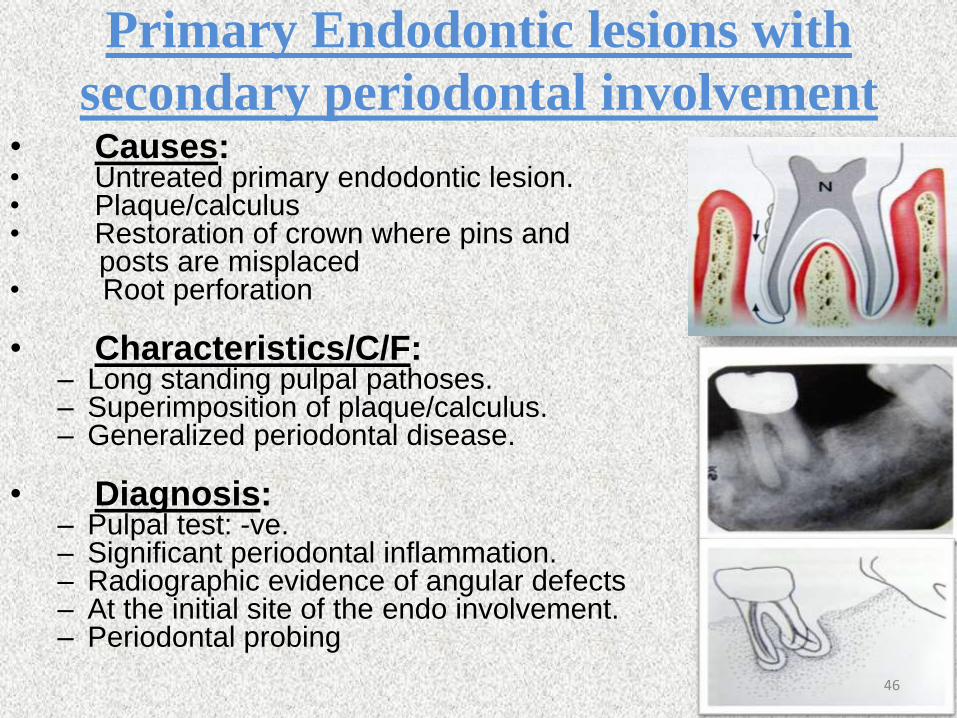
Primary Endodontic lesions with
secondary periodontal involvement• Causes:• Untreated primary endodontic lesion.• Plaque/calculus• Restoration of crown where pins and
posts are misplaced • Root perforation
• Characteristics/C/F:– Long standing pulpal pathoses.– Superimposition of plaque/calculus.– Generalized periodontal disease.
• Diagnosis:– Pulpal test: -ve.– Significant periodontal inflammation.– Radiographic evidence of angular defects– At the initial site of the endo involvement.– Periodontal probing
46
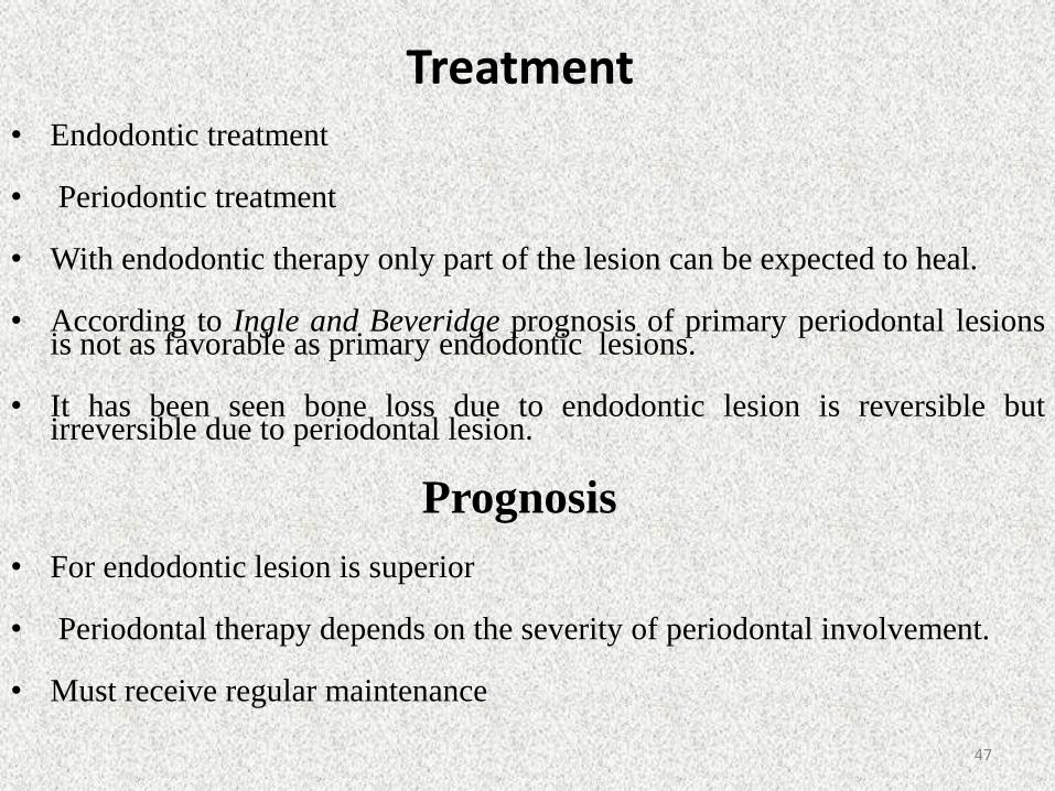
Treatment• Endodontic treatment
• Periodontic treatment
• With endodontic therapy only part of the lesion can be expected to heal.
• According to Ingle and Beveridge prognosis of primary periodontal lesionsis not as favorable as primary endodontic lesions.
• It has been seen bone loss due to endodontic lesion is reversible butirreversible due to periodontal lesion.
Prognosis
• For endodontic lesion is superior
• Periodontal therapy depends on the severity of periodontal involvement.
• Must receive regular maintenance
47
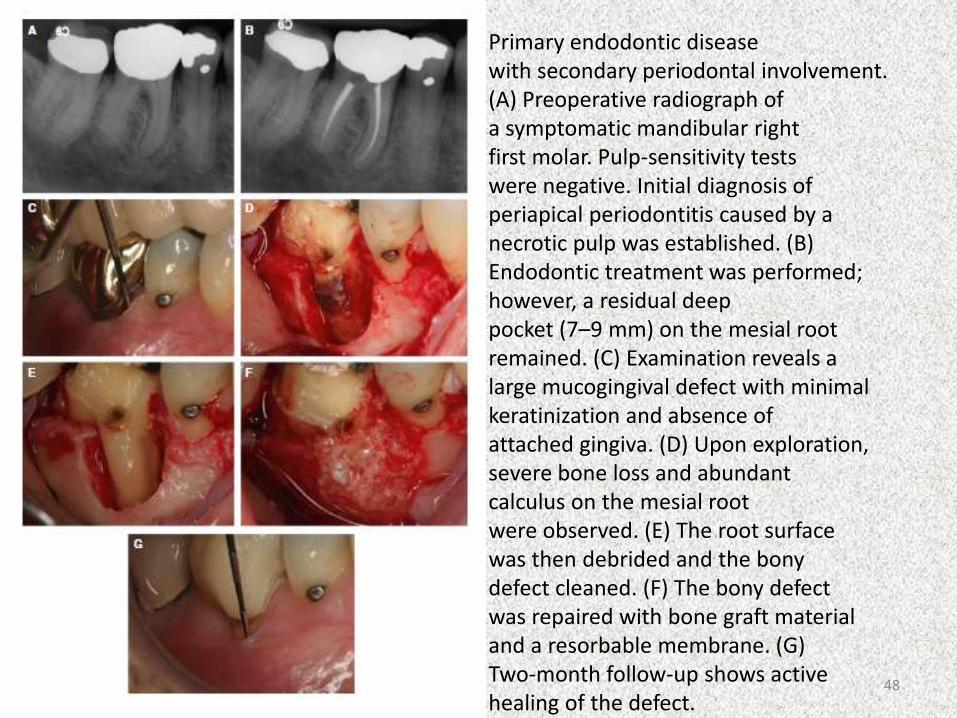
Primary endodontic diseasewith secondary periodontal involvement.(A) Preoperative radiograph ofa symptomatic mandibular rightfirst molar. Pulp-sensitivity testswere negative. Initial diagnosis ofperiapical periodontitis caused by anecrotic pulp was established. (B)Endodontic treatment was performed;however, a residual deeppocket (7–9 mm) on the mesial rootremained. (C) Examination reveals alarge mucogingival defect with minimalkeratinization and absence ofattached gingiva. (D) Upon exploration,severe bone loss and abundantcalculus on the mesial rootwere observed. (E) The root surfacewas then debrided and the bonydefect cleaned. (F) The bony defectwas repaired with bone graft materialand a resorbable membrane. (G)Two-month follow-up shows activehealing of the defect.
48

Primary Periodontal lesions Characteristics
• Teeth vital
• Generalized bone loss
• Calculus/plaque
• Soft tissue inflammation
• Broad based pockets
• Mobility
• Periodontal abscess during acute phase
Diagnosis
• Visual examination
• Probing
• Pulp testing- POSITIVE
• Radiographs49

Treatment
• Periodontal therapy
• Root amputation
• Guided tissue regeneration
• Root canal therapy(advance cases)
Prognosis
• Depends on the outcome of periodontal therapy.
• Depends on the extent of the periodontitis and on patients ability to comply with potential long term treatment and maintenance therapy.
50
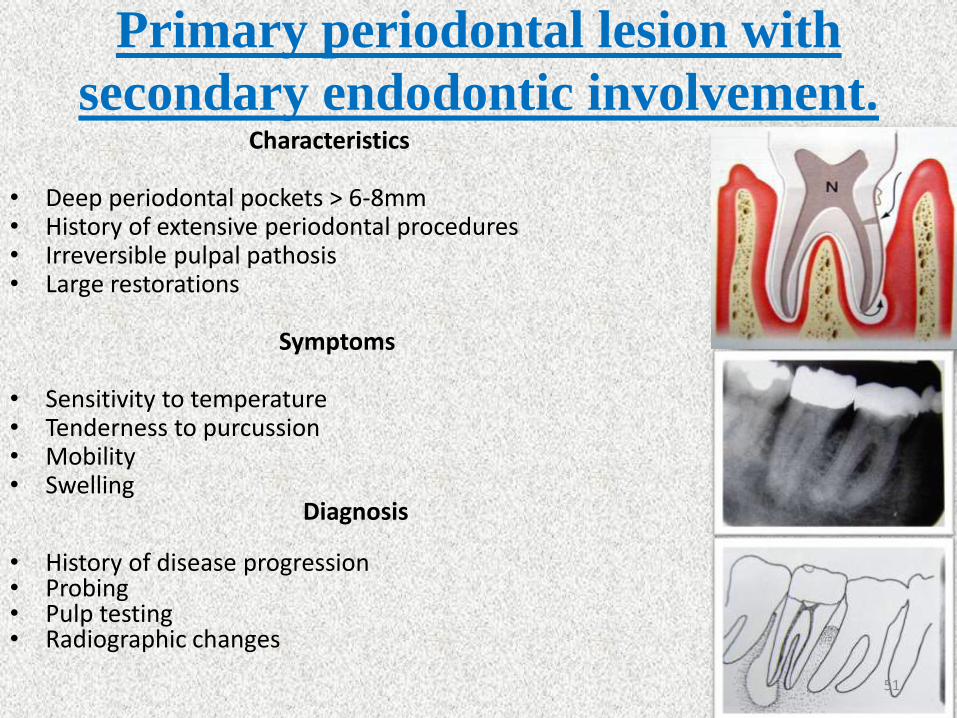
Primary periodontal lesion with
secondary endodontic involvement.Characteristics
• Deep periodontal pockets > 6-8mm• History of extensive periodontal procedures• Irreversible pulpal pathosis• Large restorations
Symptoms
• Sensitivity to temperature• Tenderness to purcussion• Mobility• Swelling
Diagnosis
• History of disease progression• Probing• Pulp testing• Radiographic changes
51

Different diagnosis
• Presence of generalized periodontal disease.
• Failure of probing G.P point to reach the apex.
• Presence of pulp vitality.
• Class IV lesion will deteriorate unless periodontal treatment is instituted.
Treatment
• Periodontal therapy• Root amputation • Guided tissue regeneration• Root canal therapy
Prognosis
• Depends on continuing periodontal treatment subsequent to endodontic therapy.
52
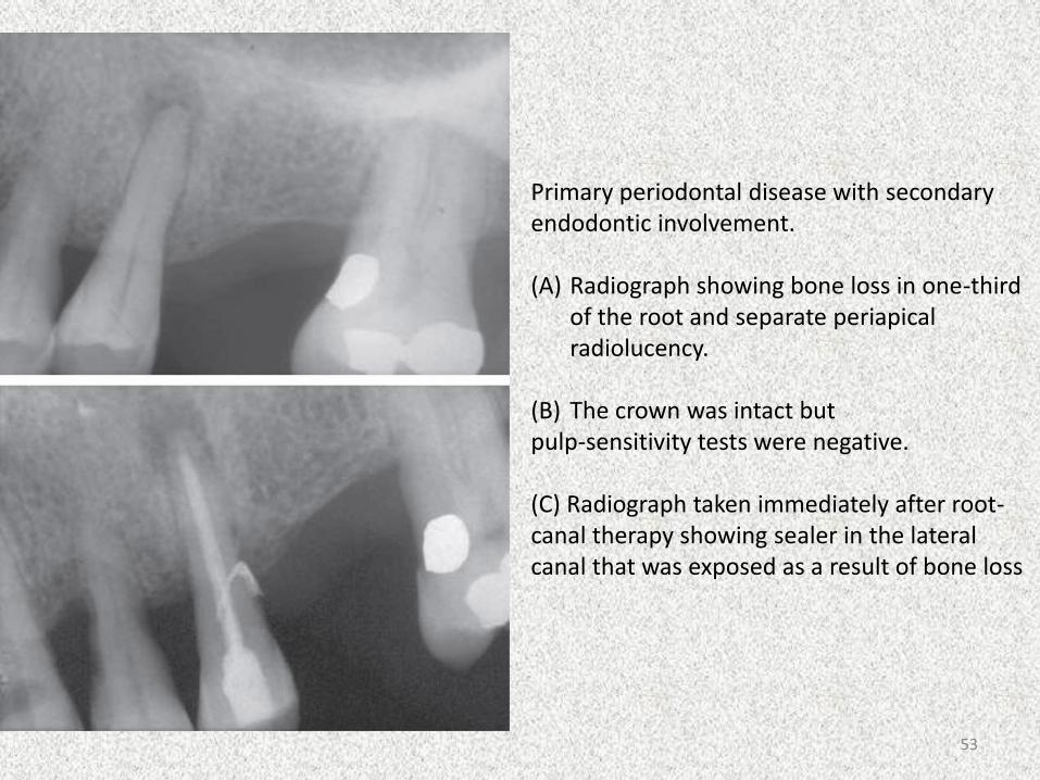
Primary periodontal disease with secondaryendodontic involvement.
(A) Radiograph showing bone loss in one-third of the root and separate periapicalradiolucency.
(B) The crown was intact butpulp-sensitivity tests were negative.
(C) Radiograph taken immediately after root-canal therapy showing sealer in the lateral canal that was exposed as a result of bone loss
53

True combined lesions
• It arises when an endodontic disease progressing coronally joins with an infected periodontal pocket progressing apically.
Characteristics
• Once the pulpal and periodontal lesions coalesce, they may be clinically indistinguishable.
• Necrotic pulp/ failed endodontic treatment, plaque, calculus and periodontitis will be present in varying degrees.
Diagnosis• Probing• Radiographs • Pulpal testing: negative
54
Treatment
• Endodontic therapy
• Periodontal therapy.
• Hemisection
• Bicuspidization
• Root amputation
Prognosis
• Depends on the each individual factor.
• Adequate root canal therapy results in resolution of the periapical lesion
• Prognosis of the affected tooth then depends totally on the outcome of periodontal therapy.
55
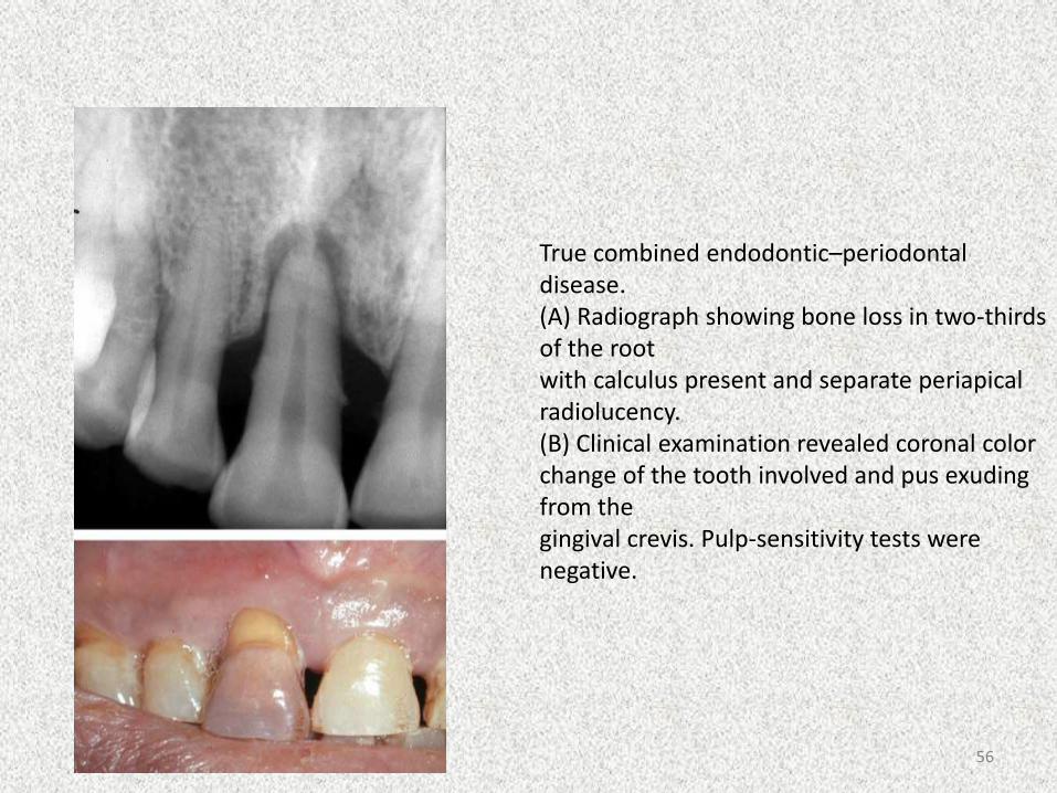
True combined endodontic–periodontal disease.(A) Radiograph showing bone loss in two-thirds of the rootwith calculus present and separate periapicalradiolucency.(B) Clinical examination revealed coronal colorchange of the tooth involved and pus exuding from thegingival crevis. Pulp-sensitivity tests were negative.
56
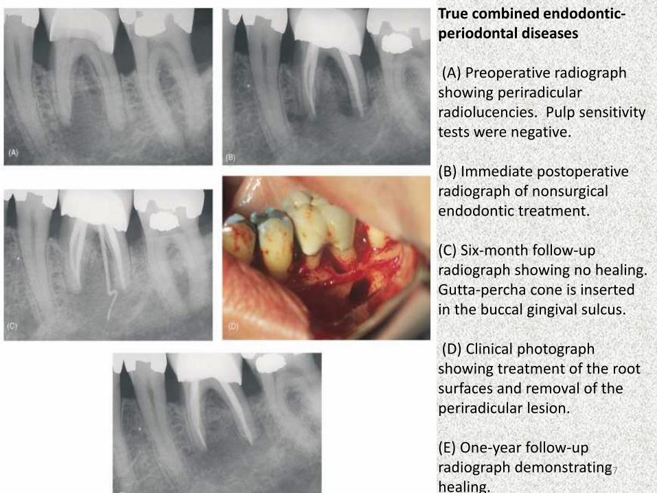
True combined endodontic-periodontal diseases
(A) Preoperative radiograph showing periradicularradiolucencies. Pulp sensitivity tests were negative.
(B) Immediate postoperative radiograph of nonsurgical endodontic treatment.
(C) Six-month follow-up radiograph showing no healing. Gutta-percha cone is inserted in the buccal gingival sulcus.
(D) Clinical photograph showing treatment of the root surfaces and removal of the periradicular lesion.
(E) One-year follow-up radiograph demonstrating healing.
57

DIAGNOSIS CHARACTERSTICS CLINICAL FINDINGS TREATMENT
ENDODONTICLESION
•Periapical bone loss•Drainage through • sulcus.•History /signs of • extensive restorative • treatment.•Gingival swelling.•Furcation bone loss
•Pulp test negative.•Pd probing yields narrow, • isolated pocket.•Evidence of inadequate root canal treatment.•Rapid onset.
Endodontic Treatment
ENDODONTICLESION WITH PERIODONTAL
DISEASE
•Necrotic pulp.•Generalized • periodontitis with • plaque & calculus.
•Pulp test negative, evidence of inflammation/necrosis.•Generalized increase in pocket depth & attachment loss.•Radiographic evidence of pulp & periodontal disease.
First: Endodontictreatment,evaluate in 2-3months.Then: periodontaltreatment.
PERIODONTAL LESION
•Deep pockets.•Extensive attachment • loss.•No evidence of pulpal • disease.
•H/o disease progression/therapy.•Deep probing.•Pulp test positive.
Periodontal Therapy only
58
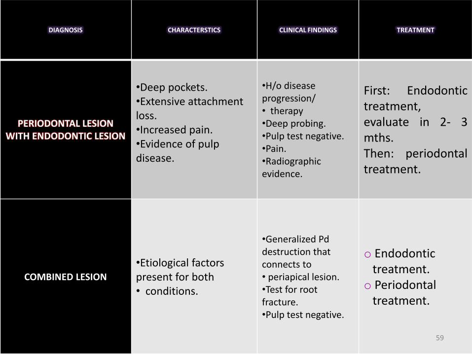
DIAGNOSIS CHARACTERSTICS CLINICAL FINDINGS TREATMENT
PERIODONTAL LESION WITH ENDODONTIC LESION
•Deep pockets.•Extensive attachment loss.•Increased pain.•Evidence of pulp disease.
•H/o disease progression/• therapy•Deep probing.•Pulp test negative.•Pain.•Radiographic evidence.
First: Endodontictreatment,evaluate in 2- 3mths.Then: periodontaltreatment.
COMBINED LESION
•Etiological factors present for both • conditions.
•Generalized Pddestruction that connects to• periapical lesion.•Test for root fracture.•Pulp test negative.
o Endodontictreatment.
o Periodontaltreatment.
59
ENDODONTIC PROCEDURAL COMPLICATIONS
AND THEIR EFFECTS ON PERIODONTIUM
• PERFORATION
• TOOTH FRACTURE
• SODIUM HYPOCHLORITE ACCIDENT
• RESORPTIVE DEFECTS
• DENTAL ANOMALIES
• ULTRASONIC DEVICES
60
PERFORATIONSIatrogenic perforation
Preparation of the access cavity
Instrumentation of canal space
Preparation of the tooth for post (improper post space)
Extensive gouging of a tooth with rotary instruments during periodontal surgical procedures.
Treatment
• Minimize the time from perforation till its sealed.
• Treatment prognosis of root perforations depends on the size, location, time of diagnosis and treatment, degree of periodontal damage and the sealing ability and biocompatibility of the repair material.
61
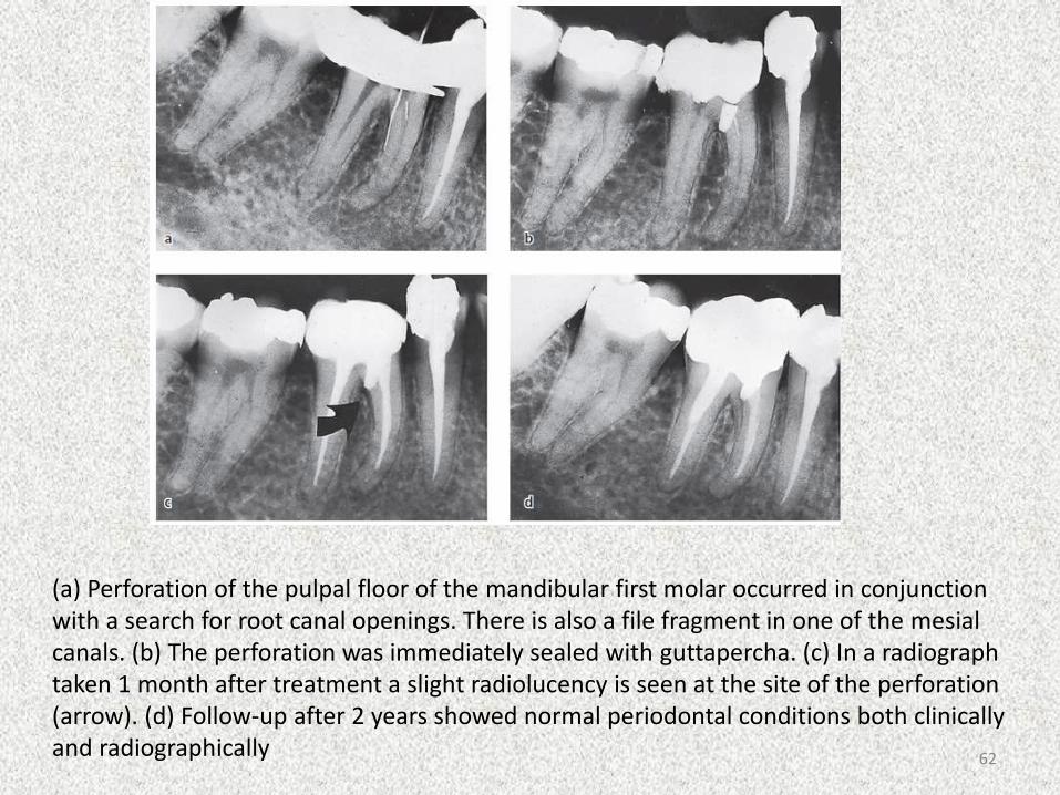
(a) Perforation of the pulpal floor of the mandibular first molar occurred in conjunction with a search for root canal openings. There is also a file fragment in one of the mesialcanals. (b) The perforation was immediately sealed with guttapercha. (c) In a radiograph taken 1 month after treatment a slight radiolucency is seen at the site of the perforation (arrow). (d) Follow-up after 2 years showed normal periodontal conditions both clinically and radiographically 62

Tooth fracture
• Crown-Root Fractures
• Horizontal Root Fractures in the Coronal Area
• Horizontal Fractures in the Midroot Area
• Horizontal Fractures in the Apical Area
• Vertical root fractures
63

64
Sodium hypochlorite accident
• Conc. 0.5-6% (2.5% most commonly used)
• Extrusion of sodium hypochlorite into the periradicular tissues results in rapid diffuse swelling and extreme pain.
CAUSES
• Forcing the irrigant into surronding periradiculartissues.
• Iatrogenic perforations
• overinstrumentation
65
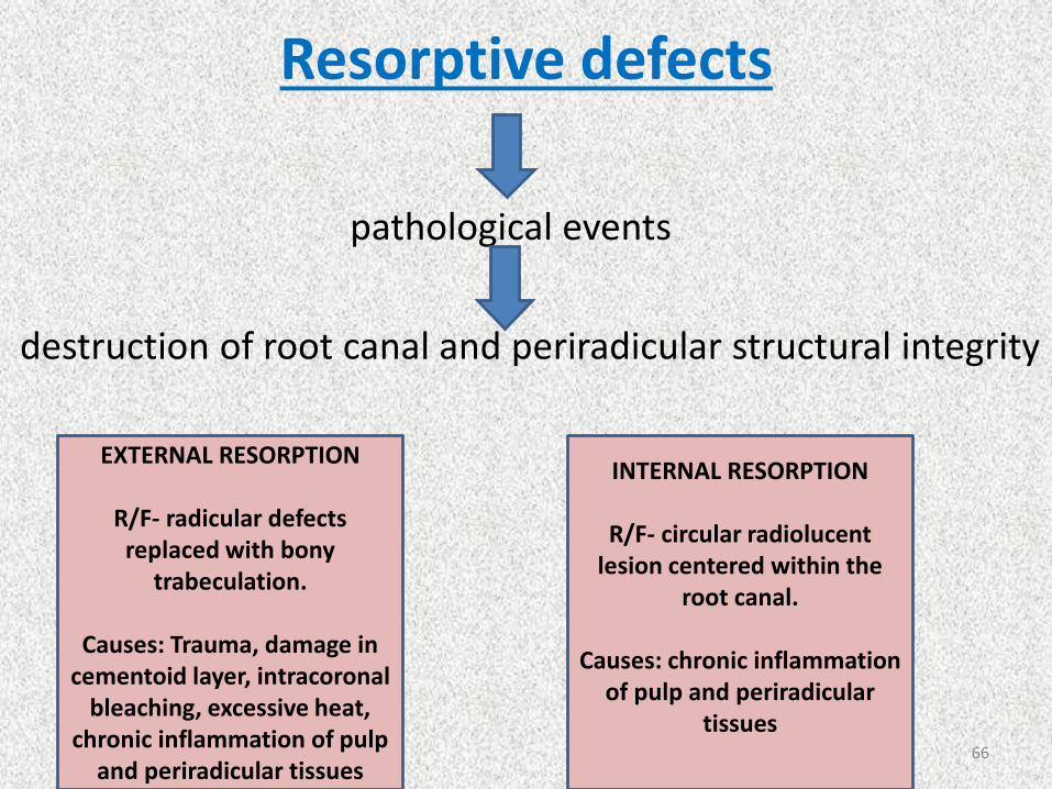
Resorptive defects
pathological events
destruction of root canal and periradicular structural integrity
EXTERNAL RESORPTION
R/F- radicular defects replaced with bony
trabeculation.
Causes: Trauma, damage in cementoid layer, intracoronal
bleaching, excessive heat, chronic inflammation of pulp
and periradicular tissues
INTERNAL RESORPTION
R/F- circular radiolucent lesion centered within the
root canal.
Causes: chronic inflammation of pulp and periradicular
tissues66
Treatment
• Root canal treatment – complete resolution of the resorptive process.
• combined with MTA in case of perforation repair.
• Recent studies have also assessed the potential use of Bisphosphonates –inhibits bone resorption by acting directly on osteoclasts.
67
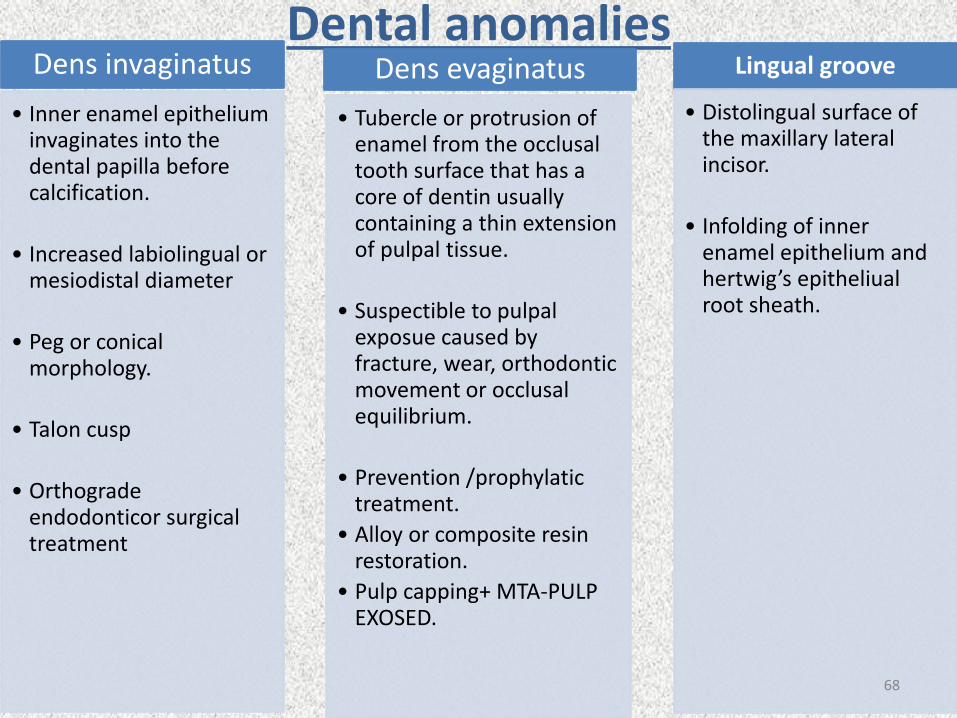
Dental anomaliesDens invaginatus
• Inner enamel epithelium invaginates into the dental papilla before calcification.
• Increased labiolingual or mesiodistal diameter
• Peg or conical morphology.
• Talon cusp
• Orthogradeendodonticor surgical treatment
Dens evaginatus
• Tubercle or protrusion of enamel from the occlusal tooth surface that has a core of dentin usually containing a thin extension of pulpal tissue.
• Suspectible to pulpalexposue caused by fracture, wear, orthodontic movement or occlusal equilibrium.
• Prevention /prophylatictreatment.
• Alloy or composite resin restoration.
• Pulp capping+ MTA-PULP EXOSED.
Lingual groove
• Distolingual surface of the maxillary lateral incisor.
• Infolding of inner enamel epithelium and hertwig’s epitheliualroot sheath.
68

![Temecula Valley MicroEndodontics · Combined endodontic]periodontic lesion Root amputation prior to endodontic treatment No medical problem (ASA Class No history of anesthesia problems](https://static.fdocuments.net/doc/165x107/5f15737df573a4457e7f5816/temecula-valley-microendodontics-combined-endodonticperiodontic-lesion-root-amputation.jpg)


