Empiric Treatment: Pneumonia. Overview of Pneumonia diseases.asp?did=38
118
Empiric Treatment: Pneumonia
-
Upload
wilfred-horton -
Category
Documents
-
view
224 -
download
1
Transcript of Empiric Treatment: Pneumonia. Overview of Pneumonia diseases.asp?did=38
Empiric Treatment: Pneumonia
Overview of Pneumonia
• http://www.virtualrespiratorycentre.com/diseases.asp?did=38
• Link• Link• Link• Link• LINK
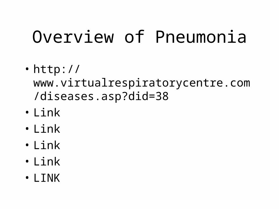
What is pneumonia?
• Pneumonia is an inflammatory illness of the lung.
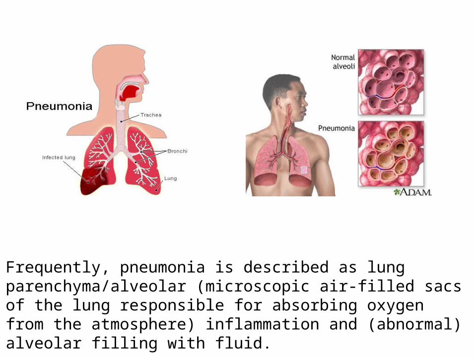
Frequently, pneumonia is described as lung parenchyma/alveolar (microscopic air-filled sacs of the lung responsible for absorbing oxygen from the atmosphere) inflammation and (abnormal) alveolar filling with fluid.
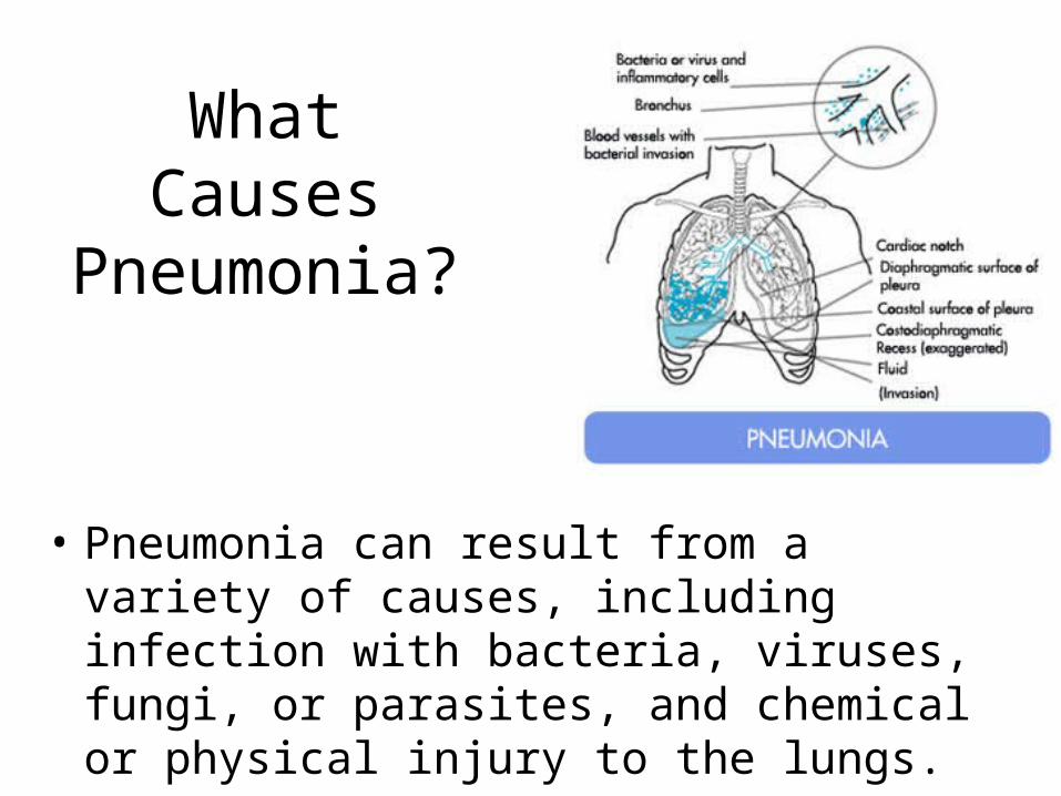
What Causes Pneumonia?
• Pneumonia can result from a variety of causes, including infection with bacteria, viruses, fungi, or parasites, and chemical or physical injury to the lungs.
Pneumonia
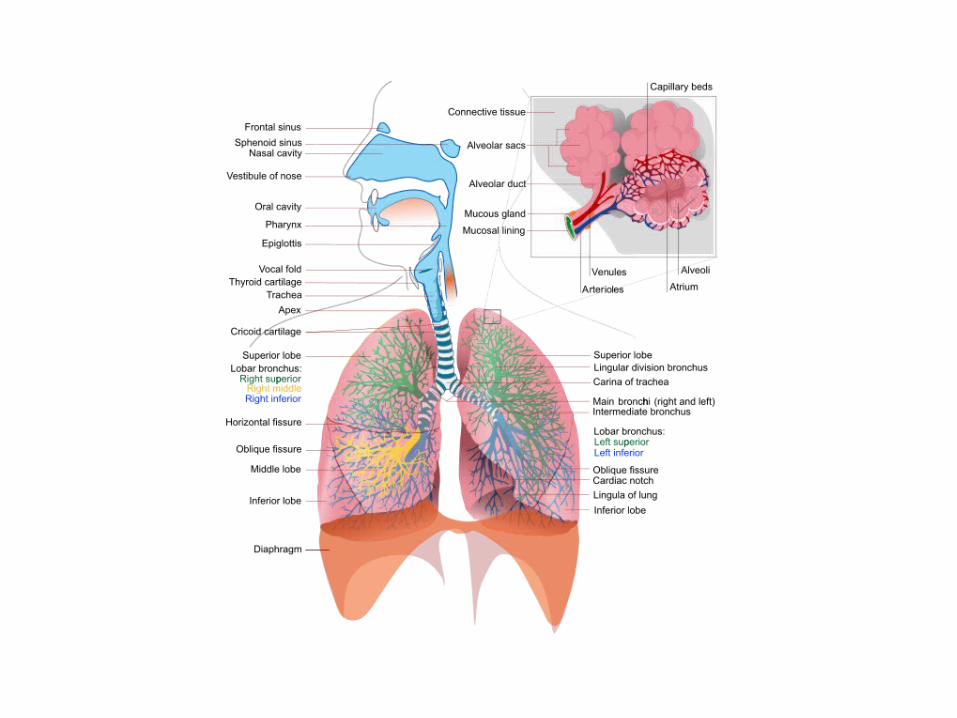
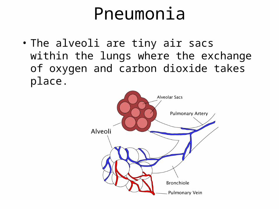
• The alveoli are tiny air sacs within the lungs where the exchange of oxygen and carbon dioxide takes place.
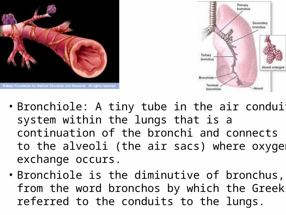
• Bronchiole: A tiny tube in the air conduit system within the lungs that is a continuation of the bronchi and connects to the alveoli (the air sacs) where oxygen exchange occurs.
• Bronchiole is the diminutive of bronchus, from the word bronchos by which the Greeks referred to the conduits to the lungs.
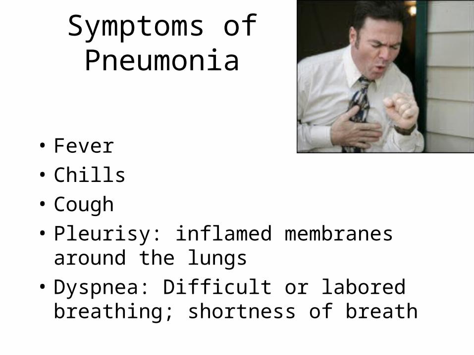
Symptoms of Pneumonia
• Fever
• Chills
• Cough
• Pleurisy: inflamed membranes around the lungs
• Dyspnea: Difficult or labored breathing; shortness of breath
Diagnosis of Pneumonia
• Pneumonia usually produces distinctive sounds; these abnormal sounds are caused by narrowing of airways or filling of the normally air-filled parts of the lung with inflammatory cells and fluid, a process called consolidation.
Diagnosis of Pneumonia
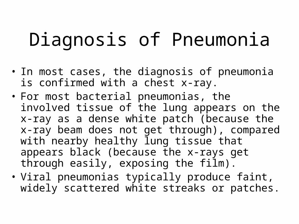
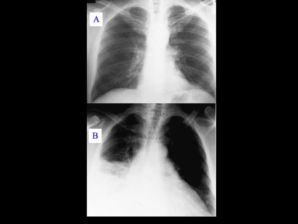
• In most cases, the diagnosis of pneumonia is confirmed with a chest x-ray.
• For most bacterial pneumonias, the involved tissue of the lung appears on the x-ray as a dense white patch (because the x-ray beam does not get through), compared with nearby healthy lung tissue that appears black (because the x-rays get through easily, exposing the film).
• Viral pneumonias typically produce faint, widely scattered white streaks or patches.
Two Types of Pneumonia
• Community-Acquired Pneumonia (CAP): individual residing in their homes
• Hospital-Acquired Pneumonia (HAP): individuals residing in hospitals
Community-Acquired Pneumonia
• Typical: Sudden onset of fever, chills, pleuritic chest pain, productive cough– Streptococcus pneumoniae– Haemophilus influenzae
• Atypical: often preceded by mild respiratory illness– Legionella spp.– Mycoplasma pneumoniae– Chlamydophila pneumoniae

CAP: typical
Streptococcus pneumoniae
Gram +
Usually susceptible to penicillin
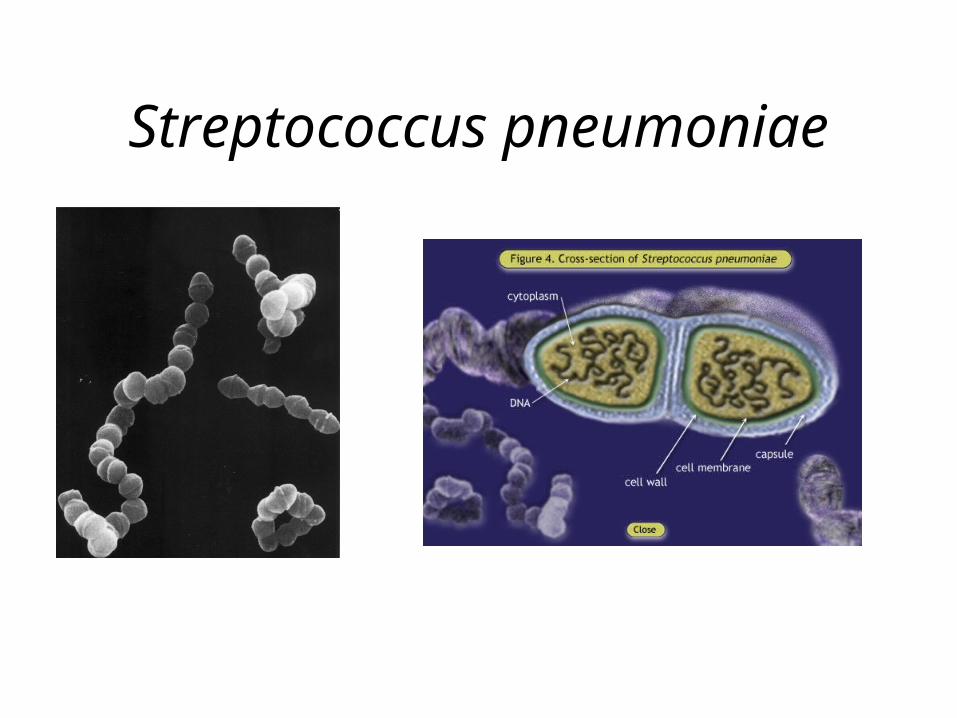
Streptococcus pneumoniae
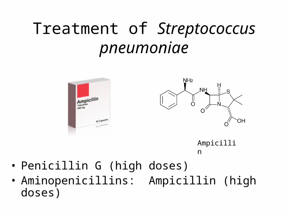
Treatment of Streptococcus pneumoniae
• Penicillin G (high doses)• Aminopenicillins: Ampicillin (high doses)
Ampicillin
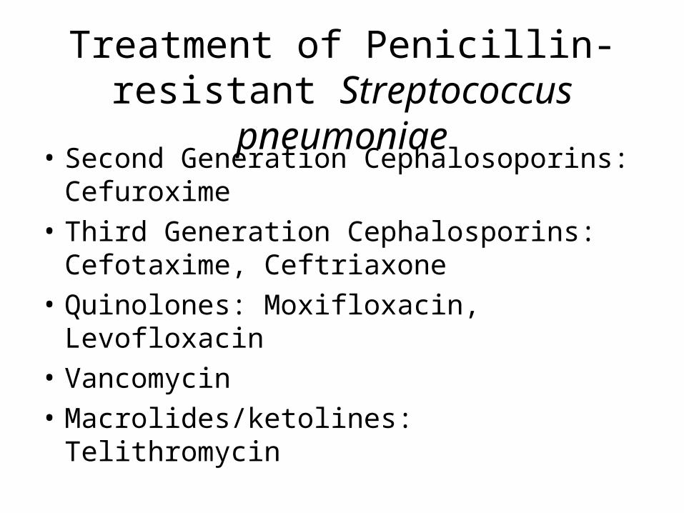
Treatment of Penicillin-resistant Streptococcus pneumoniae
• Second Generation Cephalosoporins: Cefuroxime
• Third Generation Cephalosporins: Cefotaxime, Ceftriaxone
• Quinolones: Moxifloxacin, Levofloxacin
• Vancomycin
• Macrolides/ketolines: Telithromycin
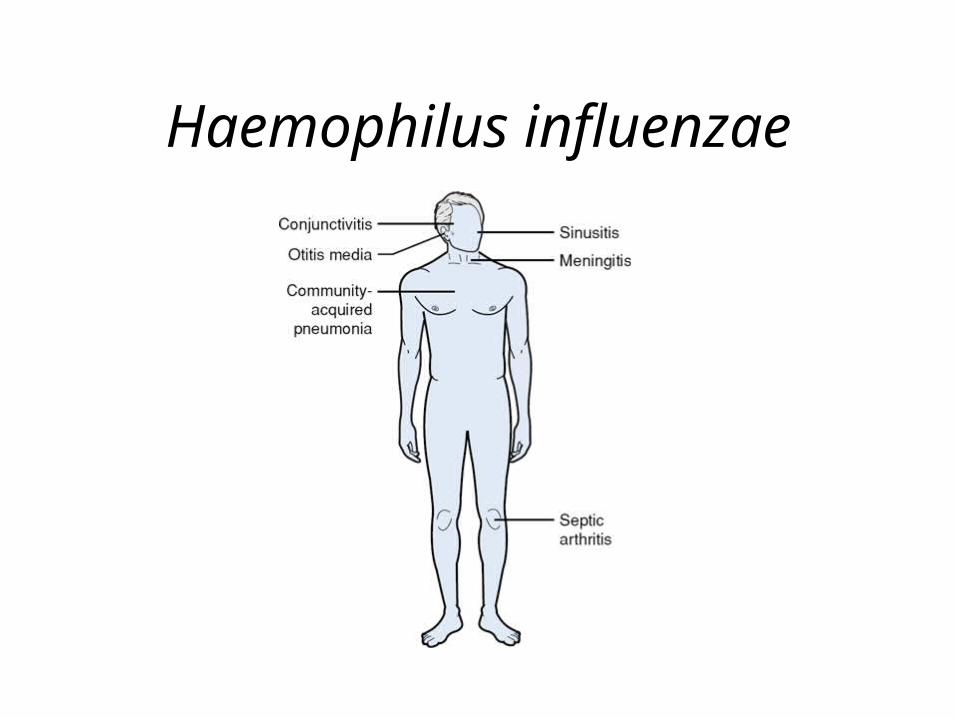
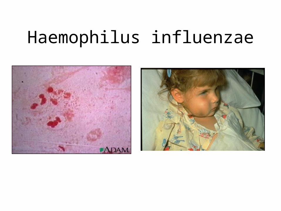
Haemophilus influenzae
Haemophilus influenzae
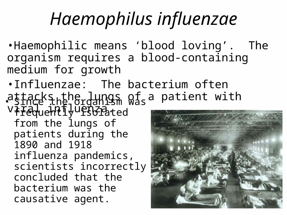
• Since the organism was frequently isolated from the lungs of patients during the 1890 and 1918 influenza pandemics, scientists incorrectly concluded that the bacterium was the causative agent.
•Haemophilic means ‘blood loving’. The organism requires a blood-containing medium for growth•Influenzae: The bacterium often attacks the lungs of a patient with viral influenza.
Haemophilus influenzae
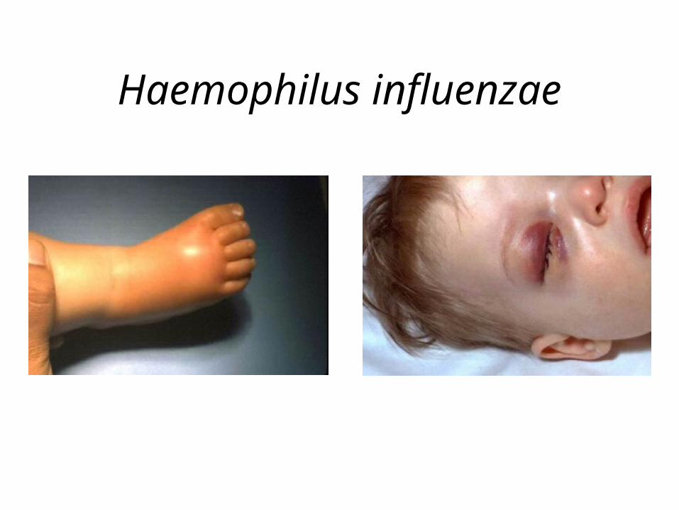
Haemophilus influenzae
Treatment of Infections Caused by Haemophilus influenzae
• Aminopenicillins + -lactamase inhibitor:– Amoxicillin/clavulanate– Ampicillin/sulbactam
• Second-generation cephalosporin– Cefuroxime
• Third-generation cephalosporin– Ceftriaxone– Cefotaxime
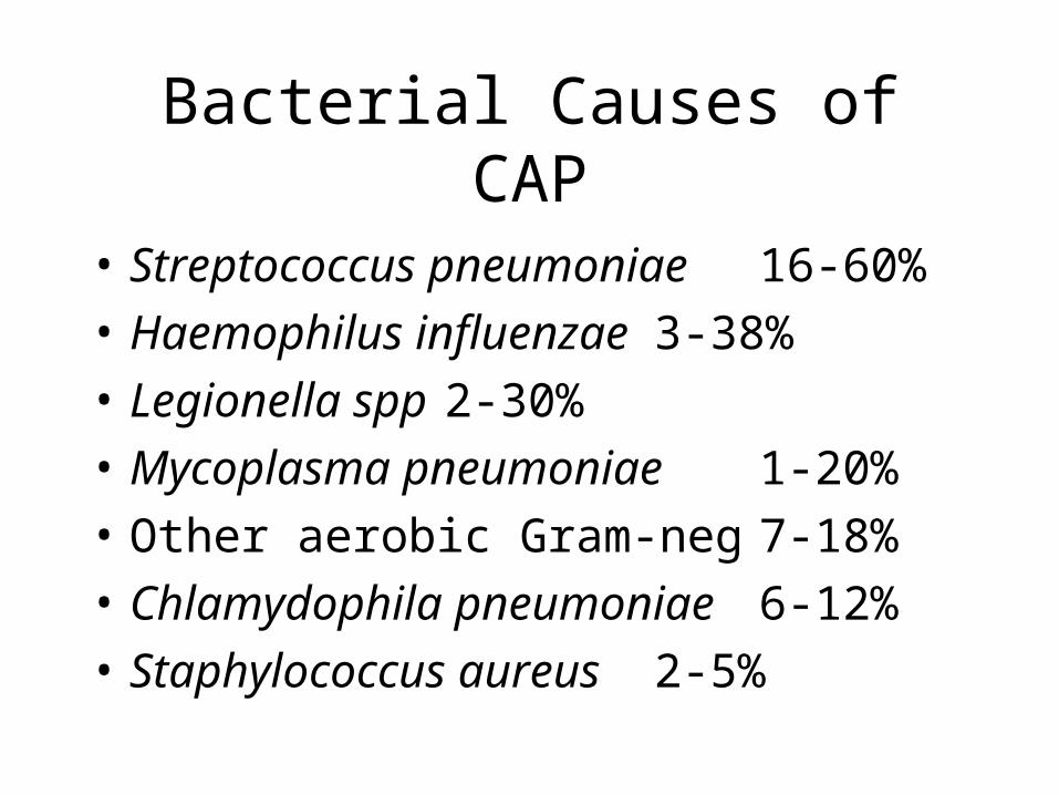
Bacterial Causes of CAP
• Streptococcus pneumoniae 16-60%• Haemophilus influenzae 3-38%• Legionella spp 2-30%• Mycoplasma pneumoniae 1-20%• Other aerobic Gram-neg 7-18%• Chlamydophila pneumoniae 6-12%• Staphylococcus aureus 2-5%
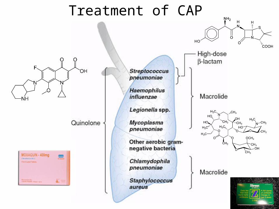
Treatment of CAP
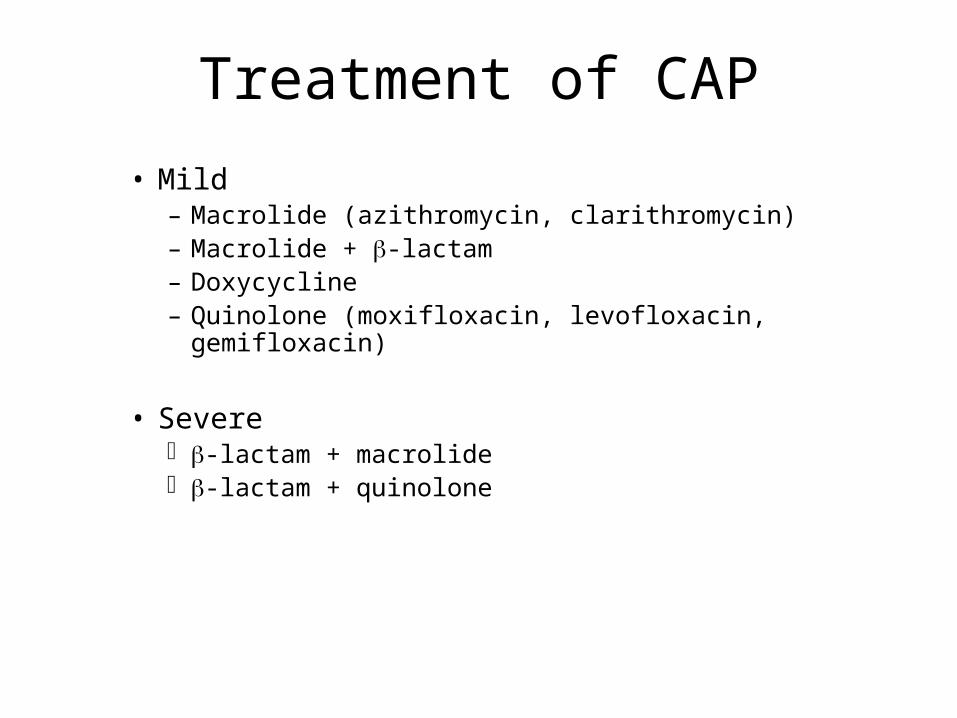
Treatment of CAP
• Mild– Macrolide (azithromycin, clarithromycin)– Macrolide + -lactam– Doxycycline– Quinolone (moxifloxacin, levofloxacin,
gemifloxacin)
• Severe -lactam + macrolide -lactam + quinolone
Treatment of CAP
• Severe -lactam + macrolide -lactam + quinolone
HAP is also divided into two classes:
• Early onset HAP: occurs within first five days of hospitalization
• Late onset HAP: occurs after 5 days of hospitalization
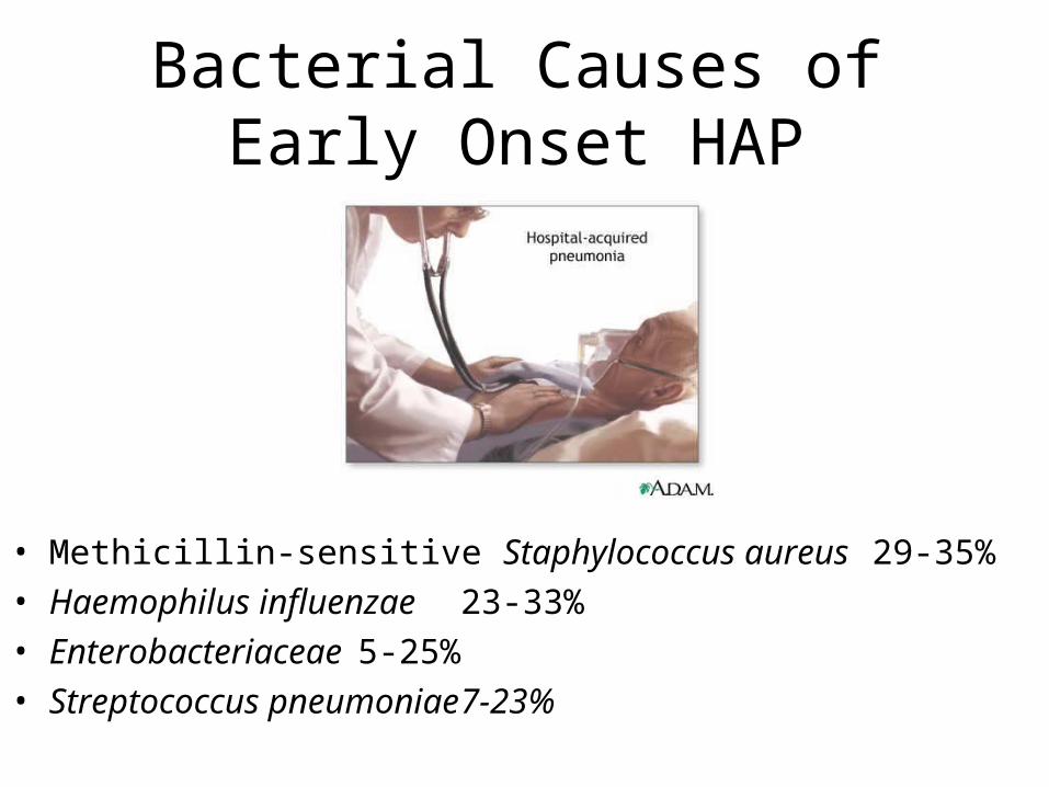
Bacterial Causes ofEarly Onset HAP
• Methicillin-sensitive Staphylococcus aureus 29-35%
• Haemophilus influenzae 23-33%
• Enterobacteriaceae 5-25%• Streptococcus pneumoniae 7-23%
Bacterial Causes of Late Onset HAP
• Pseudomonas aeruginosa 39-64%
• Acinetobacter spp. 6-26%
• Enterobacteriaceae 16-31%
• Methicillin-resistant S. aureus 0-2%
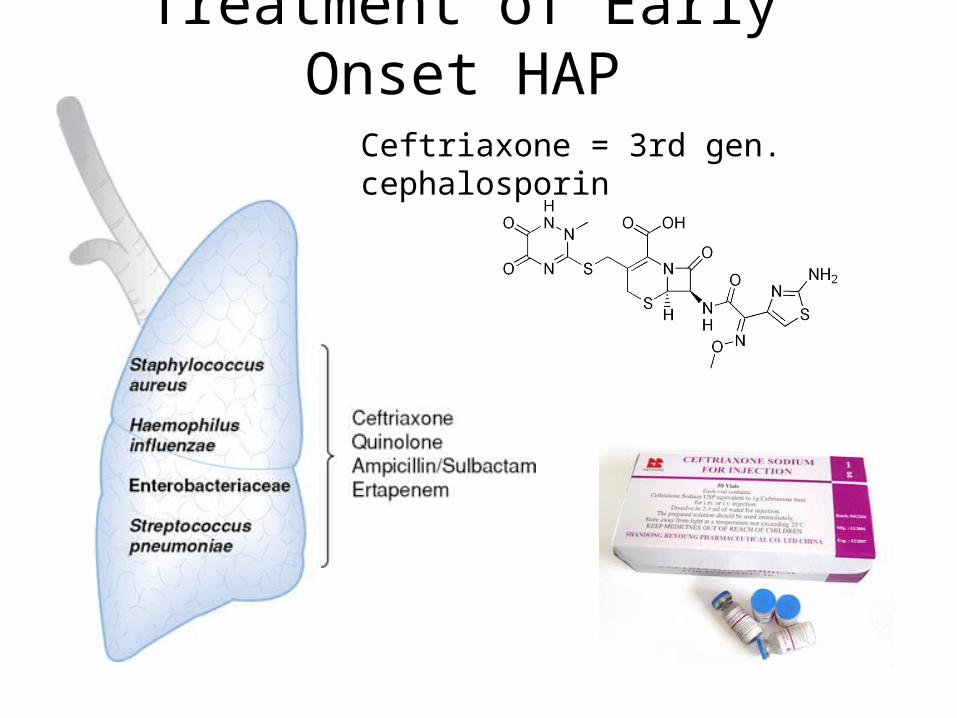
Treatment of Early Onset HAP
Ceftriaxone = 3rd gen. cephalosporin
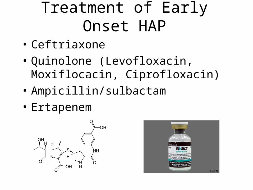
Treatment of Early Onset HAP
• Ceftriaxone
• Quinolone (Levofloxacin, Moxiflocacin, Ciprofloxacin)
• Ampicillin/sulbactam
• Ertapenem
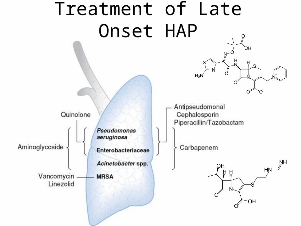
Treatment of Late Onset HAP
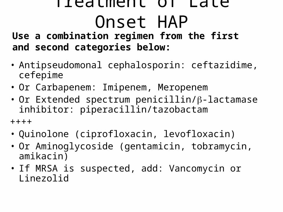
Treatment of Late Onset HAP
• Antipseudomonal cephalosporin: ceftazidime, cefepime• Or Carbapenem: Imipenem, Meropenem• Or Extended spectrum penicillin/-lactamase inhibitor:
piperacillin/tazobactam++++• Quinolone (ciprofloxacin, levofloxacin)• Or Aminoglycoside (gentamicin, tobramycin, amikacin)• If MRSA is suspected, add: Vancomycin or Linezolid
Use a combination regimen from the first and second categories below:

Urinary Tract Infections
• http://www.virtualrenalcentre.com/diseases.asp?did=281
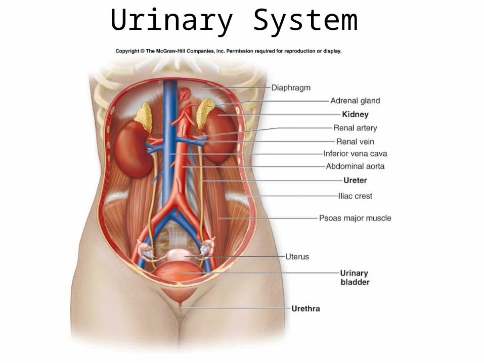
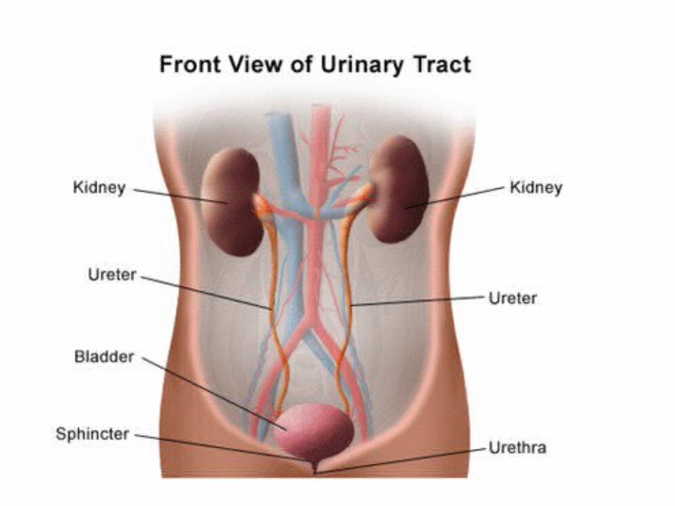
Urinary System
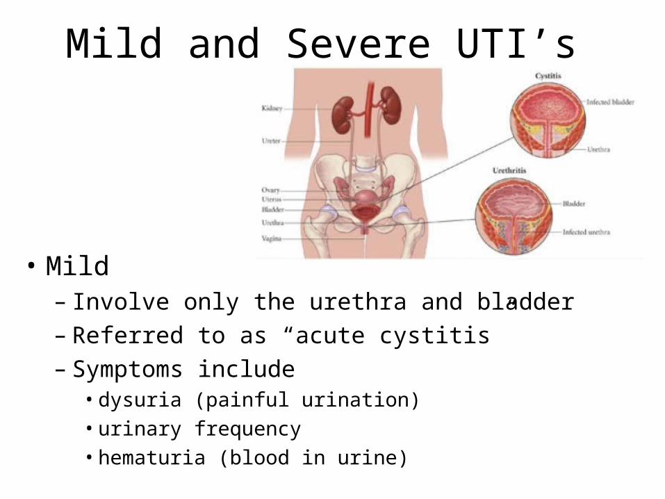
Mild and Severe UTI’s
• Mild– Involve only the urethra and bladder– Referred to as “acute cystitis”– Symptoms include
• dysuria (painful urination)• urinary frequency• hematuria (blood in urine)
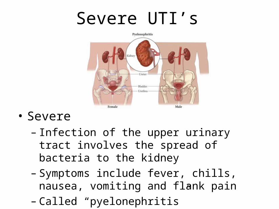
Severe UTI’s
• Severe– Infection of the upper urinary tract involves the
spread of bacteria to the kidney – Symptoms include fever, chills, nausea,
vomiting and flank pain– Called “pyelonephritis”
‘Complicated’ and ‘Uncomplicated’ UTI’s
• Uncomplicated: Less likely to recur. Occur in young, healthy, nonpregnant women
• Complicated: All other UTI’s. More likely to recur.
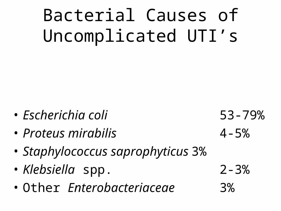
Bacterial Causes of Uncomplicated UTI’s
• Escherichia coli 53-79%
• Proteus mirabilis 4-5%
• Staphylococcus saprophyticus3%
• Klebsiella spp. 2-3%
• Other Enterobacteriaceae 3%
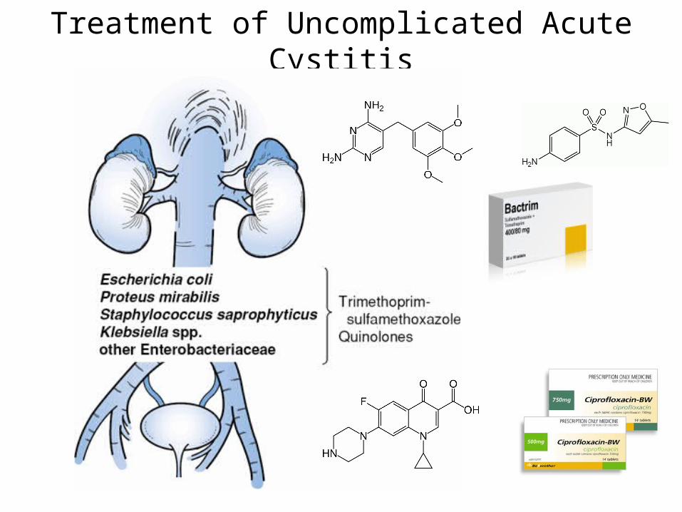
Treatment of Uncomplicated Acute Cystitis
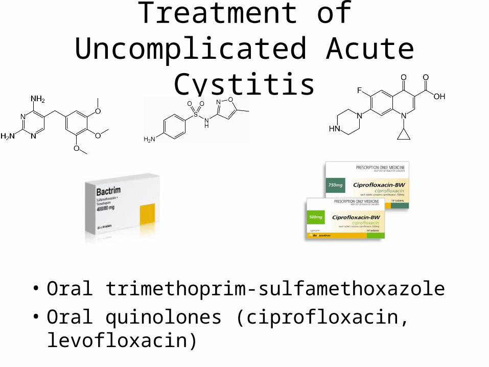
Treatment of Uncomplicated Acute Cystitis
• Oral trimethoprim-sulfamethoxazole
• Oral quinolones (ciprofloxacin, levofloxacin)
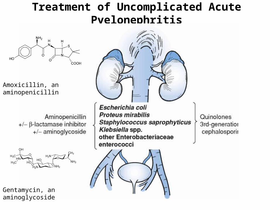
Treatment of Uncomplicated Acute Pyelonephritis
Gentamycin, an aminoglycoside
Amoxicillin, an aminopenicillin
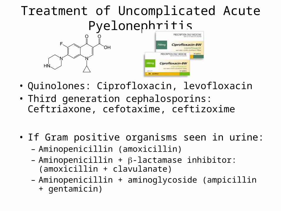
Treatment of Uncomplicated Acute Pyelonephritis
• Quinolones: Ciprofloxacin, levofloxacin• Third generation cephalosporins: Ceftriaxone,
cefotaxime, ceftizoxime
• If Gram positive organisms seen in urine:– Aminopenicillin (amoxicillin)– Aminopenicillin + -lactamase inhibitor: (amoxicillin +
clavulanate)– Aminopenicillin + aminoglycoside (ampicillin +
gentamicin)
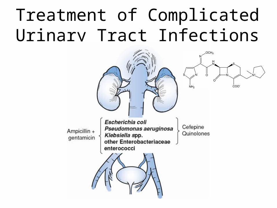
Treatment of Complicated Urinary Tract Infections
Treatment of Complicated Urinary Tract Infections
• Fourth generation cephalosporins (cefepime)
• Quinolones: Ciprofloxacin, Levofloxacin
• If Gram-positive bacteria seen in urine:– Aminopenicillin + aminoglycoside:
Ampicillin + gentamicin
Pelvic Inflammatory Disease
• Link

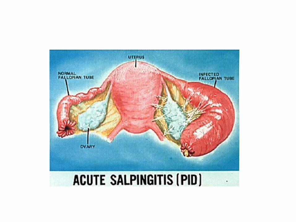
Female Reproductive Organs
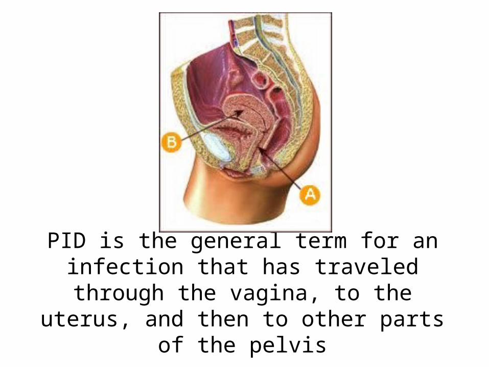
PID is the general term for an infection that has traveled through the vagina, to the
uterus, and then to other parts of the pelvis
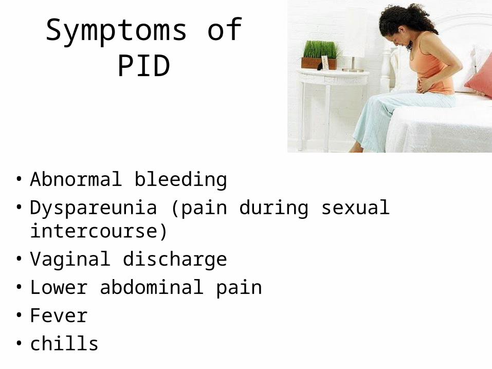
Symptoms of PID
• Abnormal bleeding
• Dyspareunia (pain during sexual intercourse)
• Vaginal discharge
• Lower abdominal pain
• Fever
• chills
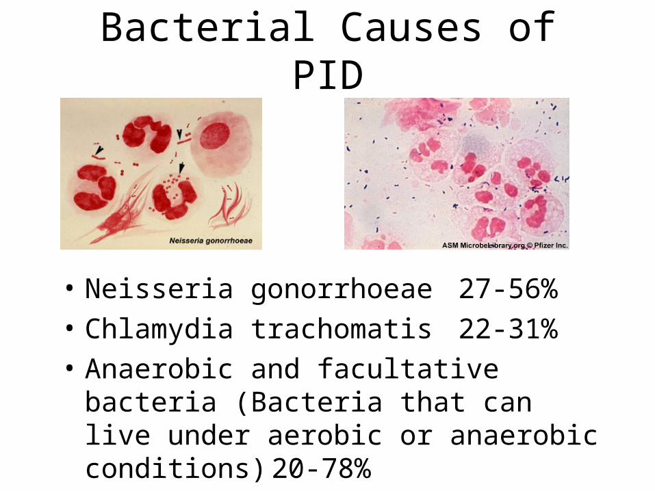
Bacterial Causes of PID
• Neisseria gonorrhoeae 27-56%
• Chlamydia trachomatis 22-31%
• Anaerobic and facultative bacteria (Bacteria that can live under aerobic or anaerobic conditions) 20-78%
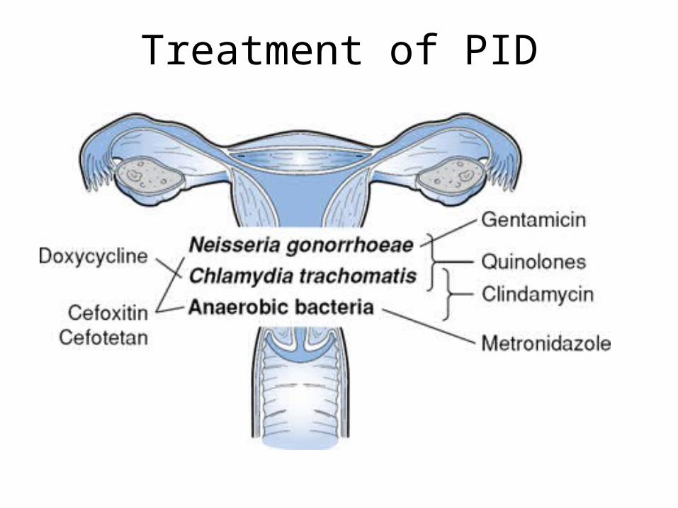
Treatment of PID
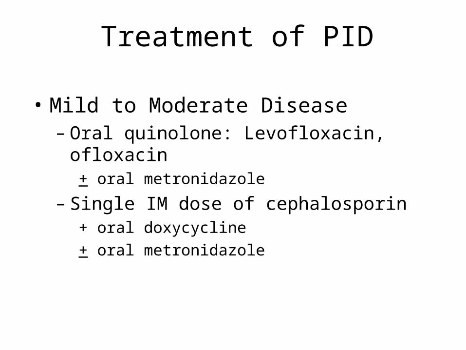
Treatment of PID
• Mild to Moderate Disease– Oral quinolone: Levofloxacin, ofloxacin
+ oral metronidazole
– Single IM dose of cephalosporin+ oral doxycycline
+ oral metronidazole
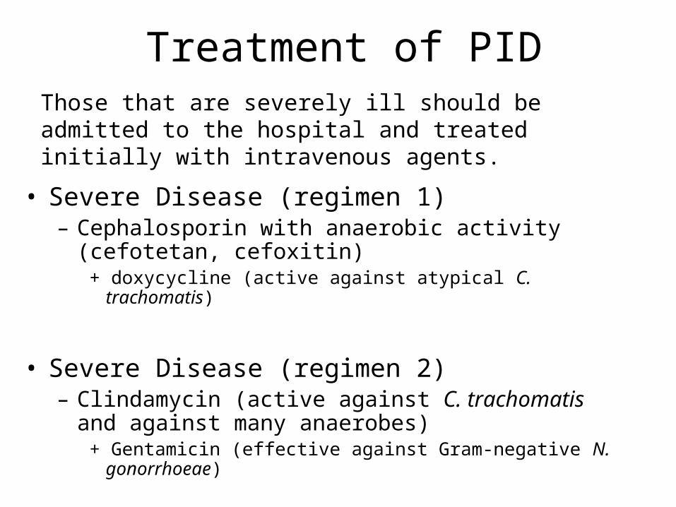
Treatment of PID
• Severe Disease (regimen 1)– Cephalosporin with anaerobic activity (cefotetan,
cefoxitin)+ doxycycline (active against atypical C. trachomatis)
• Severe Disease (regimen 2)– Clindamycin (active against C. trachomatis and against
many anaerobes)+ Gentamicin (effective against Gram-negative N. gonorrhoeae)
Those that are severely ill should be admitted to the hospital and treated initially with intravenous agents.
Meningitis
• Link
• Link
• Link
Meningitis
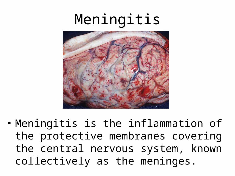
• Meningitis is the inflammation of the protective membranes covering the central nervous system, known collectively as the meninges.
• Meningitis may develop in response to a number of causes, most prominently bacteria, viruses and other infectious agents, but also physical injury, cancer, or certain drugs.
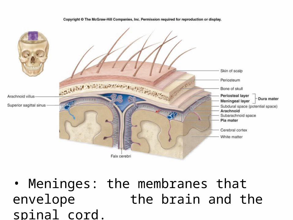
• Meninges: the membranes that envelope the brain and the spinal cord.

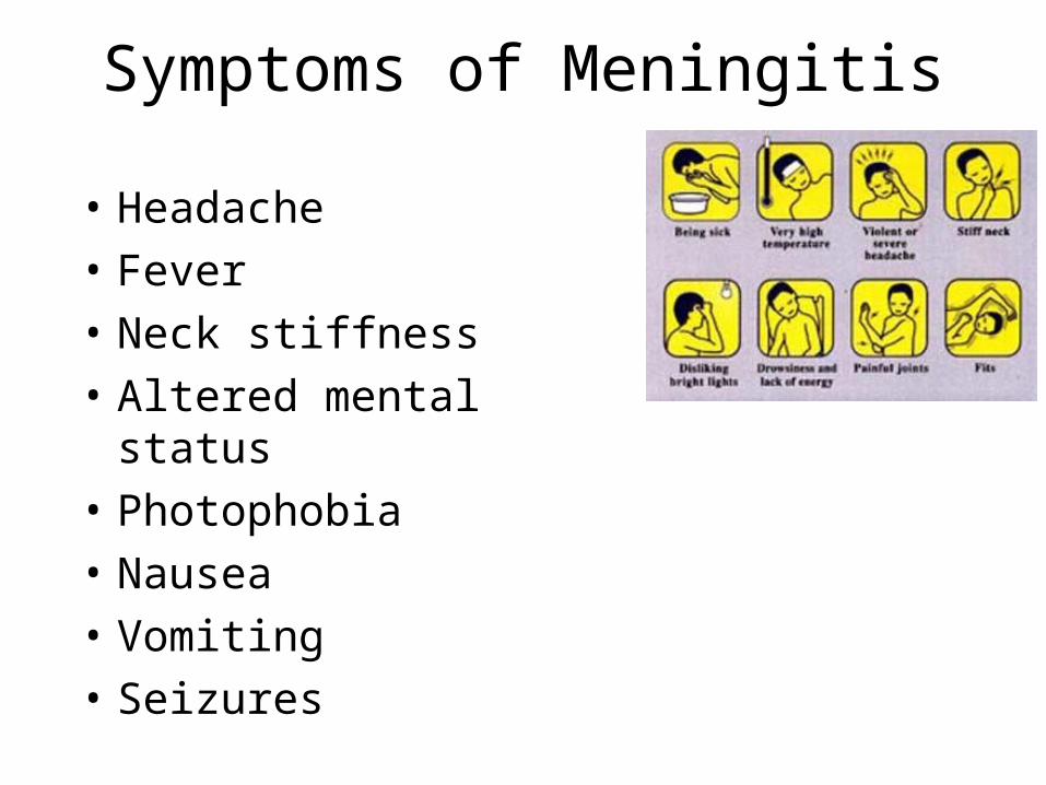
Symptoms of Meningitis
• Headache
• Fever
• Neck stiffness
• Altered mental status
• Photophobia
• Nausea
• Vomiting
• Seizures
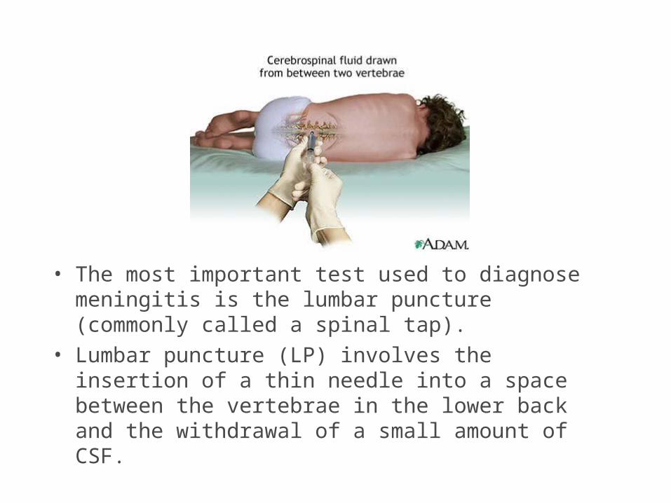
• The most important test used to diagnose meningitis is the lumbar puncture (commonly called a spinal tap).
• Lumbar puncture (LP) involves the insertion of a thin needle into a space between the vertebrae in the lower back and the withdrawal of a small amount of CSF.
Lumbar puncture
• http://www.virtualcancercentre.com/investigations.asp?sid=13
Diagnosis of Meningitis
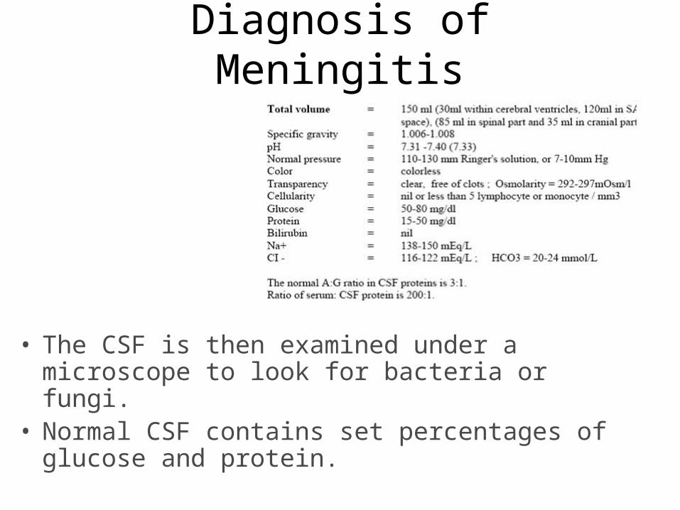
• The CSF is then examined under a microscope to look for bacteria or fungi.
• Normal CSF contains set percentages of glucose and protein.
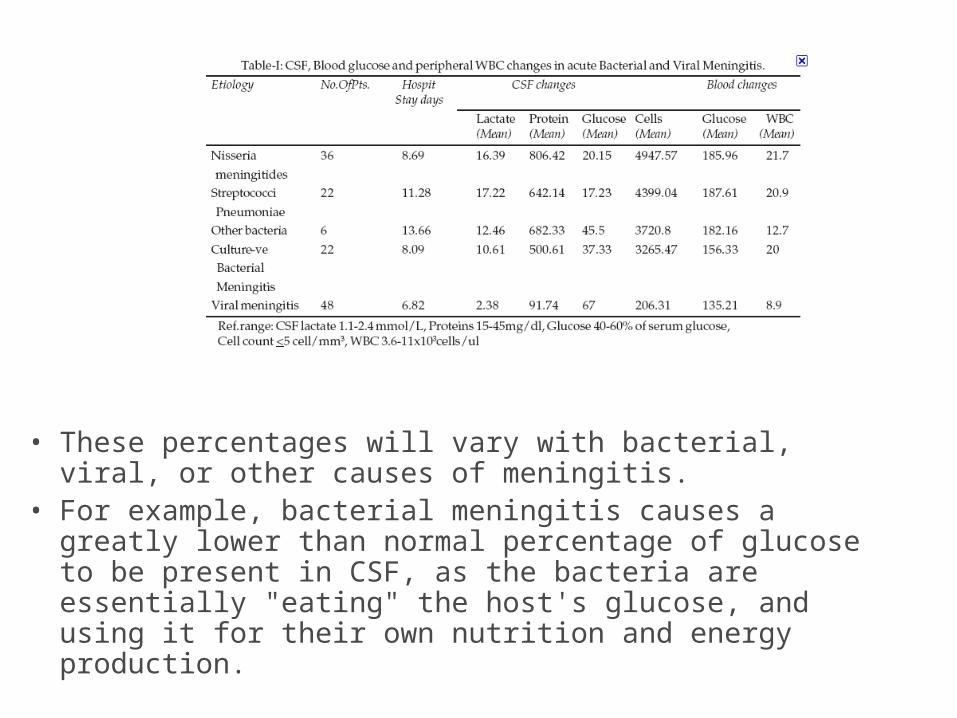
• These percentages will vary with bacterial, viral, or other causes of meningitis.
• For example, bacterial meningitis causes a greatly lower than normal percentage of glucose to be present in CSF, as the bacteria are essentially "eating" the host's glucose, and using it for their own nutrition and energy production.
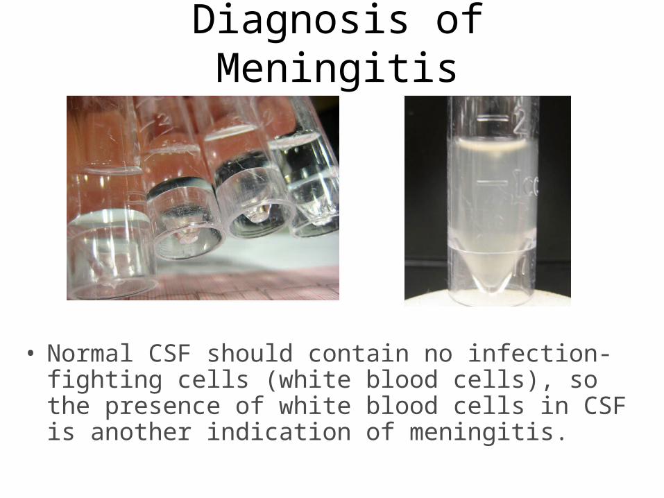
Diagnosis of Meningitis
• Normal CSF should contain no infection-fighting cells (white blood cells), so the presence of white blood cells in CSF is another indication of meningitis.
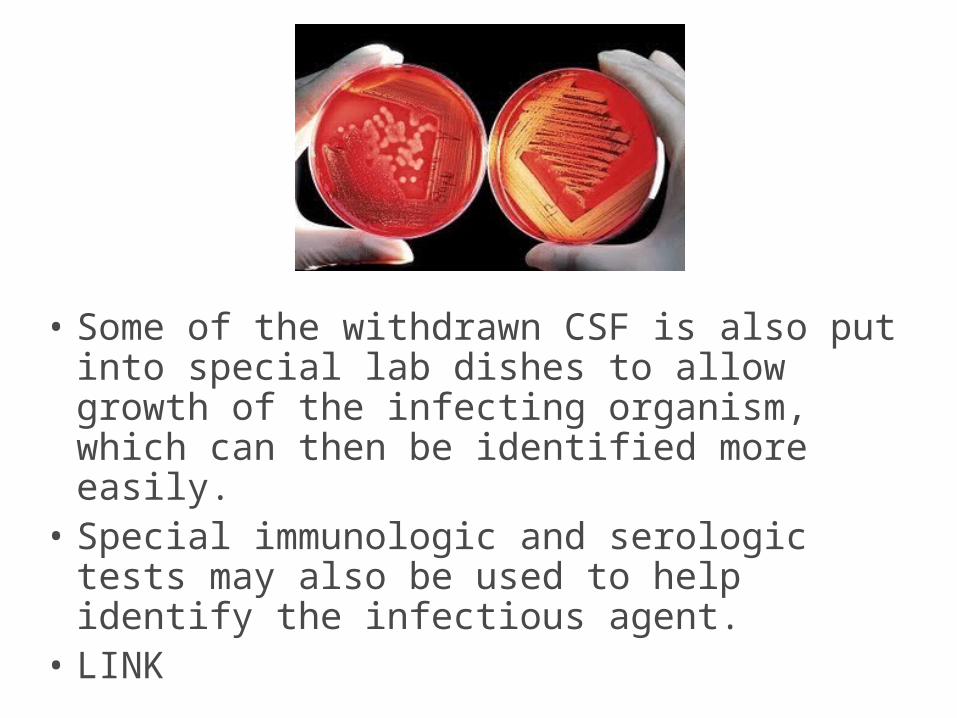
• Some of the withdrawn CSF is also put into special lab dishes to allow growth of the infecting organism, which can then be identified more easily.
• Special immunologic and serologic tests may also be used to help identify the infectious agent.
• LINK
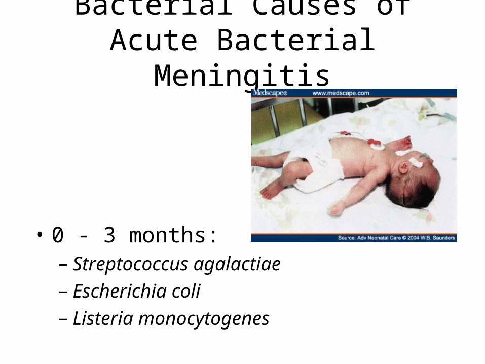
Bacterial Causes of Acute Bacterial Meningitis
• 0 - 3 months: – Streptococcus agalactiae– Escherichia coli– Listeria monocytogenes
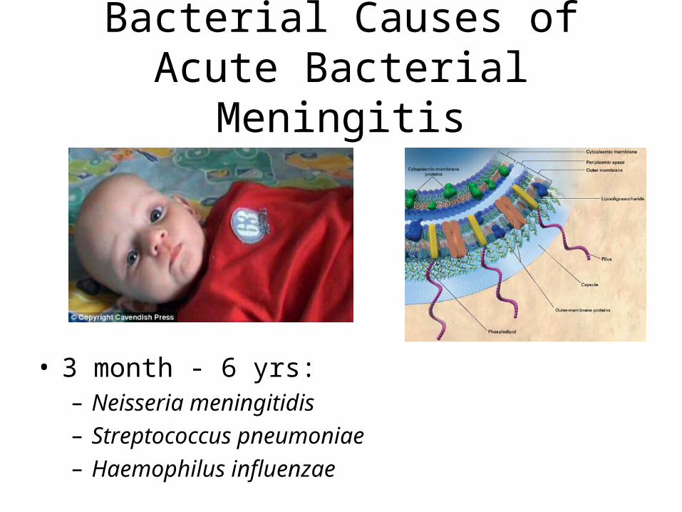
Bacterial Causes of Acute Bacterial Meningitis
• 3 month - 6 yrs:– Neisseria meningitidis– Streptococcus pneumoniae– Haemophilus influenzae
Bacterial Causes of Acute Bacterial Meningitis
• 16 yrs - 50 yrs– Streptococcus
pneumoniae– Neisseria
meningitidis
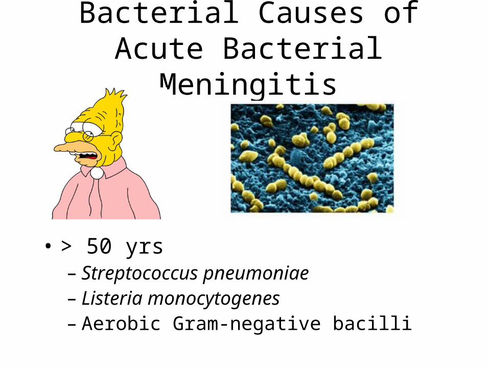
Bacterial Causes of Acute Bacterial Meningitis
• > 50 yrs– Streptococcus pneumoniae– Listeria monocytogenes– Aerobic Gram-negative bacilli
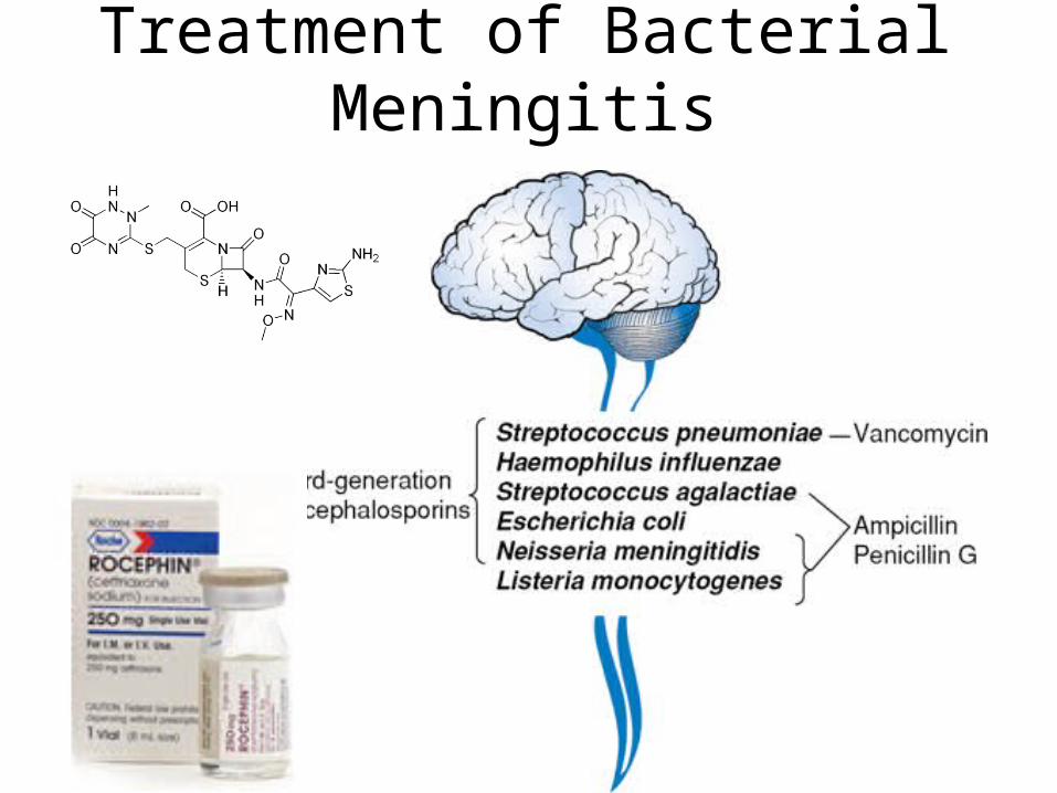
Treatment of Bacterial Meningitis
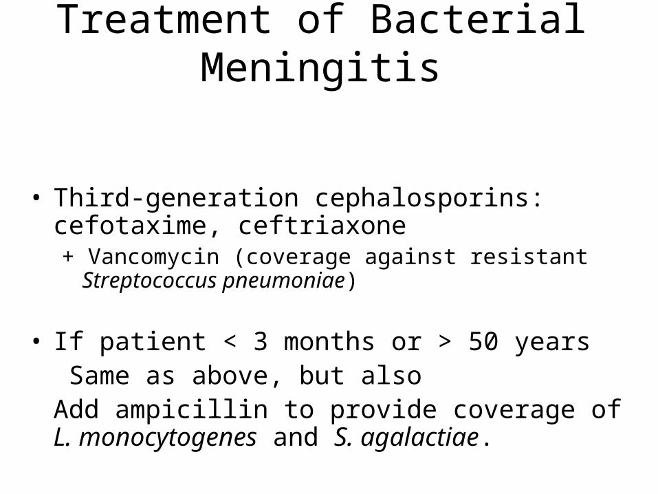
Treatment of Bacterial Meningitis
• Third-generation cephalosporins: cefotaxime, ceftriaxone+ Vancomycin (coverage against resistant Streptococcus
pneumoniae)
• If patient < 3 months or > 50 years Same as above, but alsoAdd ampicillin to provide coverage of L. monocytogenes and S. agalactiae.
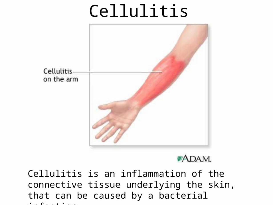
Cellulitis
Cellulitis is an inflammation of the connective tissue underlying the skin, that can be caused by a bacterial infection.
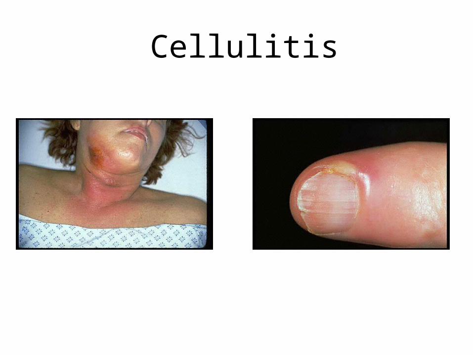
Cellulitis
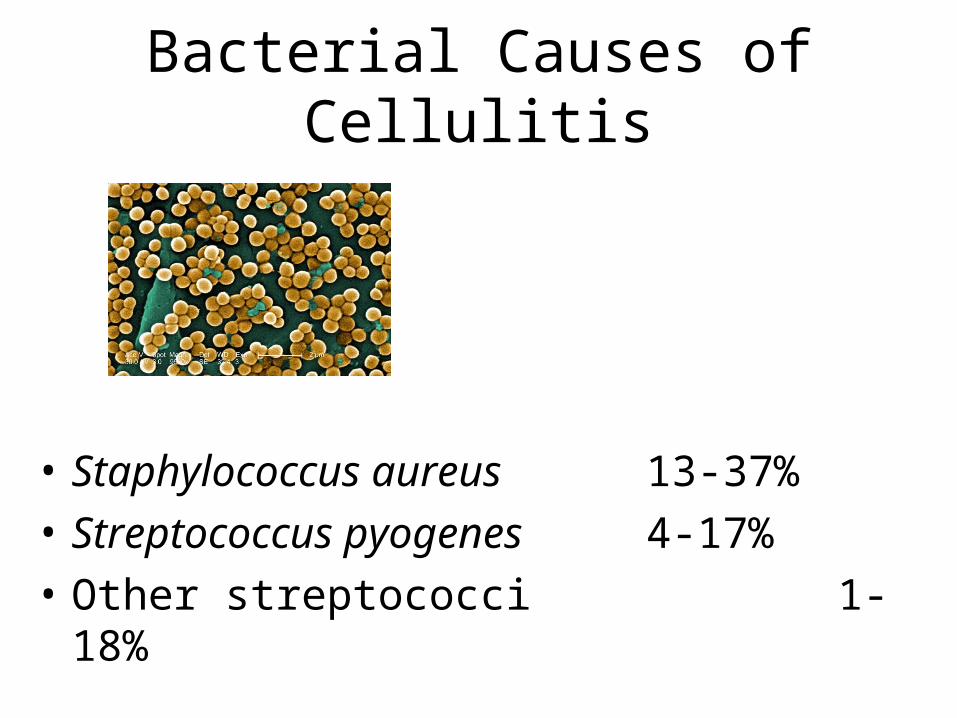
Bacterial Causes of Cellulitis
• Staphylococcus aureus 13-37%
• Streptococcus pyogenes 4-17%
• Other streptococci 1-18%
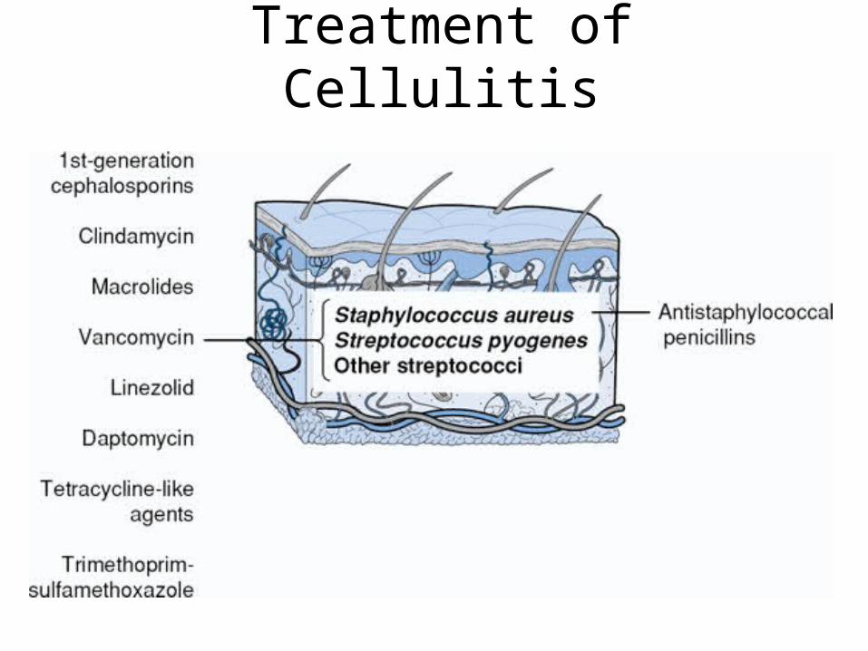
Treatment of Cellulitis
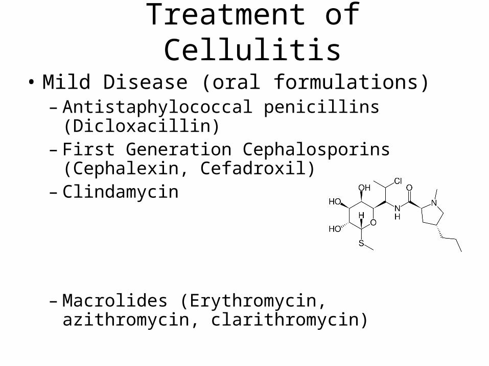
Treatment of Cellulitis
• Mild Disease (oral formulations)– Antistaphylococcal penicillins (Dicloxacillin)– First Generation Cephalosporins
(Cephalexin, Cefadroxil)– Clindamycin
– Macrolides (Erythromycin, azithromycin, clarithromycin)
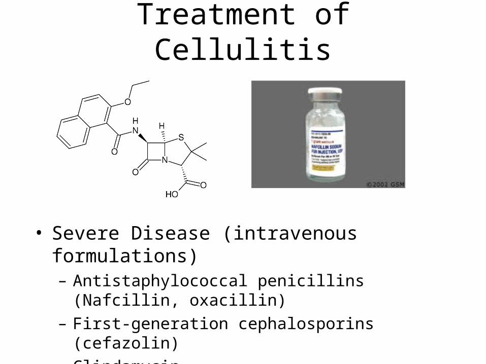
Treatment of Cellulitis
• Severe Disease (intravenous formulations)– Antistaphylococcal penicillins (Nafcillin, oxacillin)– First-generation cephalosporins (cefazolin)– Clindamycin
Treatment of Cellulitis
• If MRSA is suspected– Vancomycin– Linezolid– Daptomycin– Tetracyclines (Tigecycline, doxycycline)– Sulfa drugs (Trimethoprim-
sulfamethoxazole)– Clindamycin

• http://www.virtualrespiratorycentre.com/diseases.asp?did=879
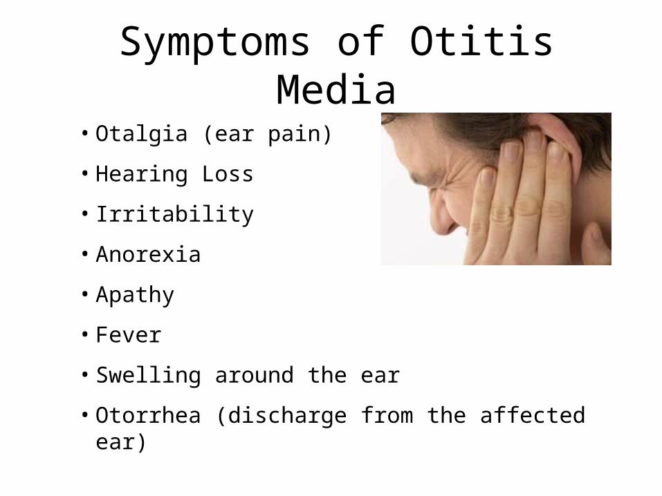
Symptoms of Otitis Media
• Otalgia (ear pain)
• Hearing Loss
• Irritability
• Anorexia
• Apathy
• Fever
• Swelling around the ear
• Otorrhea (discharge from the affected ear)
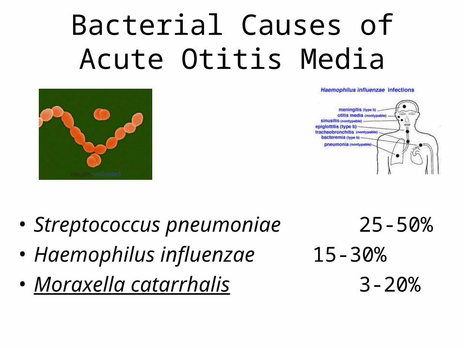
Bacterial Causes of Acute Otitis Media
• Streptococcus pneumoniae 25-50%
• Haemophilus influenzae 15-30%
• Moraxella catarrhalis 3-20%
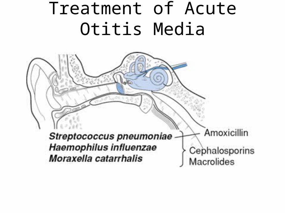
Treatment of Acute Otitis Media
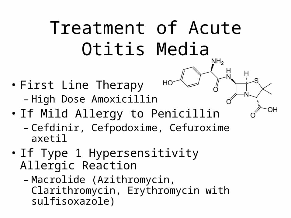
Treatment of Acute Otitis Media
• First Line Therapy– High Dose Amoxicillin
• If Mild Allergy to Penicillin– Cefdinir, Cefpodoxime, Cefuroxime axetil
• If Type 1 Hypersensitivity Allergic Reaction– Macrolide (Azithromycin, Clarithromycin,
Erythromycin with sulfisoxazole)
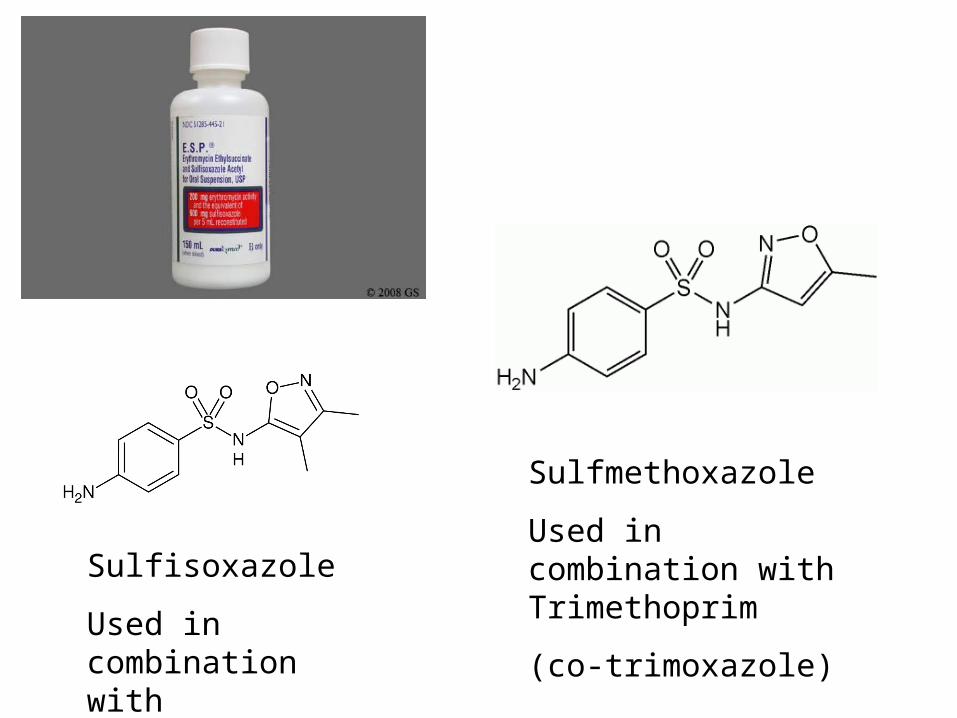
Sulfisoxazole
Used in combination with Erythromycin
Sulfmethoxazole
Used in combination with Trimethoprim
(co-trimoxazole)
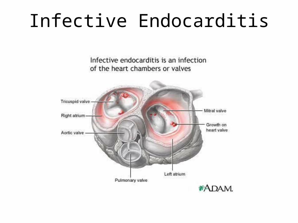
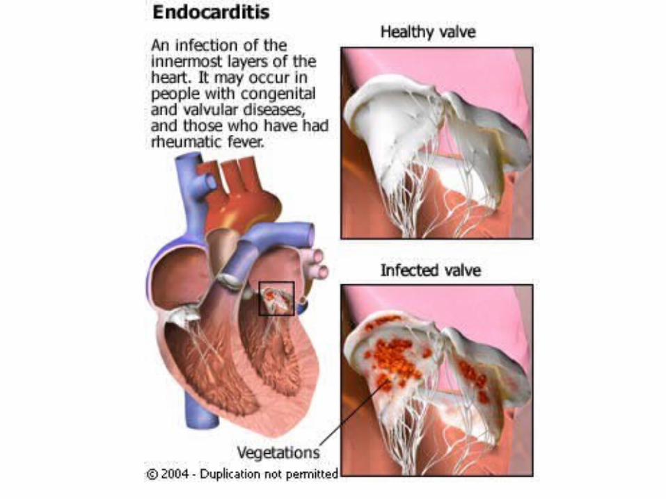
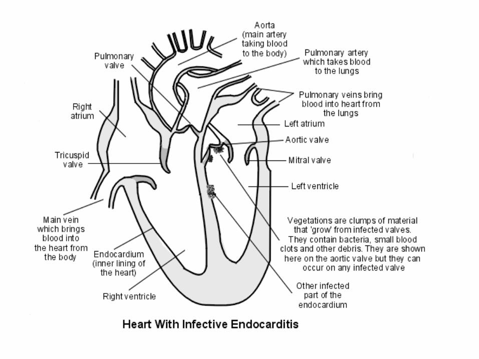
Infective Endocarditis
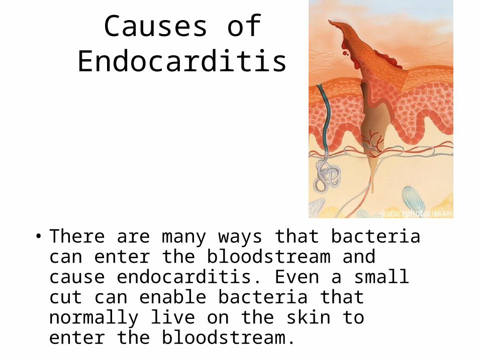
Causes of Endocarditis
• There are many ways that bacteria can enter the bloodstream and cause endocarditis. Even a small cut can enable bacteria that normally live on the skin to enter the bloodstream.
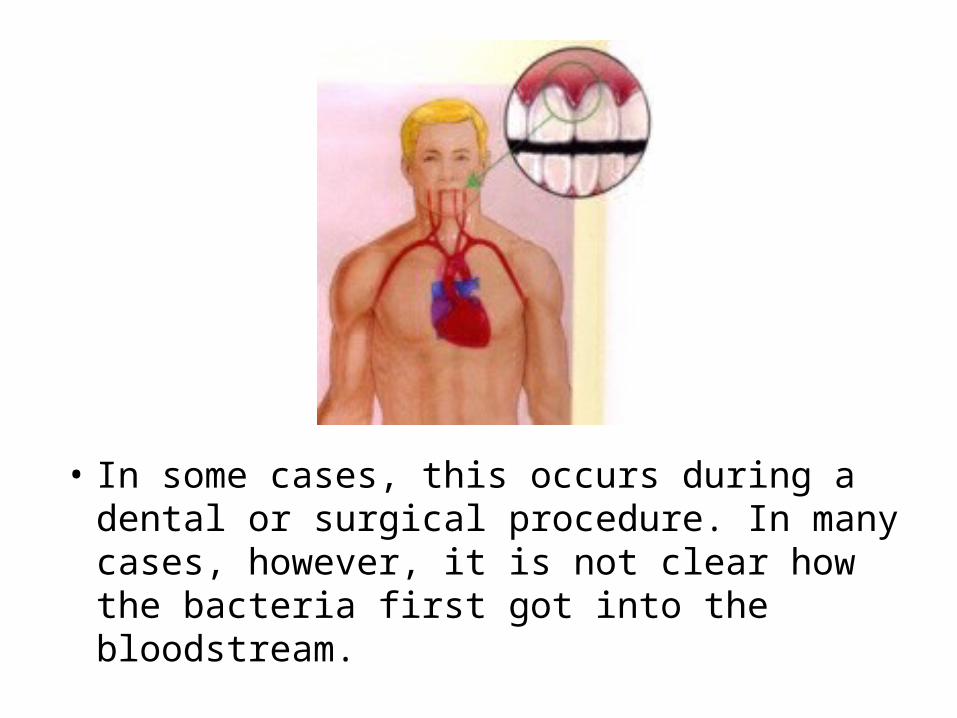
• In some cases, this occurs during a dental or surgical procedure. In many cases, however, it is not clear how the bacteria first got into the bloodstream.
Symptoms of Endocarditis
• Symptoms are non-specific, making endocarditis difficult to diagnose:
• Fatigue• Malaise• Weakness• Weight loss• Fever• Chills• Dyspnea on exertion (shortness of breath)
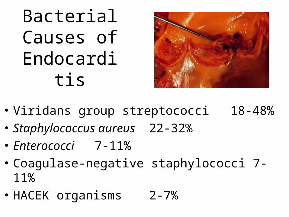
Bacterial Causes of
Endocarditis
• Viridans group streptococci 18-48%
• Staphylococcus aureus 22-32%
• Enterococci 7-11%
• Coagulase-negative staphylococci 7-11%
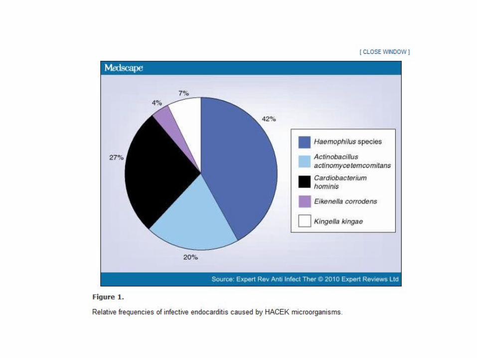
• HACEK organisms 2-7%
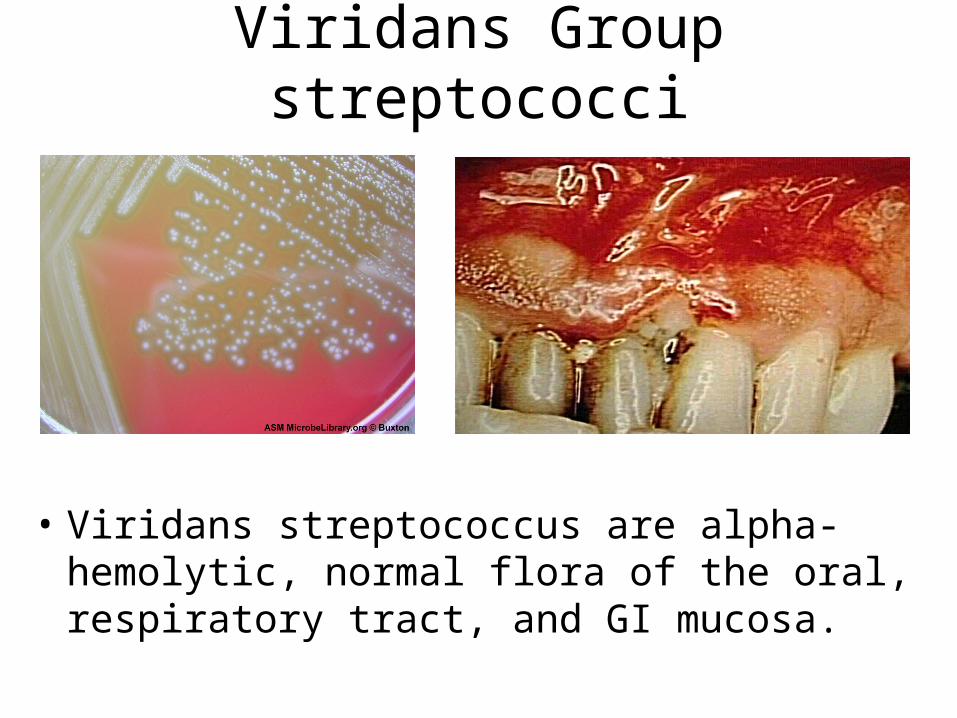
Viridans Group streptococci
• Viridans streptococcus are alpha-hemolytic, normal flora of the oral, respiratory tract, and GI mucosa.
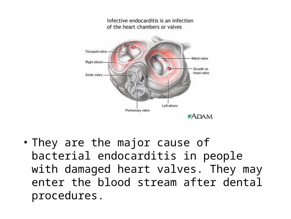
• They are the major cause of bacterial endocarditis in people with damaged heart valves. They may enter the blood stream after dental procedures.
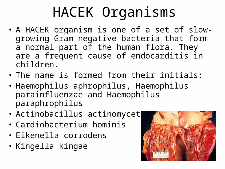
HACEK Organisms• A HACEK organism is one of a set of slow-growing
Gram negative bacteria that form a normal part of the human flora. They are a frequent cause of endocarditis in children.
• The name is formed from their initials:• Haemophilus aphrophilus, Haemophilus
parainfluenzae and Haemophilus paraphrophilus• Actinobacillus actinomycetemcomitans• Cardiobacterium hominis• Eikenella corrodens• Kingella kingae
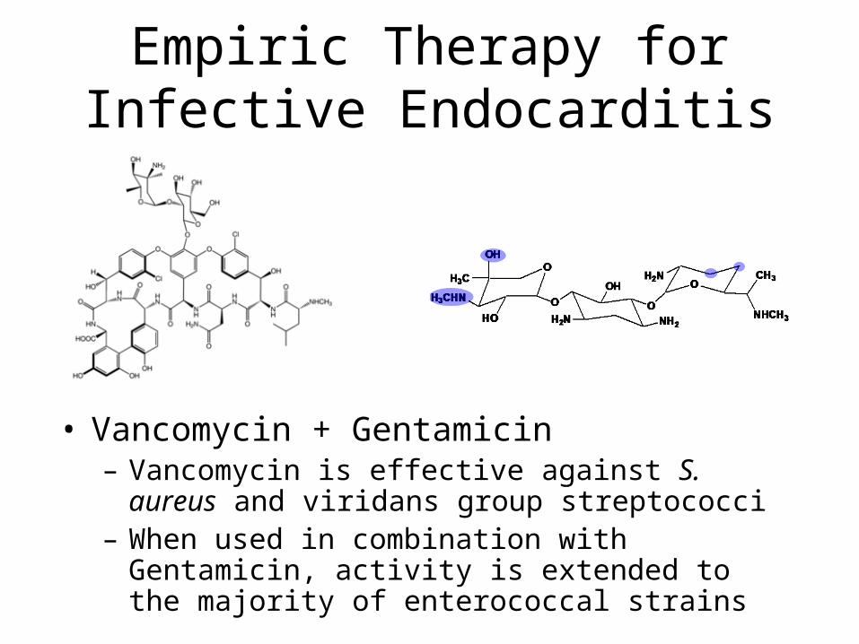
Empiric Therapy for Infective Endocarditis
• Vancomycin + Gentamicin– Vancomycin is effective against S. aureus and
viridans group streptococci– When used in combination with Gentamicin,
activity is extended to the majority of enterococcal strains
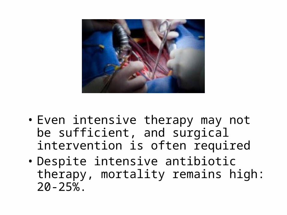
• Even intensive therapy may not be sufficient, and surgical intervention is often required
• Despite intensive antibiotic therapy, mortality remains high: 20-25%.
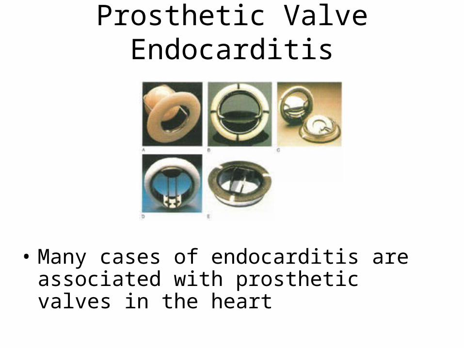
Prosthetic Valve Endocarditis
• Many cases of endocarditis are associated with prosthetic valves in the heart
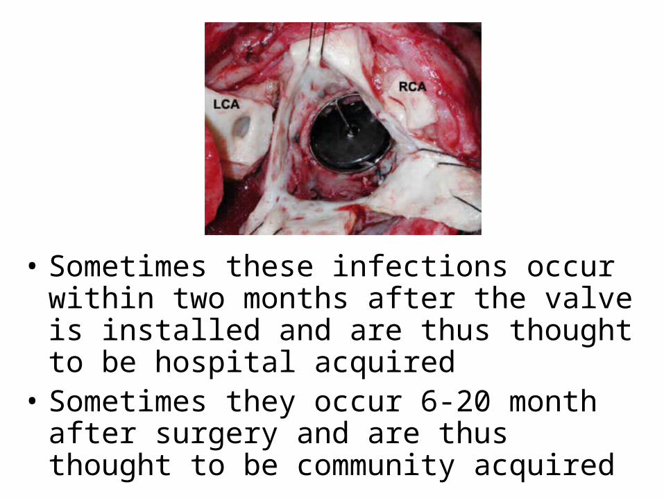
• Sometimes these infections occur within two months after the valve is installed and are thus thought to be hospital acquired
• Sometimes they occur 6-20 month after surgery and are thus thought to be community acquired

Treatment of Prosthetic Valve Endocarditis
• Vancomycin + Gentamicin + Rifampin– With or without cefepime or ceftriaxone
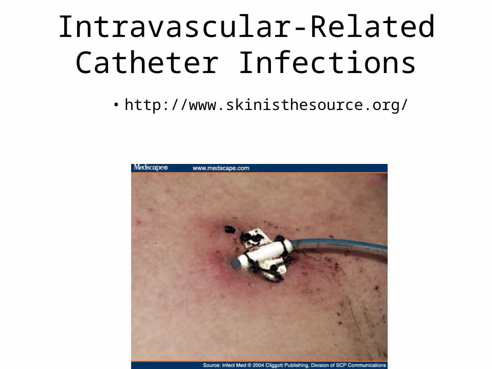
Intravascular-Related Catheter Infections
• http://www.skinisthesource.org/
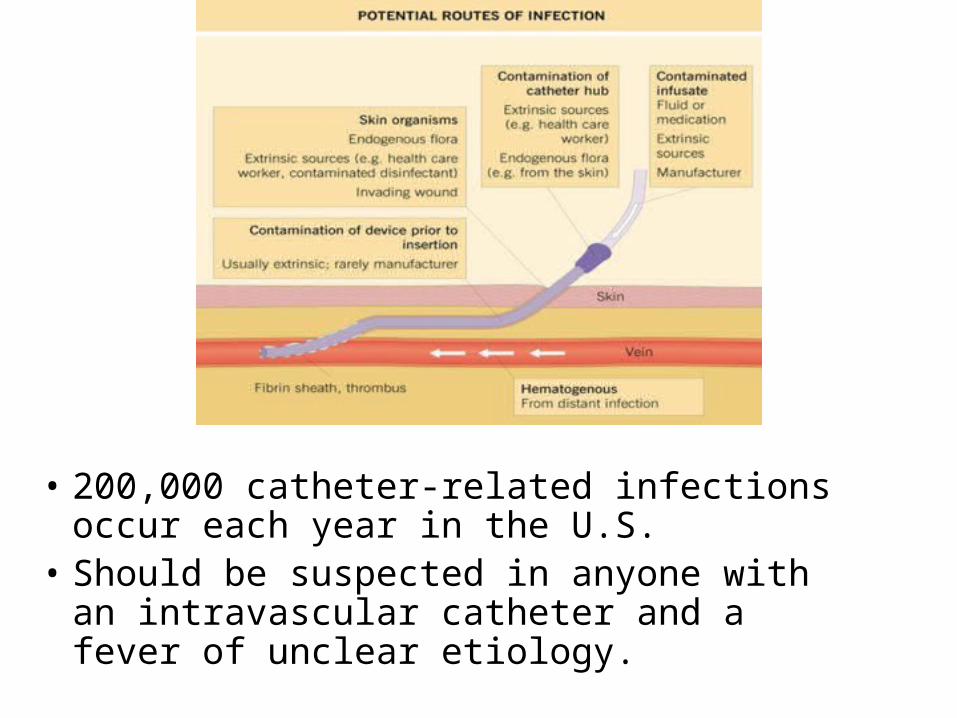
• 200,000 catheter-related infections occur each year in the U.S.
• Should be suspected in anyone with an intravascular catheter and a fever of unclear etiology.
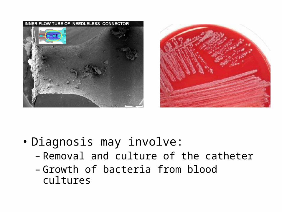
• Diagnosis may involve:– Removal and culture of the catheter– Growth of bacteria from blood cultures
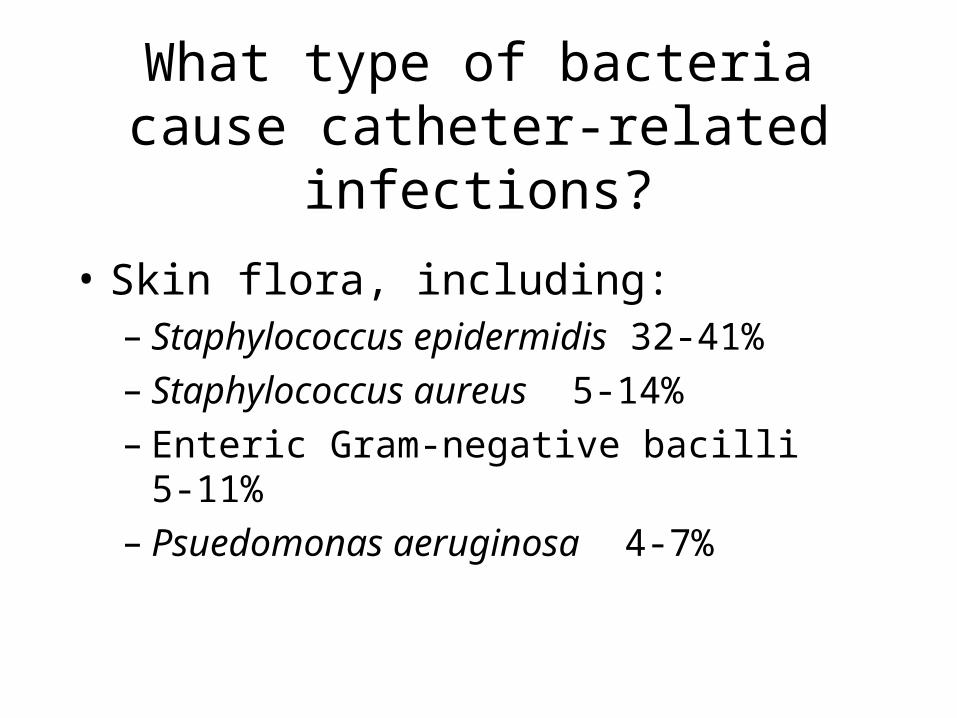
What type of bacteria cause catheter-related infections?
• Skin flora, including:– Staphylococcus epidermidis 32-41%– Staphylococcus aureus 5-14%– Enteric Gram-negative bacilli 5-11%– Psuedomonas aeruginosa 4-7%
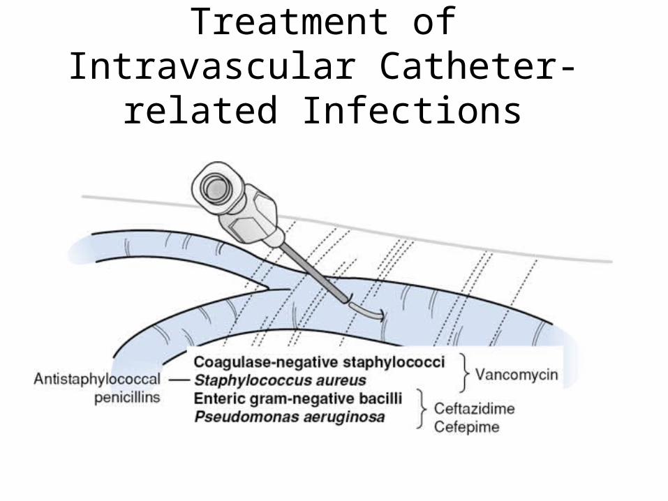
Treatment of Intravascular Catheter-related Infections
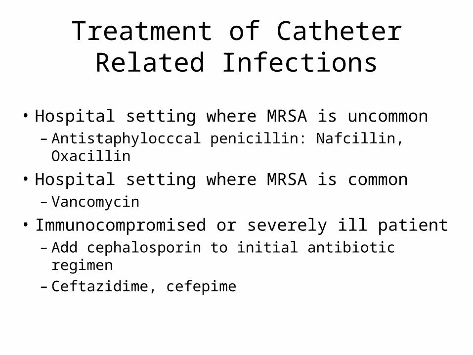
Treatment of Catheter Related Infections
• Hospital setting where MRSA is uncommon– Antistaphylocccal penicillin: Nafcillin, Oxacillin
• Hospital setting where MRSA is common– Vancomycin
• Immunocompromised or severely ill patient– Add cephalosporin to initial antibiotic regimen– Ceftazidime, cefepime
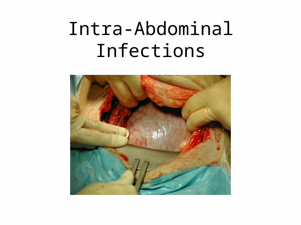
Intra-Abdominal Infections
Causes of Intra-abdominal infections
• Usually caused by contamination of the usually sterile abdomen with microbial flora of the bowel
• Can be quite severe, leading to sepsis and death
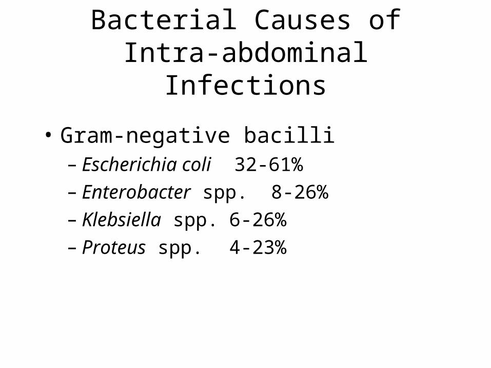
Bacterial Causes of Intra-abdominal Infections
• Gram-negative bacilli– Escherichia coli 32-61%– Enterobacter spp. 8-26%– Klebsiella spp. 6-26%– Proteus spp. 4-23%
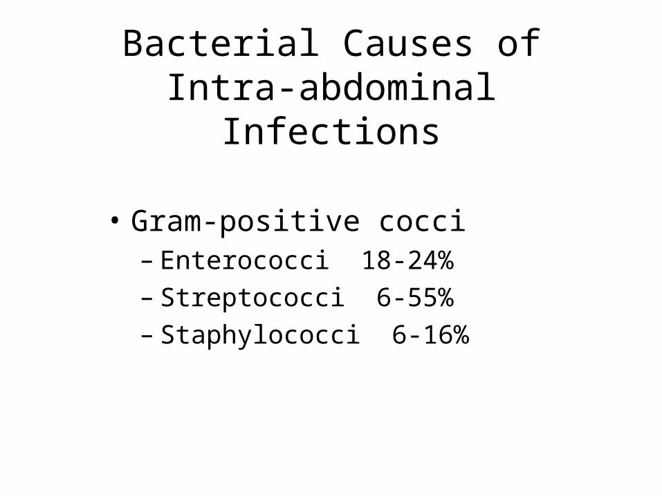
Bacterial Causes of Intra-abdominal Infections
• Gram-positive cocci– Enterococci 18-24%– Streptococci 6-55%– Staphylococci 6-16%
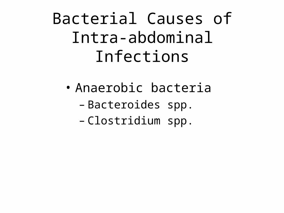
Bacterial Causes of Intra-abdominal Infections
• Anaerobic bacteria– Bacteroides spp.– Clostridium spp.
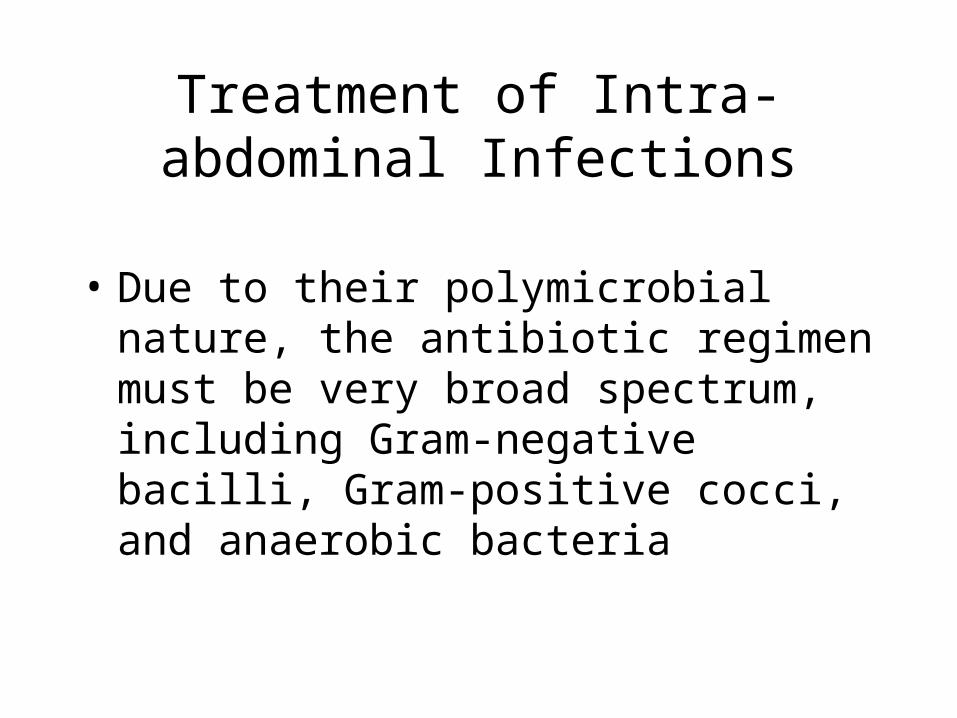
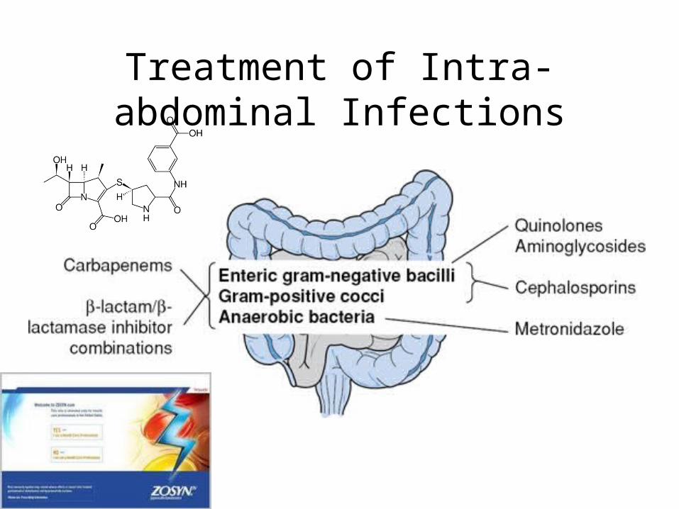
Treatment of Intra-abdominal Infections
• Due to their polymicrobial nature, the antibiotic regimen must be very broad spectrum, including Gram-negative bacilli, Gram-positive cocci, and anaerobic bacteria
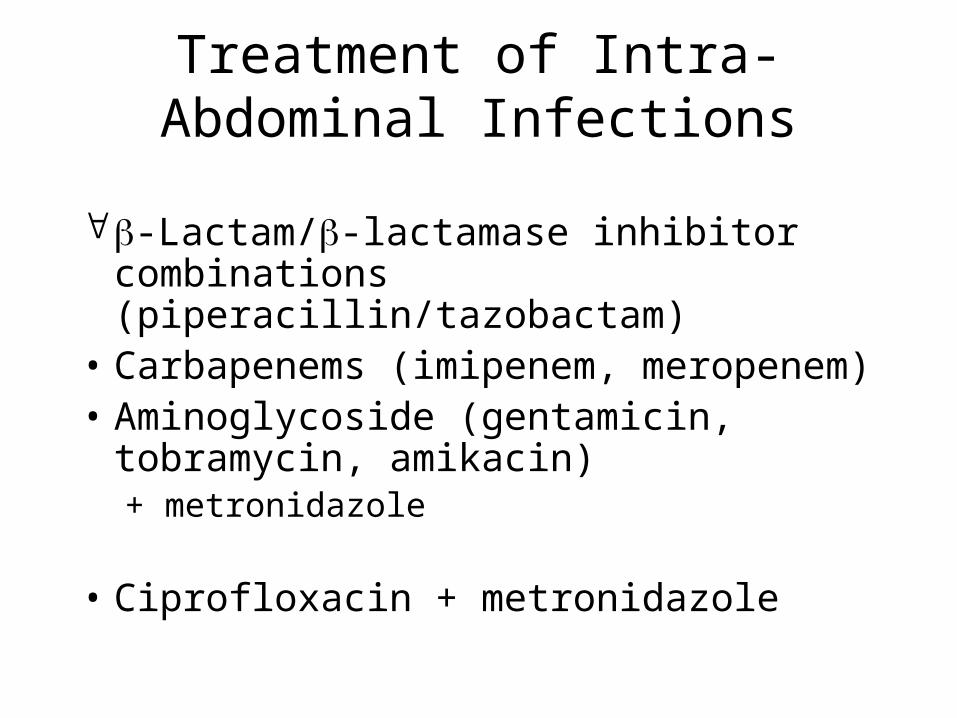
Treatment of Intra-Abdominal Infections
-Lactam/-lactamase inhibitor combinations (piperacillin/tazobactam)
• Carbapenems (imipenem, meropenem)• Aminoglycoside (gentamicin,
tobramycin, amikacin)+ metronidazole
• Ciprofloxacin + metronidazole
Treatment of Intra-abdominal Infections