EMBRYOLOGY Brachial plexus (BP) is developed at 5 weeks of gestation Afferent fibers develop from...
51
BRACHIAL PLEXUS INJURY INVESTIGATION , LOCALIZATION AND TREATMENT
-
Upload
garett-bartell -
Category
Documents
-
view
218 -
download
0
Transcript of EMBRYOLOGY Brachial plexus (BP) is developed at 5 weeks of gestation Afferent fibers develop from...
BRACHIAL PLEXUS INJURYINVESTIGATION , LOCALIZATION AND TREATMENT

EMBRYOLOGY
Brachial plexus (BP) is developed at 5 weeks of gestation
Afferent fibers develop from neuroblast located alongside neural tube
Efferent fibers originate from neuroblast in the basal plate of tube from where they grow outside
Afferent and efferent fibers join to form the nerve
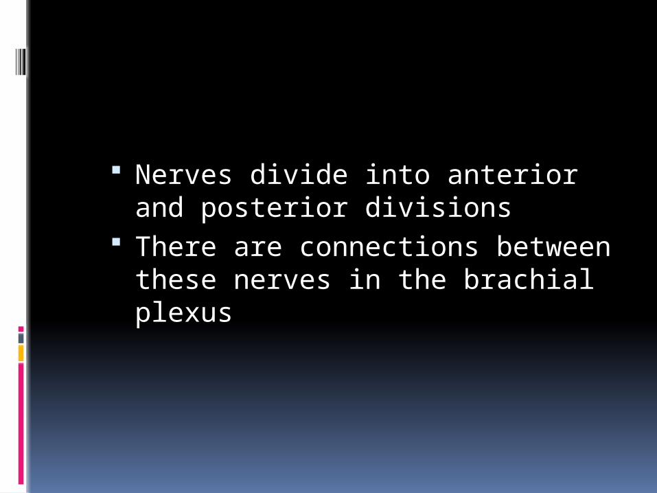
Nerves divide into anterior and posterior divisions
There are connections between these nerves in the brachial plexus

commons.wikimedia.org/wiki/File:Brachial_plexus.jpg
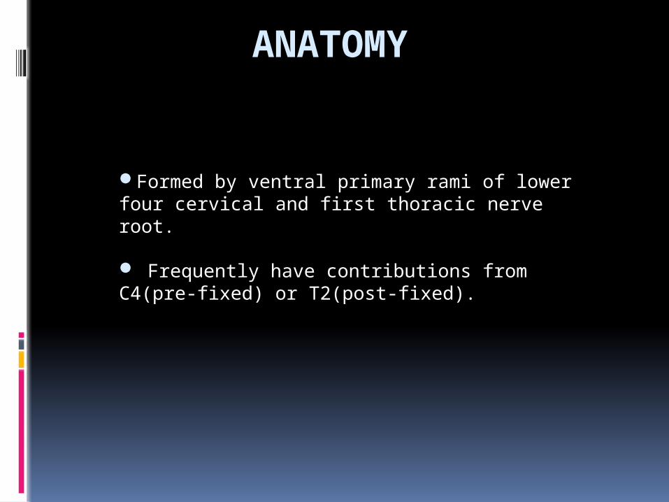
ANATOMY
Formed by ventral primary rami of lower four cervical and first thoracic nerve root.
Frequently have contributions from C4(pre-fixed) or T2(post-fixed).
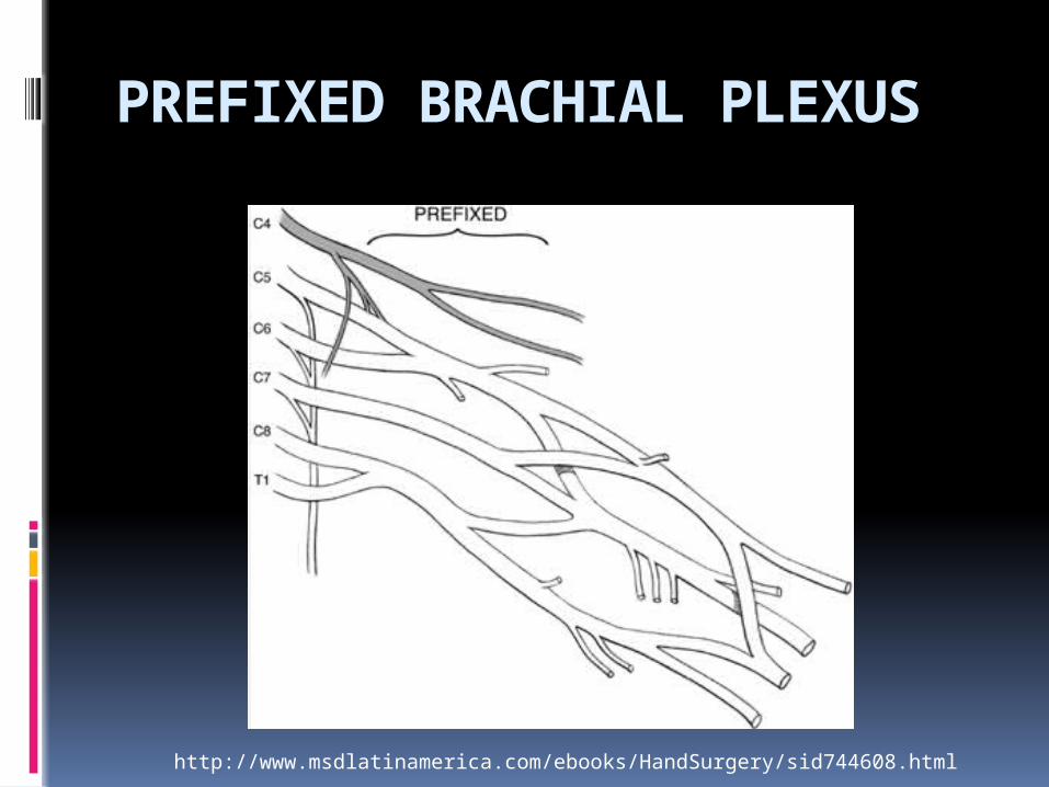
PREFIXED BRACHIAL PLEXUS
http://www.msdlatinamerica.com/ebooks/HandSurgery/sid744608.html

Post-fixed plexus
http://www.msdlatinamerica.com/ebooks/HandSurgery/sid744608.html
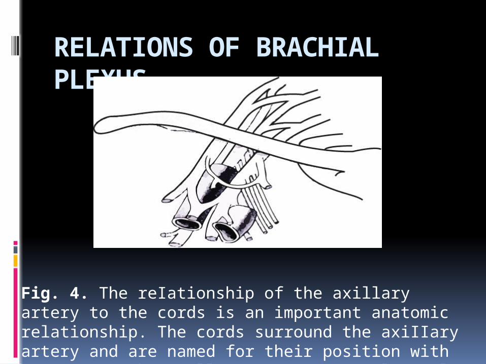
RELATIONS OF BRACHIAL PLEXUS
Fig. 4. The reIationship of the axillary artery to the cords is an important anatomic relationship. The cords surround the axiIIary artery and are named for their position with respect to the axillary artery. L.C. lateral cord MC. Medial cord: PC . posterior Cord.
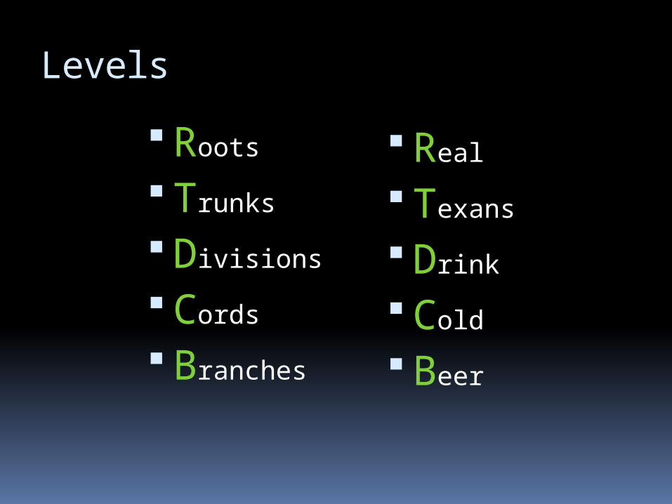
Levels
Roots
Trunks
Divisions
Cords
Branches
Real
Texans
Drink
Cold
Beer
C5 and C6 roots form upper trunk C8 and T1 roots the lower trunk C7 forms the middle trunk Joining point of C5-C6 roots is
ERB”S POINT Each trunk divides into an anterior
and a posterior division and passes beneath the clavicle
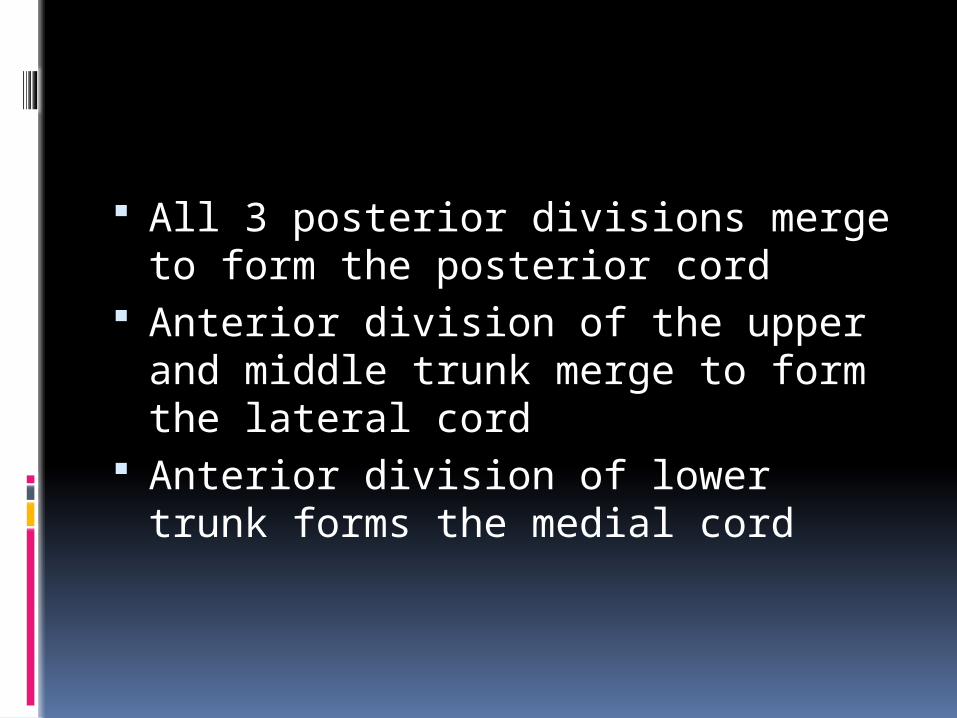
All 3 posterior divisions merge to form the posterior cord
Anterior division of the upper and middle trunk merge to form the lateral cord
Anterior division of lower trunk forms the medial cord
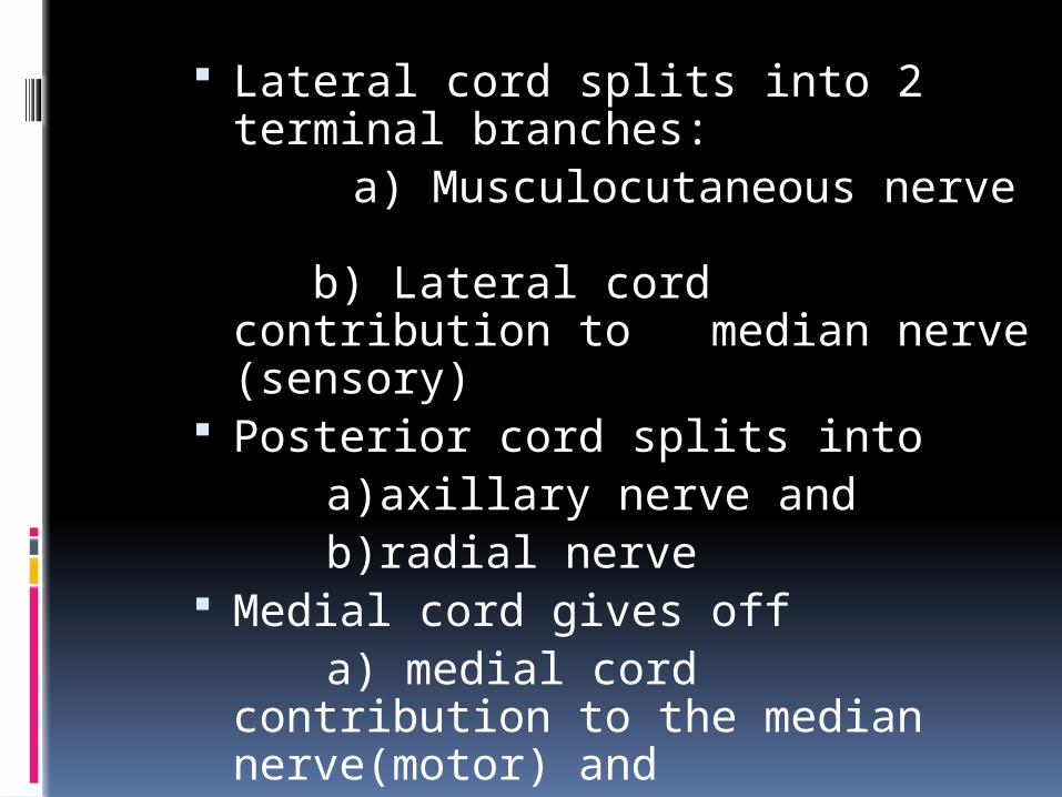
Lateral cord splits into 2 terminal branches:
a) Musculocutaneous nerve b) Lateral cord contribution to median nerve (sensory)
Posterior cord splits into a)axillary nerve and b)radial nerve Medial cord gives off a) medial cord contribution to the
median nerve(motor) and b)ulnar nerve
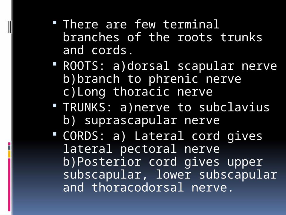
There are few terminal branches of the roots trunks and cords.
ROOTS: a)dorsal scapular nerve b)branch to phrenic nerve c)Long thoracic nerve
TRUNKS: a)nerve to subclavius b) suprascapular nerve
CORDS: a) Lateral cord gives lateral pectoral nerve b)Posterior cord gives upper subscapular, lower subscapular and thoracodorsal nerve.
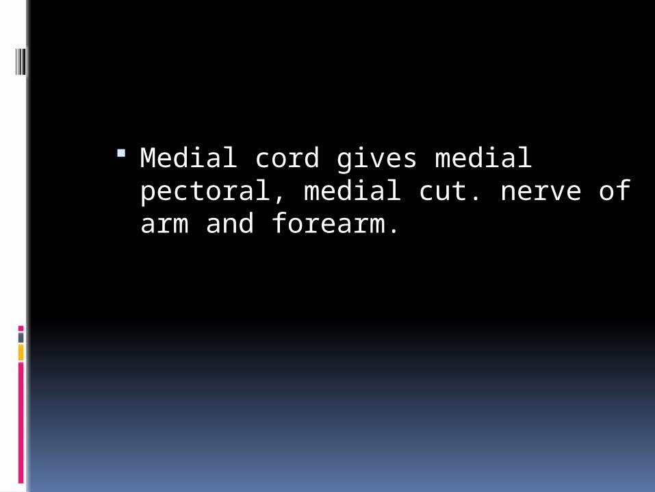
Medial cord gives medial pectoral, medial cut. nerve of arm and forearm.

Variations
Found in around 50% Most commonly pre-fixed(28-62%)
and post-fixed(16-73%)
Patho-anatomy
Anatomy of rootlets, roots and vertebral foramen contribute to the type of injury
Rootlets forming the cervical roots are intraspinal and lack connective tissue or meningeal envelope.
This feature makes them vulnerable to traction and susceptibility to avulsion at the level of cord.
The spinal nerve is able to move freely in the foramina due to non attachment to it.
There is fibrous attachment of spinal nerves to the transverse process seen in the 4th through 7th cervical roots
This explains the high incidence of root avulsions in C8-T1 roots
Preganglionic Tearing of
rootlets proximal to dorsal root ganglia
a) central b) peripheral
Postganglionic Injury distal to
DRG
Pathogenesis
Most patients are men and boys between 15- 25 years
70% of traumatic BPI secondary to motor vehicle accidents
Of these 70% involve motorcycles and bicycles
Other major injuries usually associated in 70%
They are usually closed injuries 95% traction injuries, 5% compression
injuries Supraclavicular more common than
infraclavicular involvement Roots and trunks most commonly
involved Root avulsions: 2 mechanisms peripheral- common central- rare
Traction injuries head and neck
move away from shoulder, usually involve C5,C6 andC7
C8- T1 involved in hyperabduction injuries
Other mechanisms- penetrating injuries iatrogenic injuries
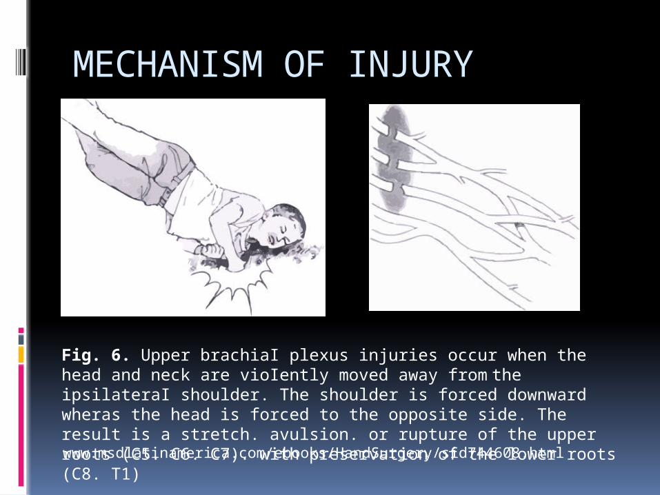
MECHANISM OF INJURY
Fig. 6. Upper brachiaI plexus injuries occur when the head and neck are vioIently moved away from the ipsilateraI shoulder. The shoulder is forced downward wheras the head is forced to the opposite side. The result is a stretch. avulsion. or rupture of the upper roots (C5. C6. C7). with preservation of the lower roots (C8. T1)
www.msdlatinamerica.com/ebooks/HandSurgery/sid744608.html
Clinical features
High degree of suspicion in injury to shoulder girdle, first rib and axillary artery
Median, ulnar and radial nerves can be evaluated by examining finger and wrist motion
Elbow flexion and extension can be used to examine musculocutaneous nerve and high radial nerve function
Injury to posterior cord may affect deltoid function and muscles innervated by radial nerves
Latissimus dorsi innervated by thoracodorsal nerve is palpated posterior axillary fold
Medial and lateral pectoral nerves are branches of medial and lateral cord respectively and supply sternal and clavicular head of pectoralis major respectively
Supra scapular nerve function shoulder extension, rotation and elevation
Look for Horner syndrome, injury to long thoracic nerve and dorsal scapular nerve to differentiate between pre and post ganglionic lesion
Assess spinal accessory nerve Active passive range of motion Rule out cord injury Tinel’s sign Vascular examination Fractures

PREOPERATIVE PLANNING
Box 1. Preoperative planning priorities for brachial plexus injury surgery
1. Review clinical examinations2. Scrutinize electrodiagnostic studies 3. Review CT myelography/imaging 4. Assemble operative team, plan for
intraoperative electrodiagnostic studies 5. Plan a preoperative conference, including
priorities and contingency plans 6. Prepare patient’s expectations
IMAGING
Radiography- cervical spine views - shoulder view- AP and axillary -X- Ray chest CT myelography- gold standard for root
injury, done at 3 to 4 weeks to see for pseudo meningocoele formation
MRI shows whole of brachial plexus, cord injury and neuroma formation
ELECTRODIAGNOSTIC STUDIES
Can help confirm a diagnosis Localize lesions Define severity of axon loss and
completeness of lesion Serve as an important adjunct to
thorough history, physical exam and imaging study
For closed injuries EMG and NCV can best be performed 3 to 4 weeks after the injury because wallerian degeneration will occur by this time
EMG
Denervation changes(fibrillation potentials) can be seen in proximal muscles 10 to 14 days and 3to6 weeks post injury in most distal muscles
Reduced MUP(motor unit potential) recruitment can be shown immediately after weakness from LMN injury
Presence of active motor units with voluntary effort and few fibrillations at rest has good prognosis
Can help in distinguishing preganglionic from postganglionic lesions
NCS
In post traumatic BPI the amplitude of compound muscle action potentials (CMAP) are generally low
SNAP important in localizing a lesion as pre or postganglionic
INTRA OP TESTING
NAP (nerve action potential SEP (somatosensory evoked
potential) CMAP (compound muscle action
potential)

Management
Proximal rupture, distal avulsion 60%
Complete 5 level injury 50% Five level avulsion 30% Supraclavicular 70-75% Upper trunk 35% C4-T1 avulsion 10%
C6-C8avulsion 8% C8/T1 isolated 3%
Whole limb injury 45%Intraclavicular 25-33% Single/combined cord injury 30%
Isolated peripheral nerve injury 25%
Fig. I. Distribution of location and iv of hrachical plesus injuries.
Management
15% of supraclavicular injuries have concomitant segmental injuries at or below the clavicle where the peripheral nerve branch from the plexus
Musculocutaneous, axillary and suprascapular nerves are particularly vulnerable to traction injury because of soft tissue tethering near their origin

Infraclavicular injuries constitute 25-
33% of BPI usually occur at cords or peripheral nerves and usually are incomplete
Usually caused by shoulder fracture or dislocation
5- 25% of infraclavicular injuries are associated with axillary artery injury
Penetrating injuries are usually infraclavicular
Timing of intervention
A - acute exploration concomitant vascular injury open injury by sharp laceration crush or contaminated wound B - early exploration (1- 2 weeks) unequivocal complete C5- T1 avulsion injuries C - delayed exploration > 3 months recommended for complete injuries with no
recovery by clinical examination or EMG at 12 weeks post injury
candidates showing distal recovery without regaining clinical or electrical evidence of proximal muscle function

Prioritization
Clearly understand the anatomy of injured plexus
What is available for nerve transfer Function priority- elbow flexion is the
most important to restore followed by abduction, external rotation and scapular stabilization
Long thoracic nerve should be performed whenever possible
Radial nerve motor function can often be restored with triceps function more likely to return
Surgical options
Neurolysis Nerve repair Nerve graft Nerve transfer or neurotization Functional free muscle transfer
Neurolysis
Effective only if scar tissue seen around nerve or inside epineurium, preventing recovery or causing pain
Pre and post neurolysis direct nerve stimulation is mandatory to evaluate improvement in nerve conduction
Nerve repair
Used in sharp transection with excellent fascicular pattern and minimal scar

Nerve graft
Indicated for well defined nerve ends without segmental injuries
Intraoperatively a good fascicular pattern should be seen after the neuroma is excised
Possible sources: sural, brachial cutaneous nerve, radial sensory and possibly ulnar nerve
Before implantation graft orientation should be reversed to minimize axonal branch loss
Surgical technique is considered the most important factor in nerve graft
Neurotization
A- intraplexal B- extraplexal Plexoplexal options are undamaged
roots Other options include medial pectoral
nerves and medial cord ulnar nerve Extraplexal options- spinal accessory,
intercostal, phrenic and motor branch of deep cervical plexus
For upper arm type avulsion, a reliable neurotization strategy has included phrenic-suprascapular nerve transfer and spinal accessory nerve (with an interpositional nerve graft) to musculocutaneous nerve.
For complete avulsion injury, a strategy of multiple neurotizations using spinal accessory, phrenic and contralateral C7 nerves provides a patient with a framework to obtain hook grip.
OBERLIN TECHNIQUE
For upper trunk injury with intact lower trunk- 1 to 2 fascicles of ulnar nerve are anastomosed to biceps
Contra lateral C7 is used in pan brachial plexopathy with multiple avulsions and limited donor possibility
Contra lateral C7 root can be extended by means of vascularised ulnar nerve graft in patient with C8 T1 avulsion and median nerve is the most frequent recipient
Another option is transferring nerve to long head of triceps to anterior branch of axillary nerve
Realistic targets to reinnervation Spinal accessory to suprascapular or
musculocutaneous Phrenic to axillary nerve Intercostal to musculocutaneous long
thoracic, radial and median nerve Long head of triceps nerve to
anterior branch of axillary nerve
Functioning free muscle transfer
Usually gracillis is used- single or double gracillis transfer
PROGNOSIS
Highly dependent on pattern of injury Complete C4 to T1 injuries are
considered most severe and virtually irreparable
Avulsion injuries from C5 toT1 amenable to restoration of shoulder and elbow function only
Ideal candidate for surgery are patients with proximal rupture or avulsion and sparing of lower trunk
AIIMS STUDY
Since 1995 to2002 , 505 patients were studied for functional and occupational outcome after surgery for BPI
In India BPI is most common due to RTA with Rt side involved in 2/3
40% cases have pan BPI 85% of cable graft yielded
improvement in motor power compared 68% in neurotized nerve and 66% in patients undergoing neurolysis
Most effective donor nerve for musculocutaneous neurotization was medial pectoral nerve (63.6%) patient improved
Accessory nerve was most effective for neurotization of suprascapular nerve (100%)
Thoracodorsal axillary neurotization gave (66.7% improvement)
50% patients either remained unemployed or had to change there jobs

Thank you