
Elisa 1 copy.ppt
30
Jinu Janet Varghese Group: 4 Tbilisi State Medical University
-
Upload
tbilisi-state-medical-university -
Category
Health & Medicine
-
view
187 -
download
0
Transcript of Elisa 1 copy.ppt

Jinu Janet Varghese Group: 4
Tbilisi State Medical University

History Before the development of the ELISA, the only option
for conducting an immunoassay was radio-immunoassay, a technique using radioactively-labeled antigens or antibodies.
In radio-immunoassay, the radioactivity provides the signal, which indicates whether a specific antigen or antibody is present in the sample.
Radio-immunoassay was first described in a paper by Rosalyn Sussman Yalow and Solomon Berson published in 1960.

When enzymes (such as peroxidase) react with appropriate substrates (such as ABTS or 3,3’,5,5’-tetramethylbenzidine), a change in color occurs, which is used as a signal. However, the signal has to be associated with the presence of antibody or antigen.
This linking process was independently developed by Stratis Avrameas and G. B. Pierce. Since it is necessary to remove any unbound antibody or antigen by washing, the antibody or antigen has to be fixed to the surface of the container; i.e., the immunosorbent has to be prepared. A technique to accomplish this was published by Wide and Jerker Porath in 1966.

What is ELISA? ELISA stands for enzyme-linked immunosorbent
assay. It is a commonly used laboratory test to detect antibodies in the blood.
It is a plate-based assay designed for detecting and quantifying substances such as peptides, proteins, antibodies and hormones.
This test is usually used to detect infection with HIV. It has a high sensitivity.
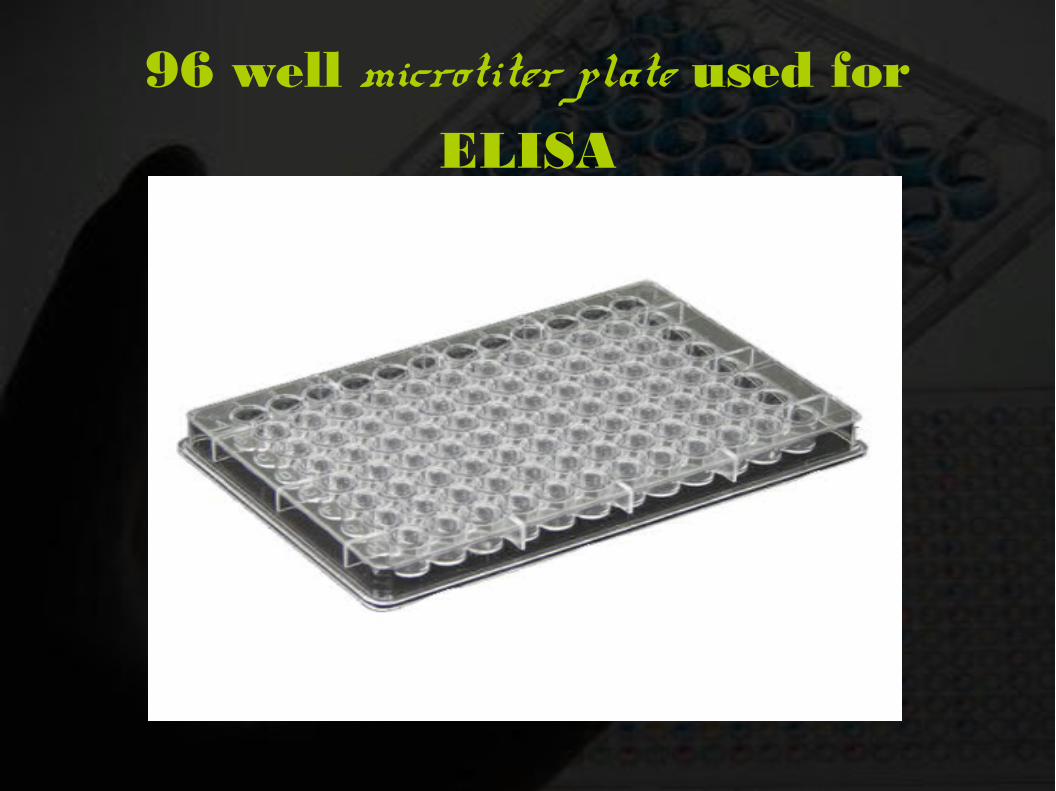
96 well microtiter plate used for
ELISA

Components of
ELISA Antibody: IgG fraction of serum purified by affinity chromatography.
Enzyme: Horse Radish Peroxidase (HRP) MW 44,000, glycoprotein with 4 lysine residues.
Substrate: TMB(3,3', 5,5', tetramethylbenzidine). The enzyme acts as a catalyst to oxidize substrate in the presence of Hydrogen peroxide to produce a blue colour. Reaction is stopped with dilute acid to cause complex to turn yellow.

ELISA Reader

Principles
The sample with an unknown amount of antigen is immobilized on a solid support either:
A) Non-specifically or,
B) Specifically
The detection antibody is added, forming a complex with the antigen

The detection antibody can be covalently linked to an enzyme, or can itself be detected by a secondary antibody which is linked to an enzyme forming a complex.
Between each step, the plate is typically washed with a mild detergent solution to remove any proteins or antibodies that are not specifically bound.
After the final wash step, the plate is developed by adding an enzymatic substrate to produce a visible signal, which indicates the quantity of antigen in the sample.


Types of ELISA
Direct ELISA
Indirect ELISA
Sandwich ELISA
Competitive ELISA

Direct ELISA

A specific monoclonal antibody is first adsorbed onto the walls of a microtiter plate.
A suspension of serum or other fluid is added to test the presence of complementary antigen.
If the antigen is present it will bind to the antibodies that are attached to the wall.
This binding is strong enough to withstand the rinsing of test fluid and unbound antigen.

Another aliquot of the monoclonal antibody which carries reporter enzyme is added to indicate a color change when the enzyme reacts with it's substrate.
Again the sample is rinsed to remove the unbound antibodies. If antigen is present, a complex that includes the antibody bound to well, the antigen and the enzyme-conjugated antibody will be formed.
The enzyme's substrate is added. A color change reveals the presence of enzyme-labeled antibody and it's bound antigen.

Indirect ELISA

It is the one that determines whether a specific antibody( e.g., HIV antibody) is present in a sample such as serum. In this case, appropriate antigen is first adsorbed to the walls of the microtiter plate.
Serum that may contain antibodies against the antigen is added to the well. Antibodies present in the sample will bind to the antigens that are adsorbed to the wall. If the sample contains only non-specific antibodies, they will not bind to the antigen.
Rinsing removes any antibodies that do not specifically attach to the antigen absorbed to the wall.

Addition of an antibody-enzyme conjugate can detect if antibodies have attached to the antigen. These reporter antibodies bind to immunoglobulins(generally IgG). Since the sample contains human antibodies, we use enzyme-conjugated anti-human IgG antibodies for the reporter.
The present antibody binds to the adsorbed antigen and enzyme-conjugated antibody will bind to this complex. Sample is rinsed to removed unbound antibodies.
Substrate for the enzyme is added and color changes indicates the antibody reaction with the original antigen.

Sandwich ELISA

Plate is coated with a capture antibody.
Sample is added and any antigen present binds to capture antibody.
Detecting antibody is added and binds to antigen.
Enzyme-linked secondary antibody is added and binds to detecting antibody.
Substrate is added and is converted by enzyme to detectable form.

Competitive ELISA

Unlabeled antibody is incubated in the presence of its antigen.
These bound antibody/antigen complexes are then added to an antigen-coated well.
The plate is washed, so that unbound antibody is removed. (The more antigen, the less antibody bind to the antigen in the well, hence "competition.")
The secondary antibody, specific to the primary antibody is added. This second antibody is coupled to the enzyme.
A substrate is added, and remaining enzymes elicit a chromogenic or fluorescent signal.

Applications Evaluates the antigen or antibody in a sample. (Eg.,
For screening donated blood for evidence of viral contamination)
Measuring hormone levels( HCG, LH, TSH, T3 & T4)
Detecting potential food allergens
In toxicology, used as a presumptive screen for certain classes of drugs.
Measuring “ rheumatoid factors ” and other autoantibody in autoimmune diseases like Lupus Erythematosus.

Advantages Sensitive assay Equipments are widely available.
No radiation hazards.
Reagents are cheap with long shelf life.
Adaptable to automation and high speed.
Qualitative and quantitative.
Reproducible.
ELISA can be used on most types of biological samples, such as plasma, serum, urine, and cell extracts

Disadvantages Only monoclonal antibodies can be used as matched
pairs
Monoclonal antibodies can cost more than polyclonal antibodies
Monoclonal antibodies more difficult to find
Negative controls may indicate positive results if blocking solution is ineffective [secondary antibody or antigen (unknown sample) can bind to open sites in well]
Enzyme/substrate reaction is short term so microwells must be read as soon as possible

Troubleshooting

If the negative controls are
giving positive results:
Contamination of the substrate solution, enzyme-labeled antibody, control themselves.
Inadequate rinsing of plates.
Inadequate blocking of plates.

If no color has developed for the
positive controls or for the
samples: Check all reagents for dating and storage conditions.
Microwell plates not coated properly.
Reagents applied in wrong order or step omitted.
Enzyme conjugate defective or inhibited by contaminant.

If very little color has developed
for positive controls and the test
samples: Check the dilution of the enzyme labeled antibody.
The concentration of the substrate.
Wash buffer not adequately drained after every wash step.
Inadequate incubation times.
Enzyme conjugate defective or inhibited by contaminant, substrate defective or contaminated, Micro well plates poorly coated.

If colour has developed for the test samples but
not the positive controls :
Check the source of positive controls, their expiry date and storage.
If the color can be seen, but the
absorbance is not high as expected : check the wave length.


![Barcode-Enabled_Medication_Administration[1] - Copy.ppt](https://static.fdocuments.net/doc/165x107/563db9c7550346aa9a9fd78e/barcode-enabledmedicationadministration1-copyppt.jpg)