Electrophoresis (More) - University of...
15
Biochemistry 3100 Lecture 4 Slide 1 Electrophoresis Electrophoresis (More) (More) Voet & Voet: Voet & Voet: Pages 144-151 Pages 144-151
Transcript of Electrophoresis (More) - University of...
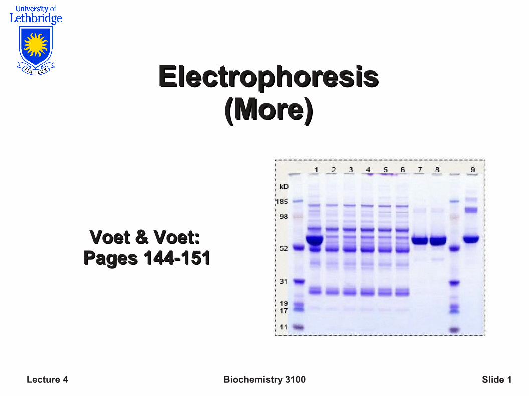
Biochemistry 3100Lecture 4 Slide 1
Electrophoresis Electrophoresis (More)(More)
Voet & Voet: Voet & Voet: Pages 144-151Pages 144-151
Biochemistry 3100Lecture 4 Slide 2
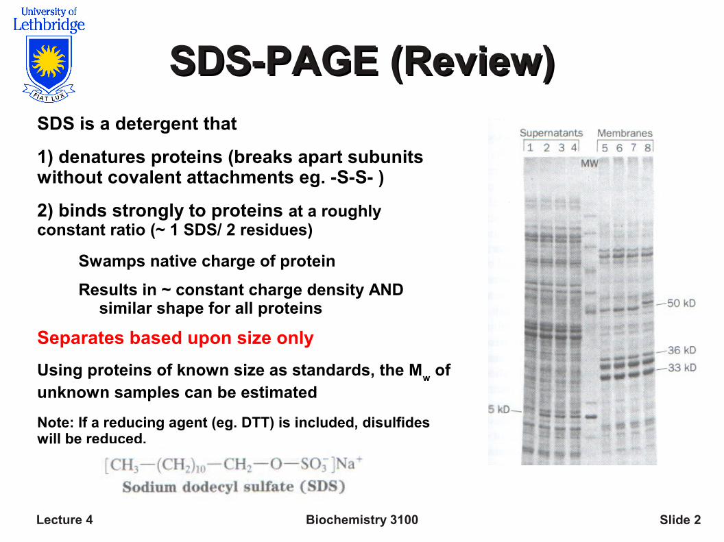
SDS-PAGE (Review)SDS-PAGE (Review)SDS is a detergent that
1) denatures proteins (breaks apart subunits without covalent attachments eg. -S-S- )
2) binds strongly to proteins at a roughly constant ratio (~ 1 SDS/ 2 residues)
Swamps native charge of protein
Results in ~ constant charge density AND similar shape for all proteins
Separates based upon size only
Using proteins of known size as standards, the Mw of
unknown samples can be estimated
Note: If a reducing agent (eg. DTT) is included, disulfides will be reduced.
Biochemistry 3100Lecture 4 Slide 3
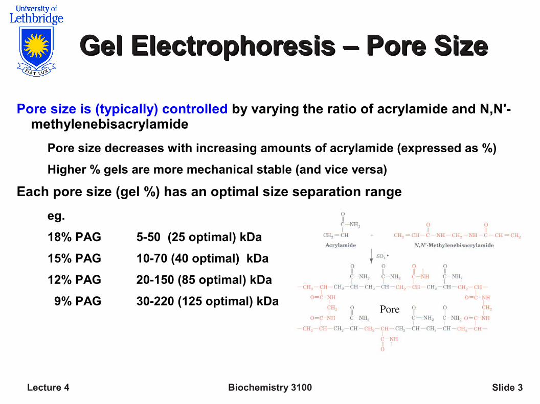
Gel Electrophoresis – Pore SizeGel Electrophoresis – Pore Size
Pore size is (typically) controlled by varying the ratio of acrylamide and N,N'-methylenebisacrylamide
Pore size decreases with increasing amounts of acrylamide (expressed as %)
Higher % gels are more mechanical stable (and vice versa)
Each pore size (gel %) has an optimal size separation range
eg.
18% PAG 5-50 (25 optimal) kDa
15% PAG 10-70 (40 optimal) kDa
12% PAG 20-150 (85 optimal) kDa
9% PAG 30-220 (125 optimal) kDaPore
Biochemistry 3100Lecture 4 Slide 4
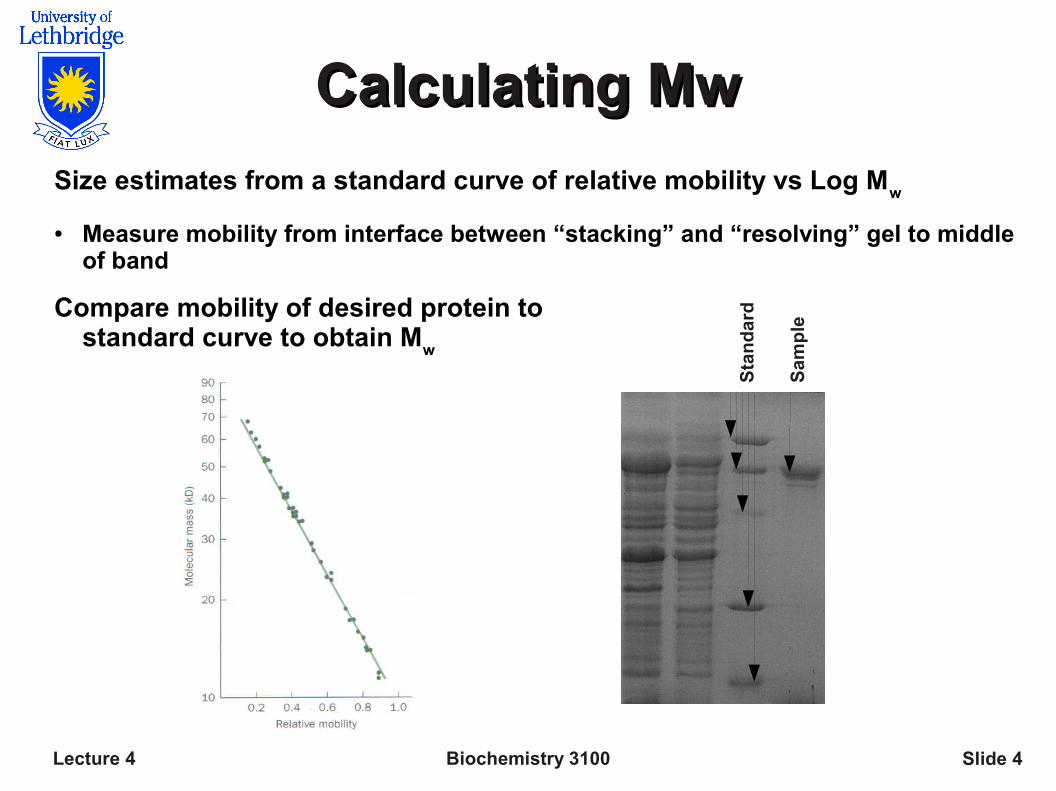
Calculating MwCalculating Mw
Size estimates from a standard curve of relative mobility vs Log Mw
● Measure mobility from interface between “stacking” and “resolving” gel to middle of band
Compare mobility of desired protein to standard curve to obtain M
w
Sta
nd
ard
Sam
ple
Biochemistry 3100Lecture 4 Slide 5
Western BlotsWestern Blots
Improved detection method based upon immunoblotting
Up to 1000x more sensitive than Coomassie Brilliant Blue Stain
Detect as little as 0.1 ng (eg. < 10 fM) using chemiluminescense (described below) or radioactivity
Requires additional steps:
1) transfer (blot) proteins from gel to membrane (nitrocellulose or PVDF)
2) Block unoccupied binding sites on membrane
3) Incubate with Ab (primary) that specifically binds protein of interest
4) Wash and incubate enzyme linked Ab; (secondary) that binds primary Ab
5) Assay enzyme linked Ab with chemiluminescence reaction
Detection is not solely based upon sample quantity
Biochemistry 3100Lecture 4 Slide 6
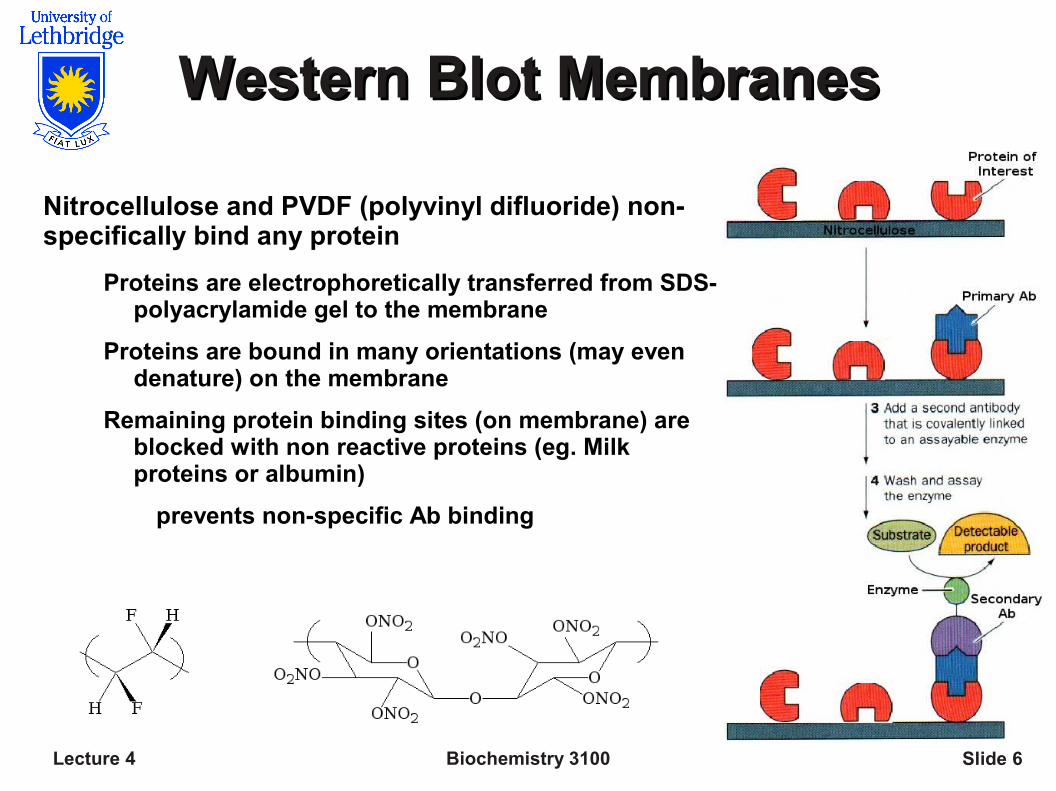
Western Blot MembranesWestern Blot Membranes
Nitrocellulose and PVDF (polyvinyl difluoride) non-specifically bind any protein
Proteins are electrophoretically transferred from SDS-polyacrylamide gel to the membrane
Proteins are bound in many orientations (may even denature) on the membrane
Remaining protein binding sites (on membrane) are blocked with non reactive proteins (eg. Milk proteins or albumin)
prevents non-specific Ab binding
Biochemistry 3100Lecture 4 Slide 7
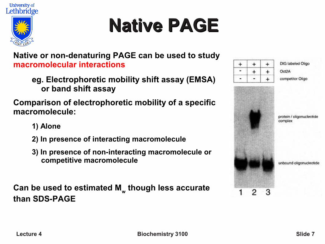
Native PAGENative PAGE
Native or non-denaturing PAGE can be used to study macromolecular interactions
eg. Electrophoretic mobility shift assay (EMSA) or band shift assay
Comparison of electrophoretic mobility of a specific macromolecule:
1) Alone
2) In presence of interacting macromolecule
3) In presence of non-interacting macromolecule or competitive macromolecule
Can be used to estimated Mw though less accurate
than SDS-PAGE
Biochemistry 3100Lecture 4 Slide 8
More Native PAGEMore Native PAGE
In the absence of SDS, the net charge of the macromolecules under investigation is an important consideration
For acidic and neutral macromolecules, the normal (basic) SDS-PAGE buffers and current polarity (cathode at top of gel) allow migration of samples into gel (Note: acidic macromolecules have pI < 7)
For basic macromolecules, acidic PAGE buffers and a reversed current polarity are required to allow migration of samples into the gel (Note: basic macromolecules have pI > 7)
Typically gels are run for several hours at a relatively low voltage to prevent heating (which may lead to dissociation)
May even cool gel rig by placing it on ice
Technique often (almost always) requires optimization of buffers, voltage and temperature to obtain a high quality gel
Biochemistry 3100Lecture 4 Slide 9
Isoelectric Focusing (IEF)Isoelectric Focusing (IEF)
Isoelectric focusing separates proteins based upon upon charge (pI)
Requires a solution or gel that maintains a stable pH gradient
Under these conditions, samples will move to a position in the pH gradient that corresponds to their isoelectric point
Produces exceptionally sharp and narrow bands (0.01 pH units wide)
Two strategies for establishing a stable pH gradient:
1) Mix together many buffers (ampholytes) with different pKas
In an electric field, each buffer in the mixture migrates to its isoelectric point establishing a gradient of pHs
2) Acrylamide derivatives of varying pKa are mixed together and
polymerized in varying ratios
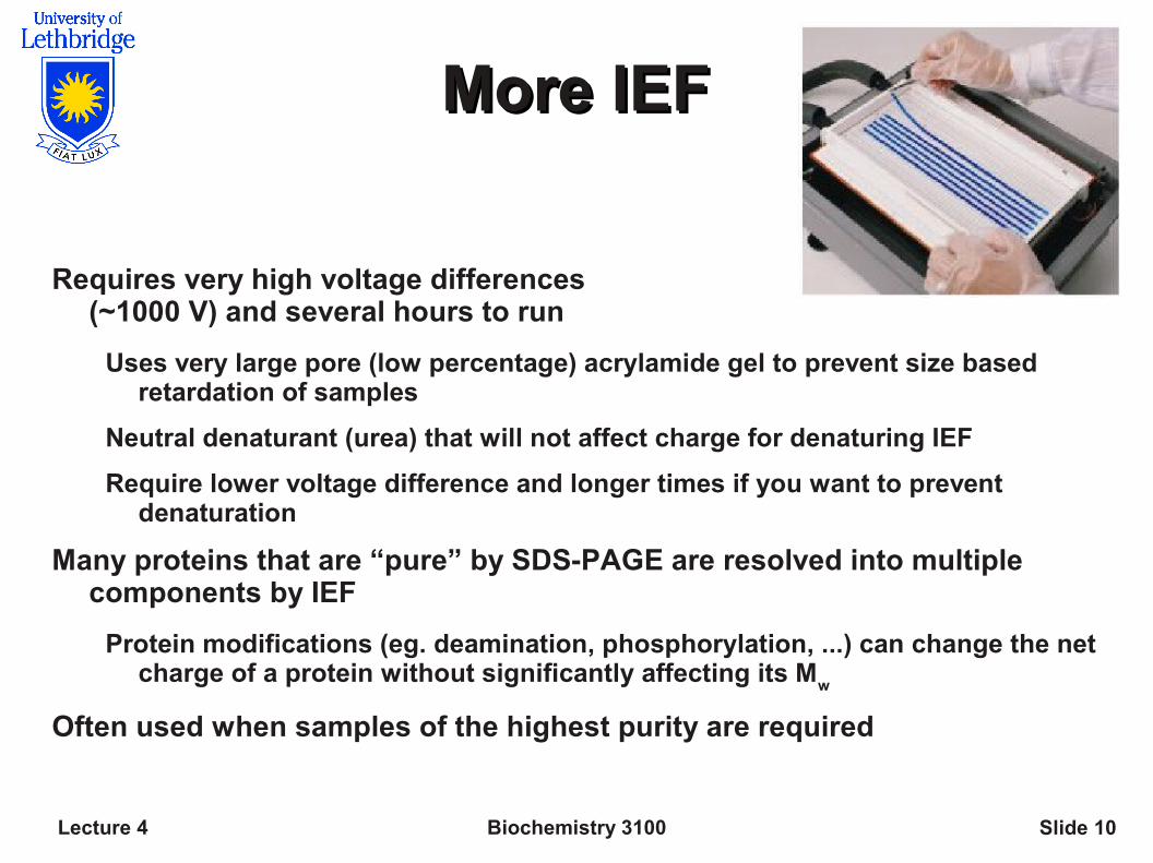
Biochemistry 3100Lecture 4 Slide 10
More IEFMore IEF
Requires very high voltage differences (~1000 V) and several hours to run
Uses very large pore (low percentage) acrylamide gel to prevent size based retardation of samples
Neutral denaturant (urea) that will not affect charge for denaturing IEF
Require lower voltage difference and longer times if you want to prevent denaturation
Many proteins that are “pure” by SDS-PAGE are resolved into multiple components by IEF
Protein modifications (eg. deamination, phosphorylation, ...) can change the net charge of a protein without significantly affecting its M
w
Often used when samples of the highest purity are required
Biochemistry 3100Lecture 4 Slide 11
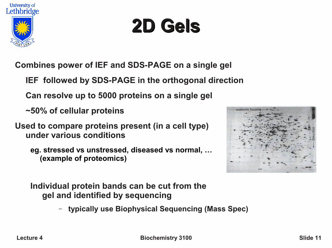
2D Gels2D Gels
Combines power of IEF and SDS-PAGE on a single gel
IEF followed by SDS-PAGE in the orthogonal direction
Can resolve up to 5000 proteins on a single gel
~50% of cellular proteins
Used to compare proteins present (in a cell type)under various conditions
eg. stressed vs unstressed, diseased vs normal, … (example of proteomics)
Individual protein bands can be cut from the gel and identified by sequencing
– typically use Biophysical Sequencing (Mass Spec)
Biochemistry 3100Lecture 4 Slide 12
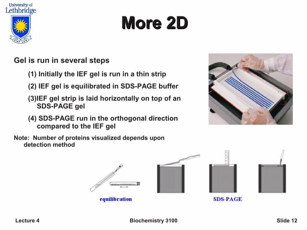
More 2DMore 2D
Gel is run in several steps
(1) Initially the IEF gel is run in a thin strip
(2) IEF gel is equilibrated in SDS-PAGE buffer
(3)IEF gel strip is laid horizontally on top of an SDS-PAGE gel
(4) SDS-PAGE run in the orthogonal direction compared to the IEF gel
Note: Number of proteins visualized depends upon detection method
Biochemistry 3100Lecture 4 Slide 13
Capillary ElectrophoresisCapillary Electrophoresis
Electrophoresis in an extremely thin tube (< 0.1 mm)
Rapid heat dissipation allows the use of higher voltage (100-300 V cm-1) and shortens separation time from an hour to minutes.
Small tube limits convective mixing and produces exceptional sharp peaks
Much lower detection limit
Can be automated
Exclusively an analytical tool as you cannot load enough protein for preparative work
IEF, Native or SDS-PAGE separations may be carried out using capillary electrophoresis
Biochemistry 3100Lecture 4 Slide 14
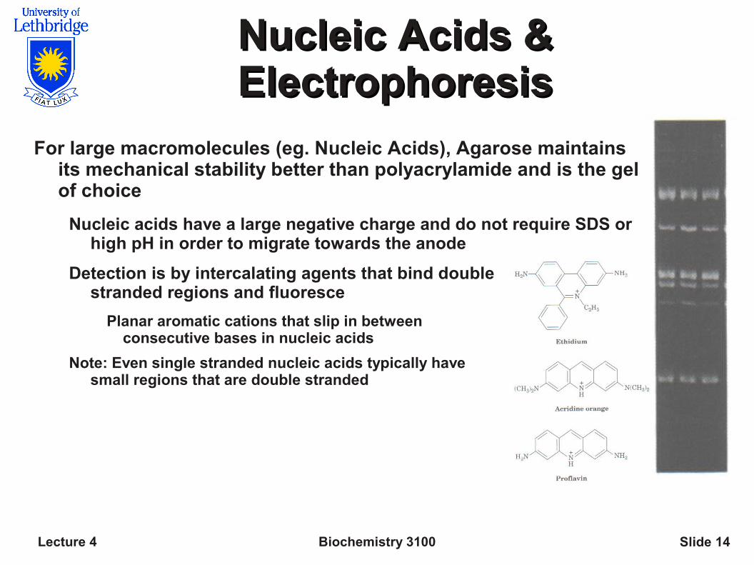
Nucleic Acids & Nucleic Acids & ElectrophoresisElectrophoresis
For large macromolecules (eg. Nucleic Acids), Agarose maintains its mechanical stability better than polyacrylamide and is the gel of choice
Nucleic acids have a large negative charge and do not require SDS or high pH in order to migrate towards the anode
Detection is by intercalating agents that bind double stranded regions and fluoresce
Planar aromatic cations that slip in between consecutive bases in nucleic acids
Note: Even single stranded nucleic acids typically have small regions that are double stranded
Biochemistry 3100Lecture 4 Slide 15
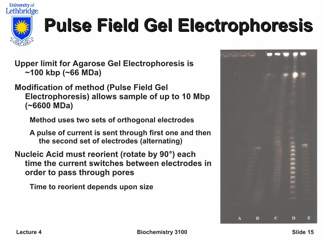
Pulse Field Gel ElectrophoresisPulse Field Gel Electrophoresis
Upper limit for Agarose Gel Electrophoresis is ~100 kbp (~66 MDa)
Modification of method (Pulse Field Gel Electrophoresis) allows sample of up to 10 Mbp (~6600 MDa)
Method uses two sets of orthogonal electrodes
A pulse of current is sent through first one and then the second set of electrodes (alternating)
Nucleic Acid must reorient (rotate by 90°) each time the current switches between electrodes in order to pass through pores
Time to reorient depends upon size