Electrochemical Polishing of 316L Stainless Steel Slotted ... · 72 Vol. 8, No. 2, June 2003...
12
70 Vol. 8, No. 2, June 2003 Progress in Biomedical Research Introduction Metallic coronary stents are medical devices that can provide endovascular scaffolding to relieve vascular obstructions. They exert a continuous radial pressure on the diseased coronary artery, resulting in a com- pression of atherosclerotic plaques, sealing of dissec- tions, and expansion of the coronary vessel [1,2]. When used as an adjunct to conventional balloon angioplasty, they improve vessel patency [3,4]. The surface roughness of stents is an important deter- minant of their thrombogenicity and tissue reaction [5], i.e., the nature of the metal surface is crucial to blood compatibility [6]. A smooth surface can help prevent the activation and aggregation of platelets, which is recognized as one component of the thrombosis process. Previous animal experiments [5,7-9] have shown that surface treatment using electrochemical polishing can improve the performance of stents. The results of implantation of the polished stainless steel coronary stents were compared with those of non-pol- ished stents. The conclusion is that metallic surface Electrochemical Polishing of 316L Stainless Steel Slotted Tube Coronary Stents: An Investigation of Material Removal and Surface Roughness H. ZHAO, J. VAN HUMBEECK Department of Metallurgy and Materials Engineering, Katholieke Universiteit Leuven, Leuven, Belgium J. SOHIER Precision Cutting Systems nv, Lubbeek, Belgium I. DE SCHEERDER Department of Cardiology, University Hospital Gasthuisberg, Katholieke Universiteit Leuven, Leuven, Belgium Summary Surface roughness is one of the properties that determines the performance of coronary stents. Therefore, in the production and application of stents, surface polishing is of paramount importance. In the present study, electro- chemical polishing was performed on 316L stainless steel slotted tube coronary stents made by laser cutting. Additionally, both acid pickling and annealing, as the pretreatment of electrochemical polishing, were also con- ducted. Material removal of the stents (both weight loss and strut width change) in the process of both pickling and electrochemical polishing was investigated. Both qualitative and quantitative roughness measurements were made to evaluate stent surface quality. Furthermore, material characterization of the stents was determined by means of composition analysis, metallographic characterization, and hardness measurement. The removal of material dur- ing both pickling and electrochemical polishing was within an acceptable range. Pickling caused a decrease in roughness of the cutting zone. Annealing resulted in an increase in roughness of the stent surfaces. Electrochemical polishing caused a smooth stent surface, which is comparable to commercially available stents. Moreover, the annealing treatment caused the stents to change from a deformed microstructure to a homogeneous structure, which is the major determining factor for the expandability of balloon stents. Key Words Coronary stents, electrochemical polishing, surface roughness of stents
Transcript of Electrochemical Polishing of 316L Stainless Steel Slotted ... · 72 Vol. 8, No. 2, June 2003...

70 Vol. 8, No. 2, June 2003
Progress in Biomedical Research
Introduction
Metallic coronary stents are medical devices that canprovide endovascular scaffolding to relieve vascularobstructions. They exert a continuous radial pressureon the diseased coronary artery, resulting in a com-pression of atherosclerotic plaques, sealing of dissec-tions, and expansion of the coronary vessel [1,2].When used as an adjunct to conventional balloonangioplasty, they improve vessel patency [3,4].The surface roughness of stents is an important deter-minant of their thrombogenicity and tissue reaction [5],
i.e., the nature of the metal surface is crucial to bloodcompatibility [6]. A smooth surface can help preventthe activation and aggregation of platelets, which isrecognized as one component of the thrombosisprocess. Previous animal experiments [5,7-9] haveshown that surface treatment using electrochemicalpolishing can improve the performance of stents. Theresults of implantation of the polished stainless steelcoronary stents were compared with those of non-pol-ished stents. The conclusion is that metallic surface
Electrochemical Polishing of 316L Stainless SteelSlotted Tube Coronary Stents:
An Investigation of Material Removal and Surface Roughness
H. ZHAO, J. VAN HUMBEECK Department of Metallurgy and Materials Engineering, Katholieke Universiteit Leuven, Leuven, Belgium
J. SOHIERPrecision Cutting Systems nv, Lubbeek, Belgium
I. DE SCHEERDERDepartment of Cardiology, University Hospital Gasthuisberg, Katholieke Universiteit Leuven, Leuven, Belgium
Summary
Surface roughness is one of the properties that determines the performance of coronary stents. Therefore, in theproduction and application of stents, surface polishing is of paramount importance. In the present study, electro-chemical polishing was performed on 316L stainless steel slotted tube coronary stents made by laser cutting.Additionally, both acid pickling and annealing, as the pretreatment of electrochemical polishing, were also con-ducted. Material removal of the stents (both weight loss and strut width change) in the process of both pickling andelectrochemical polishing was investigated. Both qualitative and quantitative roughness measurements were madeto evaluate stent surface quality. Furthermore, material characterization of the stents was determined by means ofcomposition analysis, metallographic characterization, and hardness measurement. The removal of material dur-ing both pickling and electrochemical polishing was within an acceptable range. Pickling caused a decrease inroughness of the cutting zone. Annealing resulted in an increase in roughness of the stent surfaces. Electrochemicalpolishing caused a smooth stent surface, which is comparable to commercially available stents. Moreover, theannealing treatment caused the stents to change from a deformed microstructure to a homogeneous structure,which is the major determining factor for the expandability of balloon stents.
Key Words
Coronary stents, electrochemical polishing, surface roughness of stents

Vol. 8, No. 2, June 2003 71
Progress in Biomedical Research
Acid PicklingPickling was performed on the cut stents by immersingthem in an acid solution (Table 1), consisting of HF,HNO3, and H2O, at 40 °C for 40 min. A thermostat wasused to establish a constant temperature. After thispickling treatment, the pickled stents were cleanedultrasonically using distilled water and then ethanol forat least 15 min, and were then dried by air blowing.
AnnealingAnnealing was conducted on the pickled stents in avacuum furnace (Leybold Heraeus PD 400, Germany).As shown in Figure 1, the stents were initially heatedin a vacuum at a heating rate of 3 °C/min, from roomtemperature to the annealing temperature of 1000 °C,and were then kept at 1000 °C for 1 h. Finally, theywere cooled in a vacuum at room temperature. Afterannealing, the annealed stents were cleaned by rinsingthem ultrasonically in distilled water and then ethanolfor at least 15 min, and dried by air blowing.
Electrochemical PolishingElectrochemical polishing was conducted on theannealed stents. The device used for electrochemicalpolishing was a self-construction, as illustrated inFigure 2. A 400 ml glass beaker was used as a cell. ADC rectifier (Polipower, Struers, Denmark) was usedas a power supply (30 V maximum). The stents were
treatment with electrochemical polishing effectivelyresults in a decreased thrombogenicity. The mostimportant clinical problem after stent implantation isstill neointimal hyperplasia within the stent, whichresults in a significant 16% – 30% narrowing of thestent [2,3,10-12]. Further efforts to improve the clini-cal results of coronary stents should focus on decreas-ing this neointimal hyperplasia. Previous studies withanimal models [7,8] have shown that the mestallic sur-face treatment using electrochemical polishingdecreased not only thrombogenicity but also neointi-mal hyperplasia.Electrochemical polishing is a process in which a metal-lic surface is smoothed by polarizing it anodically in asuitable electrolyte [13]. It is understood as two process-es: anodic leveling and anodic brightening [14]. Anodicleveling results from a difference in the dissolution ratebetween the peaks and valleys on a rough metal surface,depending on the current distribution or mass-transportconditions. On the other hand, anodic brightening isassociated with suppressing the influence of the metalmicrostructure on the dissolution rate. A smooth elec-trochemically polished surface, which appears bright tothe naked eye, results from a combination of these twofactors [14]. Optimization of electrochemical polishingof 316L stainless steel slotted tube coronary stents pro-duced by laser cutting has been investigated [15]. Thefocus of the present study is on the investigation ofmaterial removal and the surface roughness of the 316Lstainless steel slotted tube coronary stents during elec-trochemical polishing. The objective is to evaluate theeffect of electrochemical polishing on the 316L stainlesssteel slotted tube coronary stents.
Materials and Methods
MaterialsThe original material used in this study was 316Lstainless steel slotted tube coronary stents producedfrom a stainless steel tube (surgical grade stainlesssteel, ASTM F138) by laser cutting (Precision CuttingSystems nv, Belgium), which had a length of about15.0 mm and a diameter of 1.6 mm. The wall thicknessof the stents was 95.0 µm. Additionally, two commer-cial coronary stents, a Cook stent and an ACS stent,were used for the sake of comparison. The receivedsamples were cleaned using distilled water and thenethanol in an ultrasonic agitation bath for at least 15 min, and dried by air blowing.
Heating rate 3°/min
Pre
ssur
e (m
bar)
Tem
pera
ture
(°C
)
No. of samples
Vac
uum
Figure 1. Condition for annealing. See text for detailedinformation.
Table 1. Composition of pickling solution.

72 Vol. 8, No. 2, June 2003
Progress in Biomedical Research
used as an anode, and the cathode was a 316L stainlesssteel sheet (15 cm long, 4 cm wide, and 0.2 cm thick).The polishing temperature was controlled with a ther-mostat. The electrolyte is summarized in Table 2. Itconsists of H3PO4 (85wt%, weight percentage), glyc-erol, and H2O. The conditions for electrochemical pol-ishing, which were determined by experiment, are pre-sented in Table 3. After electrochemical polishing, thepolished stents were cleaned ultrasonically using dis-tilled water and then ethanol for at least 15 min, andwere then dried by air blowing.
Measurements of Weights and Dimensions of StentsMeasurement of the weights of a cut stent, a pickledstent, and an electrochemically polished (ECP) stentwas carried out with an electronic analytical balance
(Mettler AE 100, Switzerland). The widths of the stentstruts (as a cut stent, a pickled stent, and an ECP stent)were measured using a micrometer (Filar MicrometerEyepiece, American Optical 426C, USA) in conjunc-tion with a light optical microscope (Metalloplan,Leitz, Germany). Five measuring sites of the stentstruts were arbitrarily chosen to conduct the widthmeasurement, and a mean value was calculated.Weight loss and strut width reduction of the stents dur-ing the pickling and electrochemical polishing processwere calculated as follows:
weight loss = (weightbefore – weightafter)/weightbefore
reduction = (widthbefore – widthafter)/widthbefore
Roughness Measurements of StentsRoughness measurements were performed qualitative-ly and quantitatively to the stents, consisting of the cut,pickled, annealed, ECP, ACS, and Cook stents.Evaluation of the surface morphologies (qualitativeroughness measurement) was performed by means ofscanning electron microscopy (SEM) (Philips 515 SEM, The Netherlands). Pictures were taken withthe SEM. The quantitative roughness was measured bymeans of profilometry (WYKO NT 3300 Profiling sys-tem, Veeco, The Netherlands). The long cutoff wave-length (sampling) and the short cutoff wavelengthwere 25 µm and 0.2 µm, respectively. The evaluationlength was 125 µm (five sampling lengths). Approxi-mately 150 measuring lines were used, and thus themean roughness values could be obtained.
Material Characterization of StentsComposition analysis of the cut stent was performedusing energy dispersive spectrometry (EDS) in con-junction with SEM (Philips 515 SEM, theNetherlands). Metallographic characterization wasperformed on the transversal section of the cut andECP stents. First, grinding was done with waterproofabrasive paper (1200, 4000). Mechanical polishingwas then performed gradually with 3 µm and 1 µm dia-mond pastes and finally with a slurry of 0.05 µm SiO2
suspension. After this mechanical polishing, the sam-ples were rinsed ultrasonically using distilled waterand were then dried by air blowing. Etching was con-ducted on the mechanically polished samples using anetchant (Glyceregia) consisting of 10 ml HNO3 (65%),30 ml HCl (36% – 38%), and 30 ml glycerol. Lightoptical microscopy (LOM) (Polyvar met, Reichert-
Figure 2. Set-up for electrochemical polishing.
Table 2. Electrolyte for electrochemical polishing. wt% =weight percentage.
Table 3. Condition for electrochemical polishing.
Vol. 8, No. 2, June 2003 73
Progress in Biomedical Research
and a considerably smooth surface was revealed(Figure 3d). The surface quality was largely improvedcompared to both the pickled stent and the annealedstent. Figure 4 provides SEM pictures of the surfacemorphologies of the two commercial stents, the Cookstent (Cook Group, USA), and the ACS stent (Guidant,USA). As can be seen, although the cutting zonerevealed relatively less smoothness, these two com-
Jung, Germany) was then performed to observe themicrostructures. Pictures were taken with the LOM.Vickers hardness was measured with a Micro-Vickerstester (Leitz) on the cross section of the cut and ECPstents with a load of 50 g.
Results
Removal of Stent MaterialTable 4 presents the weights, the mean strut widths,and the calculated weight loss and width reduction ofthe cut, pickled, and ECP stents. As shown, the cutstent had a weight of about 13.0 mg and a strut widthof approximately 138.5 µm. After pickling, there wasa weight loss of about 7.7%, and the strut widthdecreased by 5.4% to about 131.0 µm. Using thesemeasured values together with the original wall thick-ness of the stent (95.0 µm), the strut thickness could becalculated from the relationship between the weightand volume, assuming that the strut length remainedconstant before and after pickling. By this calculation,a strut thickness decrease of approximately 2.4% wasobtained, i.e., after pickling, the strut thickness wasabout 92.7 µm. Due to electrochemical polishing, theweight and strut width of the stent decreased by 16.7%and 6.0%, respectively. The calculated result showedthat the strut thickness had decreased by 11.4%, i.e.,the strut thickness was approximately 82.1 µm.Therefore, after these two chemical processes, the finaldimensions of the stent strut were approximately 123.2 µm wide and 82.1 µm thick.
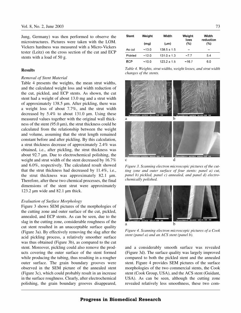
Evaluation of Surface MorphologyFigure 3 shows SEM pictures of the morphologies ofthe cutting zone and outer surface of the cut, pickled,annealed, and ECP stents. As can be seen, due to theslag in the cutting zone, considerable roughness of thecut stent resulted in an unacceptable surface quality(Figure 3a). By effectively removing the slag after theacid pickling process, a relatively smoother surfacewas thus obtained (Figure 3b), as compared to the cutstent. Moreover, pickling could also remove the prod-ucts covering the outer surface of the stent formedwhile producing the tubing, thus resulting in a rougherouter surface. The grain boundary grooves wereobserved in the SEM picture of the annealed stent(Figure 3c), which could probably result in an increasein the surface roughness. Clearly, after electrochemicalpolishing, the grain boundary grooves disappeared,
a
c
b
d
Figure 3. Scanning electron microscopic pictures of the cut-ting zone and outer surface of four stents: panel a) cut, panel b) pickled, panel c) annealed, and panel d) electro-chemically polished.
a b
Figure 4. Scanning electron microscopic pictures of a Cookstent (panel a) and an ACS stent (panel b).
Table 4. Weights, strut widths, weight losses, and strut widthchanges of the stents.

74 Vol. 8, No. 2, June 2003
Progress in Biomedical Research
mercial stents had a smooth surface. Comparing thesetwo surface morphologies with the ECP stent (Figure3d), it can be concluded that the surface quality of theECP stent was acceptable.Figure 5 shows the three-dimensional surface status,obtained by profilometry, of the outer surface of the cutstent, pickled stent, annealed stent, ECP stent, Cookstent, and ACS stent. These were the areas on whichthe quantitative roughness was measured. It is apparentthat the ECP stent (Figure 5d) had a smoother outersurface, compared to both the Cook stent (Figure 5e)and the ACS stent (Figure 5f). The polished stents,consisting of the ECP stent, the Cook stent, and theACS stent, had a much smoother outer surface than thenon-polished stents, i.e., the cut, pickled, and annealedstents. Figure 6 shows the three-dimensional surfacestatus of the cutting zone of the cut, pickled, annealed,and ECP stents. Clearly, the ECP stent had a muchsmoother surface in the cutting zone than the non-pol-ished stents, consisting of the cut, pickled, andannealed stents. These results further demonstrated theeffectiveness of electrochemical polishing.
Quantitative Roughness MeasurementFigure 7 exhibits the profiles of the outer surface of thecut stent, pickled stent, annealed stent, ECP stent,Cook stent, and ACS stent. Figure 8 shows the profilesof the cutting zone of the cut, pickled, annealed, andECP stents. Roughness can be determined from theseprofiles. Table 5 presents the average roughness Ra val-ues of the cut, pickled, annealed, and ECP stents.Figure 9 shows how this average roughness valuechanged using the different treatments: pickling,annealing, and electrochemical polishing. As can beseen, in the original cut stent, the average roughness Ra
of the cutting zone was four times greater than theouter surface. It was the largest roughness among allthe measurements. By removing the slag, the rough-ness of the cutting zone of the pickled stent appearedto be two times smaller compared to that of the cutstent. Meanwhile, the roughness of the outer surfaceincreased somewhat, as the product covering the outersurface during the tubing production was removed. Asseen from the surface morphology of the annealedstent (Figure 3c), after annealing, grain boundarygrooves appeared on the stent surface. This resulted inan increase in roughness, as compared to the pickledstent. By this quantitative roughness measurement, it isclearly demonstrated that electrochemical polishing
c
e
d
a b
f
Figure 5. Three-dimensional display of the outer surface ofthe six stents: panel a) cut, panel b) pickled, panel c)annealed, panel d) electrochemically polished, panel e)Cook, and panel f) ACS.
a
c
b
d
Figure 6. Three-dimensional display of the cutting zone ofthe four stents: panel a) cut, panel b) pickled, panel c)annealed, and panel d) electrochemically polished.

Vol. 8, No. 2, June 2003 75
Progress in Biomedical Research
provided a large decrease in stent roughness. Mean-while, the cutting zone of the ECP stent had almost thesame roughness as the outer surface, i.e., it had a uni-form surface status.Figure 10 shows that the average roughness Ra of theouter surface of the ECP stent and the ACS stent wasapproximately three times smaller than the Cook stent.As seen from the SEM images in Figure 4, the cuttingzone of both the ACS stent and the Cook stent was vis-
Hei
ght
(nm
)
Evaluation length (µm)
Hei
ght
(nm
)
Evaluation length (µm)a b
Hei
ght
(nm
)
Evaluation length (µm)
Hei
ght
(nm
)
Evaluation length (µm)c d
Hei
ght
(nm
)
Evaluation length (µm)
Hei
ght
(nm
)
Evaluation length (µm)e f
Figure 7. Profiles of the outer surface of the six stents: panel a) cut, panel b) pickled, panel c) annealed, panel d) electro-chemically polished, panel e) Cook, and panel f) ACS.
Table 5. Roughness Ra of the stents determined by profilom-etry. SD = standard deviation.

76 Vol. 8, No. 2, June 2003
Progress in Biomedical Research
ibly rougher than their outer surfaces. Therefore, it canbe demonstrated that in respect to smoothness the sur-face quality of the ECP stents obtained by applyingelectrochemical polishing was completely acceptable.Topography is the geometrical surface structure,including roughness, form errors, waviness, and vein-ing. According to the British Standard, roughness is
defined as "The irregularities in the surface texturewhich are inherent in the production process butexcluding waviness and errors of form" [16]. Theroughness measurement technologies are divided intoqualitative measurement and quantitative measure-ment. SEM gives a good qualitative image of surfaceroughness. Features magnified to about 10 nm can be
Hei
ght
(nm
)
Evaluation length (µm)
Hei
ght
(nm
)
Evaluation length (µm)a b
Hei
ght
(nm
)
Evaluation length (µm)
Hei
ght
(nm
)
Evaluation length (µm)c d
Figure 8. Profiles of the cutting zone of the six stents: panel a) cut, panel b) pickled, panel c) annealed, and panel d) electro-chemically polished.
Figure 9. Comparison of roughness between the different treated stents. Panel a) outer surface, panel b) cutting zone. ECP = electrochemical polished.
a b

Vol. 8, No. 2, June 2003 77
Progress in Biomedical Research
parameters are defined. Profile filters separate profilesinto long-wave and short-wave components. The λc
profile filter separates the roughness profile from long-wave components (e.g., waviness). The cutoff wave-length λc of a profile filter determines which wave-lengths belong to roughness and which ones to wavi-ness. The sampling length is the reference for rough-ness evaluation. Its length is equal to the cutoff wave-length λc. The evaluation length is the part of thelength over which the values of surface roughness aredetermined. The standard roughness evaluation lengthcomprises five consecutive sampling lengths. Different
observed [17]. Quantifying the degrees of roughnesscan be very complex, although various methods havebeen used to provide a quantitative description of theroughness, such as contact profilometry and opticalnon-contact profilometry.Various roughness values have been defined in theInternational Standard [18], which are used to quanti-tatively describe the roughness of a material surface,some of which are shown in Figure 11, including aver-age roughness Ra, root mean square roughness Rq, andmaximum peak-to-valley height Rmax. In theInternational Standard [19], some surface profiling
Figure 10. Comparison of the outer surface roughnessbetween the electrochemical polished (ECP) stent and twocommercial stents: ACS stent and Cook stent.
Table 6. Relationship between standard roughness and eval-uation length. S/E length = sample/evaluation length.
Figure 11. Illustration of definitions of roughness. z = outer surface; x = cutting zone.

78 Vol. 8, No. 2, June 2003
Progress in Biomedical Research
roughness is related to the selection of cutoff λc of aprofile filter. Table 6 shows the standard measurementsdefined in the International Standard [20]. In thisstudy, the roughness values of these stents werebeyond the ranges of the International Standard in theevaluation length due to the very small strut dimen-sions. They can also be valuable for a comparison ofthe surface roughness between the different stent sur-face states, but they must be obtained in the same eval-uation length.
Material CharacterizationTable 7 shows the result of the composition analysisobtained using EDS. These values could provide a pre-liminary expression of the stent composition. As canbe seen, no carbon was detected. This is due to theshortcomings of the employed analysis method. Theelement with an element number below 8 could not bedetected using EDS. Moreover, the expected concen-tration of carbon is too low to be detected by EDS.Figure 12 shows two LOM pictures of the microstruc-ture of the cross section of a cut stent. Figure 13 showsthe microstructure of the cross section of an ECP stent.Figure 14 shows the Vickers hardness of the cut stentand the ECP stent. As can be seen, the cut stent had a"deformation" microstructure, which was formedwhile producing the tube. Due to the annealing treat-ment prior to electrochemical polishing, the ECP stentrevealed homogeneous grains. The annealing treat-ment provided a decrease in the Vickers hardness from363.69 ± 10.80 to 159.26 ± 5.76. As can be observedin Figure 12b, the microstructure at the cutting edgewas the same as in the bulk. It is known that the ther-mal effect of laser cutting can cause a heat-affectedzone (HAZ) formation at the cutting edge showing adifferent microstructure from the bulk. Therefore, itmight be assumed that the HAZ resulting from theemployed laser cutting was very small. Obviously, ascan be seen in Figure 13b, with such a small initialHAZ, the ECP stent revealed no HAZ after electro-chemical polishing.
Discussion
Acid PicklingThe laser cutting process causes slag (burrs and depo-sitions) to appear on the surface of the stents, resultingin a rough surface. Before electrochemical polishing ofthe stents, as was earlier discussed for electrochemicalpolishing of NiTi alloy and Ta stents [21], the slagmust be removed. The surface quality of the polishedstents without a pre-treatment to remove the slag waseven worse than that of non-polished stents. The slag
Table 7. Composition of the 316L stainless steel coronarystent determined by energy-dispersive spectrometry. at% = atom percentage, wt% = weight percentage.
a b
Figure 12. Microstructures of a cut stent. Scale: panel a) 10 µm, panel b) 5 µm.
a b
Figure 13. Microstructures of an electrochemically polishedstent. Scale: panel a) 10 µm, panel b) 5 µm.
Figure 14. Vickers hardness (HV) of an cut stent and anelectrochemically polished stent (ECP).

Vol. 8, No. 2, June 2003 79
Progress in Biomedical Research
trolled on the basis of the anodic current density and,in some cases, on the basis of the applied voltage [26].In this study, applied voltage and anodic current wereused as the controlling parameters during the electro-chemical polishing process. All the parameters weredetermined experimentally. As discussed in some liter-ature, electrochemical polishing generally occurs at thelimiting current density (a current maximum or plateauin the current voltage curve) [26-29]. The rate of dis-solution at the limiting current is controlled by thetransport of cationic reaction products from the anodeinto the electrolyte [29]. Within the limiting-currentplateau region, the applied voltage also plays an impor-tant role in the resulting surface finishing [28]. Thetemperature is also a critical parameter for electro-chemical polishing. Polishing time is a parameter thatinfluences the removal of material from the stents incase other parameters are fixed.
Material RemovalThe strut dimensions of stents can directly determinetheir mechanical strength after deployment in the ves-sels, and in turn influence the final healing effects.Therefore, during electrochemical polishing of stents,it is important to control material removal, i.e., in thecase of achieving a satisfactory polishing effect, mate-rial removal should be as limited as possible. Weightloss and dimension decreases are two parameters forthe removal of material. Obviously, the final dimen-sions of the polished stents are determined from theiroriginal dimensions. Therefore, during pickling, a pre-treatment of polishing, overpickling should be avoid-ed. Overpickling, underpickling, and pitting usuallyare the direct results of lack of control over processvariables in the pickling of stainless steel [23]. Afterselecting a pickling solution, the pickling time, whichcan be determined experimentally, is a critical parame-ter for controlling material removal [30]. Additionally,the pickling temperature can have a pronounced influ-ence on the pickling rate. An increase in temperaturemay cause an increase in the pickling rate [23]. Duringelectrochemical polishing, the current density is notonly a determinant of the final effect, but also a mea-surement of the polishing rate [26-29], which in turnaffects the removal of material.
Surface Roughness MeasurementsThe roughness measurements not only create an imageof stent surface status, but also provide a quantitative
adhered to the surface of the stents after polishing,resulting in a worse surface quality. In the literature[22], it is also mentioned that the burrs and the deposi-tions must be removed after laser cutting in the nextproduction steps. Acid pickling is an effective methodfor the chemical removal of the surface oxides andother contaminants from metallic materials by immer-sion in an aqueous acid solution [23]. In this study, acidpickling was employed as a pre-treatment of electro-chemical polishing of the 316L stainless steel slottedtube coronary stents. The slag was effectivelyremoved, and thus a clean stent surface was preparedfor electrochemical polishing.
AnnealingThe original cut stents are made from the stainless steeltubing, which is too rigid to be balloon-expandable.Therefore, the cut stents must be made balloon-expandable before implantation. Annealing is an effec-tive method for softening the stent material. In thisstudy, an annealing treatment was accomplished undervacuum conditions at 1000 °C for 1 h, resulting in bal-loon-expandable stents, which were verified in a bal-loon expansion trial. The purpose of using a vacuumenvironment is to reduce the occurrence of oxidationduring the heat treatment. Since the carbon content(0.03% max) of low-carbon austenitic stainless steel,including 316L, is low enough to reduce precipitationof chromium carbides that markedly decrease the resis-tance to intergranular corrosion, it does not require aquenching treatment [24]. Therefore, in this study,annealing was performed and furnace cooling wasapplied. In addition, annealing results in the appear-ance of grain boundary grooves on the stent surface,which increases the surface roughness of the stentscompared to the non-annealed (pickled) stent.
Electrochemical PolishingElectrochemical polishing is a method of brighteningand smoothing the surface of metals [14,25] byimmersing the parts in an electrolyte and applying pos-itive direct current to the sample. The main electricalparameters for electrochemical polishing are the anod-ic potential, the anodic current density, and the appliedvoltage. As the nature and rate of any electrochemicalreaction are both determined by the electrode potential,the electrochemical polishing process should also becontrolled on the basis of the anodic potential. In prac-tice, the electrochemical polishing process is con-

80 Vol. 8, No. 2, June 2003
Progress in Biomedical Research
surface roughness of the stents. The initial surfacestate, together with the factors of current density andpolishing time, determines the final surface smooth-ness [26]. Therefore, determining the initial surfaceroughness demonstrates its importance. Most impor-tantly, the roughness measurements clearly exhibit thefinal surface texture of a polished stent, which is ameasurement that furthers medical and other investiga-tions, i.e., provides an opportunity to investigate quan-titatively the influence of roughness on stent biocom-patibility.
Conclusion
As concluded in a previous study [15], the sequenceof pickling and annealing prior to electrochemicalpolishing is of importance and should be reversed.The surface quality of 316L stainless steel slottedtube coronary stents is largely improved by means ofelectrochemical polishing. The surface roughness ofelectrochemically polished stents is in an acceptablerange, which is comparable to commercial stents.Using roughness measurements, combined withmaterial removal measurement, an optimal conditioncan be determined, under which relatively smallroughness and along with large strut dimensions canbe obtained. More importantly, roughness measure-ments not only determine the parameters for electro-chemical polishing, but also evaluate precisely thefinal effectiveness of the electrochemical polishingprocess. Due to the pretreatment of annealing, thepolished stent has a homogeneous microstructure anduniform hardness, and thus has good balloon expand-ability.
Acknowledgments
The authors gratefully acknowledge the technical staff at the Department of Metallurgy and MaterialsEngineering, Katholieke Universiteit Leuven,Belgium, especially Paul Crabbé, Dries Moons, MarcPeeters, and Rudy de Vos, for their technical support. The authors also acknowledge the Institutefor Encouraging of Innovation by Science andTechnology in Flanders, Belgium, for providing financial support for the project "Drug-ElutingCardiovascular Stents" of the Department ofMetallurgy and Materials Engineering together withPrecision Cutting Systems nv.
References
[1] Puel J, Juilliere Y, Bertrand ME, et al. Early and late assess-ment of sterosis geometry after coronary arterial stenting. AmJ Cardiol. 1988; 61: 546-553.
[2] Serruys PW, Strauss BH, Beatt KJ, et al. Angiographic fol-lowing-up after placement of a self-expanding coronaryartery stent. N Engl J Med. 1991; 324: 13-17.
[3] Serruys PW, de Jaegere P, Kiemeneij F, et al. A comparisonof balloon-expandable stent implantation with balloon angio-plasty in patients with coronary artery disease. N Engl J Med.1994; 331: 489-495.
[4] Fischman DL, Leon MB, Baim DS, et al. A randomised com-parison of coronary stent placement and balloon angioplastyin treatment of coronary artery disease. N Engl J Med. 1994;331: 496-501.
[5] Wang K. Biocompatibilisation of Coronary Artery Stents(Acta Biomedica Lovaniensia , No 162). Leuven: LeuvenUniversity Press. 1997: 89-102.
[6] Steinemann SG. Metal implants and surface reactions. Injury.1996; 27(Suppl 3): SC16-SC21.
[7] De Scheerder I, Verbeken E, van Humbeeck J. Metallic sur-face modification. Semin Intervent Cardiol. 1998; 3: 139-144.
[8] De Scheerder I, Sohier J, Wang K, et al. Metallic surfacetreatment using electrochemical polishing decreases throm-bogenicity and neointimal hyperplasia of coronary stents. IntJ Cardiol. 2000; 13: 179-185.
[9] De Scheerder I, Sohier J, Verbeken E, et al. Biocompatibilityof coronary stent materials: Effect of electrochemical polish-ing. Materialwiss Werkstofftech. 2001; 32: 142-148.
[10] De Scheerder I, Strauss BH, de Feyter PJ, et al. Stenting ofvenous bypass grafts: A new treatment modality for patientswho are poor candidates for reintervention. Am Heart J.1992; 123: 1046-1054.
[11] De Scheerder I, Wang K, Kerdsinchai P. Clinical and angio-graphic experience with coronary stenting using the FreedomStent. J Invas Cardiol. 1996; 8: 418-427.
[12] Strauss B. H, Serruys PW, De Scheerder I, et al. Relative riskanalysis of angiographic predictors of restenosis within thecoronary Wallstent. Circulation. 1991; 84: 1636-1643.
[13] Vidal R, West AC. Copper electropolishing in concentratedphosphoric acid. J Electrochem Soc. 1995; 142: 2682-2694.
[14] Magaino S, Matlosz M, Landolt D. An impedance study ofstainless steel electropolishing. J Electrochem Soc. 1993;140: 1365-1373.
[15] Zhao H, Van Humbeeck J, Sohier J, et al. Electrochemicalpolishing of 316L stainless steel slotted tube coronary stents.J Material Science: Materials in Medicine. 2002; 13: 911-916.
[16] Assessment of Surface Texture, part 1, British standard 1134.London: British Standard Institute. 1972.
[17] Baro AM, Garcia N, Miranda R, et al. Characterization of sur-face roughness in titanium dental implants measured withscanning tunnelling microscopy at atmospheric pressure.Biomaterials. 1986; 7: 463-466.
[18] DIN EN ISO 4287, ASME B46.1. Berlin: Deutsches Institutfür Normung.
[19] DIN EN ISO 11562, A.B. Berlin: Deutsches Institut fürNormung.

Vol. 8, No. 2, June 2003 81
Progress in Biomedical Research
[26] Shigolev PV. Electrolytic and Chemical Polishing of Metals,Revised 2nd ed. Tel Aviv: Freund Publishing House. 1974:22-36.
[27] Mathieu JB, Mathieu HJ, Landolt D. Electropolishing of tita-nium in perchloric acid-acetic acid solution. J ElectrochemSoc. 1978; 125: 1039-1043.
[28] Piotrowski O, Madore C, Landolt D. Electropolishing of tita-nium and titanium alloys in perchloric-free electrolytes.Plating and Surface Finishing. 1998; 85: 115-119.
[29] Piotrowski O, Madore C, Landolt D. Electropolishing of tan-talum in sulfuric acid-methanol electrolytes. ElectrochemicaActa. 1999; 44: 3389-3399.
[30] Bauccio M L. Technical note 9: Descaling and special surfacetreatments. In: Boyer R, et al. (editors). Materials PropertiesHandbook: Titanium Alloys. Metals Park, Ohio: AmericanSociety for Metals, 1994: 1145.
[20] DIN EN ISO 4288, ASME B46.1. Berlin: Deutsches Institutfür Normung.
[21] Zhao H, Van Humbeeck J, De Scheerder I. Surface condition-ing of NiTi and Ta stents. Prog Biomed Res. 2001; 6: 439-448.
[22] Momma C, Knop U, Nolte S. Laser cutting of slotted tubecoronary stents: State-of-the-art and future developments.Prog Biomed Res. 1999; 4: 39-44.
[23] Hudson RM, Joniec RJ, Shatynski SR. Pickling of Iron andsteel. In: Cubberly WH, et al. (editors). Metals Handbook,Volume 5, 9th ed. Metals Park, Ohio: American Society forMetals. 1982: 68-82.
[24] Rapoport D, Byrnes ER, Crooks KL, et al. Heat treating ofstainless steels. In: Alban LE, et al (editors). MetalsHandbook, Volume 4, 9th ed. Metals Park, Ohio: AmericanSociety for Metals. 1981: 623-646.
[25] Mills K, Davis TR, Destdfani JD, et al (editors). MetalsHandbook, Volume 9, 9th ed. Metals Park, Ohio: AmericanSociety for Metal. 1985: 48.
ContactProf. Dr. ir. Jan Van HumbeeckDepartment of Metallurgy and MaterialsEngineeringKatholieke Universiteit Leuven Kasteelpark Arenberg 44 B-3001 HeverleeBelgiumPhone: +32 16 321 281Fax: +32 16 321 992E-mail: [email protected]