Electropolishing of 316L Stainless Steel for … of 316L Stainless Steel for Biomedical...
1
Electropolishing of 316L Stainless Steel for Biomedical Applications: The Influence of Potential Sajjad Habibzadeh and Sasha Omanovic Department of Chemical Engineering, McGill University, 3610 university Street, Montreal, QC, Canada H3A 2B2 Metallic coronary stents, which are made mainly of 316L stainless steel (SS), are medical devices (implants) that can provide endovascular scaffolding in order to relive the vascular obstruction and minimize a risk of myocardial infraction (heart attack). However, the surface of a SS stent is thrombogenic and provokes tissue reaction. One component of the process of thrombosis is the activation and aggregation of platelets. Since nature of a metal surface is crucial to the blood compatibility, employing a suitable surface treatment can improve the biocompatibility of the stent. In this work, electrochemical polishing (EP) was applied as a SS stent surface treatment method. The SS surface was anodically polarized at cell voltages of 2.5, 4 and 10 V in an appropriate electrolyte, at 60 o C. A range of experimental techniques were used in the investigation of surface roughness, topographical, morphological, electrochemical/corrosion, and chemical properties of EP surfaces such as, atomic force microscopy (AFM), scanning electron microscopy (SEM), impedance spectroscopy (EIS), anodic polarization, x-ray photoelectron spectroscopy (XPS). In addition, the influence of electropolishing on the attachment and morphology of platelet-rich plasma (PRP) with the EP was assessed. The average surface roughness (R a ) measured by AFM depended on the voltage difference applied. The lowest value was obtained at 4 V (36 nm), whereas the values on the unmodified (control) surface, EP surfaces electropolished at 2.5V and 10 V were 123 nm, 78 nm and 67 nm, respectively. EIS results confirmed that the corrosion stability of all EP surfaces was higher than that of the control surface. XPS results revealed the chromium enrichment in the passive oxide films on the EP samples, and the oxygen content close to the outer film surface was the largest on the sample prepared at 4V. Hence, one of the origins of the increased corrosion resistance of EP samples was the formation of a thicker and more Cr-rich passive oxide film, in comparison to the film formed on the control substrate surface. PRP results (see Fig. 1) showed a relatively abundant accumulation of platelets on the control surface, while significantly fewer platelets were attached to the EP surfaces. Thus, a ca. 93% reduction of the number of attached platelets was obtained on the EP sample prepared at 4 V, in comparison to the control surface (see Fig. 2). Figure 1. SEM images of platelets attached to the a) unmodified 316L SS and the 316L SS electropolished at b) 2.5V c) 4V d) 10V. Images were taken after 60 min of static incubation in PRP. Figure. 2. Adhesion of platelets on the unmodified 316L SS (control), and 316L SS electropolished at cell voltages of 2.5V, 4V and 10V. The samples incubation time in PRP was 60 min. The results are the counted platelet on the surface mean value ± SD of three samples averaged over the entire sample surface for each bar. In conclusion, electropolishing of a stainless steel surface in an adequate electrolyte produces the surface that is more corrosion resistant and ‘unfriendly’ for platelet attachment, rendering the surface more biocompatible. a b c d 0 200 400 600 800 1000 1200 1400 1600 Control EP-2.5 V EP-4 V EP-10 V Surface platelets count Abstract #644, 223rd ECS Meeting, © 2013 The Electrochemical Society
-
Upload
nguyenkiet -
Category
Documents
-
view
215 -
download
0
Transcript of Electropolishing of 316L Stainless Steel for … of 316L Stainless Steel for Biomedical...
Electropolishing of 316L Stainless Steel for Biomedical Applications: The Influence of Potential
Sajjad Habibzadeh and Sasha Omanovic
Department of Chemical Engineering, McGill University, 3610 university Street, Montreal, QC, Canada H3A 2B2
Metallic coronary stents, which are made mainly
of 316L stainless steel (SS), are medical devices (implants) that can provide endovascular scaffolding in order to relive the vascular obstruction and minimize a risk of myocardial infraction (heart attack). However, the surface of a SS stent is thrombogenic and provokes tissue reaction. One component of the process of thrombosis is the activation and aggregation of platelets. Since nature of a metal surface is crucial to the blood compatibility, employing a suitable surface treatment can improve the biocompatibility of the stent. In this work, electrochemical polishing (EP) was applied as a SS stent surface treatment method. The SS surface was anodically polarized at cell voltages of 2.5, 4 and 10 V in an appropriate electrolyte, at 60oC.
A range of experimental techniques were used in
the investigation of surface roughness, topographical, morphological, electrochemical/corrosion, and chemical properties of EP surfaces such as, atomic force microscopy (AFM), scanning electron microscopy (SEM), impedance spectroscopy (EIS), anodic polarization, x-ray photoelectron spectroscopy (XPS). In addition, the influence of electropolishing on the attachment and morphology of platelet-rich plasma (PRP) with the EP was assessed.
The average surface roughness (Ra) measured by AFM depended on the voltage difference applied. The lowest value was obtained at 4 V (36 nm), whereas the values on the unmodified (control) surface, EP surfaces electropolished at 2.5V and 10 V were 123 nm, 78 nm and 67 nm, respectively. EIS results confirmed that the corrosion stability of all EP surfaces was higher than that of the control surface. XPS results revealed the chromium enrichment in the passive oxide films on the EP samples, and the oxygen content close to the outer film surface was the largest on the sample prepared at 4V. Hence, one of the origins of the increased corrosion resistance of EP samples was the formation of a thicker and more Cr-rich passive oxide film, in comparison to the film formed on the control substrate surface.
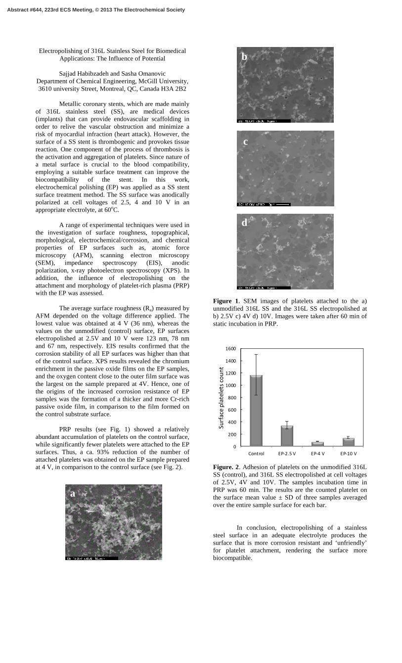
PRP results (see Fig. 1) showed a relatively
abundant accumulation of platelets on the control surface, while significantly fewer platelets were attached to the EP surfaces. Thus, a ca. 93% reduction of the number of attached platelets was obtained on the EP sample prepared at 4 V, in comparison to the control surface (see Fig. 2).
Figure 1. SEM images of platelets attached to the a) unmodified 316L SS and the 316L SS electropolished at b) 2.5V c) 4V d) 10V. Images were taken after 60 min of static incubation in PRP.
Figure. 2. Adhesion of platelets on the unmodified 316L SS (control), and 316L SS electropolished at cell voltages of 2.5V, 4V and 10V. The samples incubation time in PRP was 60 min. The results are the counted platelet on the surface mean value ± SD of three samples averaged over the entire sample surface for each bar.
In conclusion, electropolishing of a stainless steel surface in an adequate electrolyte produces the surface that is more corrosion resistant and ‘unfriendly’ for platelet attachment, rendering the surface more biocompatible.
a
b
c
d
0
200
400
600
800
1000
1200
1400
1600
Control EP-2.5 V EP-4 V EP-10 V
Su
rfa
ce p
late
lets
co
un
t
Abstract #644, 223rd ECS Meeting, © 2013 The Electrochemical Society