Electrochemical nanobiosensing in unprocessed whole...
28
Accepted Manuscript Title: Electrochemical nanobiosensing in unprocessed whole blood: recent advances Author: Mohammad Hasanzadeh, Nasrin Shadjou PII: S0165-9936(15)30031-5 DOI: http://dx.doi.org/doi: 10.1016/j.trac.2015.07.018 Reference: TRAC 14591 To appear in: Trends in Analytical Chemistry Please cite this article as: Mohammad Hasanzadeh, Nasrin Shadjou, Electrochemical nanobiosensing in unprocessed whole blood: recent advances, Trends in Analytical Chemistry (2015), http://dx.doi.org/doi: 10.1016/j.trac.2015.07.018. This is a PDF file of an unedited manuscript that has been accepted for publication. As a service to our customers we are providing this early version of the manuscript. The manuscript will undergo copyediting, typesetting, and review of the resulting proof before it is published in its final form. Please note that during the production process errors may be discovered which could affect the content, and all legal disclaimers that apply to the journal pertain.
Transcript of Electrochemical nanobiosensing in unprocessed whole...

Accepted Manuscript
Title: Electrochemical nanobiosensing in unprocessed whole blood: recent
advances
Author: Mohammad Hasanzadeh, Nasrin Shadjou
PII: S0165-9936(15)30031-5
DOI: http://dx.doi.org/doi: 10.1016/j.trac.2015.07.018
Reference: TRAC 14591
To appear in: Trends in Analytical Chemistry
Please cite this article as: Mohammad Hasanzadeh, Nasrin Shadjou, Electrochemical
nanobiosensing in unprocessed whole blood: recent advances, Trends in Analytical Chemistry
(2015), http://dx.doi.org/doi: 10.1016/j.trac.2015.07.018.
This is a PDF file of an unedited manuscript that has been accepted for publication. As a service
to our customers we are providing this early version of the manuscript. The manuscript will
undergo copyediting, typesetting, and review of the resulting proof before it is published in its
final form. Please note that during the production process errors may be discovered which could
affect the content, and all legal disclaimers that apply to the journal pertain.

1
Electrochemical nanobiosensing in unprocessed
whole blood: Recent advances
Mohammad Hasanzadeh a*
, Nasrin Shadjou b,c**
a Drug Applied Research Center, Tabriz University of Medical Sciences, Tabriz 51664,
Iran.
b Department of Nanochemistry, Nano Technology Center, Urmia University, Urmia,
Iran.
c Department of Chemistry, Faculty of Chemistry, Urmia University, Urmia, Iran
E-mail address:
(* ( [email protected], [email protected]
(** ( [email protected], [email protected]
Tel.: +98 914 3619877; fax: +98 41133632312.
Page 1 of 27

2
Highlights
Surfaces modification of nanomaterials for (bio/immune)-sensing applications in
whole blood was discussed.
Current developments in the determination of some biological molecules in
unprocessed blood were discussed.
Incorporation of biorecognition elements into nanomaterial based electrodes for
(bio/immune)-sensing in unprocessed whole blood was described.
Graphical Abstract
Abstract
Nowadays, the assay of whole blood is becoming increasingly important in modern
biomedical research such as clinical diagnostics, drug discovery, and biodefense. Whole
blood is a particularly complex mixture which contains a variety of substance such as
protein, glucose, inorganic salt, hormone, biomarkers and so on. Hence, a plentiful of
information can be obtained by investigating the whole blood, which is very helpful for
assessing the health status of the patient. Recently significant progress has been made in
electrochemical (bio/immune) sensing from whole blood using nanomaterials. This
Page 2 of 27

3
review describes fabrication and chemical modification of the surfaces of nanomaterials
for (bio/immune)-sensing applications in whole blood. We also present a comprehensive
overview of current developments and key issues in the determination of some biological
molecules with particular emphasis on evaluating the methods. We also discuss
incorporation of biorecognition elements into nanomaterial based electrodes for
immunesensing in unprocessed whole blood.
Keywords: electrochemical biosensing, nanotechnology, biomedical analysis, cancer,
enzyme, lab-on-a-chip, unprocessed and whole blood, immunesensing.
1. Introduction
Blood is one of the most informative sources for health and disease monitoring in the
human body [1] For example, monitoring levels of biomarkers in blood is known to be an
effective method for early diagnosis of various diseases such as cancer, by which better
treatment options and improved survival rates of patients can be provided [2].
Furthermore, therapeutic efficacy monitoring was demonstrated by following the levels
of chosen biomarkers in blood before and after a therapy, which facilitates the physicians
ability to determine the best treatment options [3]. Frequent monitoring of appropriate
Page 3 of 27

4
biomarkers is desirable for such purposes, since it leads to fast and timely feedback. This
approach requires an easily accessible sensory platform that can monitor the level of
biomarkers in a time-efficient and noninvasive (or negligibly invasive) manner with
comparatively low cost and minimal pain for the patients. However, few existing systems
satisfy all the aforementioned criteria. Thus, further research efforts are required toward
realization of such a system.
Nanobiosensors have the potential to meet the aforementioned criteria because of the
capability of performing rapid, label free, electrical detection with potentially low cost.
These devices utilize a capture agent on the sensor surface to bind the target biomolecules
with both selectivity and specificity. The captured biomolecules affect the electronic
properties of the nanowires, resulting in an electronically readable signal. Multiplexing
has also been demonstrated by selectively functionalizing the nanowires with receptors
for different biomarkers [4]. However, a challenge still remains toward making this
technology more clinically practical. That is, the use of whole blood as an input is not
typically investigated for biomarker detection; as such complex environments are known
to cause problems such as false signal and saturation of receptors. The use of whole blood
will allow evaluation of fragile proteins that experience prompt degradation after being
taken out of the body as well as providing a simplified sample preparation protocol to
expedite analysis.
Nowadays, the assay of whole blood is becoming increasingly important in modern
biomedical research such as clinical diagnostics, drug discovery, and biodefense. Whole
blood is a particularly complex mixture which contains a variety of substance such as
protein, glucose, inorganic salt, hormone, and so on. Hence, a plentiful of information
can be obtained by investigating the whole blood, which is very helpful for assessing the
health status of the patient. For example, glucose levels in blood can be related to the
diagnosis of diabetes, alcohol consumption, obesity and high cholesterol, while the
lactate concentration in blood is a biochemical indicator of anaerobic metabolism in
patients with circulatory failure. The determination of immunogenic tumor-associated
antigens level is very beneficial to clinical tumor diagnoses. The availability of a rapid,
simple, low cost, in situ whole blood assay with the capacity to detect a variety of
selected analytes would greatly benefit point-of-care or public health applications.
Recently significant progress has been made in biosensing from whole blood using a
capture release microfluidic chip [5] or from desalted serum; however, more effort is
needed for developing accurate and cost-effective systems that allow direct use of whole
blood samples prepared using simple tools such as finger pricks. In addition, there is still
a lack of understanding about biosensing using nanomaterials in complex media such as
serum and plasma. Therefore, this review describes fabrication and chemical
modification of the surfaces of nanomaterials for (bio/immune)-sensing applications. We
also present a comprehensive overview of current developments and key issues in the
determination of some biological molecules with particular emphasis on evaluating the
methods. This review shows how nanomaterials have made significant contributions in
the developments of electrochemical nanobiosensors, including immuno-, enzyme, DNA,
aptamer ones. More importantly, different aspects of the electrochemical biosensors such
as type of nanomaterials, detection techniques, analytes and the corresponding sensitivity
and sample matrix, as well as several noticeably prominent characteristics have been
Page 4 of 27
5
discussed in detail. Accordingly, research opportunities and future development trends in
these areas are discussed.
2. Type of application
2.1. Electrochemical enzyme nanobiosensing in unprocessed whole blood
Recently, an impressive number of inventive designs for enzyme-based electrochemical
sensing appeared. These types of sensor combine enzyme layers with electrochemical
transducers to produce a biosensor and promise to provide a simple, accurate and
inexpensive platform for patient diagnosis. Electrochemical methods are well suited to
enzyme investigation. Because electrochemical reactions give an electronic signal
directly, there is no need for expensive signal-transduction equipment. Moreover,
because immobilized probe enzyme can be readily confined to a variety of electrode
substrates, detection can be accomplished with an inexpensive electrochemical analyzer.
Indeed, portable systems for clinical testing and on-site environmental monitoring are
being developed. Sensitive electrochemical-signaling strategies based on the direct or
catalyzed oxidation of enzyme bases, and the redox reactions of reporter molecules or
enzymes recruited to the electrode surface by specific enzyme probe-target interactions
and charge-transport reactions mediated by the p-stacked base pairs have all been
demonstrated.
In this sub-section of review, we selectively review recent advances of electrochemical
enzyme nanobiosensing in unprocessed whole blood. Below, we include examples, and
compare and contrast enzyme biosensors based on nanomaterials. We also discuss the
feasibility of nanomaterials usage in enzyme biosensing, as these technologies might be
implemented to produce sensitive multiplexed assays for clinical diagnostics of diseases.
Until now, the primary methods of detecting blood glucose concentration are performed
by biochemical analyzers and glucose meters [6]. In hospital, the quantification of the
concentration of glucose is mainly carried out in serum samples by biochemical
analyzers, which are isolated from whole blood by centrifugation, but not in untreated
whole blood. The test results are influenced by the different model numbers of test
instruments and detection reagents, the treatment processes of blood samples, especially
additional centrifuging and too long measuring time from collecting a specimen of blood
to examination. As for commercial glucose meters, accurate results for glucose
concentration cannot be provided by a commercial glucose meter, due to the fact that
some defects cannot be ignored during its operation. For example, blood samples are
obtained from the fingertip peripheral but not veins, and doped easily with tissue fluid. So
the development of novel glucose biosensors for antifouling, rapid, highly sensitive, and
selective detection is of paramount importance for blood glucose concentration
monitoring in whole blood samples.
While an electrochemical biosensor directly used in whole blood, the biofouling of
electrode surface can be developed by platelet, fibrin and blood cell adhesion in the
complex environment of whole blood media. The biofouling of electrode surface will
bring catastrophic damage to the electron transfer between enzyme and electrode redox
center. As we know, anti-biofouling surfaces lie at the heart of several contemporary and
advanced technologies, ranging from coatings for biomedical implants, ship hulls, to
carriers for targeted drug delivery [7,8]. Hydrophilic surfaces of polymers like
polyethyleneglycol (PEG), with low values of polymer-water interfacial energy, show
resistance to protein adsorption and cell adhesion [9]. In this case, the hydrophilic
Page 5 of 27

6
polymer coating, carboxymethyl-PEG-carboxymethyl (CM-PEG-CM), was designed and
explored as antibiofouling surface for preparing for electrochemical glucose biosensor
that can be used directly for whole blood samples [10]. The biosensor was applied in
whole blood directly, which was based on the low values of polymer-water interfacial
energy, resistance to protein adsorption and cell adhesion. The entrapped GOx could
preserve its bioactivity and exhibited an excellent electrochemical behavior with a formal
potential of -0.29 V in phosphate buffer solution (PBS) (pH = 7.4). The results indicated
that the modified electrode can be used to determine glucose without interference from l-
ascorbic acid (AA) and uric acid (UA) with the low detection limit of 12.4 µM. The data
obtained from the biosensor showed good agreement with those from a biochemical
analyzer in hospital. The GOx biosensor modified with CM-PEG-CM will have essential
meaning and practical application in future that attributed to the effect of anti-biofouling
and good performance.
At the similar report by this research group, a novel electrochemical biosensor, which can
be evaluate the level of blood glucose with the help of antibiofouling technology, was
prepared by Sun and coworkers [11]. In this report, glucose oxidase (GOx) was
immobilizing on polyurethane-Pluronic F127 (PUF127) nanospheres modified glass
carbon electrode (GCE). Then, the electrochemical behavior of the biosensor in whole
blood was studied. The results of this report, indicated that GOx immobilized on the PU-
F127 nanospheres exhibited direct electron transfer reaction, which led to stable
amperometric biosensing for glucose with a detection limit of 11.4 µM in whole blood.
Interestingly, prposed sensor by this research group also offered suitable anti-interference
ability to ascorbic acid (AA) and uric acid (UA), especially when a detection potential of
-0.49 V was employed.
Also, small sample volume and in situ detection of glucose in human whole blood was
developed by using a screen-printed carbon electrode (SPCE) coupled with a paper disk
[12]. Interestingly, the SPCE was modified with graphene/polyaniline/Au
nanoparticles/glucose oxidase(Gr/PANI/AuNPs/GOD) composite and then covered by a
paper disk impregnated with the sample. After introducing PBS on the paper disk, the
electrochemical measurement was carriedout. The assay was based on measuring the
current decrease of flavin adenine dinucleotide (FAD) in GOD provoked by the enzyme-
substrate reaction using differential pulse voltammetry (DPV). It seem that, this new
paper-based electrochemical glucose sensor shows promise in applying point-of-care
(POC) device in whole blood tests, and particularly being appropriate for use in the
developing world and in resource-limited settings.
Interestingly, Picher et al [13] developed a lab-on-a-chip containing embedded
amperometric sensors in four microreactors that can be addressed individually and that
are coated with crystalline surface protein monolayers to provide a continuous, stable,
detection of blood glucose. It is envisioned that the microfluidic device will be used in a
feedback loop mechanism to assess natural variations in blood glucose levels during
hemodialysis to allow the individual adjustment of glucose. Reliable and accurate
detection of blood glucose is accomplished by simultaneously performing (a) blood
glucose measurements, (b) autocalibration routines, (c) mediator-interferences detection,
and (d) background subtractions. The electrochemical detection of blood glucose
variations in the absence of electrode fouling events is performed by integrating
crystalline surface layer proteins (S-layer) that function as an efficient antifouling
Page 6 of 27
7
coating, a highly-oriented immobilization matrix for biomolecules and an effective
molecular sieve with pore sizes of 4 to 5 nm. Picher and coworkers demonstrate that the
S-layer protein SbpA (from Lysinibacillus sphaericus CCM 2177) readily forms
monomolecular lattice structures at the various microchip surfaces (e.g. glass, PDMS,
platinum and gold) within 60 min, eliminating unspecific adsorption events in the
presence of human serum albumin, human plasma and freshly-drawn blood samples. The
highly isoporous SbpA-coating allows undisturbed diffusion of the mediator between the
electrode surface, thus enabling bioelectrochemical measurements of glucose
concentrations between 500 mM to 50 mM (calibration slope dI/dc of 8.7 nA/mM). Final
proof-of-concept implementing the four microfluidic microreactor design is demonstrated
using freshly drawn blood.
To work with whole blood samples, the process of separating plasma or serum is a
frequently required. Centrifugation is the most common method for this separation [14].
As an alternative, several research groups attempted to directly separate plasma from
whole blood using µ-PADs. Two general approaches for blood separation on µ-PADs
have appeared. One approach is to use heamagglutination between immobilized
antibodies and red blood cell antigen. The resulting plasma can be separated and flows
through the hydrophilic paper [15]. The other approach uses different types of paper, e.g.
blood separation membranes, connected with Whatman No.1 in either vertical [16] or
lateral platforms [17]. Currently, paper based microfluidics for blood separation has been
used for determining glucose [15], liver function [16] and, total protein [17] primarily
using colorimetric detection. However, analysis of biological markers from whole blood
sample based on ePADs few report has been reported.
For example, Songjaroen and coworkers [18] fabricated a new µ-PAD platform for
electrochemical detection of glucose from whole blood samples following plasma
isolation. Effective separation of the blood cells from the plasma fraction is essential for
reliable analysis of glucose. First, glucose levels are reduced when plasma specimens
remain in prolonged contact with red blood cells [19]. In addition, hemoglobin interferes
with the glucose assay [20]. Here, the wax dipping technique [21] was used to create the
microfluidic patterns on the paper because it allows for two different types of paper to be
joined together. The final device was composed of two blood separation zones that
directed isolated plasma to a middle detection zone. The resulting flow generated a
uniform gathering of plasma at the electrode and much better reproducibility of
electrochemical signals. The proposed ePADs work with whole blood samples with 24-
60% hematocrit without dilution, and the plasma was completely separated within 4 min.
Glucose in isolated plasma separated was detected using glucose oxidase immobilized on
the middle of the paper device. The hydrogen peroxide generated from the reaction
between glucose and the enzyme pass through to a Prussian blue modified screen printed
electrode (PB-SPEs). The currents measured using chronoamperometry at the optimal
detection potential for H2O2 (-0.1 V versus Ag/AgCl reference electrode) were
proportional to glucose concentrations in the whole blood. The linear range for glucose
assay was in the range 0-33.1 mM. The coefficients of variation (CVs) of currents were
6.5%, 9.0% and 8.0% when assay whole blood sample containing glucose concentration
at 3.4, 6.3, and 15.6 mM, respectively. Because each sample displayed intra-individual
variation of electrochemical signal, glucose assay in whole blood samples were measured
using the standard addition method. Results demonstrate that the ePAD glucose assay
Page 7 of 27

8
was not significantly different from the spectrophotometric method (p = 0.376, paired
sample t-test, n = 10).
Acknowledging the benefits of hyperbranched polymers and their nanoparticles, herein
china researchers [22] reported the design and synthesis of sulfonic acid group
functionalized hydroxyl-terminated hyperbranched polyester (H3O-SO3H) nanoparticles
and their biomedical application. In this report, a glucose biosensor was fabricated by
immobilizing the positively charged Au nanoparticles, H3O-SO3H nanoparticles and
glucose oxidase (GOx) onto the surface of glassy carbon electrode (GCE) [22]. It can be
applied in whole blood directly, which was based on the good hemocompatibility and
antibiofouling property of H30-SO3H nanoparticles. The biosensor had good
electrocatalytic activity toward glucose with a wide linear range (0.2-20 mM), a low
detection limit 12 µM in whole blood and good antiinterference property. The
development of materials science will offer a novel platform for application to substance
detection in whole blood.
To complete our discussion in this sub-section, Table 1 summarizes recent reports on
analytical parameters of some electrochemical enzyme nanobiosensing methods in
unprocessed whole blood. In conclusions of this section, it is important to point out;
nanomaterials have emerged as versatile tools for generating excellent supports for
enzyme stabilization due to their small size and large surface area. By proper surface
modification, various nanomaterials have been synthesized and successfully utilized for
protein/enzyme immobilization, which have already displayed promising effects in
practical applications. We have summarized in this sub-section the applications of
nanomaterials in enzyme immobilization and bioanalysis. The immobilized enzymes
generally show better stability towards pH and heat than the free ones and can be
recovered and reused multiple times. However, the activity of some enzymes decreases to
some extent after immobilization, which indicates that more efforts are still required to
explore the immobilization techniques
On the other hand, up to now; almost all researches on nanomaterials based enzymatic
biosensors are still in laboratory. As for practical applications, few enzymatic biosensors
appear to be commercially feasible except for some blood glucose biosensors. As for
practical applications, few enzymatic biosensors appear to be commercially feasible
except for some blood glucose and hand-held biosensors. To commercialize the
enzymatic biosensors based on nanomaterials, efforts should be made to break some key
technical barriers such as controlling the morphology of nanomaterials on device,
realizing efficient enzyme immobilization and keeping enzyme long-life bioactivity as
well as reducing matrix interference and sensor fouling.
2.2. Electrochemical nano-immunesensing in unprocessed whole blood
Recently, there was growing interest in the use of electrochemical immunosensors for
clinical diagnosis [23]. Electrochemical immunosensors utilize various types of electrode
[e.g., as screen-printed carbon electrodes (SPCEs), carbon-paste electrodes (CPEs) and
glassy-carbon electrodes (GCEs)]. The antibodies were immobilized by physical
adsorption or chemical immobilization. These types of electrodes can be used only once.
With the selected latest research articles from 2012 to May 2015, herein, we summarize
various electrochemical nano-immunosensors for detection of different biomarkers in this
Page 8 of 27

9
review. More importantly, we discuss in detail different aspects such as type of
nanomaterials, injection and detection techniques, labels, analytes and the corresponding
sample matrix, and sensitivity. Consequently, we discuss several outstanding properties
of the nano-immunosensors and their research opportunities as well as the development
potential and prospects. Also, we summarize below examples of nanostructure material
applications in electrochemical immunosensing biomarkers in whole blood reported so
far in the literature, with their advantages and limitations, and we stress their potential for
future development in this field.
To the best of our knowledge,, current methods for the quantitative detection of
antibodies, including ELISAs, Western blots, and fluorescence polarization assays, are
complex, multiplestep processes that rely on well-trained technicians working in well-
equipped laboratories [24]. In response, researchers would like to find some of other cost
effective an -
coworkers [25] described a versatile, DNA-b c c c “ w c ”
single-step measurement of specific antibodies directly in undiluted whole blood. The
design of this switch takes advantage of the occurrence of two antigen-binding sites on
each antibody, which are separated by ∼12 nm [26]. Specifically, these researchers used
DNA to engineer a switch that brings into close proximity (<4 nm) two copies of an
g ( c “ g ” or simplicity), via the formation of
a stem-loop structure. The two antigens, both linked at the extremities of the DNA strand
are thus located in the middle of the two strands of the stem (Figures 1 and 2). Upon
binding of the antibody to one of these antigens, the high effective concentration of the
second antigen provides the driving force to open the switch (more favorable bidentate
binding. [27] Thus separate the reporter elements. These researchers motivation for using
DNA as the scaffold for this switch is threefold. First, the chemistry of DNA supports the
addition of a variety of antigens ranging from small molecules to polypeptides and
proteins, either during its automated chemical synthesis or through postsynthesis
conjugation. [28] Second, DNA based switches are robust (i.e., they are not triggered by
nonspecific interactions) and relatively stable against degradation. Finally, the base-
g c DNA y w c ’ y c
the sensor achieves optimal detection limits [29].
Also, Romania researchers proposed a novel tool for screening of whole blood for early
detection of breast cancer antigen (CA153) [30]. In this report, Stefan-van Staden and
coworkers propose a stochastic microsensor based on maltodextrin with dextrose
equivalent between 4 and 7. Maltodextrin has a compact helix structure. Materials such
as maltodextrins were excellent candidates for the design of stochastic microsensors used
in the screening of whole blood for breast cancer biomarker. The results obtained for the
assay of breast cancer antigen in whole blood using this microsensor (by applying a
potential of 0.125 V vs. Ag/AgCl and performing measurements specific to stochastic
sensing described in detail in the article) were in agreement with those obtained using the
standard method, ELISA (Enzyme-Linked Immunosorbent Assay utilized a monoclonal
anti-CA15-3 antibody directed against intact breast cancer antigen, CA 153 for solid
phase immobilization on the microtiter wells.. The limit of determination obtained for
breast cancer antigen (0.5mU/mL) when assayed in whole blood, proved that the
microsensor is a good tool (in terms of sensitivity, selectivity, and fast response) for early
screening of whole blood for breast cancer.
Page 9 of 27

10
At the similar report by these Romania researchers [31], proposed six new stochastic
microsensors based on physical immobilization of oleoylethanolamide (1), (Z)- N-[(1S)-
2-hydroxy-1-(phenylmethyl)ethyl]-9octadecenamide (2), Nphenethyloleamide(3), N-[2-
(4-methoxyphenyl)ethyl]oleamide(4), N-[1]naphthyloleamide (5), N-cyclohexyloleamide
(6) in graphite paste, for the assay of carcinoembryonic antigen in the human whole
blood. According to obtained result by these researchers, the most sensitive microsensors
were based on oleamide 4, and oleamide 1, respectively immobilized in graphite paste.
The microsensors based on oleamide 1 and oleamide 4 were also presenting the lowest
limit of determination (0.1 pg/mL).
Literature review show that these researchers have another reports in design of stochastic
sensors for electrochemical biomarkers in whole blood.[32-34]
Recently, having the advantages of easy operation, fast response and low cost,
electrochemical technique based detection systems have also gained much attention for
cancer cell detection with high sensitivity even at the single cell level [35, 36].
For this purpose, Liu et al [37] developed an electrochemical impedimetric biosensor for
rapid, sensitive, and low-cost detection of cancer cells at low concentration in blood by
taking advantage of the high selectivity and affinity of antibodies as well as the large
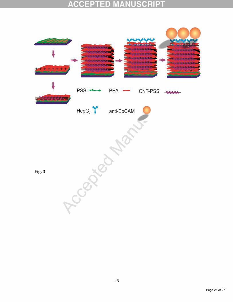
surface and good conductivity of multi-walled carbon nanotubes (MWCNTs). As shown
in Fig. 3 the multilayers of MWCNTs with the negatively charged carboxylate groups are
assembled on indium tin oxide (ITO) glass with the positively charged polyelectrolytes
by the layer-by-layer (LBL) assembly technique. The antibodies of epithelial cell-specific
markers, EpCAM antibodies, are then linked to MWCNTs in the multilayers via a
carbodiimide-mediated wet-chemistry approach. The resulting EpCAM
antibodies/MWCNT nanocomposites are used as nanoscale anchorage substrates to
effectively capture cells on the electrode surface via the specific binding between cell
surface EpCAM and EpCAM antibodies. The EpCAM expressed human liver cancer cell
(HepG2) is used as a model for circulating tumor cells, while the human cervical
carcinoma cell line (HeLa cells) and normal human hematologic cells, which do not
express EpCAM, are used as mixed cells to interfere with detection of HepG2 cells. The
electrochemical impedance of the prepared biosensors is linear with the logarithm of
concentration of the liver cancer cell line (HepG2) within the concentration range of 10 to
105 cells per mL. The detection limit for HepG2 cells is 5 cells per mL. The proposed
method by these researchers is confirmed to be simple, rapid and sensitive for real-time
measurement of the target cells in blood specimens at low concentration.
Label-free nanosensors can detect disease markers to provide point-of-care diagnosis that
is low-cost, rapid, specific and sensitive [38, 39]. However, detecting these biomarkers in
physiological fluid samples is difficult because of problems such as biofouling and non-
specific binding, and the resulting need to use purified buffers greatly reduces the clinical
relevance of these sensors. Hence, to overcome this limitation, researchers would like to
explore novel methods such as using distinct components within the sensor to perform
purification and detection.
For example, a novel label-free electrochemical aptasensors for thrombin detection in
whole blood using self-assembled multilayers with carboxymethyl-PEG-carboxymethyl
(CM-PEG-CM) and thrombin-binding aptamer (TBA) was developed [40]. In the sensing
strategy, CM-PEG-CM and TBA were assembled on the electrode surface via covalent
binding. In the presence of target, the TBA on the outermost layer of the self-assembled
Page 10 of 27

11
multilayer would catch the target on the electrode interface, which makes a barrier for
electrons and inhibits the electro-transfer, resulting in the decreased DPV signals. Using
this strategy, a wide detection range (1 pM-160 nM) for target thrombin was obtained,
with a low detection limit of 1.56×10⁻¹⁴
M. The control experiments were also carried out
by using bull serum albumin (BSA) and lysozyme in the absence of thrombin. The results
showed that the aptasensors had good specificity, stability and reproducibility to
thrombin. Moreover, the aptasensors could be used for detection of thrombin in whole
blood which could provide a promising platform for fabrication of aptamer based
biosensors in clinical application.
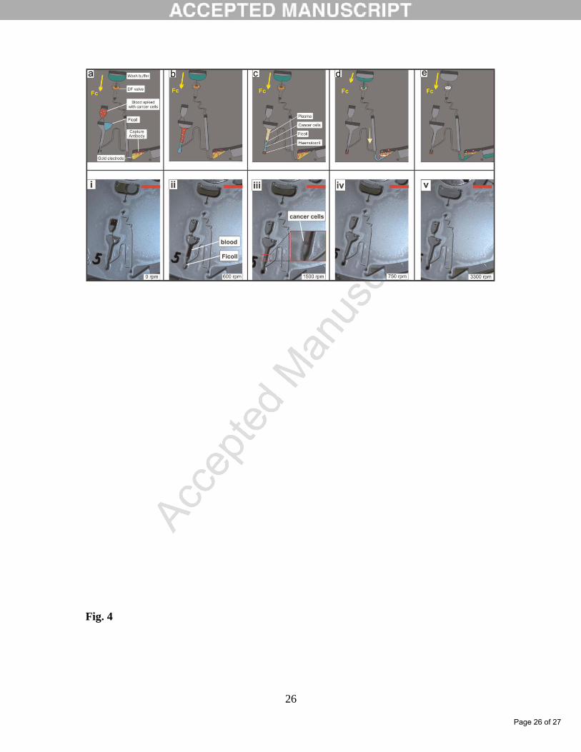
Also, an electrochemical Lab-on-a-Disc (eLoaD) platform for the automated
quantification of ovarian cancer cells (SKOV3) from whole blood was reported by
Nwankire and coworkers [41]. This centrifugal microfluidic system combines complex
sample handling, i.e., blood separation and cancer cell extraction from plasma, with
specific capture and sensitive detection using label-free electrochemical impedance. Flow
control is facilitated using rotationally actuated valving strategies including siphoning,
capillary and centrifugo-pneumatic dissolvable film (DF) valves. For the detection
systems, the thiol-containing amino acid, L-Cysteine, was self-assembled onto smooth
gold electrodes and functionalized with anti-EpCAM. By adjusting the concentration of
buffer electrolyte, the thickness of the electrical double layer was extended so the
interfacial electric field interacts with the bound cells. Schematic and image sequence of
the full assay protocol was shown in Fig. 4. Significant impedance changes were
recorded at 117.2 Hz and 46.5 Hz upon cell capture. Applying AC amplitude of 50 mV at
117.2 Hz and open circuit potential, aminimum of 214 captured cells/mm2 and 87%
capture efficiency could be recorded. The eLoaD platform can perform five different
assays in parallel with linear dynamic range between 16,400 and (2.670.0003)106 cancer
cells/mL of blood, i.e. covering nearly three orders of magnitude.
Biomarker detection based on nanowire biosensors has attracted a significant amount of
research effort in recent years. However, only very limited research work has been
directed toward biomarker detection directly from physiological fluids mainly because of
challenges caused by the complexity of media. This limitation significantly reduces the
practical impact generated by the aforementioned nanobiosensors. Recently, Chang and
coworkers [42] demonstrate an In2O3 nanowire-based biosensing system that is capable
of performing rapid, label-free, electrical detection of cancer biomarkers directly from
human whole blood collected by a finger prick (Fig. 5). Passivating the nanowire surface
successfully blocked the signal induced by nonspecific binding when performing active
measurement in whole blood. Passivated devices showed markedly smaller signals
induced by nonspecific binding of proteins and other biomaterials in serum and higher
sensitivity to target biomarkers than bare devices. The detection limit of passivated
sensors for biomarkers in whole blood was similar to the detection limit for the same
analyte in purified buffer solutions at the same ionic strength, suggesting minimal
decrease in device performance in the complex media. We then demonstrated detection
of multiple cancer biomarkers with high reliability at clinically meaningful
concentrations from whole blood collected by a finger prick using this sensing system.
To complete our discussion in this sub-section, Table 2 summarizes recent reports on
analytical parameters of some electrochemical immunesensing methods in unprocessed
whole blood.In this sub-section some of nanomaterial-based electrochemical
Page 11 of 27

12
immunosensors in whole blood are comprehensively summarized. More importantly,
different aspects of the immunosensors such as type of nanomaterials, injection and
detection techniques, labels, analytes and the corresponding sample matrix, the
sensitivity, etc. are discussed in detail. Consequently, several outstanding properties of
the whole blood immunosensors and their research opportunities as well as the
development potential and prospects are discussed. Additionally, we summarized
examples of nanomaterial based electrochemical immunosensors reported so far in the
literature, with their advantages and limitations and stress their potential for future
developments in this field.
2.3. Other paper of interest
Other than the above discussed nanomaterial-based electrochemical biosensors, in the
literature, other types of nanomaterial-based design electrochemical biosensors for
different types of analytes have been reported. In the current sub-section, we will briefly
discuss some of these nanobiosensors. In this sub-section of review, we focused our
attention on neurotransmitters, proteins and biomarkers biosensing in whole blood.
Despite their easy realization, for amperometric/voltammetric biosensors, few research
efforts have been devoted to these electrochemical nanobiosensors. Nevertheless, there is
a few numbers of promising works concerning o neurotransmitters, proteins now
available in the literature.
Neurotransmitters are the brain chemicals that are released from nerve cells which allow
At the present sub-section, more recent researches on electrochemical biosensing of
neurotransmitters in whole blood based on nanomaterials were review.
Dopamine (DA), epinephrine (EP) and norepinephrine (NE) are very important
neurotransmitters in the mammalian central nervous system, and they play an essential
role in the regulation of physiological processes in living systems. In the case of detection
of DA, EP and NE in whole blood, Stefan-van Stadena and coworkers [43] developed a
new multimode sensors for pattern recognition of neurotransmitters: DA, EP and NE in
biological fluids such as whole blood and urine with aminimum sampling of the
biological fluid (e.g., mixture of the biological fluid with an optimized buffer of pH 3.01
in a ratio1:1 (v/v)). In this report, diamond paste matrix was modified with three different
porphyrins: 5,10,15,20-tetraphenyl-21H, 23H-porphyrine,hemin and protoporphyrin IX
in order to design the multimode sensing systems. Stochastic mode represents a new
approach of electroanalysis and is the best for qualitative analysis using electrochemical
sensors; it can be used reliable also for quantitative analysis. In this report, the lowest
limits of quantification were: 10-10
mol/L for dopamine and epinephrine, and 10-11
mol/L
for norepinephrine. Also, obtained results by this group show that, the multimode
microsensors were selective over ascorbic and uric acids and the method facilitated
reliable assay of neurotransmitters in urine samples, and therefore, the pattern recognition
showed high reliability (RSD<1% for more than 6 months) for the simultaneous
determination of dopamine, epinephrine and norepinephrine from urine and whole blood
samples.Arizona State University researchers published a new article concerning
biosensing of neurotransmitters in whole blood based on nanomaterials. Dai et al [44]
utilized low-cost mesoporous carbon inks to screen print single-use disposable electrodes
for the detection of norepinephrine amperometrically. There are numerous advantages to
amperometric sensing in terms of cost, simplicity and maturity of the technologies.
However, the low concentration (90-220 pg/mL) of NE in human blood provides a
Page 12 of 27

13
significant challenge to obtaining acceptable signal to noise when only the current is
being measured to assess the concentration in blood. To overcome the lower
concentration limits for the amperometric sensing, these researchers increased the surface
area of the electrode to enhance the current signal by use of triconstituent assembly and
silica etching; surface areas from 1500 to 2300 m2/g can be produced depending upon the
carbon-silica ratio during self-assembly. To assess the role of the small micropores, one
control material without silica is also examined. The performance of these screen printed
mesoporous carbon electrodes is strongly dependent upon the surface area with greatest
sensitivity for the highest surface area carbon. To ensure NE can be effectively oxidized,
an enzyme, phenylethanolamine N-methyl transferase, with a cofactor, S-(5ˊ-Adenosyl)-
l-methionine chloride dihydrochloride, to active the enzyme is used to catalyze the
reaction. The resultant sensors can detect NE at concentrations as low as 100 pg/mL-1
in
rabbit whole blood.While highly sensitive protein detection methods have been achieved
in serum samples, biomarker detection in unprocessed whole blood remains very
challenging due to non-specific binding of cells and particulates to the sensor surface.
One approach to perform biosensing in whole blood samples has been demonstrated by
Stern et al., [45] who have developed a microfabricated microfluidic purification chip to
c 10μL w b 20 H w v c q e only achieves
500pM sensitivity, which is 106 times less sensitive than the techniques established for
detection in serum. Moreover, this level of sensitivity is not pertinent to most protein
biomarkers, whose serum concentrations in healthy individuals range between 0.01-100
pM. Hence, researchers would like to explore some of high efficient method for detection
of proteins.
For example Taipei Medical University researchers evaluated an electrochemical
biosensor for uric acid measurement in human whole blood samples [46]. In this work, a
commercially available uric acid monitoring system that is chemically modified to reduce
interference was evaluated via clinical evaluation for its performance and interference as
compared to a centralized laboratory instrument.
There has been significant progress over a period of two decades to develop
electrochemical-sensing approaches for heparin. The early breakthroughs were by
Meyerhoff and Yang, which used liquid PVC-based membranes doped with adequate
sensing components for the direct detection of heparin and protamine in blood [47]. The
strong protamine-heparin interaction makes it possible to use a protamine-selective
electrode in heparin assays, which also minimizes possible biases arising from weaker
nonspecific interactions of heparin with other blood components. Unfortunately,
spontaneous extraction of the polyion into the membrane made these types of sensors
operationally irreversible.
In subsequent years, a number of groups explored dynamic electrochemistry-based
methods to render the heparin or protamine detection operationally reversible. [48]
Shvarev group used mainly controlled current techniques for this purpose. While
protocols mainly useful as titration end points were successful early on, [49] linear
calibration curves less dependent on the background electrolyte of the sample were
achieved with chronopotentiometry. [50] An improved membrane formulation with a
selective membrane containing an excess of the protamine recognizing anion
dinonylnaphthalene sulfonate (DNNS) was recently shown to exhibit the required
Page 13 of 27

14
sensitivity and selectivity as well as linear calibration curves for use in whole blood
samples. [51]
Thin layer coulometry with ion-selective membranes has recently been explored in
Bakker group for the detection of a variety of ions [52, 53]. While a thin layer allows one
to work with just a few microliters of sample, coulometry is a very promising
interrogation platform since the absolute counting approach reduces the need for frequent
calibration. The present work aims to dramatically reduce the required sample volume
and to introduce a heparin sensing principle that is potentially calibration free.
Crespo and corkers [54] explore a potentially calibration-free methodology for the
detection of protamine (and, by titration, heparin) in whole of human blood in the
therapeutic concentration range from 20 to 120 mg/L. The use of a thin layer sample (5.8
μL) c b w b c v b Ag/AgC w
achieves an exhaustive depletion from the sample. In this report, coulometry detection
was chosen for the interrogation of the thin layer, employing a double pulse technique
with 120 s for each pulse. Protamine calibration curves were recorded at physiological
concentrations and in undiluted human blood. Heparin-protamine titrations were
performed in undiluted human blood samples, mimicking the final application with
patients undergoing critical care. The observed values correlate satisfactorily with those
of an alternative technique, so-called flash-chronopotentiometry on planar membranes.
Researches of this work, believed that further progress should involve (1) an
improvement of the biocompatibility of the inner electrode element in contact with blood,
(2) the microfabrication of a precise and accuracy thin layer compartment accompanied
with a tuning of the electrochemical protocol to reduce the intercept of the calibration
curve as much as possible, and (3) replacing the inner liquid solution by a solid ion-to-
electron transducer. Further, such progress may result in a point-of-care analysis system
for heparin that will be as simple to use as an off-the shelf glucose test strip.
3. Conclusions and future prospects
Nanomaterial-based electrochemical (bio/immune)-sensors will play an increasing role in
electroanalytical science in the near future. For electroanalytical applications, research is
required into the development of protocols for synthesis and functionalization of
nanomaterials. For bioanalytical applications, research should focus on increasing the
sensitivity and the selectivity of the nanobiosensors and on lifetime-based detection
methodologies. The fast advancement of nanomaterial-based electrochemical
(bio/immune)-sensors highlights their future applications in diverse scientific fields. The
major impacts appear to be in early detection of diseases, genetic mutations and
biotargets. Electrochemical (bio/immune)-sensors based nanomaterial provide fast,
simple, sensitive detection systems for cancer that may provide robust tools for
anticancer biosensor research. Despite significant advances in electrochemical
(bio/immune) based on nanomaterials, there are still challenges to explore new protocols
and strategies for improving the sensitivity and practical applications of the
electrochemical (bio/immune), as follows:
(1) Until now, the notable characteristics of electrochemical (bio/immune) sensing (e.g.,
disposable electrode array, label-free, multiplex analysis and microfluidic flow injection)
were rarely mentioned in whole/unpreceded blood. These characteristics indicate the
great opportunities for researchers in these scientific disciplines. The intrinsic
Page 14 of 27

15
electrochemical properties of nanomaterials would provide excellent theoretical support
for the development of label-free electrochemical immunosensors.
(2) Results raise doubts about repeatability, reproducibility, and comparability of
nanomaterials based-electrochemical (bio/immune)-sensing; as with every emerging
technology, standards need to be established to avoid doubts about the lack of
reproducibility, repeatability, and compatibility across platforms and laboratories.
(2) There is no comparison of the robustness of the nanomaterials for electrochemical
cytosensing in the literature. Obviously, electrochemists have a great deal to do to
address the performance of nanomaterials based-electrochemical (bio/immune)-sensing
in the future.
(3) Designing methodology for proper immobilization of cells on a nanomaterials based
compounds is a complex mixture of sciences and arts. There are often conflicting
requirements from stability and activity. Diversity of the process conditions necessarily
requires the design of specific, immobilized cells that can match the corresponding
requirements for the desired application.
(4) Another puzzling challenge is to develop a novel, simple method for immobilizing
enzymes on nanomaterials to reduce further electron-transfer resistance and to improve
stability, sensitivity, selectivity and life-span of electrodes.
(5) The long-term goals associated with incorporating nanomaterials into cytosensing
technology suggest that further research is required before these devices reach sufficient
performance standards.
(6) The use of nanomaterial tags for detecting proteins, neurotransmitters is still in its
infancy. Due to the minority of research being on development new nanomaterial-based
electrochemical biosensors for detecting proteins, neurotransmitters n whole blood , more
electrochemical techniques should be involved in this area. With nanomaterial, it will be
possible for biosensors to be applied to pre-warning and real-time detection of diseases.
(7) Unfortunately, minor reports (discussed above) can be seen to apply label-free
methods for electrochemical nanobiosensing. But, above examples shows that label-free
methods play a key role in manipulating various intracellular processes for
electrochemical biosensing applications. In general, until now, the remarkable
characteristics of label-free electrochemical biosensors were rarely mentioned. These
remarkable characteristics nicely indicate the great opportunities for researchers. The
intrinsic electrochemical properties of label-free electrochemical biosensors will provide
excellent theoretical support for the development of these biosensors.
(8) The biofouling of electrode surface will bring catastrophic damage to the electron
transfer between enzyme and electrode redox center. Therefore researchers should be
solving this problem using application of novel nanomaterials on structure of
electrochemical biosensors.
From the above examples, we conclude that there is a bright opportunity for further
advances and developments of nanomaterials-based (bio/immune)-sensing devices based
on electrochemical methods, especially through further miniaturization and integration
into lab-on-chip systems. The design of implantable (bio/immune)-sensing with the
ability to in vivo detection of cells and real time analysis is promising for the application
of electrochemical (bio/immune)-sensors, even though it is yet to be explored. Therefore,
electrochemists have a great deal to for to address electrochemical (bio/immune)-sensors
behavior based on nanomaterials in the future. In general, electrochemical (bio/immune)-
Page 15 of 27

16
sensors based on nanomaterials show great promise for future applications in health-care
testing and disease diagnostics. Given the impressive progress in nanomaterials-based
electrochemical systems, there is no doubt that they will have major impact on POC
clinical diagnostics. In future research, special attention will probably be given to
implantable electrochemical (bio/immune)-sensors based on nanomaterials for detection
and determination several of substance such as protein, glucose, inorganic salt, hormone,
biomarkers and so on. Future research will most probably explore the use of combination
nanomaterials with biomarkers, proteins, enzymes and so on in order to improve the
performance of implantable electrochemical immunosensors.
ACKNOWLEDGMENTS
Partial support through Drug Applied Research Center, Tabriz University of Medical
Sciences and Nano Technology Center, Faculty of Chemistry, Urmia University are
greatly acknowledged.
4. References:
[1] K. Fujii, T. Nakano, M. Kanazawa, S. Akimoto, T. Hirano, H. Kato, T. Nishimura,
Clinical-scale high-throughput human plasma proteome clinical analysis: Lung
adenocarcinoma, Proteomics 5 (2005) 1150-1159.
[2] J.A. Borgia, S. Basu, L.P. Faber, A.W. Kim, J.S. Coon, K.A. Kaiser-Walters, C.
Fhied, S. Thomas, Establishment of a multi-analyte serum biomarker panel to identify
lymph node Metastases in non-small cell lung cancer, J. Thorac. Oncol. 4 (2009) 338-
347.
[3] B.M. Nolen, J.R. Marks, S. Ta'san, A. Rand, T.M. Luong, Y. Wang, K. Blackwell,
A.E. Lokshin, Serum biomarker profiles and response to neoadjuvant chemotherapy for
Locally advanced breast cancer. Breast Cancer Res. 10 (2008) R45.
[4] M. Hasanzadeh, N. Shadjou, M. de la Guardia, Electrochemical biosensing using
hydrogel nanoparticles, Trends Anal. Chem. 62 (2014) 11-19.
[5] E.V.A. Stern, N.K. Rajan, J.M. Criscione, J. Park, B.R. Ilic, D.J. Mooney, M.A. Reed,
T.M. Fahmy, Label-free biomarker detection from whole blood. Nat. Nanotechnol. 5
(2010) 138-142.
[6] M. Hasanzadeh, N. Shadjou, M. Eskandani, M. de la Guardia, Mesoporous silica-
based materials for use in electrochemical enzyme nanobiosensors, Trends Anal. Chem.
40 (2012) 106-118.
[7] L.D. Chambers, K.R. Stokes, F.C. Walsh, R.J.K. Wood, Modern approaches to
marine antifouling coatings, Surf. Coat. Tech. 201 (2006) 3642-3652.
[8] C. Werner, M.F. Maitz, C. Sperling, Current strategies towards hemocompatible
coatings, J. Mater. Chem. 17 (2007) 3376-3384.
[9] M.N. Mar, B.D. Ratner, S.S. Yee, An intrinsically protein-resistant surface plasmon
resonance biosensor based upon a RF-plasma-deposited thin film, Sens. Actuators B:
Chem.54 (1999) 125-131.
[10] C. Suna, J. Miao, J. Yan, K. Yang, C. Mao, J. Ju, J. Shen, Applications of
antibiofouling PEG-coating in electrochemical biosensors for determination of glucose in
whole blood, Electrochimica Acta 89 (2013) 549- 554.
Page 16 of 27

17
[11] C. Sun, L. Chen, F. Xu, P. Zhu, J. Luan, C. Mao, J. Shen, Hemocompatible and
antibiofouling PU-F127 nanospheres platform for application to glucose detection in
whole blood, J. Mater. Chem. B, 1 (2013) 801-809.
[12] F.Y. Kong, S.X. Gu, W.W. Li, T.T. Chen, Q. Xu, W. Wang, A paper disk equipped
with graphene/polyaniline/Au nanoparticles/glucose oxidase biocomposite modified
screen-printed electrode: Toward whole blood glucose determination, Biosens.
Bioelectronics 56 (2014) 77-82
[13] M.M. Picher, S. Kupcu, C.J. Huang, J. Dostalek, D. Pum, U.B. Sleytr, P. Ertl,
Nanobiotechnology advanced antifouling surfaces for the continuous electrochemical
monitoring of glucose in whole blood using a lab-on-a-chip, Lab Chip 13 (2013) 1780-
1789
[14] V. VanDelinder, A. Groisman, Separation of plasma from whole human blood in a
continuous cross-flow in a molded microfluidic device, Anal. Chem. 78 (2006) 3765-
3771.
[15] X. Yang, O. Forouzan, T.P. Brown, S.S. Shevkoplyas, Integrated separation of blood
plasma from whole blood for microfluidic paper-based analytical devices, Lab Chip 12
(2012) 274-280.
[16] S.J. Vella, P. Beattie, R. Cademartiri, A. Laromaine, A.W. Martinez, S.T. Phillips,
K.A. Mirica, G.M. Whitesides, Measuring markers of liver function using a
micropatterned paper device designed for blood from a fingerstick, Anal. Chem. 84
(2012) 2883-2891.
[17] T. Songjaroen, W. Dungchai, O. Chailapakul, C.S. Henry, W. Laiwattanapaisal,
Blood separation on microfluidic paper-based analytical devices, Lab Chip 12 (2012)
3392-3398
[18] J. Noiphung, T. Songjaroen, W. Dungchai, C.S. Henry, O. Chailapakul, W.
Laiwattanapaisal, Electrochemical detection of glucose from whole blood using paper-
based microfluidic devices, Anal. Chim. Acta 788 (2013) 39-45
[19] T. Songjaroen, W. Dungchai, O. Chailapakul, W. Laiwattanapaisal, Paper-based
analytical device for quantitative urinalysis, Talanta 85 (2011) 2587-2593.
[20] D. Tompkins, J. Toffaletti, Enzymic determination of citrate in serum and urine,
with use W g “ ” v c C C 28 (1982) 192-195
[21] D. Yücel, K. Dalva, Effect of in vitro hemolysis on 25 common biochemical tests,
Clin. Chem. 38 (1992) 575-577.
[22] C. Sun, X. Chen, Q. Han, M. Zhou, C. Mao, Q. Zhu, J. Shen, Fabrication of glucose
biosensor for whole blood based on Au/hyperbranched polyester nanoparticles
multilayers by antibiofouling and self-assembly technique, Anal. Chim. Acta 776 (2013)
17- 23
[23] G.D. Lliu, J.T. Yan, G.L. Shen, R.Q. Yu, Renewable amperometric immunosensor
for complement 3 (C3) assay in human serum Original, Sens. Actuators B 80 (2001) 95-
100.
[24] M. Hasanzadeh, N. Shadjou, M. Eskandani, M. de la Guardia, E. Omidinia,
Mesoporous silica materials for use in electrochemical immunesensing, Trends Anal.
Chem. 45 (2013) 93-106
[25] A. Vallee-Belisle, F. Ricci, T. Uzawa, F. Xia, K.W. Plaxco, Bioelectrochemical
switches for the quantitative detection of antibodies directly in whole blood, J. Am.
Chem. Soc. 134 (2012) 15197-15200
Page 17 of 27

18
[26] T.R. Sosnick, D.C. Benjamin, J. Novotny, P.A. Seeger, J. Trewhella, Distances
between the antigen-binding sites of three murine antibody subclasses measured using
neutron and x-ray scattering, J. Biochemistry 31 (1992) 1779-1786.
[27] L. Tian, T. Heyduk, Bivalent ligands with long nanometer-scale flexible linkers,
Biochemistry 48 (2009) 264-275.
[28] R.P. Haugland, The Molecular Probes Handbook: A guide to fluorescent probes and
labeling technologies, 10th ed.; Invitrogen: C.A. San Diego (2005).
[29 A - , F. Ricci, K.W. Plaxco, Thermodynamic basis for the optimization
of binding-induced biomolecular switches and structure-switching biosensors. Proc. Natl.
Acad. Sci. U.S.A. 106 (2009) 13802-13807.
[30] R.I. Stefan-van Staden, J.F. van Staden, New tool for screening of whole blood for
early detection of breast cancer antigen (CA153), J. Modern Med. Chem. 1 (2013) 86-91.
[31] C.C. Negut, R.I. Stefan-van Staden, I. Moldoveanu, E.M. Ungureanu, C. Stanciu-
Gavan, New stochastic microsensors based on oleamides, Electrochem. Commun. 51
(2015) 98-102
[32] I. Moldoveanu, C. Stanciu-Gavan, R.I. Stefan-van Staden, Molecular recognition of
HER-1 in whole-blood samples, J. Mol. Recognit. 27 (2014) 653-658.
[33] R.I. Stefan-van Staden, L.A. Gugoasa, J.F. van Staden, O.C. Rusu, A genetic
screening test for obesity based on stochastic sensing, J. Electrochem. Soc. 161 (2014)
B167-B170.
[34] R.I. Stefan-van Staden, I. Moldoveanua, stochastic microsensors based on
nanostructured materials used in the screening of whole blood for hepatitis B, J.
Electrochem. Soc. 161 (2014) B3001-B3005.
[35] Z.J. Zhang, L. Wu, J.S. Wang, J.S. Ren, X.G. Qu, A Pt-nanoparticle electrocatalytic
assay used for PCR-free sensitive telomerase detection, Chem. Commun. 49 (2013)
9986-9988.
[36] L. Wu, J.S. Wang, J.S. Ren, X.G. Qu, Ultrasensitive telomerase activity detection in
circulating tumor cells based on DNA metallization and sharp solid-State electrochemical
techniques, Adv. Funct. Mater., 24 (2014) 2727-2733.
[37] Y. Liu, F. Zhu, W. Dan, Y. Fua, S. Liu, Construction of carbon nanotube based
nanoarchitectures for selective impedimetric detection of cancer cells in whole blood,
Analyst 139 (2014) 5086-5092.
[38] E. Stern, Label-free immunodetection with CMOS-compatible semiconducting
nanowires. Nature 445 (2007) 519-522.
[39] E. Stern, A. Vacic, M.A. Reed, Semiconducting nanowire field-effect transistor
biomolecular sensors, IEEE Trans. Electron. Dev. 55 (2008) 3119-3130
[40] C. Sun, X. Wang, X. Yanga, L. Xing, Bo Zhao, X. Yang , C. Mao, A label-free
electrochemical aptasensor for sensitive thrombin detection in whole blood, Electrochim.
Acta 106 (2013) 327-332.
[41] E.C. Nwankire, A. Venkatanarayanan, T. Glennon, T.E. Keyes, R.J. Forster, J.
Ducree, Label-free impedance detection of cancer cells from whole blood on an
integrated centrifugal microfluidic platform. Biosen. Bioelectron. 68 (2015) 382-389.
[42] H.K. Chang, F.N. Ishikawa, R. Zhang, R. Datar, R.J. Cote, M.E. Thompson, C.
Zhou, Rapid, label-free, Electrical whole blood bioassay based on nanobiosensor
systems. ACS Nano 5 (2011) 9883-9891.
Page 18 of 27

19
[43] R.I. Stefan-van Staden, I. Moldoveanu, J.F. van Staden, Pattern recognition of
neurotransmitters using multimode sensing J. Neuroscience Methods 229 (2014) 1-7.
[44] M. Dai, B. Haselwood, B.D. Vogt, J.T. La Belle, Amperometric sensing of
norepinephrine at picomolar concentrations using screen printed, high surface area
mesoporous carbon, Anal. Chim. Acta 788 (2013) 32- 38.
[45] E. Stern, A. Vacic, N. Rajan, J. Criscione, J. Park, B. Ilic, D. Mooney, M. Reed, T.
Fahmy, Label-free biomarker detection from whole blood. Nature Nanotechnol. 5 (2010)
138-142
[46] L.T. Liao, C.C. Liao, C.C. Liu, Ting-Ya Yang, G.C. Wang, Evaluation of an
electrochemical biosensor for uric acid measurement in human whole blood samples,
Clin. Chim. Acta 436 (2014) 72-77.
[47] S.C. Ma, V.C. Yang, B. Fu, M.E. Meyerhoff, Electrochemical sensor for heparin:
further characterization and bioanalytical applications, Anal. Chem. 65 (1993) 2078-
2084.
[48] S. Amemiya, Y. Kim, R. Ishimatsu, B. Kabagambe, Electrochemical heparin sensing
at liquid/liquid interfaces and polymeric membranes, Anal. Bioanal. Chem. 399 (2011)
571-579.
[49] A. Shvarev, E. Bakker, Reversible electrochemical detection of nonelectroactive
polyions, J. Am. Chem. Soc. 125 (2003) 11192-11193.
[50] K.L. Gemene, E. Bakker, Detection of protease activities by flash
chronopotentiometry using a reversible polycation-sensitive polymeric membrane
electrode, Anal. Biochem. 386 (2009) 276-281.
[51] G. A. Crespo, M.G. Afshar, E. Bakker, Reversible sensing of the anticoagulant
heparin with protamine permselective membranes, Angew. Chem., Int. Ed., 51 (2012)
12575-12578.
[52] A. Shvarev, B. Neel, E. Bakker, Detection limits of thin layer coulometry with
ionophore based ion-selective membranes, Anal. Chem. 84 (2012) 8038-8044.
[53] M. Sohail, R. De Marco, K. Lamb, E. Bakker, Thin layer coulometric determination
of nitrate in fresh waters. Anal. Chim. Acta, 744 (2012) 39-44.
[54] G.A. Crespo, M.G. Afshar, D. Dorokhin, E. Bakker, Thin layer coulometry based on
ion-exchanger membranes for heparin detection in undiluted human blood, Anal. Chem.
86 (2014) 1357-1360.
.
Page 19 of 27

20
Figure legends:
Fig. 1. Antibody (Ab)- c v c c c “ w c ” [41
Fig. 2. Antibody-activated switch architecture, [41].
Fig. 3: Preparation of ITO/MWCNT/PEI/anti-EpCAM and immunological recognition
between anti-EpCAM and HepG2 [68].
Fig. 4: Schematic (a-e) and image sequence (i-v) of the full assay protocol for integrated
centrifugal microfluidic platform [73].
Fig. 5: Device configuration and real-time sensing response for (a) unpassivated CA-125
nanosensor in buffer, (b) unpassivated CA-125 nanosensor in serum [74].
Table 1: Analytical parameters of some electrochemical enzyme nanobiosensing
methods in unprocessed whole blood
Type of electrode modifier Type of
analyte
Detection potential
versus Ag/AgCl
LOD Ref
carboxymethyl-PEG-
carboxymethyl (CM-PEG-CM)-
GCE
glucose -0.29 V 12.4 µM 10
polyurethane-Pluronic F127
(PUF127) nanospheres and
immobilizing glucose oxidase
(GOx) on (PU-F127)-glass
carbon electrode (GCE)
glucose -0.49 V 11.4 µM 11
graphene/polyaniline/Au
nanoparticles/glucose
oxidase(Gr/PANI/AuNPs/GOD)-
SPCE
glucose - - 13
µ-PADs (S-layer protein SbpA
(from Lysinibacillus sphaericus
CCM 2177))
glucose - 6.4 mM 13
ePADs (GOx-Prussian blue
modified screen printed
electrode (PB-SPEs).
glucose -0.1 V 3.4 mM 18
Au nanoparticles-H3O-SO3H
nanoparticles -glucose oxidase
(GOx) modified glassy carbon
electrode (GCE)
glucose - 12 µM 22
Page 20 of 27

21
Table 2: Analytical parameters of some electrochemical immunesensing methods in
unprocessed whole blood
Type of electrode modifier Type of analyte Detection
potential
versus
Ag/AgCl
LOD Ref
stochastic microsensor based on
maltodextrin with dextrose
equivalent between 4 and 7
breast cancer
antigen- CA 153
0.125 V 0.5mU/mL 30
stochastic microsensors based on
physical immobilization of
oleoylethanolamide (1), (Z)- N-
[(1S)-2-hydroxy-1-
(phenylmethyl)ethyl]-
9octadecenamide (2),
Nphenethyloleamide(3), N-[2-(4-
methoxyphenyl)ethyl]oleamide(4),
N-[1]naphthyloleamide (5), N-
cyclohexyloleamide (6) in
graphite paste
carcinoembryonic
antigen
- 0.1 pg/mL 31
stochastic microsensors based
Mn(III) with meso-tetra (4-
carboxyphenyl) porphyrin, and
maltodextrin (dextrose
equivalence between 4 and 7),
immobilized in diamond paste,
graphite paste or C60 fullerene
paste
HER-1 - 280 fg/ml
and 4.86
ng/ml
32
stochastic microsensors based on
M α-cyclodextrin and
5,10,15,20-tetraphenyl-21H,23H
leptin 1.25 × 10⁻¹⁰
33
plasminogen
activator
1 × 10-12
Page 21 of 27

22
porphyrin were immobilized in
carbon based matrices such as
diamond paste, graphite,
graphene, carbon nanotubes, and
C60 fullerenes.
inhibitor-1 (PAI-
1)
stochastic microsensors based four
electrochemical active materials:
α-cyclodextrin, maltodextrin with
dextrose equivalent, the complex
of Mn(III) with 5,10,15,20-
tetraphenyl-21H,23H-porphyrin,
and the traditional hemolysine
immobilized in diamond paste
matrices
hepatitis B - - 34
Cytosensor: EpCAM
antibodies/MWCNT
nanocomposites
HepG2 cells - 5 cells per
mL
37
self-assembled multilayers with
carboxymethyl-PEG-
carboxymethyl (CM-PEG-CM)
and thrombin-binding aptamer
(TBA)
thrombin - 1.56×10⁻¹⁴
M
40
Lab-on-a-Disc (eLoaD) platform
based on thiol-containing amino
acid, L-Cysteine, was self-
assembled onto smooth gold
electrodes and functionalized with
anti-EpCAM
ovarian cancer
cells (SKOV3)
- 214
captured
cells/mm2
41
Page 22 of 27

23
Fig. 1
Page 23 of 27

24
Fig. 2
Page 24 of 27
25
Fig. 3
Page 25 of 27
26
Fig. 4
Page 26 of 27

27
Fig. 5
Page 27 of 27
![Chapter 2 Signal Amplification for Nanobiosensing€¦ · 40 2 Signal Amplification for Nanobiosensing been used for the photonic detection of biorecognition processes [14]. This](https://static.fdocuments.net/doc/165x107/6031a8439eee3a120e15fd47/chapter-2-signal-ampliication-for-nanobiosensing-40-2-signal-ampliication-for.jpg)