EFFECTS OF FASTING AND INTRALUMINAL CONTRAST ENHANCEMENT ON ULTRASONOGRAPHIC APPEARANCE OF THE...
5
EFFECTS OF FASTING AND INTRALUMINAL CONTRAST ENHANCEMENT ON ULTRASONOGRAPHIC APPEARANCE OF THE EQUINE SMALL INTESTINE TRACY NORMAN,KEITH CHAFFIN,DAVID SCHMITZ The equine small intestine is challenging to evaluate ultrasonographically. In humans, hydrosonography has been used to improve ultrasonographic images of the small intestine. We hypothesized that fasting horses for 24 h would enhance the ability to image the small intestine transabdominally by separating intestinal loops and reducing intraluminal gas, and that the administration of intragastric contrast agent would further improve that ability. Ten healthy horses were examined ultrasonographically under three treatment conditions: (a) regular diet, (b) after a 24-h fast, and (c) fasted plus intragastric administration of water and mineral oil. During each phase of the study, 30-s video clips were obtained from four predetermined abdominal windows, and were examined to determine diagnostic quality. Fasting improved the ability to obtain high-quality images of the small intestine significantly. The addition of contrast agent resulted in qualitative improvement in image quality, but differences did not result in statistically significant improvement. r 2010 Veterinary Radiology & Ultrasound, Vol. 51, No. 6, 2010, pp 642–646. Key words: equine, image enhancement, small intestine, ultrasound. Introduction I N ADULT HORSES, gas and ingesta in the large colon and the length, position, tortuous course, and intraluminal gas of the small intestine render the bowel difficult to assess thoroughly with ultrasound. This makes critical assessment difficult or impossible, and intramural lesions may be missed. This is a serious limitation, as increased wall thick- ness is one of the most common sonographic abnormalities of the small intestine, 1 and visualization of a greater pro- portion of the small intestine increases the probability of identifying a focal or segmental lesion. Fasting horses for 8 h increases the length of jejunum visible ultrasonograph- ically compared with fed horses. 2,3 In humans, the challenges of examining the small intes- tine sonographically are similar to those in the horse. Hydrosonography of the small intestine, where a hypo- echoic contrast agent is administered per os to provide intraluminal contrast, is used occasionally to improve vi- sualization of the small intestine by separating individual intestinal loops and reducing intraluminal gas. 4–7 A variety of contrast agents have been used for hydrosonography, including water, orange juice, cellulose-based suspensions, and isotonic polyethylene glycol solutions. 4–7 We hypothesized that fasting horses for 24 h would im- prove visualization of the small intestine, and that the ad- ministration of intragastric fluid would further improve the quality of small intestinal images. Our objectives were to determine whether fasting horses for 24 h improves the overall diagnostic quality of sonographic images of the small intestine compared with fed horses, and whether an intragastric contrast agent in fasted horses would further enhance diagnostic quality. Materials and Methods Ten normal adult Quarter Horses obtained from the university teaching herd were studied. There were nine geldings and one mare, and age ranged from 2 to 17 years. Each horse was evaluated ultrasonographically three times. The first was while the horse was on a routine feeding schedule (fed horses). Fed horses were allowed free access to coastal Bermuda grass hay and given 1 kg of commercial sweet feed twice daily. The second examination was per- formed after the horses were fasted for 24 h (fasted horses). Fasted horses had free access to water while feed and hay were withheld. The third examination was performed on fasted horses 15min after 3 l of water and 1 l of mineral oil were administered by a nasogastric tube (contrast horses). Each horse acted as its own control. This paper has been submitted for presentation at the Annual Con- vention of the American Association of Equine Practitioners. Address correspondence and reprint requests to Dr. Tracy Norman, at the above address. E-mail: [email protected] Received April 15, 2010; accepted for publication May 6, 2010. doi: 10.1111/j.1740-8261.2010.01719.x From the Department of Large Animal Clinical Sciences, Texas A&M College of Veterinary Medicine and Biomedical Sciences, College Station, TX 77802. Neutrena Vitality s Ultra, Cargill Incorporated, Minneapolis, MN. 642
-
Upload
tracy-norman -
Category
Documents
-
view
215 -
download
3
Transcript of EFFECTS OF FASTING AND INTRALUMINAL CONTRAST ENHANCEMENT ON ULTRASONOGRAPHIC APPEARANCE OF THE...

EFFECTS OF FASTING AND INTRALUMINAL CONTRAST ENHANCEMENT
ON ULTRASONOGRAPHIC APPEARANCE OF THE EQUINE SMALL
INTESTINE
TRACY NORMAN, KEITH CHAFFIN, DAVID SCHMITZ
The equine small intestine is challenging to evaluate ultrasonographically. In humans, hydrosonography has
been used to improve ultrasonographic images of the small intestine. We hypothesized that fasting horses for
24 h would enhance the ability to image the small intestine transabdominally by separating intestinal loops and
reducing intraluminal gas, and that the administration of intragastric contrast agent would further improve that
ability. Ten healthy horses were examined ultrasonographically under three treatment conditions: (a) regular
diet, (b) after a 24-h fast, and (c) fasted plus intragastric administration of water and mineral oil. During each
phase of the study, 30-s video clips were obtained from four predetermined abdominal windows, and were
examined to determine diagnostic quality. Fasting improved the ability to obtain high-quality images of the
small intestine significantly. The addition of contrast agent resulted in qualitative improvement in image quality,
but differences did not result in statistically significant improvement. r 2010 Veterinary Radiology &
Ultrasound, Vol. 51, No. 6, 2010, pp 642–646.
Key words: equine, image enhancement, small intestine, ultrasound.
Introduction
IN ADULT HORSES, gas and ingesta in the large colon and
the length, position, tortuous course, and intraluminal
gas of the small intestine render the bowel difficult to assess
thoroughly with ultrasound. This makes critical assessment
difficult or impossible, and intramural lesions may be
missed. This is a serious limitation, as increased wall thick-
ness is one of the most common sonographic abnormalities
of the small intestine,1 and visualization of a greater pro-
portion of the small intestine increases the probability of
identifying a focal or segmental lesion. Fasting horses for
8 h increases the length of jejunum visible ultrasonograph-
ically compared with fed horses.2,3
In humans, the challenges of examining the small intes-
tine sonographically are similar to those in the horse.
Hydrosonography of the small intestine, where a hypo-
echoic contrast agent is administered per os to provide
intraluminal contrast, is used occasionally to improve vi-
sualization of the small intestine by separating individual
intestinal loops and reducing intraluminal gas.4–7 A variety
of contrast agents have been used for hydrosonography,
including water, orange juice, cellulose-based suspensions,
and isotonic polyethylene glycol solutions.4–7
We hypothesized that fasting horses for 24h would im-
prove visualization of the small intestine, and that the ad-
ministration of intragastric fluid would further improve the
quality of small intestinal images. Our objectives were to
determine whether fasting horses for 24h improves the
overall diagnostic quality of sonographic images of the
small intestine compared with fed horses, and whether an
intragastric contrast agent in fasted horses would further
enhance diagnostic quality.
Materials and Methods
Ten normal adult Quarter Horses obtained from the
university teaching herd were studied. There were nine
geldings and one mare, and age ranged from 2 to 17 years.
Each horse was evaluated ultrasonographically three times.
The first was while the horse was on a routine feeding
schedule (fed horses). Fed horses were allowed free access
to coastal Bermuda grass hay and given 1kg of commercial
sweet feed� twice daily. The second examination was per-
formed after the horses were fasted for 24h (fasted horses).
Fasted horses had free access to water while feed and hay
were withheld. The third examination was performed on
fasted horses 15min after 3 l of water and 1 l of mineral oil
were administered by a nasogastric tube (contrast horses).
Each horse acted as its own control.
This paper has been submitted for presentation at the Annual Con-vention of the American Association of Equine Practitioners.Address correspondence and reprint requests to Dr. Tracy Norman, at
the above address. E-mail: [email protected] April 15, 2010; accepted for publication May 6, 2010.doi: 10.1111/j.1740-8261.2010.01719.x
From the Department of Large Animal Clinical Sciences, Texas A&MCollege of Veterinary Medicine and Biomedical Sciences, College Station,TX 77802.
�Neutrena Vitalitys
Ultra, Cargill Incorporated, Minneapolis, MN.
642
In preparation for sonography, the abdomen was
clipped and cleaned with soap and water. An ultrasound
machinew with a variable frequency 2.5MHz curvilinear
tranducer was used. Adjustments to depth, power, overall
gain, and time/gain compensation were made as needed.
Images were acquired over a 30-min period. In horses re-
ceiving contrast agent, image acquisition began 15min fol-
lowing contrast agent administration and ceased 30min
later. Imaging began on the right lateral abdomen to image
the duodenum, and then proceeded to right ventral, left
lateral, and left ventral areas of the abdomen. Thirty-
second ultrasonographic video clips of the small intestine
were obtained from four discrete windows. The duodenum
was imaged on the right side of the abdomen deep to the
liver; if the duodenum was not visible at this location, it
was imaged adjacent to the cranial pole of the right kidney.
The jejunum was imaged in the right ventral abdomen, left
ventral abdomen, and left lateral abdomen deep to the
spleen. At each location, a 30-s video clip was recorded for
analysis by an investigator unaware of the imaging time
relative to fasting and contrast agent administration. All
video clips were obtained by the same investigator (T.N.).
The images were labeled with the horse’s identification and
a code to indicate the treatment phase of the study; this
code was not known to the investigator evaluating the im-
ages (K.C.). Images were shuffled before evaluation.
From each of the four abdominal windows, images were
evaluated by the investigator to determine the overall ap-
pearance and clarity of the small intestine (Table 1). Vis-
ibility was defined as whether a high-quality, diagnostic
image of the small intestine was present. Visibility was used
as a baseline or overall assessment of the images of the
small intestine. Definition was defined as whether the small
intestinal wall thickness from the lumen to the serosal
margin could be measured easily. The ability to measure
wall thickness was judged to be important because of the
number of small intestinal disorders that cause inflamma-
tion or infiltration of the bowel wall.1,3,8,9 Motility was
defined as the visible presence and relative frequency of
active contractions and dilations of the small intestine. The
visible movement of fluid or ingesta through the small in-
testine allows not only observation of serial dilations and
contractions, but potentially also of adjacent segments of
the intestine. Circumferential visibility was defined as the
ability to view the entire circumference of the small intes-
tine, including the mesenteric wall. The ability to view the
entire circumference of the wall was considered to be im-
portant because solitary mural lesions may be missed
sonographically unless the entire circumference of the wall
is visible. Dilation was defined as whether the small intes-
tine was seen to dilate to a diameter of � 3.5 cm during
the video clip. Wall thickness measurement varies with
dilation and contraction of the intestinal lumen, and ac-
curacy of such measurements is improved with full dila-
tion.8 A score was assigned by the investigator to each
parameter of each video clip separately, and the sum of the
four scores (one for each of the four abdominal quadrants)
was used to determine the total ultrasound score for that
parameter.
Data were analyzed using descriptive and inferential
methods. Summary statistics included the mean, standard
deviation, median, and range (minimum and maximum
values) for each variable measured. The data were exam-
ined graphically for violation of normal distribution by
plotting the observed values vs. standard normal quantiles;
no evidence of deviation from normality was noted. For
inferential data analysis, the study was designed to test two
hypotheses. The first null hypothesis was that sonographic
parameters did not differ significantly between fed horses
and fasted horses. The second null hypothesis was that
sonographic parameters did not differ significantly between
fasted horses and horses that were fasted and then received
contrast agent. Linear mixed-effects models were fit to the
data to test these hypotheses, accounting for repeated
measurements on individual horses. Ultrasound variables
represented the dependent variable for statistical analysis,
with treatment group (fasted, fed, or contrast) treated as a
fixed effect and individual horse as a random effect. A
significance level of Po0.05 was used for analysis, which
was performed using S-PLUS statistical software.z
Results
All horses tolerated ultrasonographic examination and
the administration of contrast agent without apparent ad-
verse effects.
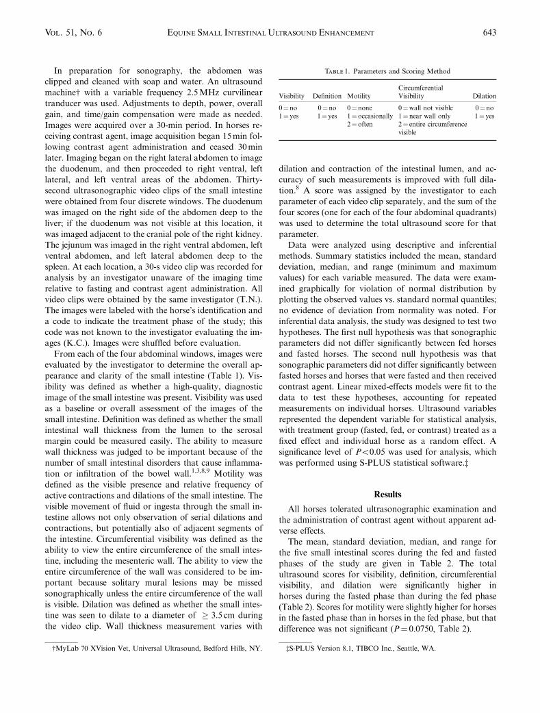
The mean, standard deviation, median, and range for
the five small intestinal scores during the fed and fasted
phases of the study are given in Table 2. The total
ultrasound scores for visibility, definition, circumferential
visibility, and dilation were significantly higher in
horses during the fasted phase than during the fed phase
(Table 2). Scores for motility were slightly higher for horses
in the fasted phase than in horses in the fed phase, but that
difference was not significant (P¼ 0.0750, Table 2).
Table 1. Parameters and Scoring Method
Visibility Definition MotilityCircumferentialVisibility Dilation
0¼no 0¼no 0¼ none 0¼wall not visible 0¼no1¼ yes 1¼ yes 1¼ occasionally 1¼near wall only 1¼ yes
2¼ often 2¼ entire circumferencevisible
wMyLab 70 XVision Vet, Universal Ultrasound, Bedford Hills, NY. zS-PLUS Version 8.1, TIBCO Inc., Seattle, WA.
643EQUINE SMALL INTESTINALULTRASOUND ENHANCEMENTVol. 51, No. 6

The mean, standard deviation, median, and range for
the five small intestinal scores of horses during the fasted
and contrast agent phases of the study are given in Table 3.
When the total ultrasound scores of small intestinal images
from the fasting and contrast agent phases were compared,
total ultrasound scores for visibility, definition, motility,
and circumferential visibility tended to be higher for horses
when they received contrast agent, but these differences
were not significant (Table 3). Total scores for dilation
were similar for the two groups (Table 3).
Discussion
In healthy adult horses, withholding feed for 24h sig-
nificantly improved the total scores for four clinically im-
portant parameters relating to ultrasonographic image
quality of the small intestine.
That at least 8 h of fasting improved the sonographic
visibility of the small intestine in healthy horses has been
documented.2 The mechanisms by which fasting enhances
the quality of small intestinal sonographic images are not
clearly understood, but it is likely that withholding feed
reduces the amount of ingesta and gas in the large colon
and cecum, thus allowing unobstructed penetration of
sound waves to the small intestine in the ventral abdomen.
It is also possible that emptying of the small intestine re-
duces intraluminal gas by reducing fermentable substrate,
thus improving circumferential visibility. In contrast to the
previously reported study,2 we did not find significant re-
duction in small intestinal movement in fasted horses. In
fact, although the difference was not statistically significant,
motility scores were higher during the fasted phase vs. the
fed phase. The reason for this difference between our study
and those previously reported is unknown, but may be due
to differences in the duration of fasting or the methods
employed to assess images between the two studies.
When water and mineral oil were administered intragas-
trically as a contrast agent, total ultrasound scores for each
parameter were not significantly improved when compared
with total scores of horses that were fasted alone. Never-
theless, there were some positive aspects of contrast agent
administration. For visibility, definition, motility, and
circumferential visibility, scores for the contrast agent
phase were higher than those for the fasted phase, but
not statistically significant. It is unclear why the adminis-
tration of contrast agent did not significantly improve the
ultrasound parameters. The small number of horses likely
played a role. Assessing more horses may have resulted in
statistical significance for some of the parameters.
Another possibility was that the contrast agent was not
suitable to provide sufficient intraluminal contrast com-
pared with intestinal fluid, provided endogenously or via
recent drinking of water, by the fasting horses. In humans,
considerable effort has been directed at identifying the ideal
contrast agent for intestinal sonography.4,5 The ideal con-
trast agent should be hypoechoic, nonabsorbable, non-
foaming, isotonic, and have the ability to adsorb and
displace intraluminal gas.4–7 Although water and orange
juice have been used to provide intraluminal contrast
in people,7 inconsistent results have been obtained,
Table 2. Fasted vs. Fed: Total (Summed) Scores of Ultrasonographic and Corresponding P-Values for Ultrasonographic Video Clips ObtainedTransabdominally in Fed vs. Fasted Conditions in 10 Adult Horses
Fed Fasted
P-ValueMean SD Median Range Mean SD Median Range
Visibility 1.0 0.816 1.0 0–2 2.2 1.033 2.0 0–4 0.013�
Definition 1.0 0.816 1.0 0–2 2.2 1.033 2.0 0–4 0.013�
Motility 3.3 1.494 3.5 1–5 4.5 1.716 4.5 2–8 0.075Circumferential Visibility 4.6 1.174 5.0 3–7 5.9 1.370 6.0 3–8 0.033�
Dilation 0.9 0.737 1.0 0–2 2.0 1.054 2.0 0–4 0.0115�
�Statistically significant difference.SD, standard deviation of the mean.
Table 3. Fasted vs. Contrast: Total (Summed) Scores and Corresponding P-Values for Ultrasonographic Video Clips Obtained Transabdominallyin Fasted vs. Contrast Agent Conditions in 10 Adult Horses
Fasted Contrast Agent
P-ValueMean SD Median Range Mean SD Median Range
Visibility 2.2 1.033 2 0–4 2.7 0.949 3 1–4 0.2887Definition 2.2 1.033 2 0–4 2.5 0.850 2.5 1–4 0.4679Motility 4.5 1.716 4.5 2–8 5.6 1.174 6 4–8 0.1022Circumferential Visibility 5.9 1.370 6 3–8 6.3 1.059 6.5 5–8 0.4620Dilation 2.0 1.054 2 0–4 2.0 0.816 2 1–3 1.000
SD, standard deviation of the mean.
644 NORMAN, CHAFFIN, and SCHMITZ 2010
presumably because of rapid transit, absorption, and in-
consistent displacement of intraluminal gas.4–7 Polyethyl-
ene glycol, methylcellulose, simethicone, cellulose, and
simethicone-coated cellulose have also been used as hydro-
sonography contrast agents, with variable results.4,5,7 The
ideal contrast agent for the horse remains unknown. Most
agents used in humans have not been evaluated for safety
in the horse, and some have been associated with abdom-
inal pain and diarrhea in humans.6,7 We chose to use water
and mineral oil because these substances are anechoic and
are known to be well tolerated in horses. Water is sono-
graphically anechoic, but when mixed in the intestinal
lumen, traps microbubbles of gas, resulting in a mixed
echogenicity. Other limitations of using water alone are its
rapid transit through the gastrointestinal tract and its rapid
absorption. Mineral oil is also sonographically anechoic
and is not absorbed by the intestinal tract of horses, but
tends to trap microbubbles of gas even more so than water.
Mineral oil may reduce the small intestinal absorption of
water, thus enhancing the ability of water to provide con-
trast. However, the mixture of the two liquids in the lumen
of the intestine results in multiple echoic interfaces, with a
sonographic outcome of a mixed echogenicity. A uniform,
purely echo-poor contrast agent would be preferable for
imaging the equine small intestine. Safety trials in horses
are needed to determine which agents would be safe for this
purpose. Additionally, the volume of contrast agent ad-
ministered to horses in this study may have been insuffi-
cient for image enhancement. We used a total volume of 4 l
based on clinical experience. Perhaps, a larger volume
would have led to enhanced images, but this is not known.
Another explanation for the absence of significant find-
ings following contrast agent administration is the timing of
the ultrasound examinations. We chose to examine horses in
the contrast agent phase during the time period 15–45-min
postadministration of contrast agent in an attempt to allow
sufficient time for partial gastric emptying. It is likely that
gastric emptying and small intestinal transit time varies be-
tween horses and conditions, and thus we may not have
imaged every horse at the optimal time. It is unknown what
amount of delay, if any, would be the most appropriate
following the administration of intragastric contrast agent
to horses. In humans, immediate examination following
contrast agent administration is recommended.6,7
Additionally, it is possible that administration of con-
trast agent, regardless of the agent itself, volume, or timing
of ultrasonographic examination, simply does not enhance
the quality of ultrasound images of the small intestine of
horses over that achieved following fasting alone.
A limitation of this study was that the horses were ex-
amined using the same order of treatments. Horses were
scanned for the fed treatment phase as they entered the
study, and then had food withheld for 24h and reexamined
for the fasted phase. Immediately following this fasted ex-
amination, horses received the contrast agent and then
were rescanned 15min later. This order was chosen for
convenience, eliminating the need to fast the horses mul-
tiple times. However, some bias may have been introduced
using this same order in every horse. Similarly, during each
phase of the study, ultrasound examinations were per-
formed systematically in the same order; the right lateral
abdomen was examined first, followed by the right ventral,
left lateral, and left ventral abdomen. It is unknown
whether the uniform order of examination of the abdom-
inal quadrants introduced bias.
Although the investigator scoring the images was un-
aware of the phase of the study, the presence or absence of
contrast agent was likely obvious, thus allowing the study
phase of that image to be known. We suspect the impact of
this limitation is minimal even though intraluminal intes-
tinal fluid was clearly visible in the images because similar
contents are likely seen in individual horses, regardless of
whether they are fed, fasted, or received contrast agent.
Furthermore, when intraluminal fluid is identified sono-
graphically, its source cannot be determined.
The investigator obtaining the images was aware of the
phase of the study when the images were acquired. To
minimize any bias introduced in this way, we standardized
our method of performing the examinations and acquiring
video clips before starting the study. In addition, a strict
time limit (30min) was set for the acquisition of images to
yield consistent results. We acknowledge this limitation,
but feel that minimal bias was introduced.
Finally, the video clips may not have represented the
entire examination. Ultrasound scores were based strictly
upon images available on the 30-s video clips, even though
the actual examination included 7.5min per window. In
addition, the time limitations set for image acquisition do
not necessarily represent the conditions of a clinical exam-
ination. The investigator performing the examinations at-
tempted, as best as possible, to collect video clips that
effectively represented the findings of the entire examina-
tion. Thus, we believe this limitation also had minimal
influence.
Our results of this study demonstrate that fasting horses
for 24h significantly improves the ability to attain high-
quality diagnostic ultrasonographic images of the small
intestine. Fasting resulted in improved visibility, definition,
circumferential visibility, and dilation of the small intestine.
Administration of water and mineral oil as an intraluminal
contrast agent did not improve any of the ultrasound pa-
rameters significantly, although there were higher scores
for some parameters.
ACKNOWLEDGMENTS
The authors thank Noah Cohen for providing the statistical analysisfor this project, and Heather Quiram and Michael O’Connor for theirtechnical assistance.
645EQUINE SMALL INTESTINALULTRASOUND ENHANCEMENTVol. 51, No. 6

REFERENCES
1. Whitcomb MB. Equine abdominal ultrasound. Proceedings of the24th Annual ACVIM Forum, Louisville, KY, Richmond Hill, Ontario, CN:Content Management, May 31–June 3, 2006.
2. Mitchell CF, Malone ED, Sage AM, Nikisch K. Evaluation of gas-trointestinal activity patterns in healthy horses using B mode and Dopplerultrasonography. Can Vet J 2005;46:134–140.
3. Sage AM. New ultrasonographic approaches to chronic colic. Pro-ceedings of the 24th Annual ACVIM Forum, Louisville, KY, Richmond Hill,Ontario, CN: Content Management, May 31–June 3, 2006.
4. Lev-Toaff AS, Lnager JE, Rubin DL, et al. Safety and efficacy of anew oral contrast agent for sonography: a phase II trial. Am J Roentgenol1999;173:431–436.
5. Lund PJ, Fritz TA, Unger EC, Hunt RK, Fuller E. Cellulose as agastrointestinal contrast agent. Radiology 1992;185:783–788.
6. Maconi G, Radice E, Bareggi E, Porro GB. Hydrosonography of thegastrointestinal tract. Am J Roentgenol 2009;193:700–708.
7. Nylund K, Ødegaard S, Hausken T, et al. Sonography of the smallintestine. World J Gastroenterol 2009;15:1319–1330.
8. Fontaine GL, Hanson RR, Rodgerson DH, Steiger R. Ultrasound eva-luation of equine gastrointestinal disorders. Compendium 1999;21:253–262.
9. Whitcomb MB. Ultrasonographic evaluation of chronic equine ab-dominal pain. Proceedings of the North American Veterinary Conference,Orlando, FL, Gainesville, FL, Eastern States Veterinary Association, Jan-uary 13–17, 2007.
646 NORMAN, CHAFFIN, and SCHMITZ 2010