Effect of pterostilbene on palmitic acid-induced secretion ...
7
临床与病理杂志 J Clin Pathol Res 2021, 41(8) hps://lcbl.csu.edu.cn 1728 收稿日期 (Date of reception):2021–04–27 通信作者 (Corresponding author):杨永玉,Email: [email protected] 基金项目 (Foundation item):湖南省自然科学基金 (2019JJ50923);湖南省中医药管理局科研计划项目 (2020039)。is work was supported by the Natural Science Foundation of Hunan Province (2019JJ50923) and Hunan Provincial Administration of Traditional Chinese Medicine Scientific Research Project (2020039), China. · 论著· doi: 10.3978/j.issn.2095-6959.2021.08.002 View this article at: hps://dx.doi.org/10.3978/j.issn.2095-6959.2021.08.002 紫檀芪对棕榈酸诱导巨噬细胞炎症因子分泌的影响 陈卓 1 ,李子铭 1 ,杨永玉 2 (1. 中南大学湘雅三医院老年医学科,长沙 410013;2. 中南大学湘雅二医院药学部,长沙 410011) [摘 要] 目的:研究紫檀芪(pterostilbene ,PTE) 对棕榈酸(palmitic acid ,PA) 诱导巨噬细胞炎症因子分泌的 影响及可能机制。方法:采用MTT法检测PTE对巨噬细胞RAW264.7 活性的影响;先用不同浓度的 PTE预处理RAW264.7 ,后用PA处理,ELISA法检测巨噬细胞培养上清液中炎症因子IL-18 、IL-1β和 TNF-α 水平,采用蛋白质印迹法检测巨噬细胞中NLRP3 、凋亡相关斑点样蛋白(apoptosis-associated speck-like protein containing CARD,ASC) 、半胱天冬酶1(caspase-1) 、核转录因子-κB p65(NF-κB p65) 、p-NF-κB p65 的表达。结果:与对照组比较,0~200 μmol/L 浓度的PTE 对巨噬细胞的活力无 影响。50 μmol/L和100 μmol/L的PTE可显著抑制PA诱导的巨噬细胞炎症因子IL-18 、IL-1β 和TNF-α 水平的升高,降低PA 诱导巨噬细胞中NLRP3 、ASC、caspase-1 蛋白表达的升高。PTE 可减少PA诱 导胞质中p-IKK和p-NF-κB p65 表达的升高,以及IκBα 表达的降低。PTE 还可降低PA诱导的p-NF-κB p65 的核转位。结论:PTE可抑制PA诱导巨噬细胞炎症因子表达的升高,其机制可能与其抑制炎症 小体活化的通路有关。 [关键词] 紫檀芪;巨噬细胞;炎症小体;核转录因子-κB Effect of pterostilbene on palmitic acid-induced secretion of inflammatory factors in macrophages CHEN Zhuo 1 , LI Ziming 1 , YANG Yongyu 2 (1. Department of Geriatrics, ird Xiangya Hospital, Central South University, Changsha 410013; 2. Department of Pharmacy, Second Xiangya Hospital, Central South University, Changsha 410011, China) Abstract Objective: To study the effect of pterostilbene (PTE) on palmitic acid (PA)-induced inflammatory factor secretion in macrophages and its possible mechanism. Methods: M method was used to detect the effect of pterostilbene on the activity of W264.7 cells. W264.7 cells were pretreated with different concentrations of pterostilbene and then treated by PA. ELISA kit was used to detect the levels of IL-18, IL-1β and TNF-α in the supernatant of macrophages, and Western bloing was used to detect the expression of NLRP3, apoptosis-
Transcript of Effect of pterostilbene on palmitic acid-induced secretion ...

临床与病理杂志J Cl in Path ol R e s 2021, 41(8) https://lcbl.csu.edu.cn
1728
收稿日期 (Date of reception):2021–04–27
通信作者 (Corresponding author):杨永玉,Email: [email protected]
基金项目 (Foundation item):湖南省自然科学基金 (2019JJ50923);湖南省中医药管理局科研计划项目 (2020039)。This work was supported by
the Natural Science Foundation of Hunan Province (2019JJ50923) and Hunan Provincial Administration of Traditional Chinese Medicine Scientific Research
Project (2020039), China.
·论著· doi: 10.3978/j.issn.2095-6959.2021.08.002View this article at: https://dx.doi.org/10.3978/j.issn.2095-6959.2021.08.002
紫檀芪对棕榈酸诱导巨噬细胞炎症因子分泌的影响
陈卓1,李子铭1,杨永玉2
(1. 中南大学湘雅三医院老年医学科,长沙 410013;2. 中南大学湘雅二医院药学部,长沙 410011)
[摘 要] 目的:研究紫檀芪(pterostilbene,PTE)对棕榈酸(palmitic acid,PA)诱导巨噬细胞炎症因子分泌的
影响及可能机制。方法:采用MTT法检测PTE对巨噬细胞RAW264.7活性的影响;先用不同浓度的
PTE预处理RAW264.7,后用PA处理,ELISA法检测巨噬细胞培养上清液中炎症因子IL -18、IL -1β和
TNF-α水平,采用蛋白质印迹法检测巨噬细胞中NLRP3、凋亡相关斑点样蛋白(apoptosis-associated
speck-like protein containing CARD,ASC)、半胱天冬酶1(caspase-1)、核转录因子-κB p65(NF-κB
p65)、p-NF-κB p65的表达。结果:与对照组比较,0~200 μmol/L浓度的PTE对巨噬细胞的活力无
影响。50 μmol/L和100 μmol/L的PTE可显著抑制PA诱导的巨噬细胞炎症因子IL -18、IL -1β和TNF-α
水平的升高,降低PA诱导巨噬细胞中NLRP3、ASC、caspase-1蛋白表达的升高。PTE可减少PA诱
导胞质中p-IKK和p-NF-κB p65表达的升高,以及IκBα表达的降低。PTE还可降低PA诱导的p-NF-κB
p65的核转位。结论:PTE可抑制PA诱导巨噬细胞炎症因子表达的升高,其机制可能与其抑制炎症
小体活化的通路有关。
[关键词] 紫檀芪;巨噬细胞;炎症小体;核转录因子-κB
Effect of pterostilbene on palmitic acid-induced secretion of inflammatory factors in macrophages
CHEN Zhuo1, LI Ziming1, YANG Yongyu2
(1. Department of Geriatrics, Third Xiangya Hospital, Central South University, Changsha 410013; 2. Department of Pharmacy,
Second Xiangya Hospital, Central South University, Changsha 410011, China)
Abstract Objective: To study the effect of pterostilbene (PTE) on palmitic acid (PA)-induced inflammatory factor
secretion in macrophages and its possible mechanism. Methods: MTT method was used to detect the effect of
pterostilbene on the activity of RAW264.7 cells. RAW264.7 cells were pretreated with different concentrations
of pterostilbene and then treated by PA. ELISA kit was used to detect the levels of IL-18, IL-1β and TNF-α in
the supernatant of macrophages, and Western blotting was used to detect the expression of NLRP3, apoptosis-

紫檀芪对棕榈酸诱导巨噬细胞炎症因子分泌的影响 陈卓,等 1729
肥胖是一种慢性低级别炎症反应,低度炎症
与 机 体 代 谢 紊 乱 以 及 其 他 的 慢 性 疾 病 的 发 生 有
关 [ 1 ]。 肥胖发生时,脂肪组织中巨噬细胞和游离
自由脂肪酸(free fatt y acid,FFA)的含量增加 [2]。
研究 [3]表明:M1型巨噬细胞是脂肪组织中炎症因
子的主要来源细胞;FFA中的饱和脂肪酸如棕榈酸
(palmitic acid,PA)可激活M1型巨噬细胞,增加机
体的炎症反应 [ 4 ]。因此,降低PA诱导巨噬细胞炎
症因子分泌对肥胖等代谢性炎症及其并发症有保
护作用。
紫檀芪( pterost i lbene,P TE)是从葡萄、蓝莓
和花生中提取的二苯乙烯类化合物,是一种天然
抗氧化剂,与白藜芦醇相比,具有较高的生物利
用度 [ 5 ]。研究 [ 6 ]发现: P T E 对细胞炎症、氧化应
激和细胞凋亡有抑制作用。P T E能够调节能量摄
入、改善脂肪细胞功能和脂肪组织炎症,干预肥
胖的发生 [ 7 ]。在T N F- α诱导的3 T 3 -L 1脂肪细胞炎
症模型 [8]中,PTE通过抑制核转录因子-κB(nuclear f a c to r o f κ B,N F- κ B)和核转录因子 - κ B抑制蛋白
(inhibitor of NF-κB,IκB)的磷酸化,降低炎症因子
如COX-2、iNOS、IL -6、IL -1β、IL -12的表达。但
目前尚未有研究探讨过P TE对PA诱导巨噬细胞炎
症因子产生的作用。因此,本文研究P TE对PA诱
导巨噬细胞炎症因子分泌的影响,探讨PTE在肥胖
等代谢性炎症中的可能药理机制。
1 材料与方法
1.1 材料
R AW264.7巨噬细胞购自中国科学院上海细胞
库;PA(P5585)、DMEM高糖培基(D6429)、牛血
清白蛋白(SRE0098)和胎牛血清(F8687)购自美国
Sigma公司;PTE(P108000)购自上海阿拉丁试剂有
限公司;RIPA裂解液(P0013C)、BCA法蛋白定量
试剂盒 ( P 0 0 1 0 S) 、核蛋白提取试剂盒 ( P 0 0 2 7 ) 以
及 I L - 1 8 E L I S A检测试剂盒(P I 5 5 3 )均购自上海碧
云天生物技术公司;anti-NLRP3(BA3677)、anti-c a s p a s e - 1 ( B A 2 2 2 0 ) 、 a n t i - L a m i n B ( P B 9 6 1 1 ) 、
I L - 1 β ( E K 0 3 9 4 ) 和 T N F - α ( E K 0 5 2 7 ) E L I S A 检 测
试 剂 盒 购 自 武 汉 博 士 德 生 物 公 司 ; β - a c t i n 抗 体
( B M 0 6 2 7 ) 、辣根过氧化物酶结合羊抗兔或羊抗
鼠 二 抗 购 自 北 京 中 杉 金 桥 生 物 技 术 有 限 公 司 ;
a n t i - p - I K K (s c - 2 1 6 6 1 ) 购自美国 S a n t a c r u z 公司;
p -N F- κ B p 6 5 (ab 7 6 3 0 2 )、ant i -A SC (ab 1 7 5 4 4 9 )、
anti-IκBα(ab32518)抗体购自英国Abcam公司。
1.2 方法
1.2.1 细胞培养
使 用 含 1 0 % 胎 牛 血 清 、 1 0 0 μ g / m L 链 霉 素 和 100 U/mL青霉素的DME M培养基,在37 ℃、5% CO2培养箱中常规培养RAW264.7细胞。当细胞生长
至融合度为80%时传代,取对数生长期的细胞用于
实验。
1.2.2 MTT 法检测 PTE 对细胞活性的影响
将 R AW 2 6 4 . 7 细胞 ( 密度约为 1 × 1 0 4个 / m L) 接
种至96孔板,200 μL/孔。设溶媒对照组与加药组
(12.5、25、50、100、200 μmol/L),每个组设6个
复孔。用二甲基亚砜(dimethyl sulfox ide,DMSO)溶解药物(DMSO含量<0.1%)。用不同浓度(12.5、
2 5、5 0、1 0 0、2 0 0 μ m o l / L)的P T E处理细胞2 4 h后 , 弃 上 清 液 , 每 孔 加 2 0 0 μ L 含 1 0 % 的 M T T 溶
液,继续孵育4 h后,弃上清液,每孔加200 μL的
DMSO,振荡溶解。测定490 nm处吸光度,计算细
胞的相对活力。
1.2.3 PTE 对炎症因子分泌的影响
将 R AW 2 6 4 . 7 细胞 ( 密度约为 1 × 1 0 4个 / m L) 接
associated speck-like protein containing CARD (ASC), caspase-1, Nuclear factor of κB p65 (NF-κB p65),
p-NF-κB p65 in macrophages. Results: Compared with the control group, MTT test results showed that PTE
at a concentration of 0–200 μmol/L had no effect on the activity of macrophages. PTE at 50 and 100 μmol/L
could significantly inhibit the PA-induced the expression of IL-18, IL-1β and TNF-α, and reduce PA-induced
the expression of NLRP3, ASC, caspase-1 in macrophages. Meanwhile, PTE inhibited the expression of p-IKK
and p-NF-κB p65, and reduced the decrease of the expression of IκBα in cytoplasm of PA-induced RAW264.7
cells. PTE can also reduce the nuclear translocation of p-NF-κB p65 induced by PA. Conclusion: Pterostilbene
can inhibit PA-induced increase of inflammatory factor expression in macrophages, and its mechanism may be
related to its inhibition of the pathway of inflammasomes.
Keywords pterostilbene; macrophages; inflammasome; nuclear factor of κB

临床与病理杂志, 2021, 41(8) https://lcbl.csu.edu.cn1730
种 于 6 孔 板 中 , 常 规 培 养 1 2 h 。 设 立 溶 媒 对 照 组
( 1 0 % B S A )、PA组和药物组,细胞用药物预孵育
12 h后,PA处理24 h。收集上清液用ELISA法测定
炎症因子IL -1β、IL -18以及TNF-α表达,收集细胞
测定炎症小体相关蛋白NLRP3、ASC、caspase-1、
p-IKK、p-NF-κB p65和IκBα的表达。
1.2.4 PA-BSA 溶液配制
用少量乙醇溶解PA后,加入10%的无FFA-BSA溶液,65 ℃水浴加热溶解,过滤、备用。
1.2.5 ELISA 法检测炎症因子
严格按照 E L I S A 试剂盒说明书操作,吸取适
量各组上清液,依次加入生物素抗原以及亲和素
抗原,在37 ℃培养箱中孵育60 min,最后依次加
入显色剂、终止液,于450 nm波长下进行测定OD值。绘制标准曲线并求得的回归方程,计算各组
上清液中IL -1β、IL -18以及TNF-α的含量。
1.2.6 蛋白质印迹法
提取细胞中的总蛋白,离心后取上清液加入
5 ×上样缓冲液,再于沸水中煮沸变性。用1 0 %的
S D S - PA G E 凝胶分离蛋白质,采用湿法转膜的方
法将蛋白转移至P V D F膜上。用5 %脱脂奶粉封闭
1 h后,加入NLR P3(1:500)、caspase-1(1:500)、
p - I K K ( 1 : 5 0 0 ) 、 p - N F - κ B p 6 5 ( 1 : 5 0 0 ) 、
I κ B α ( 1 : 5 0 0 )、A S C ( 1 : 5 0 0 )、β - a c t i n ( 1 : 1 0 0 0 )或
L amin B(1:500)一抗,于4 ℃孵育过夜,TBST清
洗 , 分 别 加 入 相 应 的 二 抗 : H R P 标 记 的 羊 抗 鼠
IgG(1:5 000)和HRP标记的羊抗兔IgG(1:5 000),室
温孵育1 h,TBST洗净,ECL显影液(A液:B液=1:1)曝光显影。Image J图像分析系统对光密度进行分
析。目的蛋白表达量=目的蛋白的灰度值/内参蛋
白的灰度值。
1.2.7 细胞核蛋白提取 严 格 依 据 试 剂 盒 说 明 书 进 行 提 取 , 收 集 细
胞,加入适量抽提试剂 A ,涡旋后冰浴 2 0 m i n ,
加入抽提试剂B,冰浴1 min,于4 ℃下以12 000×g的 速 度 离 心 5 m i n 。 留 取 沉 淀 , 加 入 适 量 细 胞 核
蛋白抽提试剂,涡旋后冰浴3 0 m i n,于4 ℃下以 12 000×g离心10 min,测定浓度,变性,备用。
1.2.8 免疫荧光分析
处理RAW 264.7细胞后弃上清,用PBS洗3次后
用4 %多聚甲醛固定15 min。加入PBS-T溶液,室温
孵育10 min,后用PBS-B溶液封闭1 h。加入NF-κB
p65抗体,于4 ℃下过夜。加入抗兔Cy3二抗孵育 1 h,后用DAPI染核10 min,在荧光显微镜下观察
拍照。
1.3 统计学处理
使 用 S P S S 2 1 . 0 软 件 进 行 数 据 分 析 , 数 据 以x±SE M描述,多个样本均数间的比较采用单因素
方差分析,P<0.05为差异有统计学意义。
2 结果
2 .1 PTE 减少 PA 诱导巨噬细胞炎性因子分泌的
升高
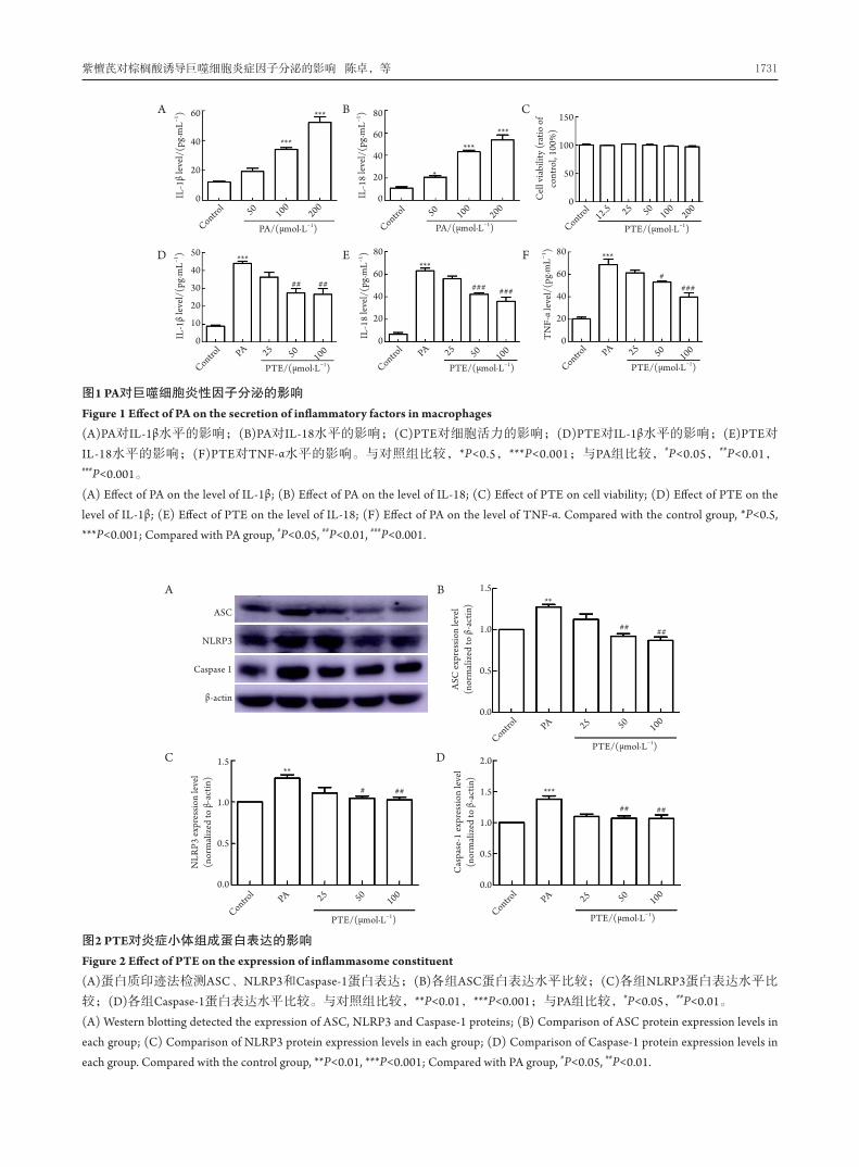
用不同浓度的PA处理细胞24 h,100 μmol/L和 2 0 0 μ m o l / L P A 可 显 著 增 加 炎 症 因 子 的 分 泌 (图1A、1B)。MTT结果显示:与对照组相比,不
同浓度的PTE对细胞活力没有影响(图1C)。用不同
浓度的PTE预处理细胞12 h后,用200 μmol/L PA处
理24 h。与对照组相比,PA可显著增加巨噬细胞炎
症因子的分泌,50 μmol/L和100 μmol/L PTE可显
著降低PA诱导的巨噬细胞炎症因子IL -1β、IL -18以
及TNF-α表达的升高(图1D~1F)。
2.2 PTE 降低 PA 诱导 NLRP3、ASC 和 caspase-1表达的升高
与对照组相比,PA增加巨噬细胞中炎症小体
相关蛋白NLR P3、A SC和caspase-1的表达;与PA组比,PTE 50 μmol/L和100 μmol/L均能显著降低
NLRP3、ASC和caspase-1蛋白的表达(图2)。
2.3 PTE 降低 PA 诱导细胞质中 NF-κB p65 的活化
与对照组相比, PA 诱导 R AW 2 6 4 . 7 细胞质中
p-IKK和p-NF-κB p65表达升高,降低IκBα的表达;
而PTE可减少PA诱导胞质中p-IKK和NF-κB p65蛋白
的表达,增加IκBα的表达(图3)。
2.4 PTE 降低 PA 诱导 p-NF-κB p65 的核转位
与对照组相比,PA诱导巨噬细胞胞核中p-NF-κ B p 6 5 表 达 升 高 ; 而 P T E 可 抑 制 巨 噬 细 胞 胞 核
p-NF-κB p65的表达。免疫荧光显示:不同浓度的
P T E 可减少PA 诱导N F - κ B p 6 5在细胞核中的表达 (图4)。
紫檀芪对棕榈酸诱导巨噬细胞炎症因子分泌的影响 陈卓,等 1731
图1 PA对巨噬细胞炎性因子分泌的影响
Figure 1 Effect of PA on the secretion of inflammatory factors in macrophages
(A)PA对IL-1β水平的影响;(B)PA对IL-18水平的影响;(C)PTE对细胞活力的影响;(D)PTE对IL-1β水平的影响;(E)PTE对
IL-18水平的影响;(F)PTE对TNF-α水平的影响。与对照组比较,*P<0.5,***P<0.001;与PA组比较,#P<0.05,##P<0.01,###P<0.001。
(A) Effect of PA on the level of IL-1β; (B) Effect of PA on the level of IL-18; (C) Effect of PTE on cell viability; (D) Effect of PTE on the
level of IL-1β; (E) Effect of PTE on the level of IL-18; (F) Effect of PA on the level of TNF-α. Compared with the control group, *P<0.5,
***P<0.001; Compared with PA group, #P<0.05, ##P<0.01, ###P<0.001.
图2 PTE对炎症小体组成蛋白表达的影响
Figure 2 Effect of PTE on the expression of inflammasome constituent
(A)蛋白质印迹法检测ASC、NLRP3和Caspase-1蛋白表达;(B)各组ASC蛋白表达水平比较;(C)各组NLRP3蛋白表达水平比
较;(D)各组Caspase-1蛋白表达水平比较。与对照组比较,**P<0.01,***P<0.001;与PA组比较,#P<0.05,##P<0.01。
(A) Western blotting detected the expression of ASC, NLRP3 and Caspase-1 proteins; (B) Comparison of ASC protein expression levels in
each group; (C) Comparison of NLRP3 protein expression levels in each group; (D) Comparison of Caspase-1 protein expression levels in
each group. Compared with the control group, **P<0.01, ***P<0.001; Compared with PA group, #P<0.05, ##P<0.01.
Control
Control
Control
PA
PAPA
25
2525
50
5050
PTE/(μmol·L−1)
PTE/(μmol·L−1)PTE/(μmol·L−1)
**
**
***
##
#
## ##
##
##
100
100100
ASC
NLRP3
Caspase 1
β-actin
1.5
1.0
0.5
0.0
2.0
1.5
1.0
0.5
0.0
1.5
1.0
0.5
0.0
NLR
P3 e
xpre
ssio
n le
vel
(nor
mal
ized
to β
-act
in)
ASC
exp
ress
ion
leve
l (n
orm
aliz
ed to
β-a
ctin
)C
aspa
se-1
exp
ress
ion
leve
l (n
orm
aliz
ed to
β-a
ctin
)
A
C
B
D
Control
Control
Control
Control
Control
Control
PAPAPA 252525
50 5012.5
505050
100100 25
PTE/(μmol·L−1)
PTE/(μmol·L−1)
PTE/(μmol·L−1)PTE/(μmol·L−1)
PA/(μmol·L−1)
***
******
***
***
***
*
## ####
### #####
***
PA/(μmol·L−1)
100100
100
200200
20010050
60
40
20
0
50
40
30
20
10
0
80
60
40
20
0
150
100
50
0
80
60
40
20
0
80
60
40
20
0
IL-1
β le
vel /
(pg ·
mL− 1
)IL
-1β
leve
l/(p
g·m
L−1)
IL-1
8 le
vel /
(pg ·
mL− 1
)
Cel
l via
bilit
y (r
atio
of
cont
rol,
100%
)T
NF-
α le
vel/
(pg ·
mL− 1
)
IL-1
8 le
vel /
(pg ·
mL− 1
)
A
D
C
F
B
E

临床与病理杂志, 2021, 41(8) https://lcbl.csu.edu.cn1732
图3 PTE对PA诱导的胞质NF-κB p65活性的影响
Figure 3 Effect of PTE on PA-induced NF-κB in cytoplasm (A)蛋白质印迹法检测p-NF-κB p65、p-IKK和IκBα蛋白表达;(B)各组p-NF-κB p65蛋白表达水平比较;(C)各组p-IKK蛋白表达水平比较;(D)各组IκBα蛋白表达水平比较。与对照组比较,**P<0.01;与PA组比较,#P<0.05,##P<0.01。(A) Western blotting detected the expression of p-NF-κB p65、p-IKKand IκBα proteins; (B) Comparison of p-NF-κB p65 protein expression levels in each group; (C) Comparison of p-IKK protein expression levels in each group; (D) Comparison of IκBα protein expression levels in each group. Compared with the control group, **P<0.01; Compared with PA group, #P<0.05, ##P<0.01.
图4 PTE对PA诱导p-NF-κB p65核转位的影响
Figure 4 Effect of PTE on PA-induced p-NF-κB p65 nuclear translocation (A)蛋白质印迹法检测胞核p-NF-κB p65蛋白表达;(B)各组p-NF-κB p65蛋白表达水平比较;(C)免疫荧光显示不同浓度PTE对PA诱导NF-κB p65在胞核中表达的影响。与对照组比较,**P<0.01;与PA组比较,#P<0.05,##P<0.01。(A) Western blotting detected the expression of p-NF-κB p65 proteins in nucleus; (B) Comparison of p-NF-κB p65 protein expression levels in each group; (C) Immunofluorescence showed Effect of different concentrations of PTE on the expression of NF-κB p65 in the nucleus induced by PA. Compared with the control group, **P<0.01; Compared with PA group, #P<0.05, ##P<0.01.
p-NF-κB p65
p-IKK
IκBα
β-actin
Control
**
**
**
#
#
# ##
## ## ##
####
Control
Control
PA
PAPA
25
2525
50
5050
PTE/(μmol·L−1)
PTE/(μmol·L−1)PTE/(μmol·L−1)
100
100100
2.0
1.5
1.0
0.5
0.0
2.0
1.5
1.0
0.5
0.0
1.5
1.0
0.5
0.0
p-N
F-κ B
p65
exp
ress
ion
leve
l (n
orm
aliz
ed to
β-
actin
)
IκBα
exp
ress
ion
leve
l (n
orm
aliz
ed to
β-a
ctin
)
p-IK
K e
xpre
ssio
n le
vel
(nor
mal
ized
to β
-act
in)
A
C D
B
p-NF-κB p65
Lamin B
Control PA 25 50
PTE/(μmol·L−1)
PTE
(25 μmol/L)
PTE
(100 μmol/L)
PTE
(50 μmol/L)
对照组 PA
50 μm
50 μm 50 μm
50 μm 50 μm
100
2.5
2.0
1.5
1.0
0.5
0.0p-N
F-κ B
p65
exp
ress
ion
leve
l (n
orm
aliz
ed to
β-a
ctin
) **
# ##
A
C
B

紫檀芪对棕榈酸诱导巨噬细胞炎症因子分泌的影响 陈卓,等 1733
3 讨论
FFA是脂肪细胞中三酰甘油的主要组成部分,
也是机体主要能量来源之一。FFA包括饱和脂肪酸
和不饱和脂肪酸,通常饱和脂肪酸诱导巨噬细胞
产生炎症反应[9],而不饱和脂肪酸(如油酸)具有抑
制炎症的作用 [10]。低度炎症与机体代谢紊乱以及
其他的慢性疾病的发生有关,肥胖或胰岛素抵抗
时,脂解反应增加,循环系统或脂肪组织中饱和
脂肪酸PA的浓度显著升高 [11]。PA可诱导巨噬细胞
炎症因子分泌增加,导致炎症反应增加 [12]。因此
降低PA诱导的巨噬细胞炎症因子分泌增加对肥胖
相关的代谢性疾病具有益处。本研究结果显示:
与对照组相比,50 μmol/L或100 μmol/L浓度的PTE可显著降低 PA 诱导的巨噬细胞炎症因子 I L - 1 β 、
IL -18和TNF-α的升高,提示PTE对改善代谢性炎症
有积极作用。
炎性小体在 I L - 1 β、 I L - 1 8等促炎性细胞因子
和 趋 化 因 子 的 释 放 中 起 关 键 作 用 [ 1 3 ]。 N L R P 3 是
NOD样受体(NOD -l i ke receptor,NLR)家族成员
之一,其与A SC和前胱天蛋白酶-1( pro-caspase-1)形 成 多 种 蛋 白 复 合 物 的 炎 症 小 体 [ 1 3 ]。 N L R P 3 炎
症小体激活后可活化caspase-1,活化的caspase-1裂解前 I L - 1 β 和前 I L - 1 8 生成,并释放 I L - 1 β 和 I L -18,从而诱导炎症反应 [14]。在肥胖小鼠脂肪组织
中,NLR P3的活性明显升高且与胰岛素抵抗的发
生密切相关 [15]。PA可激活内皮细胞NLRP3炎症小 体 [16],脂多糖可通过TLR/NF-κB激活NLRP3 [17]。
本 研 究 结 果 显 示 : P T E 可 降 低 PA 诱 导 巨 噬 细 胞
NLRP3、ASC和caspase-1的表达升高,提示PTE可
抑制PA诱导的NLRP3炎症小体激活。在正常情况
下,NF-κB二聚体与IκB家族构成三聚体而存在于
细胞质中,当细胞受到外界因素,如PA或炎症细胞
因子、脂多糖等刺激后,激活核转录因子-κB抑制
蛋白激酶(IκB kinase,IKK),使IκB发生磷酸化并迅
速降解,NF-κB被释放,磷酸化后进入细胞核,与
特异性DNA位点结合,进行基因转录调节[18]。已有
研究[19]发现:在脓毒血症时,脂多糖可通过NF-κB通路活化N L R P 3炎症小体。我们测定了P T E对胞
质以及胞核中NF-κB的影响,结果显示:PTE明显
降低细胞质中PA诱导的p-NF-κB p65的表达,说明
PTE抑制了PA诱导的胞质中NF-κB p65的活化;同
时也降低细胞核中p-NF-κB p65的表达,说明PTE抑制了PA诱导NF-κB p65的核转位。说明PTE可能
通过NF-κB/NLRP3信号通路抑制NLRP3炎症小体
激活,从而发挥抗炎作用,但其具体机制需要进
一步研究。
综上,本文研究了PTE对巨噬细胞炎症因子分
泌的影响,发现P TE可明显降低PA诱导巨噬细胞
炎症因子的分泌,其机制可能与其抑制炎症小体
活化通路有关。
参考文献
1. Amin MN, Hussain MS, Sarwar MS, et al. How the association between
obesity and inflammation may lead to insulin resistance and cancer[ J].
Diabetes Metab Syndr, 2019, 13(2): 1213-1224.
2. Kim SJ, Feng D, Guillot A, et al. Adipocyte death preferentially induces
liver injury and inflammation through the activation of chemokine (C-C
Motif) receptor 2-positive macrophages and lipolysis[ J]. Hepatology,
2019, 69(5): 1965-1982.
3. Engin AB, Engin A, Gonul II, et al. The effect of adipocyte-macrophage
crosstalk in obesity-related breast cancer[ J]. J Mol Endocrinol, 2019,
62(3): R201-R222.
4. Sunil Gowda SN, Raviraj R, Nagarajan D, et al. Radiation-induced lung
injury: impact on macrophage dysregulation and lipid alteration - a
review[ J]. Immunopharmacol Immunotoxicol, 2019, 41(3): 370-379.
5. Estrela JM, Ortega A, Mena S, et al. Pterostilbene: Biomedical
applications[ J]. Crit Rev Clin Lab Sci, 2013, 50(3): 65-78.
6. Remsberg CM, Yáñez JA, Ohgami Y, et al. Pharmacometrics of
pterostilbene: preclinical pharmacokinetics and metabolism, anticancer,
antiinflammatory, antioxidant and analgesic activity[ J]. Phytother Res,
2008, 22(2): 169-179.
7. Pan MH, Wu JC, Ho CT, et al. Antiobesity molecular mechanisms of
action: resveratrol and pterostilbene[ J]. Biofactors, 2018, 44(1): 50-60.
8. Hsu CL, Lin YJ, Ho CT, et al. The inhibitory effect of pterostilbene on
inflammatory responses during the interaction of 3T3-L1 adipocytes
and RAW 264.7 macrophages[ J]. J Agric Food Chem, 2013, 61(3):
602-610.
9. Korbecki J, Bajdak-Rusinek K . The effect of palmitic acid on
inflammatory response in macrophages: an overview of molecular
mechanisms[ J]. Inflamm Res, 2019, 68(11): 915-932.
10. Zeng X, Zhu M, Liu X, et al. Oleic acid ameliorates palmitic acid
induced hepatocellular lipotoxicity by inhibition of ER stress and
pyroptosis[ J]. Nutr Metab (Lond), 2020, 17: 11.
11. Kimura I, Ichimura A, Ohue-Kitano R, et al. Free fatty acid receptors in
health and disease[ J]. Physiol Rev, 2020, 100(1): 171-210.
12. Rosso C, Kazankov K, Younes R, et al. Crosstalk between adipose
tissue insulin resistance and liver macrophages in non-alcoholic fatty
liver disease[ J]. J Hepatol, 2019, 71(5): 1012-1021.
13. Zahid A, Li B, Kombe AJK, et al. Pharmacological Inhibitors of the

临床与病理杂志, 2021, 41(8) https://lcbl.csu.edu.cn1734
NLRP3 inflammasome[ J]. Front Immunol, 2019, 10: 2538.
14. Garrido W, Jara C, Torres A, et al. Blockade of the adenosine A3
receptor attenuates caspase 1 activation in renal tubule epithelial cells
and decreases interleukins IL-1β and IL-18 in diabetic rats[ J]. Int J Mol
Sci, 2019, 20(18): 4531.
15. Rheinheimer J, de Souza BM, Cardoso NS, et al. Current role of the
NLRP3 inflammasome on obesity and insulin resistance: A systematic
review[ J]. Metabolism, 2017, 74: 1-9.
16. Qi Y, Du X, Yao X, et al. Vildagliptin inhibits high free fatty acid (FFA)-
induced NLRP3 inflammasome activation in endothelial cells[ J]. Artif
Cells Nanomed Biotechnol, 2019, 47(1): 1067-1074.
17. Yu X, Lan P, Hou X, et al. HBV inhibits LPS-induced NLRP3
inflammasome activation and IL-1β production via suppressing the
NF-κB pathway and ROS production[ J]. J Hepatol, 2017, 66(4):
693-702.
18. Kanigur Sultuybek G, Soydas T, Yenmis G. NF-κB as the mediator of
metformin's effect on ageing and ageing-related diseases[ J]. Clin Exp
Pharmacol Physiol, 2019, 46(5): 413-422.
19. Lu o M , Yan D, Su n Q, e t a l . G i n s en o s i d e R g 1 attenu ate s
cardiomyocyte apoptosis and inflammation via the TLR4/NF-kB/
NLRP3 pathway[ J]. J Cell Biochem, 2020, 121(4): 2994-3004.
本文引用:陈卓, 李子铭, 杨永玉. 紫檀芪对棕榈酸诱导巨噬细
胞炎症因子分泌的影响[J]. 临床与病理杂志, 2021, 41(8): 1728-1734.
doi: 10.3978/j.issn.2095-6959.2021.08.002
Cite this article as: CHEN Zhuo, LI Ziming, YANG Yongyu. Effect of
pterostilbene on palmitic acid-induced secretion of inflammatory factors
in macrophages[ J]. Journal of Clinical and Pathological Research, 2021,
41(8): 1728-1734. doi: 10.3978/j.issn.2095-6959.2021.08.002