Effect of Cardiac Glycosides on Action Potential...
38
JPET/2003/049189—R2 1 Effect of Cardiac Glycosides on Action Potential Characteristics and Contractility in Cat Ventricular Myocytes: Role of Calcium Overload Stuart R. Ruch, Manabu Nishio, and J Andrew Wasserstrom Departments of Medicine (Cardiology) and Molecular Pharmacology and Biological Chemistry and the Feinberg Cardiovascular Research Institute, Northwestern University Medical School, Chicago IL Copyright 2003 by the American Society for Pharmacology and Experimental Therapeutics. JPET Fast Forward. Published on September 3, 2003 as DOI:10.1124/jpet.103.049189 This article has not been copyedited and formatted. The final version may differ from this version. JPET Fast Forward. Published on September 3, 2003 as DOI: 10.1124/jpet.103.049189 at ASPET Journals on May 1, 2020 jpet.aspetjournals.org Downloaded from
Transcript of Effect of Cardiac Glycosides on Action Potential...
JPET/2003/049189—R2
1
Effect of Cardiac Glycosides on Action Potential Characteristics
and Contractility in Cat Ventricular Myocytes: Role of Calcium Overload
Stuart R. Ruch, Manabu Nishio, and J Andrew Wasserstrom
Departments of Medicine (Cardiology) and Molecular Pharmacology and Biological Chemistry
and the Feinberg Cardiovascular Research Institute, Northwestern University Medical School,
Chicago IL
Copyright 2003 by the American Society for Pharmacology and Experimental Therapeutics.
JPET Fast Forward. Published on September 3, 2003 as DOI:10.1124/jpet.103.049189This article has not been copyedited and formatted. The final version may differ from this version.JPET Fast Forward. Published on September 3, 2003 as DOI: 10.1124/jpet.103.049189
at ASPE
T Journals on M
ay 1, 2020jpet.aspetjournals.org
Dow
nloaded from

JPET/2003/049189—R2
2
Running title: Cardiac Glycoside Effects on Cat Myocytes
Address for Correspondence:
J. Andrew Wasserstrom, PhD
Division of Cardiology – S203
Ward 3-105
Northwestern University Medical School
303 E. Chicago Ave.
Chicago IL 60611
(312) 503-1384 (voice)
(312) 503-3891 (fax)
email: [email protected]
Number of Text Pages: 17
Number of Tables: 1
Number of Figures: 9
Number of References: 27
Number of words in Abstract: 252
Number of words in Introduction: 423
Number of words in Discussion: 1407
List of Non-standard Abbreviations: NCX, sodium-calcium exchanger; RMP, resting membrane
potential; FxS, fractional shortening; AP, action potential; APD, action potential duration; APD50..
APD90, action potential duration at 50% (or 90%) of repolarization; ICa, calcium current (L-type);
IK1, inward rectifying potassium current; KHB, Krebs-Henseleit buffer; DHO, dihydroouabain;
HEPES, (N-[2-Hydroxyethyl]piperazine-N’-[2-ethanesulfonic acid]); EGTA,
This article has not been copyedited and formatted. The final version may differ from this version.JPET Fast Forward. Published on September 3, 2003 as DOI: 10.1124/jpet.103.049189
at ASPE
T Journals on M
ay 1, 2020jpet.aspetjournals.org
Dow
nloaded from

JPET/2003/049189—R2
3
Ethylenediaminetetraacetic acid; BAPTA, 1,2-bis(2-Aminophenoxy)ethane-N,N,N’,N’-tetraacetic
acid
Section Assignment: Cardiovascular
This article has not been copyedited and formatted. The final version may differ from this version.JPET Fast Forward. Published on September 3, 2003 as DOI: 10.1124/jpet.103.049189
at ASPE
T Journals on M
ay 1, 2020jpet.aspetjournals.org
Dow
nloaded from

JPET/2003/049189—R2
4
Abstract
There is increasing evidence that cardiac glycosides act through mechanisms distinct
from inhibition of the sodium pump but which may contribute to their cardiac actions. To more
fully define differences between agents indicative of multiple sites of action, we studied changes
in contractility and action potential (AP) configuration in cat ventricular myocytes produced by
six cardiac glycosides (ouabain, ouabagenin, dihydroouabain, actodigin, digoxin, and
resibufogenin). AP shortening was observed only with ouabain and actodigin. There was
extensive inotropic variability between agents, with some giving full inotropic effects before
automaticity occurred whereas other produced minimal inotropy before toxicity. AP shortening
was not a result of alterations in calcium current or the inward rectifier potassium current, but
correlated with an increase in steady state outward current (Iss), which was sensitive to KB-
R7943, a Na+-Ca2+ exchange (NCX) inhibitor. Interestingly, Iss was observed following exposure
to ouabain and dihydroouabain, suggesting that an additional mechanism is operative with
dihydroouabain that prevents AP shortening. Further investigation into differences in inotropy
between ouabagenin, dihydroouabain and ouabain revealed almost identical responses under
AP voltage clamp. Thus all agents appear to act on the sodium pump and thereby secondarily
increase the outward reverse mode NCX current, but the extent of AP duration shortening and
positive inotropy elicited by each agent is limited by development of their toxic actions. The
quantitative differences between cardiac glycosides suggest that mechanisms independent of
sodium pump inhibition may result from an altered threshold for calcium overload possibly
involving direct effects on calcium release from the sarcoplasmic reticulum.
This article has not been copyedited and formatted. The final version may differ from this version.JPET Fast Forward. Published on September 3, 2003 as DOI: 10.1124/jpet.103.049189
at ASPE
T Journals on M
ay 1, 2020jpet.aspetjournals.org
Dow
nloaded from

JPET/2003/049189—R2
5
IInnttrroodduuccttiioonn
The well-described action of cardiac glycosides to inhibit the Na+,K+-ATPase (Na+ pump,
sodium pump) is believed by many investigators to be the mechanism of central importance in
both inotropic and toxic effects of these agents (Akera et.al.,1970; Lee and Dagostino, 1982;
Gadsby et al., 1985; Steimers et al., 1990). Indeed, studies supporting the sodium pump
inhibition hypothesis are so frequent in the literature that the implication is that the action of
cardiac glycosides on the sodium pump is the only significant effect mediated by cardiac
glycosides in the myocardium. However, there is also substantial evidence that cardiac
glycosides may act at sites other than the sodium pump, and that these alternative sites may
account for important differences between agents. Support for alternative mechanisms
underlying inotropic and / or toxic effects of cardiac glycosides include findings that cardiac
glycoside analogs differ significantly in their effects on intracellular [Na+], action and resting
membrane potentials (Wasserstrom et al., 1991), and toxic to therapeutic ratios in both isolated
preparations (Karagueuzian and Katzung, 1981) and whole animals (Mendez et.al., 1974). In
addition, studies directed at defining alternative cardiac glycoside mechanisms have revealed
intracellular sites of action independent of the sodium pump, including the sarcoplasmic
reticulum (Fujino and Fujino, 1982; Isenberg, 1984) and its calcium release channel (Rardon
and Wasserstrom, 1990; McGarry and Williams, 1993; Sagawa et al., 2002).
Despite growing evidence in support of alternative mechanisms and their role in
determining differences in the effects of cardiac glycoside analogs, few studies have compared
cardiac glycoside analog actions using isolated cardiac myocytes. In contrast to multicellular
preparations, the use of single cardiac cells provides considerable advantages in evaluating
cardiac glycoside effects. Of key importance is the elimination of extracellular diffusion barriers
that confound measurement of membrane currents and other cellular processes dependent on
ion gradients. In addition, nearly all previous studies have used a variety of multicellular
preparations under an equally varied number of experimental conditions, making comparisons
This article has not been copyedited and formatted. The final version may differ from this version.JPET Fast Forward. Published on September 3, 2003 as DOI: 10.1124/jpet.103.049189
at ASPE
T Journals on M
ay 1, 2020jpet.aspetjournals.org
Dow
nloaded from

JPET/2003/049189—R2
6
between agents extremely difficult. The purpose of this study was to characterize the
electrophysiological and inotropic effects of different cardiac glycosides in isolated cardiac
myocytes in a standardized manner in order to determine if there are indeed differences in
cellular actions of these agents. Six cardiac glycoside analogs were chosen for study based on
specific differences in their molecular structures. The study was designed first to characterize
differences between these six cardiac glycoside analogs on action potential (AP) configuration
and contractility in isolated cat ventricular myocytes, and then to define the underlying
mechanisms that account for the differences in their actions.
This article has not been copyedited and formatted. The final version may differ from this version.JPET Fast Forward. Published on September 3, 2003 as DOI: 10.1124/jpet.103.049189
at ASPE
T Journals on M
ay 1, 2020jpet.aspetjournals.org
Dow
nloaded from

JPET/2003/049189—R2
7
Methods
Isolation of Cat Ventricular Myocytes:
After anesthesia by sodium pentobarbital (approx. 45 mg/kg, IP), the hearts from adult
cats were excised and perfused through the aorta and coronary arteries with Ca2+-free modified
Krebs Henseleit buffer (KHB) for 5min. The heart was then perfused with 0.08% collagenase in
KHB solution (Worthington CLS II) for 8-10 minutes at 36oC. Ventricular tissue was minced and
incubated in a shaker bath for 5 min in KHB-collagenase solution at 36oC. Cells were then filtered
(200 µm nylon sieve), washed free of collagenase and placed in KHB containing 1% albumin and
1mM Ca2+ for 15 min. Cells were stored in 1.8 mM Ca2+ Tyrode’s solution at room temperature
and were used up to 36 hours after isolation. Typical cell yields are 60-70% with this technique.
Only rod-shaped, Ca2+-tolerant myocytes with visible cross-striations were used for study.
Solutions and Drugs:
(Modified) Tyrode’s Solution (all concentrations in mM): 140 NaCl, 5.4 KCl, 0.5 MgCl2, 5
HEPES, 0.4 NaH2PO4, 11 glucose, 1.8 CaCl2, pH = 7.4 (NaOH). ICa Extracellular Solution: 0.2
BaCl2 added to Tyrode’s to block Ik1. Ramp Protocol Extracellular Solution: 0.1 CdCl2 added to
Tyrode’s solution to block ICa. High Resistance Microelectrode Solution: 500 KCl. Whole Cell
Pipette Solution: 10 NaCl, 120 K+ -aspartate, 25 KCl, 20 HEPES, 0.5 MgCl2, 4 K2ATP, 0.056 or
1.0 EGTA. Cardiac glycosides were purchased from Sigma (St Louis, MO) with the exception of
resibufogenin, which was obtained from Tientsin First Central Pharmaceutical (Tientsin, China).
cardiac glycoside stock solutions of 1-10 mM were made in distilled water (DHO, resibufogenin),
or 50% EtOH/water (ouabain, ouabagenin, actodigin, digoxin). KB-R7943 was a gift of Kanebo,
Ltd. (Osaka, Japan). In the experiments in which ICa was measured, a low concentration of Ba2+
(0.2mM) was sufficient to block background K+ current but had little effect on magnitude and
kinetics of ICa in the presence of 1.8mM CaCl2. We have used this approach previously (Nishio
This article has not been copyedited and formatted. The final version may differ from this version.JPET Fast Forward. Published on September 3, 2003 as DOI: 10.1124/jpet.103.049189
at ASPE
T Journals on M
ay 1, 2020jpet.aspetjournals.org
Dow
nloaded from

JPET/2003/049189—R2
8
et al., 2002) and have found it to be a useful means to measure ICa under conditions in which
normal physiological conditions are otherwise maintained (physiological [Ca2+], temperature, K+
gradient).
Molecular Structures of the Six Cardiac Glycosides Studied
To more accurately define differences between cardiac glycoside agents and further
evaluate cardiac glycoside structure-activity relationships, six cardiac glycosides (ouabain,
ouabagenin, dihydroouabain (DHO), actodigin, digoxin, and resibufogenin) were chosen for
electrophysiology and contractility studies in single cat ventricular myocytes. Figure 1 shows
the molecular structure of these agents. Of particular note, DHO and ouabagenin differ from the
prototypical analog ouabain by saturation of the lactone ring and omission of the carbohydrate
moiety, respectively. Actodigin differs from ouabain in the site of attachment of the lactone ring
at C-17 of the steroid nucleus. Resibufogenin, which unlike the other analogs is isolated from
an animal source (frog skin), has a unique 6-membered lactone ring at C-17, and also lacks the
sugar at C-3. Digoxin, chosen primarily for its importance in clinical medicine, shares many
similarities with ouabain but differs in the type of carbohydrate at C-3, and the number and
position of hydoxyl groups attached to the steroid nucleus, thereby making it the most
hydrophobic of the group.
Measurement of Voltage, Current, and Fractional Shortening in Single Ventricular Myocytes:
Several drops of cell suspension were placed in an experimental chamber (0.5 ml
volume) mounted on the stage of an inverted microscope (Nikon Diaphot). After allowing 10
minutes for the cells to adhere to the chamber bottom, they were superfused (2-3 ml/min) with
Tyrode's solution maintained at 36 + 1oC using a Peltier device.
For experiments which included measurement of fractional shortening, the video image
of the long axis of the cell was aligned with the rasters of the video edge detector (Crescent
This article has not been copyedited and formatted. The final version may differ from this version.JPET Fast Forward. Published on September 3, 2003 as DOI: 10.1124/jpet.103.049189
at ASPE
T Journals on M
ay 1, 2020jpet.aspetjournals.org
Dow
nloaded from

JPET/2003/049189—R2
9
Electronics) which continuously monitored cell length along a single line. Fractional shortening
was defined as the ratio of cell shortening to resting cell length. Intracellular access was then
obtained using either high-resistance microelectrodes or whole-cell patch pipettes.
High-Resistance Microelectrode Method: A microelectrode (15-30 MΩ resistance) was pressed
gently against the cell surface forming a relatively low-resistance seal. Access was then
obtained by briefly turning up the capacitance compensation, causing the amplifier circuitry to
ring. Immediately following access, negative holding current (~1nA) was applied to maintain a
resting membrane potential (RMP) of –70 to –80mV. A high resistance seal spontaneously
formed within a few minutes and RMP became more negative, indicating successful
impalement. Background holding current was then turned off. The primary advantage of this
technique is that it allows electrical access of the cell without appreciable diffusion between the
cytoplasm and the electrode solution. The result is greater stability in measurement of ICa and
contractility.
Whole-Cell Suction Pipette Method: A 2-3 MΩ pipette was pressed gently against the cell
surface and suction was applied (50-100 cm H2O), allowing formation of a gigaohm seal.
Application of a brief suction pulse then ruptured the membrane patch giving electrical and
physical access to the cell interior. Advantages of this technique include low resistance access,
and the ability to control the ionic composition of the cytoplasm (diffusion through a relatively
wide-bore pipette). Disadvantages include ICa “run-down” that interferes with stable
measurement of ICa and contractility.
After obtaining access to the cell interior, voltage- and current-clamp protocols were
directed by pCLAMP6 software (Axon Instruments). For voltage clamp experiments,
discontinuous single-electrode voltage clamp mode of the Axoclamp-2A was used, which allowed
This article has not been copyedited and formatted. The final version may differ from this version.JPET Fast Forward. Published on September 3, 2003 as DOI: 10.1124/jpet.103.049189
at ASPE
T Journals on M
ay 1, 2020jpet.aspetjournals.org
Dow
nloaded from

JPET/2003/049189—R2
10
simultaneous measurement of both current and actual (not command) membrane potential. In
addition, series resistance compensation is unnecessary because current passage does not occur
during voltage measurement since each task is performed during alternate phases of every duty
cycle. Voltage, current, and cell length signals were digitized by a TL-1 DMA interface (Axon
Instruments) at 7-10 kHz and channeled into the Clampex data acquisition program, for later
analysis using the Clampfit program (pCLAMP6). Additional details of the use of the switch clamp
with high resistance electrodes can be found in previous publications (see Salata and
Wasserstrom, 1988; Wasserstrom and Salata, 1988).
Statistical Analysis:
Concentration-response curves were analyzed using one-way ANOVA followed by the
Student-Newman-Keuls test if criteria for significance were met. Data sets that did not fit
parametric criteria were analyzed using non-parametric ranks analysis, followed by Dunn’s test
where appropriate. All means represent the results of 5-11 experiments, unless stated
otherwise. Data were considered significant when p < 0.05.
This article has not been copyedited and formatted. The final version may differ from this version.JPET Fast Forward. Published on September 3, 2003 as DOI: 10.1124/jpet.103.049189
at ASPE
T Journals on M
ay 1, 2020jpet.aspetjournals.org
Dow
nloaded from

JPET/2003/049189—R2
11
Results
Effect of Cardiac Glycosides on Contractility and Action Potential Characteristics in Cat
Ventricular Myocytes
Using high-resistance microelectrodes, single cat ventricular myocytes were paced at 1
Hz while APs and contractility (unloaded cell shortening) were recorded simultaneously. Figure
2 shows data from three separate experiments, demonstrating the effects of 3 µM ouabain (left),
10 µM DHO (middle) and 10 µM ouabagenin (right). The high inotropic concentrations of all
three glycosides presented here produce maximal contractile responses. Spontaneous
contractions and delayed afterdepolarizations indicative of cardiotoxicity were observed
immediately after these recordings were made. Note that despite similar inotropic responses
elicited by ouabain and DHO, only ouabain shortens the AP before the onset of spontaneous
contractions. Shortening of the AP observed with ouabain and actodigin was statistically
significant for both AP duration at 50% (AP50) and 90% (AP90) of repolarization, as shown in
Figure 3. Ouabagenin, resibufogenin, and digoxin had no significant effect on AP duration.
DHO was unique in that it induced slight AP prolongation at concentrations approaching toxicity.
Concentration-effect curves for inotropy for the six cardiac glycoside analogs are
illustrated in Figure 4. Calculated values for effective concentration at 50% of maximal effect
(EC50) are shown in the tables below. The only significant difference in maximal inotropic
response lies between ouabain and ouabagenin. Comparison of maximal inotropy (before the
onset of toxicity) indicates that ouabain produces a significantly greater increase in cell
shortening than its aglycone ouabagenin (2.16 ± 0.14 vs. 1.43 ± 0.05 respectively; n=11 and 7,
respectively, p<0.05).
Additional electrophysiology data are presented in Table 1. Control AP parameters
(table 1) did not differ between experimental groups. In general, 1-3 mV of RMP depolarization
This article has not been copyedited and formatted. The final version may differ from this version.JPET Fast Forward. Published on September 3, 2003 as DOI: 10.1124/jpet.103.049189
at ASPE
T Journals on M
ay 1, 2020jpet.aspetjournals.org
Dow
nloaded from

JPET/2003/049189—R2
12
and a 5-10 mV decrease in AP amplitude was observed at maximal cardiac glycoside
concentrations, with no significant differences noted between agents.
Effect of Ouabain on ICa
Finding significant differences between cardiac glycosides on AP shortening prompted
an investigation into membrane currents that might explain these observed differences. Voltage
clamp studies were conducted to evaluate effects of ouabain on ICa as a possible mechanism for
AP shortening using high-resistance microelectrodes. As demonstrated in Figure 5A, exposure
to 3µM ouabain has little effect on the magnitude of ICa, although in this example the rate of
inactivation appears to be increased. With longer exposure times (10 min) there is a small
decrease in ICa; however this decrease was not statistically significant (Figure 5B). DHO shows
a similar effect on ICa (Figure 5C), with a small time-dependent decrease in magnitude. Thus
the difference in action potential changes induced by these agents cannot be explained on the
basis of alterations in ICa. Further experiments evaluating the stability of ICa measurements
under control conditions (without cardiac glycoside) showed no time-dependent diminution of ICa
(Figure 5D) demonstrating that the slight decrease in current magnitude is in fact a result of
actions of both drugs.
Effect of Ouabain on Ik1
Further voltage clamp studies were conducted to evaluate effects of ouabain on the
inward rectifier current (Ik1) as a possible mechanism for action potential shortening. Figure 6A
shows representative Ik1 data obtained with high-resistance electrodes. The voltage clamp
protocol is presented in the top trace. From a holding potential of –40mV, hyperpolarizing and
depolarizing voltage steps were used to assess both the inward and outward Ik1 currents,
measured at the end of the test voltage step. Although ICa was not blocked in these
experiments, values for Ik1 at voltages positive to –40mV were assumed to be fairly accurate at
This article has not been copyedited and formatted. The final version may differ from this version.JPET Fast Forward. Published on September 3, 2003 as DOI: 10.1124/jpet.103.049189
at ASPE
T Journals on M
ay 1, 2020jpet.aspetjournals.org
Dow
nloaded from

JPET/2003/049189—R2
13
the end of the voltage clamp steps after ICa was mostly inactivated (300msec). Current
recordings at hyperpolarizing potentials show large inward currents; depolarizing pulses yielded
progressively diminishing outward currents typical of cat ventricular Ik1. Exposure to 3 µM
ouabain caused no change in inward currents and a slowly developing outward current during
depolarization This outward current component increased with exposure time and was
investigated further using a ramp protocol, discussed below. Composite voltage-current data
(Figure 6B) again shows no change in steady-state Ik1 over the voltage range of –140 to 30mV,
with a small increase in outward current at voltage steps above 40 mV, which did not achieve
statistical significance.
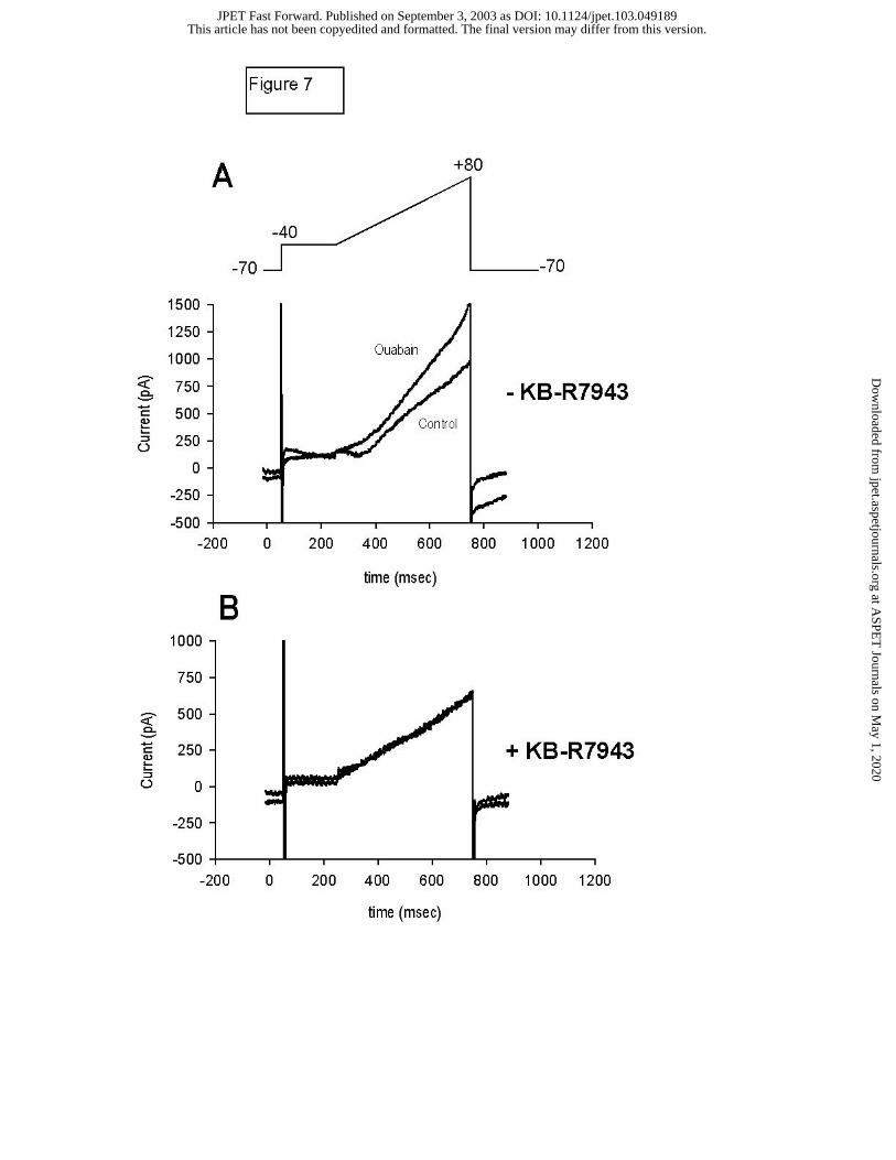
Effect of Ouabain and DHO on the Steady State Outward current (Iss):
A ramp protocol was used to evaluate the late outward current component noted above
using high resistance electrodes. The voltage protocol and representative data are presented in
Figure 7. Cd2+ (0.1 mM) was added to block ICa. As illustrated in Figure 7A, 10 minutes of
exposure to 3 µM ouabain induced a pronounced increase in outward current (previously
termed “outward steady state current”, Iss; Levi, 1993) that is observed at voltages above –20
mV. However, pretreatment of cells with 10 µM KB-R7943, a blocker of reverse mode Na+-Ca2+
exchange (NCX), markedly attenuated the increase in this current (Figure 7B), suggesting that
the observed current is the result of increased outward (reverse mode) NCX. Composite data
show that this cardiac glycoside-sensitive current (Figure 8; represented as a difference current
between control and cardiac glycoside treated conditions) is increased during exposure to
maximal inotropic concentrations of both ouabain and DHO but is suppressed by pretreatment
with 10 µM KB-R7943. The fact that there is an apparent voltage dependence to this outward
current, which increases with test potential, is also consistent with the notion that the
electrogenic NCX current may be involved. The finding that both ouabain and DHO are capable
This article has not been copyedited and formatted. The final version may differ from this version.JPET Fast Forward. Published on September 3, 2003 as DOI: 10.1124/jpet.103.049189
at ASPE
T Journals on M
ay 1, 2020jpet.aspetjournals.org
Dow
nloaded from

JPET/2003/049189—R2
14
of increasing outward NCX current, however, does not explain the divergent effects of ouabain
and DHO on the AP.
Effect of Ouabain and DHO on APD with Increased Intracellular Ca2+ Buffering.
Consideration of the possibility that the observed differences between ouabain and DHO
on APD were secondary to changes in intracellular Ca2+ prompted a series of experiments that
minimized the cardiac glycoside-induced rise in internal Ca2+. First, a series of experiments was
performed with patch electrodes using an internal [Na+] of 10 mM and near physiological Ca2+
buffering (0.056mM EGTA, measured [Ca2+]i = 82nM, Vites and Wasserstrom, 1996). Under
these conditions (which permitted dialysis and equilibration of the intracellular space with the
patch pipette internal solution), APD was observed to shorten upon exposure to 3 µM ouabain
(Figure 9A), but not 10 µM DHO (Figure 9B), just as we had observed previously using high-
resistance electrodes (Figure 2). The experiment was then repeated with markedly increased
internal Ca2+ buffering (1mM EGTA). As shown in Figure 9C and D, separate cells were
allowed to equilibrate for 20 minutes with internal solution containing the high EGTA
concentration before addition of the cardiac glycoside. During this equilibration time the APD
shortened, and then stabilized. Addition of a maximal inotropic concentration of both ouabain
(Figure 9C) and DHO (Figure 9D) was now able to cause further shortening of the AP. Under
these conditions, the decreases in APD50 and APD90 observed upon exposure to both 3 µM
ouabain or 10 µM DHO was statistically significant, as indicated in Figure 9E.
Inotropic Action of Ouabain, DHO, and Ouabagenin under AP clamp
To further characterize the observed diminished inotropic action of ouabagenin
compared with ouabain under conditions independent of resting and action potentials, myocytes
were voltage clamped using a protocol which approximated normal RMP (-78 mV) and AP
This article has not been copyedited and formatted. The final version may differ from this version.JPET Fast Forward. Published on September 3, 2003 as DOI: 10.1124/jpet.103.049189
at ASPE
T Journals on M
ay 1, 2020jpet.aspetjournals.org
Dow
nloaded from

JPET/2003/049189—R2
15
configuration. High resistance microelectrodes were employed to maintain contractility during
the experiment. Under these conditions, ouabain, ouabagenin and DHO did not differ from each
other significantly in the extent of their maximal inotropic action; all three cardiac glycosides
were able to achieve similar inotropic effects; ouabain increased cell shortening from 4.1+0.77%
to 10.4+0.76% of resting cell length (n=9); ouabagenin increased shortening from 4.9+0.87% to
11.4+0.27% (n=5); DHO 4.7+0.27% to 12.1+0.61% (n=4). Unlike the results obtained with the
free-running action potential stimulation of contraction, there were no significant differences in
the magnitude of the positive inotropic effects between these three agents. In addition, the
magnitude of the current during the plateau phase of the AP clamp increased with exposure to
all three of the cardiac glycosides but did not differ significantly between agents (data not
shown) as would be expected for a general increase in NCX current. This result suggests that
inotropic differences between agents, particularly ouabagenin and ouabain, are minimized
under conditions that control resting and excitation potentials.
This article has not been copyedited and formatted. The final version may differ from this version.JPET Fast Forward. Published on September 3, 2003 as DOI: 10.1124/jpet.103.049189
at ASPE
T Journals on M
ay 1, 2020jpet.aspetjournals.org
Dow
nloaded from

JPET/2003/049189—R2
16
Discussion
Our investigation of the actions of six cardiac glycoside analogs on action potential
configuration and contractility in single cat ventricular myocytes revealed that: 1) Of all agents
tested only ouabain and actodigin induce AP shortening at high inotropic concentrations. 2)
Ouabain-induced AP shortening is associated temporally with the appearance of ISS (reverse
mode NCX current; blocked by KB-R7943). 3) DHO is capable of producing AP shortening only
if Ca2+ overload is prevented or delayed by increased Ca2+i buffering, suggesting that cardiac
glycoside-induced cardiotoxicity is mediated by more than one mechanism; sodium pump
inhibition with resulting reverse mode NCX, and a second mechanism separate from sodium
pump inhibition. 4) Ouabagenin and certain other cardiac glycosides produce less maximal
contractile response before the onset of Ca2+ overload toxicity than ouabain, but only when the
native action potentials trigger excitation-contraction coupling. 5) The apparent difference in
inotropic action between cardiac glycosides is significantly reduced under AP voltage clamp,
suggesting that a putative second mechanism underlying cardiac glycoside induced
cardiotoxicity is at least partially voltage dependent.
Cardiac Glycoside Effects on Action Potentials, Inotropy and Toxicity
Differences in activity among cardiac glycosides have been described both in intact
animals and isolated cardiac preparations. However, attempts to explain these differences in
action based on specific features of cardiac glycoside molecular structure have produced limited
results. Part of this limitation stems from the fact that the majority of cardiac glycoside structure-
activity studies are focused on Na+/K+ - ATPase binding (DePover and Godfraind, 1982; Brown
and Erdman, 1984), and few address physiological actions in intact systems. Notable
exceptions include studies in guinea pig atria showing stimulation of the sodium pump at low
concentrations of ouabain and ouabagenin, but not DHO (Ghysel-Burton and Godfraind, 1979),
and the demonstration of differences in toxic to therapeutic ratios of cardiac glycosides in intact
This article has not been copyedited and formatted. The final version may differ from this version.JPET Fast Forward. Published on September 3, 2003 as DOI: 10.1124/jpet.103.049189
at ASPE
T Journals on M
ay 1, 2020jpet.aspetjournals.org
Dow
nloaded from

JPET/2003/049189—R2
17
dogs, dependent on the position of the attachment of the lactone ring (Mendez et.al., 1974).
However, none of these studies (individually or collectively) provides a comprehensive
foundation for understanding cardiac glycoside structure-activity relationships, and it is likely
that multiple factors in structure determine the specific response elicited by a given agent.
Information provided by our investigation indicates that subtle differences in structure (including
a simple saturation of the lactone ring) cause significant differences in physiological activity. If
intracellular sites are indeed important in determining differences in action between cardiac
glycoside agents (Fujino and Fujino, 1982; Isenberg, 1984; Sagawa et al., 2002), including the
threshold for Ca2+-overload, one could speculate that the relative ability of a given glycoside to
cross the sarcolemma (either passively or actively, Nunez-Duran et al., 1986) and affinity for
specific intracellular sites would be important additional determinants of action. In addition, if
the ryanodine receptor is an important determinant of this intracellular mechanism as postulated
by some investigators (Isenberg, 1984; Rardon and Wasserstrom, 1990; McGarry and Williams,
1993, Sagawa et al., 2002), then differences in binding affinity to this receptor between cardiac
glycosides might contribute to the observed differences in action.
Mechanisms Underlying Shortening of APD
Many studies evaluating the effects of cardiac glycosides on isolated myocardial
preparations or single cells have shown a shortening of APD (McDonald et al., 1975; Levi,
1993). Explanations for this finding are reflected in contrasting theories that attempt to
characterize a cardiac glycoside-induced outward current. The first theory (Luk and Carmeliet,
1990) invokes an outward current that is increased by 10-100 µM ouabain in guinea pig cardiac
myocytes with similar properties to a single channel current found in inside-out patches. This
current is proposed to be a Na+-activated K+ current by virtue of the fact that it is dependent on
the presence of Na+i, displays rectification that is dependent on the K+ gradient, and has a
reversal potential near the K+ equilibrium potential. The second theory (Levi, 1993) suggests the
This article has not been copyedited and formatted. The final version may differ from this version.JPET Fast Forward. Published on September 3, 2003 as DOI: 10.1124/jpet.103.049189
at ASPE
T Journals on M
ay 1, 2020jpet.aspetjournals.org
Dow
nloaded from

JPET/2003/049189—R2
18
involvement of an outward current elicited by 50 µM strophanthidin also in guinea pig cardiac
cells with reversal potential of –54 mV. This current was shown to be consistent with reverse
mode NCX by being more pronounced at positive voltages, abolished by removing external Ca2+
or addition of Ni2+, and unaffected by increasing intracellular Ca2+ buffering (BAPTA) or addition
of K+ current blockers (Ba2+, TEA, and 4-AP). Data from our experiments indicate the cardiac
glycoside-induced outward current is most likely due to increased reverse mode NCX because it
does not develop in the presence of KB-R7943, a reverse mode NCX blocker (Kimura et al.,
1999). In addition, AP shortening appears to be an action of some but not all cardiac
glycosides. This could be a result of other drug actions, including the possibility that certain
glycosides might also block IKs, the slowly-activating component of the delayed rectifier current
(Rocchetti et al., 2003), which could explain why DHO (and not OUA or actodigin) caused AP
prolongation. The balance of opposing direct and indirect actions on net membrane current
could then be responsible for the variety of glycoside effects on AP duration, especially in
different species with varying dependencies of repolarization on IKs.
Role of Ca2+ Overload in Development of APD Shortening and Inotropy
Data from this study suggest that Ca2+-overload is a primary determinant both for
maximal inotropic response and alterations in AP configuration, and could account for
differences in response between cardiac glycoside analogs. This is not a new concept,
particularly with regard to inotropic action as addressed by other investigators (Capogrossi et al,
1988). The basic theory, supported by experimental data, contends that the myocardium is
capable of increasing inotropy by Ca2+-dependent mechanisms until toxicity develops as
indicated by spontaneous sarcoplasmic reticulum Ca2+ release and spontaneous contractions.
This spontaneous, uncoordinated release of Ca2+ depletes the sarcoplasmic reticulum of Ca2+
for the next contraction, thereby reducing the inotropic state as well as any contributions of
Ca2+-dependent conductances (including NCX current) to AP duration.
This article has not been copyedited and formatted. The final version may differ from this version.JPET Fast Forward. Published on September 3, 2003 as DOI: 10.1124/jpet.103.049189
at ASPE
T Journals on M
ay 1, 2020jpet.aspetjournals.org
Dow
nloaded from

JPET/2003/049189—R2
19
We found that Ca2+-overload in fact does influence whether or not cardiac glycosides
induced AP abbreviation. Although direct evidence for sodium pump inhibition was not
evaluated, the presence of a robust increase in reverse mode NCX current within the inotropic
range of both ouabain and DHO would suggest that sodium pump inhibition is an important
mechanism in the inotropic response of cardiac glycosides in vitro. In addition, the data suggest
that a second cardiac glycoside mechanism (independent of sodium pump inhibition) influences
the threshold for Ca2+ overload and thereby determines the maximal inotropic response
observed. For example, the weaker inotropic response and lack of AP shortening observed with
ouabagenin compared to ouabain is likely the result of earlier spontaneous SR Ca2+ release
(lower toxicity threshold) elicited by ouabagenin prior to accumulation of equal levels of
intracellular calcium.
How voltage alters spontaneous release from the SR and thereby alters the maximal
inotropic response of ouabagenin (as demonstrated in the AP clamp experiments) can only be
speculated. The most likely reason is that the positive feedback between SR Ca2+ release and
membrane depolarization is prevented under voltage clamp. Thus, differences between drugs
displaying maximal inotropic effects are likely to be blunted simply because steady-state drug
effects are more closely approximated. However, it is also possible that this observation may
be related to the idea linking membrane potential with SR release (Ferrier and Howlett, 1995).
Because the threshold for spontaneous calcium release from the SR during diastole is a primary
determinant of cardiac glycoside inotropy, it may be that voltage clamp prevents or delays
diastolic calcium release. This implies that small voltage perturbations during diastole may
contribute to spontaneous release, or that spontaneous release during diastole is at least
partially dependent on membrane potential.
Effects of Low Internal Free Mg2+ Concentration on Experimental Results
This article has not been copyedited and formatted. The final version may differ from this version.JPET Fast Forward. Published on September 3, 2003 as DOI: 10.1124/jpet.103.049189
at ASPE
T Journals on M
ay 1, 2020jpet.aspetjournals.org
Dow
nloaded from

JPET/2003/049189—R2
20
It is possible that certain of our experimental conditions might influence the results found
in this study. One such issue is the low free [Mg2+] concentration in the internal solution (about
10-5M). This is likely to have important effects on Mg2+-dependent process in the cell.
However, it should be noted that even with this low [Mg2+], there are still pronounced differences
between different cardiac glycosides just as expected from data obtained using high resistance
microelectrodes in which the intracellular environment is closer to physiological. In addition, the
rectifying characteristics of IK1 are largely unaffected by the buffering of internal Mg2+ (data not
shown), suggesting that effective concentrations in critical regions of the cytoplasm remain at
normal regulatory levels despite calculated changes in bulk concentration. This fact suggests
that it is difficult to extrapolate bulk calculated [Mg2+] to true free concentration at regulatory
sites.
This article has not been copyedited and formatted. The final version may differ from this version.JPET Fast Forward. Published on September 3, 2003 as DOI: 10.1124/jpet.103.049189
at ASPE
T Journals on M
ay 1, 2020jpet.aspetjournals.org
Dow
nloaded from

JPET/2003/049189—R2
21
Footnotes
Dr. Stuart Ruch was supported in part by a NIH Program Project Grant awarded to the
University of Illinois at Chicago Departments of Physiology and Cardiology. Additional support
for this work was provided by NIH 30724 (JAW).
This article has not been copyedited and formatted. The final version may differ from this version.JPET Fast Forward. Published on September 3, 2003 as DOI: 10.1124/jpet.103.049189
at ASPE
T Journals on M
ay 1, 2020jpet.aspetjournals.org
Dow
nloaded from

JPET/2003/049189—R2
22
References
Akera T, Larson FS, Brody TM (1970) Correlation of cardiac sodium- and potassium-
activated adenosine triphosphate activity with ouabain-induced inotropic stimulation. J
Pharmacol Exp Ther 173: 145-51.
Brown L, Erdmann E. (1984) Binding of digitalis derivatives to beef, cat and human
cardiac (Na+ + K+) –ATPase. Affinity and kinetic constants. Arch Int Pharmacodyn 271:229-
240.
Capogrossi MC, Stern MD, Spurgeon HA, Lakatta EG (1988) Spontaneous Ca2+ release
from the sarcoplasmic reticulum limits Ca2+ –dependent twitch potentiation in individual cardiac
myocytes. J Gen Physiol 91:133-55.
De Pover A, Godfraind T (1982) Influence of 16ß formylation on Na, K-ATPase inhibition
by cardiac glycosides. Naunyn-Sch Arch Pharmacol 321:135-139.
Ferrier GR, Howlett SE (1995) Contractions in guinea-pig ventricular myocytes triggered
by a calcium-release mechanism separate from Na+ and L-currents. J Physiol 484:107-22.
Fujino S, Fujino M (1982) Ouabain potentiation and Ca release from sarcoplasmic
reticulum in cardiac and skeletal muscle. Can J Physiol Pharmacol 60:542-555.
Gadsby DC, Kimura J, Noma A (1985) Voltage dependence of the Na/K pump current in
isolated heart cells. Nature 315:63-5.
Ghysel-Burton J, Godfraind T (1979) Stimulation and inhibition of the sodium pump by
cardioactive steroids in relation to their binding sites and their inotropic effect on guinea pig
isolated atria. Br J Pharmacol 66:175-184.
Isenberg G. (1984) Contractility of isolated bovine ventricular myocytes is enhanced by
intracellular injection of cardioactive glycosides. Evidence for an intracellular mode of action, in
Cardiac Glycoside Receptors and Positive Inotropy (Erdmann E ed.) pp 56-71, Steinkopff,
Darmstadt.
This article has not been copyedited and formatted. The final version may differ from this version.JPET Fast Forward. Published on September 3, 2003 as DOI: 10.1124/jpet.103.049189
at ASPE
T Journals on M
ay 1, 2020jpet.aspetjournals.org
Dow
nloaded from

JPET/2003/049189—R2
23
Karagueuzian HS, Katzung BG (1981) Relative inotropic and arrhythmogenic effects of
five cardiac steroids in ventricular myocardium: Oscillatory afterpotentials and the role of
endogenous catecholamines. J Pharmacol Exp Ther 218:348-356.
Kimura J, Watano T, Kawahara M, Saki E, Yatabe J (1999) Direction-independent block
of bi-directional Na+ / Ca2+ exchange current by KB-R7943 in guinea-pig cardiac myocytes. Br J
Pharmacol 128:969-974.
Lee C, Dagostino M (1982) Effect of strophanthidin on intracellular Na+ ion activity and
twitch tension of constantly driven canine cardiac fibers. Biophys J 40:185-98.
Levi AJ (1993) A role for sodium/calcium exchange in the action potential shortening
caused by strophanthidin in guinea pig ventricular myocytes. Cardiovascular Research 27:471-
481.
Luk HN, Carmeliet E (1990) Na+ -activated K+ current in cardiac cells: rectification, open
probability, block and role in digitalis toxicity. Pflugers Arch 416:766-768.
McDonald TF, Nawrath H, Trautwein W (1975) Membrane currents and tension in cat
ventricular muscle treated with cardiac glycosides. Circ Res 37:674-682.
McGarry SJ, Williams AJ (1993) Digoxin activates sarcoplasmic reticulum Ca2+ -release
channels: a possible role in cardiac inotropy. Br J Pharmacol 108:1043-1050.
Mendez R, Pastelin G, Kabela E (1974) The influence of the position of attachment of
the lactone ring to the steroid nucleus on the action of cardiac glycosides. J Pharmacol Exp
Ther 188:189-197.
Nunez-Duran H, Riboni L, Ubaldo E, Kabela K, Barcenal-Ruiz L (1988) Ouabain uptake
by endocytosis in isolated guinea pig atria. Amer J Physiol. 255:C479-C485.
Rardon DP, Wasserstrom JA (1990) Cardiotonic steroids activate cardiac sarcoplasmic
reticulum calcium release channels. Circulation 82:III-342 (abstract).
This article has not been copyedited and formatted. The final version may differ from this version.JPET Fast Forward. Published on September 3, 2003 as DOI: 10.1124/jpet.103.049189
at ASPE
T Journals on M
ay 1, 2020jpet.aspetjournals.org
Dow
nloaded from

JPET/2003/049189—R2
24
Rocchetti M, Besana A, Mostacciuolo G, Ferrari, P, Micheletti R, Zaza A. (2003) Diverse
toxicity associated with cardiac Na+/K+ pump inhibition: Evaluation of electorphysiogogical
mechanisms. J Pharmacol Exp Ther 305:765-771.
Sagawa T, Sagawa K, Kelly JE, Tsushima RG, Wasserstrom JA (2002) Activation of
cardiac ryanodine receptors by cardiac glycosides. Amer J Physiol 282:H1118-H1126.
Salata JJ, Wasserstrom JA. (1988) Effects of quinidine on action potentials and ionic
currents in isolated canine ventricular myocytes. Circ Res 62:324-337.
Silver LH, Gomez AM, Lederer WJ, Houser SR (1983) Isolation and morphology of
calcium-tolerant feline ventricular myocytes. Am J Physiol 245:H891-H896.
Steimers JR, Lobaugh LA, Liu S, Shigeto N, Lieberman M (1990) Intracellular sodium
affects ouabain interaction with the Na/K pump in cultured chick cardiac myocytes. J Gen
Physiol 95:77-95.
Vites AM, Wasserstrom JA (1996) Fast sodium influx provides an initial step to trigger
contractions in cat ventricle. Amer J Physiol 271:H674-H686.
Wasserstrom JA, Farkas DE, Norell MA, Vereault DV (1991) Effects of different cardiac
steroids on intracellular sodium, inotropy and toxicity in sheep Purkinje fibers. J Pharmacol Exp
Ther 258:918-925.
Wasserstrom JA, Salata JJ. (1988) Basis for tetrodotoxin and lidocaine effects on
action potentials in dog ventricular myocytes. Amer J Physiol 254:H1157-H1166.
This article has not been copyedited and formatted. The final version may differ from this version.JPET Fast Forward. Published on September 3, 2003 as DOI: 10.1124/jpet.103.049189
at ASPE
T Journals on M
ay 1, 2020jpet.aspetjournals.org
Dow
nloaded from

JPET/2003/049189—R2
25
Table 1 Control Action Potential Parameters of the Experimental Groups
Abbreviations: RMP, Resting Membrane Potential; APD50 and APD90, Action Potential Duration at 50 and 90 percent of repolarization; AP, Action Potential. All values are presented as Mean (SEM). No statistical differences between groups for the parameters shown.
Cardiac Glycoside Control RMP Control APD50 Control APD90 Control AP Amplitude
Ouabain -72.3 (1.4) 254 (12) 312 (13) 114 (2.3) Dihydroouabain -71.4 (1.5) 229 (16) 288 (13) 116 (4.0) Ouabagenin -72.8 (2.1) 269 (23) 311 (25) 120 (6.2) Actodigin -74.7 (1.0) 272 (19) 320 (19) 116 (2.7) Digoxin -72.3 (2.3) 245 (29) 307 (28) 109 (8.6)
Resibufogenin -73.0 (2.7) 273 (24) 343 (21) 111 (3.6)
This article has not been copyedited and formatted. The final version may differ from this version.JPET Fast Forward. Published on September 3, 2003 as DOI: 10.1124/jpet.103.049189
at ASPE
T Journals on M
ay 1, 2020jpet.aspetjournals.org
Dow
nloaded from

JPET/2003/049189—R2
26
Figure Legends
Figure 1. Molecular Structure of the Six Cardiac Glycosides used in the Study
Figure 2. Differential Effects of Ouabain and DHO on Cat Ventricular Myocytes: Action
Potentials and Contractions. High inotropic concentration of ouabain (3 µM; upper left), but not
DHO (10 µM; upper right), elicits action potential shortening. Effect of cardiac glycoside is
indicated by the broken line trace in each figure. Both ouabain (left) and DHO (middle) are
capable of producing equivalent inotropic responses, as indicated by comparable increases in
active cell shortening upon addition of the agents. In contrast, maximal inotropic concentration
of ouabagenin has less inotropic effect than either ouabain, or DHO (right). Similar to DHO,
ouabagenin has minimal effect on APD.
Figure 3. Concentration – APD Response Curves for Six Cardiac Glycosides. Effect of
increasing cardiac glycoside concentration on APD50 (panels A and C) and APD90 (panels B and
D) is shown. Ouabain, DHO and ouabagenin concentration response curves are presented in
the upper panels (A and B). Digoxin, actodigin, and resibufogenin are presented in lower
panels (C and D). APD values are given as a fraction of control values (error bars = SEM). As
noted, ouabain and actodigin elicit action potential shortening, while DHO elicits a small but
significant prolongation of the AP at high inotropic concentrations. * p<0.05 indicates significant
difference from control.
Figure 4. Concentration – Inotropic Response Curves for Six Cardiac Glycosides. Inotropic
response to increasing cardiac glycoside concentrations, expressed as a fraction of control
shortening are presented for ouabain, ouabagenin, DHO in panel A. Digoxin, actodigin,
This article has not been copyedited and formatted. The final version may differ from this version.JPET Fast Forward. Published on September 3, 2003 as DOI: 10.1124/jpet.103.049189
at ASPE
T Journals on M
ay 1, 2020jpet.aspetjournals.org
Dow
nloaded from

JPET/2003/049189—R2
27
resibufogenin are presented in panel B. The majority of preparations became toxic at the
highest cardiac glycoside concentration indicated in each curve. Tables below each graph
denote EC50 values and maximal inotropic response (FxShMax; expressed as a fraction of control
FxSh) for each agent. EC50 values were derived from a sigmoidal fit of each concentration-
response curve. In contrast, FxShMax values were calculated from the maximal inotropic
response of each preparation (independent of the drug concentration) where FxShMax was
observed, and only from preparations that subsequently demonstrated toxicity (spontaneous
contractions and / or decreased developed force) thereby ensuring that the maximum inotropic
response had been reached. For this reason, the FxShMax values presented in the table closely
resemble the apparent maximum of the curves but do not correlate exactly. Of note,
ouabagenin produces significantly less maximal positive inotropic response than ouabain
(p<0.05).
Figure 5. Effect of Ouabain on Calcium Current (ICa). Panel A: Representative current traces
measured with high resistance microelectrodes are presented. ICa elicited by voltage steps from
–40 mV to 0 mV is shown for control condition and following a 5-minute exposure to 3 µM
ouabain. As noted, there is no effect of the cardiac glycoside on current magnitude. Inset
shows voltage protocol used for ICa voltage clamp experiments. Panel B. 3 µM ouabain has
minimal effect on ICa for the first five minutes of exposure, following which there is a small
decrease in maximal current amplitude (p>0.05, NS). Panel B. Similarly, exposure to 10 µM
DHO results in a small, time-dependent decrease in ICa, which fails to reach statistical
significance. Panel C. Myocytes not exposed to cardiac glycosides maintain control current
levels 10 minutes or longer without decrement, indicating high-resistance electrode
measurement of ICa is stable and relatively free of “rundown”.
This article has not been copyedited and formatted. The final version may differ from this version.JPET Fast Forward. Published on September 3, 2003 as DOI: 10.1124/jpet.103.049189
at ASPE
T Journals on M
ay 1, 2020jpet.aspetjournals.org
Dow
nloaded from

JPET/2003/049189—R2
28
Figure 6. Effect of Ouabain and DHO on IK1. A. Raw current traces showing IK1 in control
condition and following the addition of 3 µM ouabain. The increase in outward current noted at
positive voltage steps develops over 5-10 minutes after addition of the cardiac glycoside. Inset
shows the voltage clamp protocol used for IK1 experiments. B. IK1 data averaged from several
experiments showing the effect of 3 µM ouabain (error bars = SEM). The trend towards
increased outward current at positive voltages following addition of ouabain was not statistically
significant.
Figure 7. Ramp Protocol Experiments: Steady State Outward Current. Representative current
traces elicited by a voltage clamp ramp protocol (inset). As indicated, the protocol consisted of
a step to –40 mV for 200 ms, then a ramp up to +80 mV over 500 ms before returning to the
holding potential of -70 mV. Cd2+ and Ba2+ were added to the external solution to diminish ICa
and K+ currents. Panel A. Ten-minute exposure to 3 µM ouabain produced a dramatic increase
in steady state outward current. An increase in inward current upon return to the holding
potential was also observed following exposure to ouabain (noted by arrow). Panel B. Pre-
treatment with KB-R7943 effectively eliminated the development of this cardiac glycoside
sensitive steady state current.
Figure 8. Effect of DHO and Ouabain on cardiac glycoside Sensitive Steady State Outward
Current. Averaged data from ramp protocol experiments is presented. Glycoside-sensitive
current is defined as a subtraction (experimental minus control) current. The data summarize
the effect of ouabain (3 µM, empty circles) and DHO (10 µM, filled circles) on cardiac glycoside
sensitive current 10 minutes of exposure demonstrates the time dependent development of the
current. Pre-treatment with 10 µM KB-R7943 eliminates the effect of ouabain on development
of outward steady state current (triangles) producing significantly less current at all test voltages
This article has not been copyedited and formatted. The final version may differ from this version.JPET Fast Forward. Published on September 3, 2003 as DOI: 10.1124/jpet.103.049189
at ASPE
T Journals on M
ay 1, 2020jpet.aspetjournals.org
Dow
nloaded from

JPET/2003/049189—R2
29
(* p<0.05 compared to ouabain alone).
Figure 9. Effect of Ouabain and DHO on Action Potential Configuration in Myocytes Dialyzed
with 1 mM EGTA. Panel A and B. Effects of ouabain and DHO on action potentials measured
in cat myocytes using patch electrodes. Panel C. Equilibration of the myocyte with pipette
solution containing increased Ca2+ buffer results in dramatic shortening of the AP (from 1 to 2).
Addition of 3 µM ouabain results in further shortening of the AP over a period of 5 to 7 minutes
(from 2 to 3). Panel D. Similar experiment using 10 µM DHO produces almost identical AP
shortening; under conditions preventing Ca2+ overload, both ouabain and DHO are able to
induce AP shortening. Panel E. Averaged data from several experiments demonstrating that
both 3 µM ouabain and 10 µM DHO produce AP shortening (indicated by a decrease in both
APD50 and APD90) under conditions of increased intracellular Ca buffering. * p<0.05 between
experimental and control values. Control APD values were obtained after 20 min of EGTA
dialysis.
This article has not been copyedited and formatted. The final version may differ from this version.JPET Fast Forward. Published on September 3, 2003 as DOI: 10.1124/jpet.103.049189
at ASPE
T Journals on M
ay 1, 2020jpet.aspetjournals.org
Dow
nloaded from

This article has not been copyedited and form
atted. The final version m
ay differ from this version.
JPET
Fast Forward. Published on Septem
ber 3, 2003 as DO
I: 10.1124/jpet.103.049189 at ASPET Journals on May 1, 2020 jpet.aspetjournals.org Downloaded from

This article has not been copyedited and form
atted. The final version m
ay differ from this version.
JPET
Fast Forward. Published on Septem
ber 3, 2003 as DO
I: 10.1124/jpet.103.049189 at ASPET Journals on May 1, 2020 jpet.aspetjournals.org Downloaded from

This article has not been copyedited and formatted. The final version may differ from this version.JPET Fast Forward. Published on September 3, 2003 as DOI: 10.1124/jpet.103.049189
at ASPE
T Journals on M
ay 1, 2020jpet.aspetjournals.org
Dow
nloaded from

This article has not been copyedited and formatted. The final version may differ from this version.JPET Fast Forward. Published on September 3, 2003 as DOI: 10.1124/jpet.103.049189
at ASPE
T Journals on M
ay 1, 2020jpet.aspetjournals.org
Dow
nloaded from
This article has not been copyedited and formatted. The final version may differ from this version.JPET Fast Forward. Published on September 3, 2003 as DOI: 10.1124/jpet.103.049189
at ASPE
T Journals on M
ay 1, 2020jpet.aspetjournals.org
Dow
nloaded from

This article has not been copyedited and formatted. The final version may differ from this version.JPET Fast Forward. Published on September 3, 2003 as DOI: 10.1124/jpet.103.049189
at ASPE
T Journals on M
ay 1, 2020jpet.aspetjournals.org
Dow
nloaded from

This article has not been copyedited and formatted. The final version may differ from this version.JPET Fast Forward. Published on September 3, 2003 as DOI: 10.1124/jpet.103.049189
at ASPE
T Journals on M
ay 1, 2020jpet.aspetjournals.org
Dow
nloaded from