EEG SIGNAL CLASSIFICATION USING SOFT COMPUTING …
5
International Journal of Advances in Science Engineering and Technology, ISSN: 2321-9009 Volume- 2, Issue-3, July-2014 EEG Signal Classification Using Soft Computing Techniques for Brain Disease Diagnosis 1 EEG SIGNAL CLASSIFICATION USING SOFT COMPUTING TECHNIQUES FOR BRAIN DISEASE DIAGNOSIS 1 L. ANJANA, 2 K. DIVYA, 3 C. DHIVYASHRI Department of Computer Science and Engineering Panimalar Engineering College Chennai, India Abstract -The project proposes an automatic support system for tumor classification using the soft computing techniques. The detection of the brain tumor is a challenging problem, due to the structure of the tumor cells. The artificial neural network is used to classify the stage of brain EEG signal that if it is the case of tumor or epilepsy or normal. The manual analysis of the signal is time consuming, inaccurate and requires intensive trained person to avoid diagnostic errors. The soft computing techniques are employed for the classification of the EEG signals as the techniques are intended to model and make possible solutions to real world tribulations. The probability of correct classification has been increased by using soft computing techniques like Principal Component Analysis with neural network and Fuzzy Logic. Back Propagation Network with image and data processing techniques is employed to implement an automated Brain Tumor classification. Decision making is performed in two stages: feature extraction using Principal Component Analysis and the classification using Back Propagation Network (BPN). The performance of the BPN classifier was evaluated in terms of training performance and classification accuracies. Back Propagation Network gives fast and accurate classification than other neural networks and it is a promising tool for classification of the Tumors. Keywords—Artificial Neural Network(ANN), Back Propagation Network, Feature Extraction, Principal Component Analysis(PCA), Electroencephalogram (EEG) I. INTRODUCTION Electroencephalography (EEG) is a non-invasive test for epilepsy during which several electrodes are placed on a L patient's scalp to record electrical impulses from the brain (brain waves). It is sometimes called a brain wave test, used for testing patients with epilepsy, a brain tumor, a brain abscess, brain trauma, subdural hematoma, meningitis, encephalitis, stroke or congenital defects of the brain. It is performed by using a device that measures the fluctuations and patterns in electrical processes within the brain. When the brain cells send messages to each other, they produce tiny electrical signals. Your brain cells communicate via electrical impulses and are active all the time, even when you're asleep. This activity shows up as wavy lines on an EEG recording. An EEG is one of the main diagnostic tests for epilepsy. In an EEG test, electrodes (flat metal discs) are placed onto your scalp using a sticky substance. These electrodes pick up the electrical signals from your brain and send them to an EEG machine, which will record the signals as wavy lines onto paper or on a computer. The EEG machine records your brain's electrical activity as a series of traces, Each trace corresponds to a different region of the brain. An electroencephalogram (EEG) is a painless procedure that takes 30 to 45 minutes with rarely causes of any side effects. The EEG shows patterns of normal or abnormal brain electrical activity. Some abnormal patterns may occur with a number of different conditions, not just seizures. For example, certain types of waves may be seen after head trauma, stroke, brain tumor, or seizures. A common example of this type is called "slowing," in which the rhythm of the brain waves is slower than would be expected for the patient's age and level of alertness. Certain other patterns indicate a tendency toward seizures. Your doctor may refer to these waves as "epileptiform abnormalities" or "epilepsy waves." These include spikes, sharp waves, and spike-and-wave discharges. Spikes and sharp waves in a specific area of the brain, such as the left temporal lobe, indicate that partial seizures might possibly come from that area. Primary generalized epilepsy, on the other hand, is suggested by spike- and-wave discharges that are widely spread over both hemispheres of the brain, especially if they begin in both hemispheres at the same time. Since the early days of automatic EEG processing, representations based on a Fourier transform have been most commonly applied. This approach is based on earlier observations that the EEG spectrum contains some characteristic waveforms that fall primarily within four frequency bands—delta (1–4 Hz), theta (4–8 Hz), alpha (8–13 Hz), and beta (13– 30 Hz). Such methods have proved beneficial for various EEG characterizations, but fast Fourier transform (FFT), suffer from large noise sensitivity. Parametric power spectrum estimation methods such as AR, reduces the spectral loss problems and gives better frequency resolution. Automated classification and detection of tumors in different medical signals is motivated bythe necessity of high accuracy when dealing with a human life. Also, the computer assistance is demanded in medical institutions due to the fact that it could improve the results of humans in such a domain where the false negative cases must be at a very low rate. It has been
Transcript of EEG SIGNAL CLASSIFICATION USING SOFT COMPUTING …

International Journal of Advances in Science Engineering and Technology, ISSN: 2321-9009 Volume- 2, Issue-3, July-2014
EEG Signal Classification Using Soft Computing Techniques for Brain Disease Diagnosis 1
EEG SIGNAL CLASSIFICATION USING SOFT COMPUTING TECHNIQUES FOR BRAIN DISEASE DIAGNOSIS
1L. ANJANA, 2K. DIVYA, 3C. DHIVYASHRI
Department of Computer Science and Engineering Panimalar Engineering College
Chennai, India
Abstract -The project proposes an automatic support system for tumor classification using the soft computing techniques. The detection of the brain tumor is a challenging problem, due to the structure of the tumor cells. The artificial neural network is used to classify the stage of brain EEG signal that if it is the case of tumor or epilepsy or normal. The manual analysis of the signal is time consuming, inaccurate and requires intensive trained person to avoid diagnostic errors. The soft computing techniques are employed for the classification of the EEG signals as the techniques are intended to model and make possible solutions to real world tribulations. The probability of correct classification has been increased by using soft computing techniques like Principal Component Analysis with neural network and Fuzzy Logic. Back Propagation Network with image and data processing techniques is employed to implement an automated Brain Tumor classification. Decision making is performed in two stages: feature extraction using Principal Component Analysis and the classification using Back Propagation Network (BPN). The performance of the BPN classifier was evaluated in terms of training performance and classification accuracies. Back Propagation Network gives fast and accurate classification than other neural networks and it is a promising tool for classification of the Tumors. Keywords—Artificial Neural Network(ANN), Back Propagation Network, Feature Extraction, Principal Component Analysis(PCA), Electroencephalogram (EEG) I. INTRODUCTION Electroencephalography (EEG) is a non-invasive test for epilepsy during which several electrodes are placed on a L patient's scalp to record electrical impulses from the brain (brain waves). It is sometimes called a brain wave test, used for testing patients with epilepsy, a brain tumor, a brain abscess, brain trauma, subdural hematoma, meningitis, encephalitis, stroke or congenital defects of the brain. It is performed by using a device that measures the fluctuations and patterns in electrical processes within the brain. When the brain cells send messages to each other, they produce tiny electrical signals. Your brain cells communicate via electrical impulses and are active all the time, even when you're asleep. This activity shows up as wavy lines on an EEG recording. An EEG is one of the main diagnostic tests for epilepsy. In an EEG test, electrodes (flat metal discs) are placed onto your scalp using a sticky substance. These electrodes pick up the electrical signals from your brain and send them to an EEG machine, which will record the signals as wavy lines onto paper or on a computer. The EEG machine records your brain's electrical activity as a series of traces, Each trace corresponds to a different region of the brain. An electroencephalogram (EEG) is a painless procedure that takes 30 to 45 minutes with rarely causes of any side effects. The EEG shows patterns of normal or abnormal brain electrical activity. Some abnormal patterns may occur with a number of different conditions, not just seizures. For example, certain types of waves may be seen after head trauma, stroke, brain tumor, or
seizures. A common example of this type is called "slowing," in which the rhythm of the brain waves is slower than would be expected for the patient's age and level of alertness. Certain other patterns indicate a tendency toward seizures. Your doctor may refer to these waves as "epileptiform abnormalities" or "epilepsy waves." These include spikes, sharp waves, and spike-and-wave discharges. Spikes and sharp waves in a specific area of the brain, such as the left temporal lobe, indicate that partial seizures might possibly come from that area. Primary generalized epilepsy, on the other hand, is suggested by spike-and-wave discharges that are widely spread over both hemispheres of the brain, especially if they begin in both hemispheres at the same time. Since the early days of automatic EEG processing, representations based on a Fourier transform have been most commonly applied. This approach is based on earlier observations that the EEG spectrum contains some characteristic waveforms that fall primarily within four frequency bands—delta (1–4 Hz), theta (4–8 Hz), alpha (8–13 Hz), and beta (13–30 Hz). Such methods have proved beneficial for various EEG characterizations, but fast Fourier transform (FFT), suffer from large noise sensitivity. Parametric power spectrum estimation methods such as AR, reduces the spectral loss problems and gives better frequency resolution. Automated classification and detection of tumors in different medical signals is motivated bythe necessity of high accuracy when dealing with a human life. Also, the computer assistance is demanded in medical institutions due to the fact that it could improve the results of humans in such a domain where the false negative cases must be at a very low rate. It has been

International Journal of Advances in Science Engineering and Technology, ISSN: 2321-9009 Volume- 2, Issue-3, July-2014
EEG Signal Classification Using Soft Computing Techniques for Brain Disease Diagnosis 2
proven that double reading of medical images could lead to better Tumor detection. But the cost implied in double reading is very high, that’s why good software to assist humans in medical institutions is of great interest nowadays. Conventional methods of monitoring and diagnosing the diseases rely on detecting the presence of particular features by a human observer. Due to large number of patients in intensive care units and the need for continuous observation of such conditions, several techniques for automated diagnostic systems have been developed in recent years to attempt to solve this problem. Such techniques work by transforming the mostly qualitative diagnostic criteria into a more objective quantitative feature classification problem.
Figure 1: Neural Networks
Automated classification of Brain signals by using some prior knowledge like intensity and some anatomical features is proposed. Currently there are no methods widely accepted therefore automatic and reliable methods for Tumor detection are of great need and interest. The application of BPN in the classification of data for EEG signals problems are not fully utilized yet. These included the feature extraction and classification techniques especially for CT images problems with huge scale of data and consuming times and energy if done manually. II. RELATED WORKS Sharanreddy et al introduced a Feature Extraction and Classification of EEG Signal Using Neural Network .Feature extraction of EEG signals is core issues on EEG based brain mapping analysis. The classification of EEG signals has been performed using features extracted from EEG signals. Many features have proved to be unique enough to use in all brain related medical application. EEG signals can be classified using a set of features like Auto-regression, Energy Spectrum Density, Energy Entropy, and Linear Complexity. However, different features show different discriminative power for different subjects or different trials. In this research, two-features are used to improve the performance of EEG signals. Neural Network based techniques are applied to feature extraction of EEG signal. This paper discuss
on extracting features based on Average method and Max & Min method of the data set. The Extracted Features are classified using Neural Network Temporal Pattern Recognition Technique. The two methods are compared and performance is analyzed based on the results obtained from the Neural Network classifier. Features were Extracted using Average method and Max_Min method. Two Features extraction methods are evaluated for their performance using Pattern Recognition tool box from the obtained results it has observed that the Max_Min feature extraction method gives better accuracy compared to the Average Feature Extraction Method and Accuracy of Max_Min method is 80%Accuracy of Average method is 41%. Nandish.Met al has proposed a Analysis and simulation of brain signal data by EEG signal processing technique using MATLAB:-EEG is brain signal processing technique that allows gaining the understanding of the complex inner mechanisms of the brain and abnormal brain waves have shown to be associated with particular brain disorders. The analysis of brain waves plays an important role in diagnosis of different brain disorders. MATLAB provides an interactive graphic user interface (GUI) allowing users to flexibly and interactively process their high-density EEG dataset and other brain signal data different techniques such as independent component analysis (ICA) and/or time/frequency analysis (TFA), as well as standard averaging methods. We will be showing different brain signals by comparing, analysing and simulating datasets which is already loaded in the MATLAB software to process the EEG signals. This project has clearly demonstrated the concepts about open source plug-ins, running under the platform MATLAB environment and its ability to process biophysical data by different way such as - using simplicity of its command line language or using the many MATLAB functions and the methodology related to the analysis of the brain signal processing through MATLAB software toolbox. It has been described in detail the procedure in the modeling of EEG signals and insight brain signals recorded during surgical procedure. III. PROPOSED WORK 1. Soft Computing Techniques Soft computing is the combination of methodologies (Neuro-Computing, Fuzzy Computing, Evolutionary and Genetic Computing and Probabilistic Computing) intended to model and make possible solutions to real world tribulations, which are not modeled or too complex for mathematical modeling. Its aspire is to utilize the tolerance for approximation (model features are similar to the real ones but not the same), uncertainty (not sure that the model features belief are the same as that of the entity), imprecision (model features quantities are not same as real ones but close

International Journal of Advances in Science Engineering and Technology, ISSN: 2321-9009 Volume- 2, Issue-3, July-2014
EEG Signal Classification Using Soft Computing Techniques for Brain Disease Diagnosis 3
to them) and partial truth in order to achieve close resemblance with human like decision making. The guiding theory of soft computing is to make use of these tolerance to achieve, robustness tractability and low solution cost. Human mind is the role model for soft computing. Some of the soft computing techniques are Artificial Neural Network (ANN), Fuzzy Logic (FL), Adaptive Neuro-Fuzzy Inference System (ANFIS), Principal Component Analysis (PCA) and evolutionary computation. EEG signal classification for brain disease diagnosis using the best among the following soft computing techniques • Artificial Neural Network(ANN) • Fuzzy Logic(FL) • Principal Component Analysis(PCA)
Figure 2: Block Diagram
2.Artificial Neural Network (ANN) A neural net is an artificial illustration of the human brain that tries to imitate its learning process. ANN is an interrelated group of artificial neurons that uses a mathematical model or computational model for information processing. ANN is a network of simple processing elements which can demonstrate complex overall performance, determined by the connections between the processing elements and element parameters. ANN is an adaptive system that changes its structure based on external or internal information that flows through the network. ANN computing approach to information processing primarily involves a learning process with an ANN architecture that adaptively responds to inputs according to a learning rule. After the NN has learned, the trained network can be used to execute certain tasks depending on the exact purpose. The talent to learn by example and simplify are the principal characteristics of ANN. Classification of signals is done by using this ANN to obtain the correct classification percentage. ANN is learned using the back propagation algorithm in which the errors for the units of the hidden layer are determined byback propagating the errors of the units of the output layer.It is a systematic method of training multi-layer ANNs. Itcontains an input layer, at least one intermediate/hidden layer and an output layer in its network. Some of the ANN learning parameters are
Threshold, Goal, Epoch, Sigmoidal function, Training type and Number of Hidden layers. 3. Fuzzy Logic (FL) Fuzzy logic is a form of many valued logic, it deals with reasoning that is approximate rather than fixed and exact. Compared to traditional binary sets (where variables may take on true or false values) fuzzy logic variables may have a truth value that ranges in degree between 0 and 1. Fuzzy logic has been extended to handle the concept of partial truth, where the truth value may range between completely true and completely false. Furthermore, when linguistic variables are used, these degrees may be managed by specific functions. Irrationality can be described in terms of what is known as the fuzzjective. The term "fuzzy logic" was introduced with the 1965 proposal of fuzzy set theory by Lotfi A. Zadeh. Fuzzy logic has been applied to many fields, from control theoryto artificial intelligence. Fuzzy logics however had been studied since the 1920s as infinite-valued logics notably by Łukasiewicz and Tarski. 4.Principal Component Analysis(PCA) The signals obtained from the electrodes are given to Principal Component Analysis for dimensionality reduction to remove the redundant variables in the data and the classified using Neural Network classifier with back propagation. In Principal Component Analysis the better classification of signals is obtained for the learning parameters like epochs as 1000, number of hidden layers as 3, goal as 0.01, sigmoidal function as tansig, threshold das 0.5 and training type. The brain signals are trained using Neural Network and the training is shown in Fig.1. During the classification of the mental tasks using Neural Network classifier, the data is misclassified at the output ie., thepercentage of correct classification is low Similarly during the classification of the mental tasks using Principal Component Analysis with Neural Network classifier, the data is perfectly classified at the output ie., the percentage of correct classification is good because of the reduction of the redundant variables in the dataset. The comparison of the results of Neural Network classifier and Principal Component Analysis with Neural Network classifier is tabulated in Table 1 to show the variation of mean square error during training, meansquare error during testing, computation time and the percentage of correctly classified data for both type of classification. 5.Back Propagation Network The back propagation algorithm is used to compute the necessary corrections, after choosing the weights of the network randomly. The algorithm can be decomposed in the following four steps: i) Feed-forward computation ii) Back propagation to the output layer iii) Back propagation to the hidden layer
International Journal of Advances in Science Engineering and Technology, ISSN: 2321-9009 Volume- 2, Issue-3, July-2014
EEG Signal Classification Using Soft Computing Techniques for Brain Disease Diagnosis 4
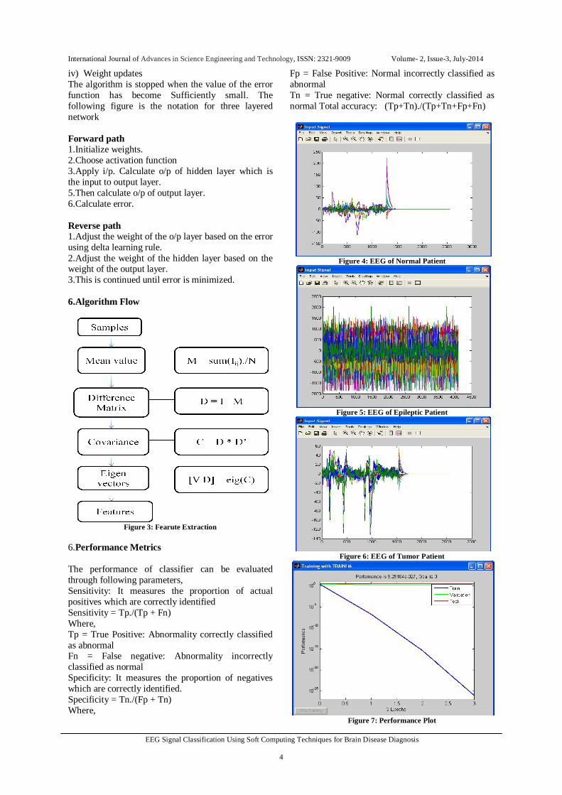
iv) Weight updates The algorithm is stopped when the value of the error function has become Sufficiently small. The following figure is the notation for three layered network Forward path 1.Initialize weights. 2.Choose activation function 3.Apply i/p. Calculate o/p of hidden layer which is the input to output layer. 5.Then calculate o/p of output layer. 6.Calculate error. Reverse path 1.Adjust the weight of the o/p layer based on the error using delta learning rule. 2.Adjust the weight of the hidden layer based on the weight of the output layer. 3.This is continued until error is minimized. 6.Algorithm Flow
Figure 3: Fearute Extraction
6.Performance Metrics The performance of classifier can be evaluated through following parameters, Sensitivity: It measures the proportion of actual positives which are correctly identified Sensitivity = Tp./(Tp + Fn) Where, Tp = True Positive: Abnormality correctly classified as abnormal Fn = False negative: Abnormality incorrectly classified as normal Specificity: It measures the proportion of negatives which are correctly identified. Specificity = Tn./(Fp + Tn) Where,
Fp = False Positive: Normal incorrectly classified as abnormal Tn = True negative: Normal correctly classified as normal Total accuracy: (Tp+Tn)./(Tp+Tn+Fp+Fn)
Figure 4: EEG of Normal Patient
Figure 5: EEG of Epileptic Patient
Figure 6: EEG of Tumor Patient
Figure 7: Performance Plot

International Journal of Advances in Science Engineering and Technology, ISSN: 2321-9009 Volume- 2, Issue-3, July-2014
EEG Signal Classification Using Soft Computing Techniques for Brain Disease Diagnosis 5
CONCLUSION The BCI problem is solved using neural network. The mental task classification is enhanced by several kinds of pre-processing, to generate the input data of the neural network. The soft computing techniques are employed for the classification of the EEG signals as the techniques are intended to model and make possible solutions to real world tribulations. The probability of correct classification has been increased by using soft computing techniques like Principal Component Analysis with neural network and Fuzzy Logic. ACKNOWLEDGEMENT We thank the Department of Computer Science and Engineering of Panimalar Engineering College for permitting to use the computational facilities by providing us most suitable environment for research and development. Specially we are very much thankful to Mrs. G. Florance and Dr. S. Malathi of Panimalar Engineering College for their contribution and support for the successful completion of this paper.
REFERENCES
[1] Pari Jahankhani, Vassilis Kodogiannis and Kenneth Revett “EEG Signal Classification Using Wavelet Feature Extraction and Neural Networks” IEEE John Vincent Atanasoff 2006 International Symposium On Modern Computing, 2006
[2] ManojThulasidas, Cuntai Guan, and Jiankang Wu “Robust Classification of EEG Signal for Brain–Computer Interface” IEEE Transactions ON Neural Systems AND Rehabilitation Engineering, VOL. 14, NO. 1, March 2006.
[3] Benjamin Blankertz, Klaus-Robert Müller, Dean J. Krusienski, GerwinSchalk, Jonathan R. Wolpaw, “The BCI Competition III: Validating Alternative Approaches to Actual BCI Problems”IEEE Transactions ON Neural Systems Rehabilitation Engineering, VOL. 14, NO. 2, JUNE 2006.
[4] Benjamin Blankertz, Guido Dornhege, Matthias Krauledat, Klaus-Robert Müller, Volker Kunzmann, Florian Losch, and Gabriel Curio, “The Berlin Brain–Computer Interface: EEG-Based
[5] Communication Without Subject Training”IEEE Transactions ON Neural Systems And Rehabilitation Engineering, VOL. 14, NO. 2, JUNE 2006.
[6] Anna Buttfield, Pierre W. Ferrez, and José del R. Millán, “Towards a Robust BCI: Error Potentials and Online Learning” IEEE Transactions ON NEURAL SYSTEMS AND Rehabilitation Engineering, VOL. 14, NO. 2, JUNE 2006.
[7] Paul R. Davidson*, Richard D. Jones, and Malik T. R. Peiris, “EEG-Based Lapse Detection With High Temporal Resolution” IEEE Transactions ON Biomedical Engineering, VOL. 54, NO. 5, MAY 2007.
[8] Alain Rakotomamonjy* and Vincent Guigue, “BCI Competition III: Dataset II- Ensemble of SVMs for BCI P300 Speller” IEEE Transactions On Biomedical ENGINEERING, VOL. 55, NO. 3, MARCH 2008.
[9] Benjamin Blankertz∗, Florian Losch, Matthias Krauledat, Guido Dornhege, Gabriel Curio, and Klaus-Robert M¨uller “The Berlin Brain–Computer Interface: Accurate Performance From First Sessionin BCI-Na¨ıve Subjects” IEEE Transactions ON BIOMEDICAL Engineering, VOL. 55, NO. 10, OCTOBER 2008.
[10] MahnazArvaneh*,Cuntai G, Kai KengAng, and Chai Quek, “Optimizing the Channel Selection and Classification Accuracy in EEG-Based BCI” IEEE TRANSACTIONS ON Biomedical Engineering, VOL. 58, NO. 6, JUNE 2011.
[11] Paul Honeine, “Online Kernel Principal Component Analysis: A Reduced-Order Model” IEEE transactions on pattern analysis and machine intelligence, vol. 34, no. 9, september 2012.
[12] Nandish.M, Stafford Michahial, Hemanth Kumar P, Faizan Ahmed, “Feature Extraction and Classification of EEG Signal Using Neural Network Based Techniques” International Journal of Engineering and Innovative Technology (IJEIT) Volume 2, Issue 4, October 2012.
[13] Kottaimalai R, Pallikonda Rajasekaran M, Selvam V, Kannapiran B, “EEG Signal Classification using Principal Component Analysis with Neural Network in Brain Computer Interface Applications” IEEE International Conference On Emerging Trends In Computing, Communication And Nanotechnology (Iceccn ), 2013.
[14] Sharanreddy, P.K. Kulkarni, “EEG signal classification for Epilepsy Seizure Detection using Improved Approximate Entropy” International Journal of Public Health Science (IJPHS) Vol. 2, No. 1, March 2013.