EEG Course 2020 - UPMC
36
EEG Course 2020 Benign Variants and EEG artifact Nirav Barot, MD, MPH.
Transcript of EEG Course 2020 - UPMC

EEG Course 2020
Benign Variants and EEG artifact
Nirav Barot, MD, MPH.


Wicket Rhythm/Waves/Spikes
• Wicket waves are not followed by slow waves, and do not disrupt the background EEG rhythms.
• Wicket Rhythm: Alpha
• Location: Temporal regions. May be unilateral or occur independently on both sides
• State: Relaxed wakefulness and drowsiness.
• Prevalence: > age 30, 0.9% in this population.
• Commonly misidentified and leads to diagnosis of epilepsy (Krauss 2005, Benbadis 2008)
• Physiological Origin: MEG analysis -> Localizing to supratemporal auditory cortex, auditory stimulation can decrease the rhythm (Tihonen 1991)


Slow Alpha Variant
• Alpha rhythm was the first recognized EEG pattern in EEG. (Berger 1929)
• The slow variant is subharmonic of alpha rhythm which is likely due to fusion of adjacent waves (Blume 2002)
• It is usually 4-5 Hz
• Seen in around 1% of normal EEG.
• Fast alpha variant: 16-20 Hz


Mu
• Somatosensory Alpha rhythm
• Greek letter “μ”: Rounded phase is positive and sharply contoured phase is negative (Blume 2002)
• Location: C3/C4, Usually occurs unilaterally with shifting asymmetry.
• May appear unilateral in setting of breach
• Physiological origin: Fusion of 10 Hz signal from SS cortex and 20 Hz signal from Premotor cortex (Hari 1997)


RMTD
• RMTD was originally called “psychomotor variant” because it resembles the rhythmic temporal theta activity seen in a temporal lobe “psychomotor” seizure.
• Does not evolve.
• Appearance: Typically 5-7 Hz, lasting for 5-10 sec
• Location: Maximal temporally, usually mid-temporal. May be unilateral or occur independently on both sides.
• State: Most common during relaxed wakefulness and drowsiness.
• Prevalence: Most common in adolescents and adults, has been reported to occur in about 2% of normal adults.

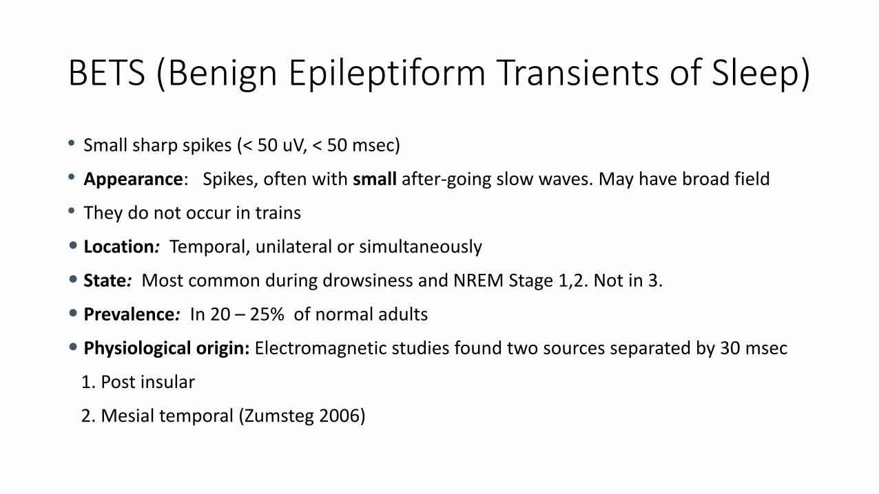
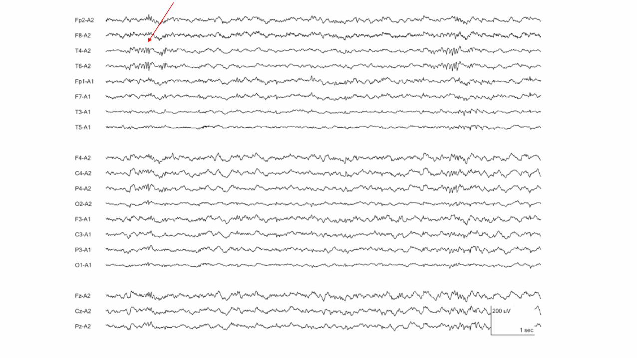
BETS (Benign Epileptiform Transients of Sleep)
• Small sharp spikes (< 50 uV, < 50 msec)
• Appearance: Spikes, often with small after-going slow waves. May have broad field
• They do not occur in trains
• Location: Temporal, unilateral or simultaneously
• State: Most common during drowsiness and NREM Stage 1,2. Not in 3.
• Prevalence: In 20 – 25% of normal adults
• Physiological origin: Electromagnetic studies found two sources separated by 30 msec
1. Post insular
2. Mesial temporal (Zumsteg 2006)


Phantom Spike/Wave
• 6 Hz spike and wave bursts.
• Appearance: The spikes are small and and difficult to see, and may not be visible in association with some of the slow waves in the burst, hence the name “phantom” spike-and-wave.
• Location: Variable, may be widespread or more focal. May be larger over the front of the head or over the back.
• State: Relaxed wakefulness and drowsiness, and go away in deep sleep.
• Prevalence: 2.5% of normal adults

• The significance of phantom spike-and-wave is unclear.
• Subdivided into two categories, with mnemonics WHAM and FOLD.
• WHAM (in Wakefulness, High-voltage, Anterior maximum, in Males) – Commonly has co-occurring epileptiform abnormality.
•
• FOLD (Female, Occipital maximum, Low-voltage, in Drowsiness) – appears to be a more benign pattern.

14 and 6 Hz Positive Spike bursts
• Appearance: Trains of arch-shaped waveforms with positive-polarity spikes and smoothly-curved negative phases (inverse of “μ”). Amp: 75 uV, Duration: 0.5-1 sec.
• Location: Post temporal. Uni or bilat.
• State: Drowsiness and light sleep. Absent in awake state and S -3 sleep. (Olofsson 1971)
• Prevalence: Common 8-14 years (Niedermeyer 1999)


Lambda Waves
• Positive occipital sharp waves in wakefulness with visual exploration.
• Clinical significance:
No increase in likelihood of Focal epileptiform discharges.
More likely to see POST and Photic driving


Posterior Slow waves of Youth
• Slow waves of inconsistent polarity lasting for 0.3-05 seconds occurring during PDR
• Common from 2-6 years and 12-21 years

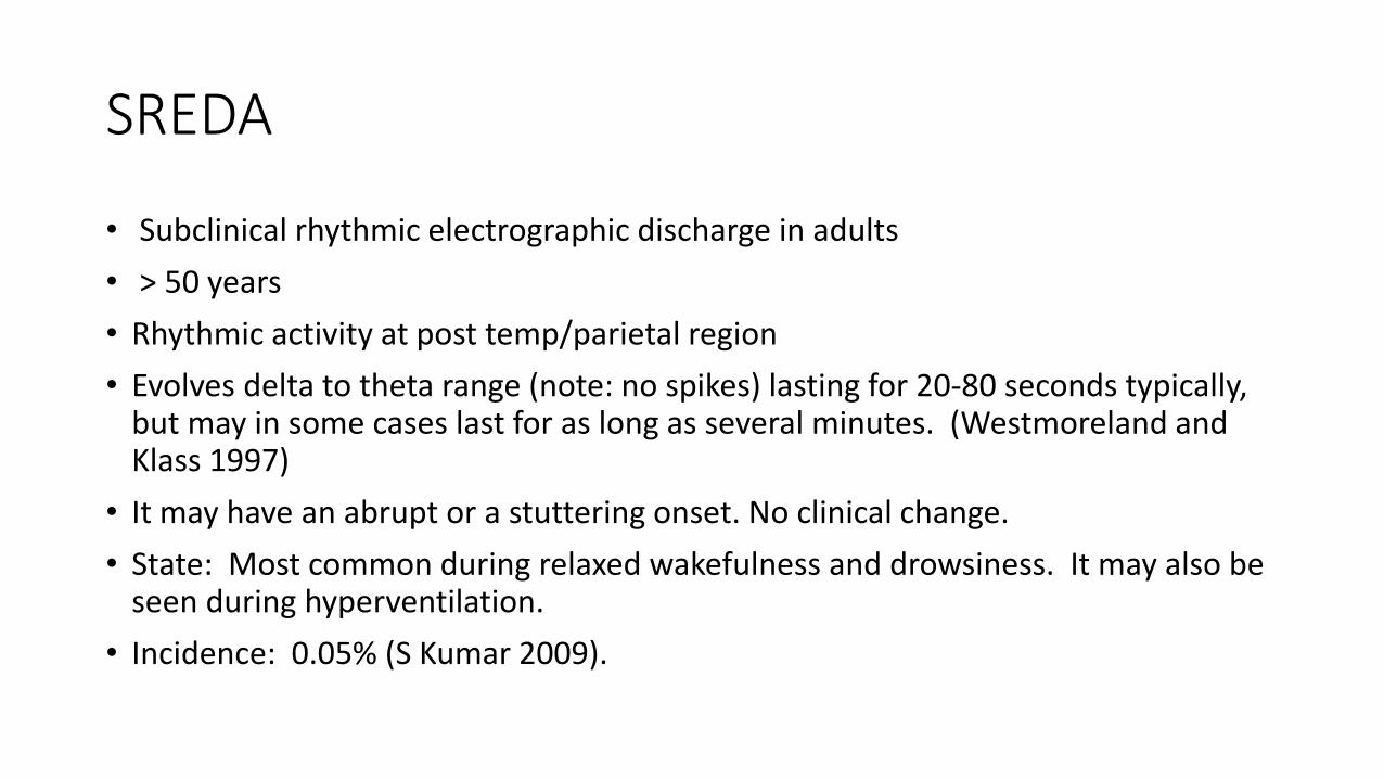
SREDA
• Subclinical rhythmic electrographic discharge in adults
• > 50 years
• Rhythmic activity at post temp/parietal region
• Evolves delta to theta range (note: no spikes) lasting for 20-80 seconds typically, but may in some cases last for as long as several minutes. (Westmoreland and Klass 1997)
• It may have an abrupt or a stuttering onset. No clinical change.
• State: Most common during relaxed wakefulness and drowsiness. It may also be seen during hyperventilation.
• Incidence: 0.05% (S Kumar 2009).

EEG Artifacts

Muscle Artifact

EKG Artifacts

Muscle Artifact-Chewing

Movement Artifact-Pulse

Rapid Eye Blinking

Electrode Artifact
• More problems with electrode artifact in the ICU• Prolonged monitoring
• Scalp integrity
• Vigilance required to keep electrodes from “going bad”• Many pts require
repair 2-3 x / day
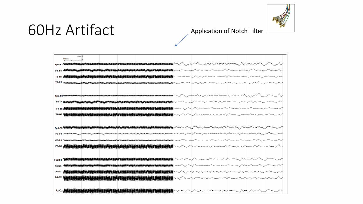
60Hz Artifact Application of Notch Filter
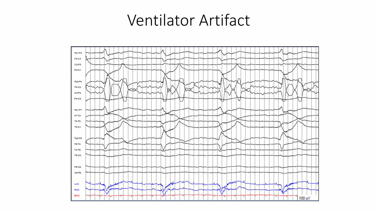
Ventilator Artifact

IV Drip Artifact
Water droplets are charged!


Questions ?
Thank you