Echocardiographic Evaluation of the Aorta - … · Echocardiographic Evaluation of the Aorta...
68
Echocardiographic Evaluation of the Aorta William F. Armstrong M.D. Director Echocardiography Laboratory Professor of Medicine University of Michigan
-
Upload
hoangkhanh -
Category
Documents
-
view
225 -
download
4
Transcript of Echocardiographic Evaluation of the Aorta - … · Echocardiographic Evaluation of the Aorta...

Echocardiographic Evaluation
of the Aorta
William F. Armstrong M.D.
Director Echocardiography Laboratory
Professor of Medicine
University of Michigan
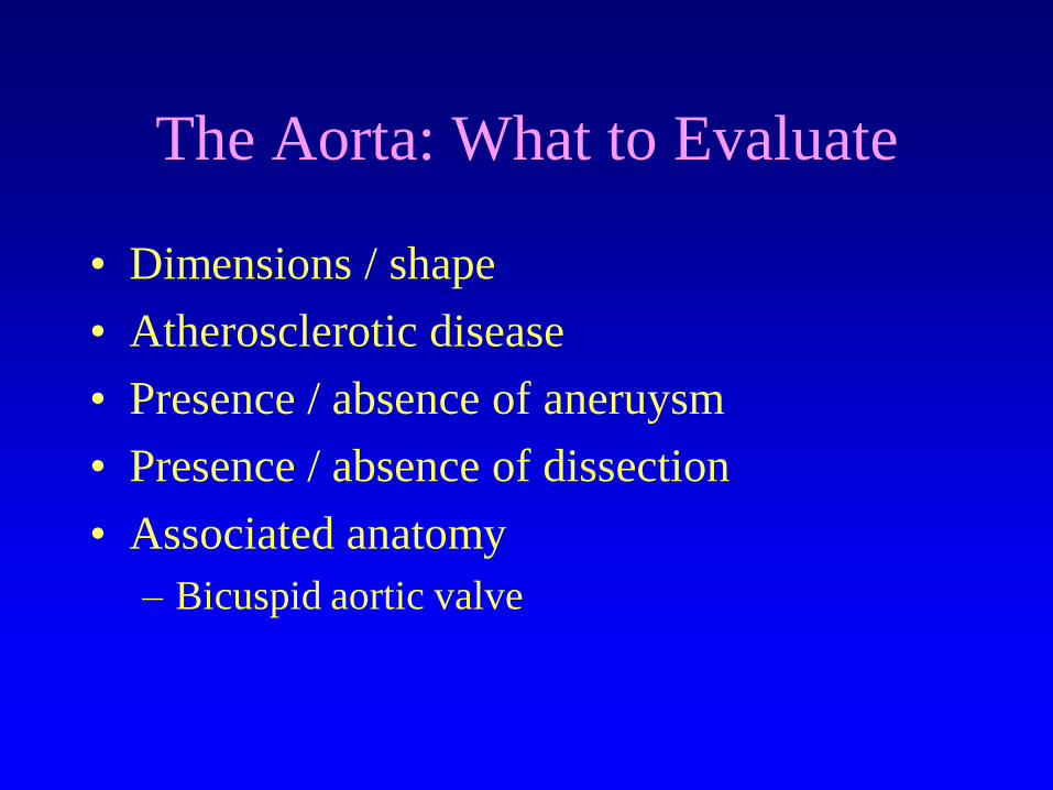
The Aorta: What to Evaluate
• Dimensions / shape
• Atherosclerotic disease
• Presence / absence of aneruysm
• Presence / absence of dissection
• Associated anatomy
– Bicuspid aortic valve

The Aorta: When to Evaluate
• Symptoms suggestive of aortic disease
• Known predisposing factor for aortic
disease
• Other test (CXR etc.) suggests aortic
disease
• First degree relatives of patients with aortic
disease
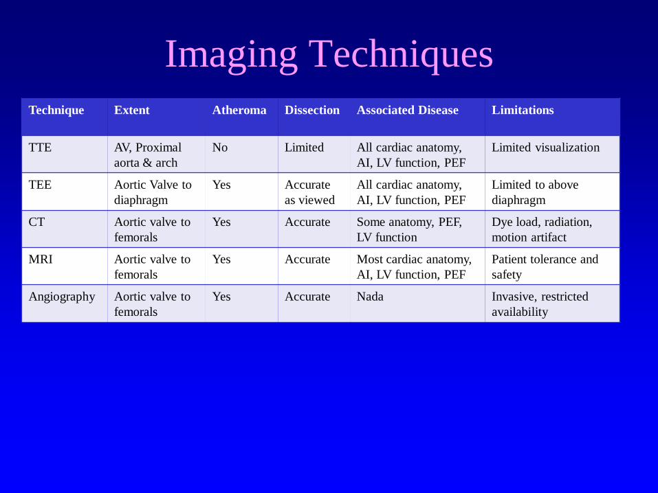
Imaging Techniques
Technique
Extent Atheroma Dissection Associated Disease Limitations
TTE AV, Proximal
aorta & arch
No Limited All cardiac anatomy,
AI, LV function, PEF
Limited visualization
TEE Aortic Valve to
diaphragm
Yes Accurate
as viewed
All cardiac anatomy,
AI, LV function, PEF
Limited to above
diaphragm
CT Aortic valve to
femorals
Yes Accurate Some anatomy, PEF,
LV function
Dye load, radiation,
motion artifact
MRI Aortic valve to
femorals
Yes Accurate Most cardiac anatomy,
AI, LV function, PEF
Patient tolerance and
safety
Angiography Aortic valve to
femorals
Yes Accurate Nada Invasive, restricted
availability
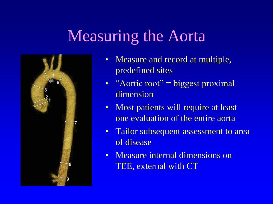
Measuring the Aorta
• Measure and record at multiple,
predefined sites
• “Aortic root” = biggest proximal
dimension
• Most patients will require at least
one evaluation of the entire aorta
• Tailor subsequent assessment to area
of disease
• Measure internal dimensions on
TEE, external with CT

Evaluating the Aorta: Which
Technique Sees What
CT / MRI TEE TTE
“Acute Aortic Syndrome”
• Acute aortic dissection
– Type A, B
• Acute intramural hematoma
• Rapidly expanding aneurysm
• Ruptured Aneurysm
• Penetrating ulcer
WG: 24 YO male
• Told of dilated aorta at age 18
– No meds and no follow-up
• Sudden chest pain at work
• Cardiac arrest in ambulance
• CPR not successful in ED
• Autopsy: type I dissection
SJ: 54 YO male
• PMHx = HTN, type 2 diabetes
• Dull retrosternal pain for 40 minutes
– Normal ECG and cardiac enzymes
• Discharged after MI excluded
• Subsequent arrest at home
• Autopsy: type I dissection
Litigation Pending

Classification of Aortic Dissection
Echocardiography 6th Edition
If the ascending aorta
is involved, call a surgeon!
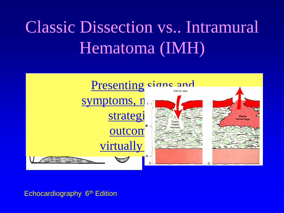
Classic Dissection vs.. Intramural
Hematoma (IMH)
Echocardiography 6th Edition
Presenting signs and
symptoms, management
strategies and
outcomes are
virtually identical

Aortic Dissection: High Risk
Conditions
* Loeys-Dietz syndrome, vascular Ehlers-Danlos syndrome, Turner syndrome, or other connective tissue disease.
†Patients with mutations in genes known to predispose to thoracic aortic aneurysms and
dissection, such as FBN1, TGFBR1, TGFBR2, ACTA2, and MYH11.
• Marfan Syndrome
• Connective tissue disease*
• Family history of aortic disease
• Known aortic valve disease
• Recent aortic manipulation (surgical or
catheter-based)
• Known thoracic aortic aneurysm
• Genetic conditions that predispose to TAD†
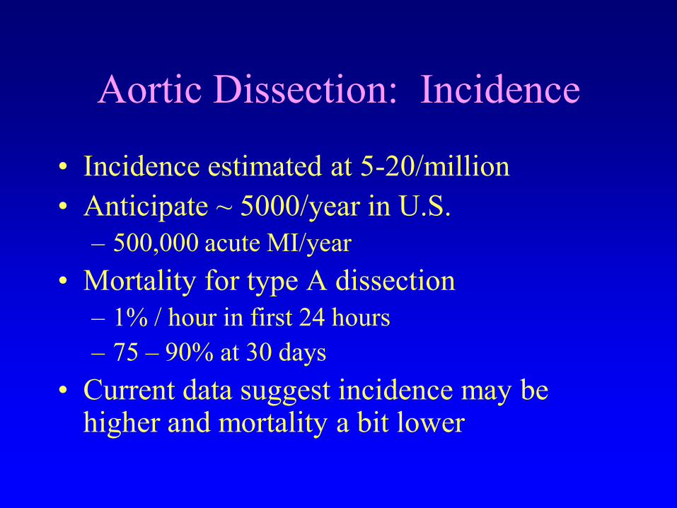
Aortic Dissection: Incidence
• Incidence estimated at 5-20/million
• Anticipate ~ 5000/year in U.S.
– 500,000 acute MI/year
• Mortality for type A dissection
– 1% / hour in first 24 hours
– 75 – 90% at 30 days
• Current data suggest incidence may be higher and mortality a bit lower
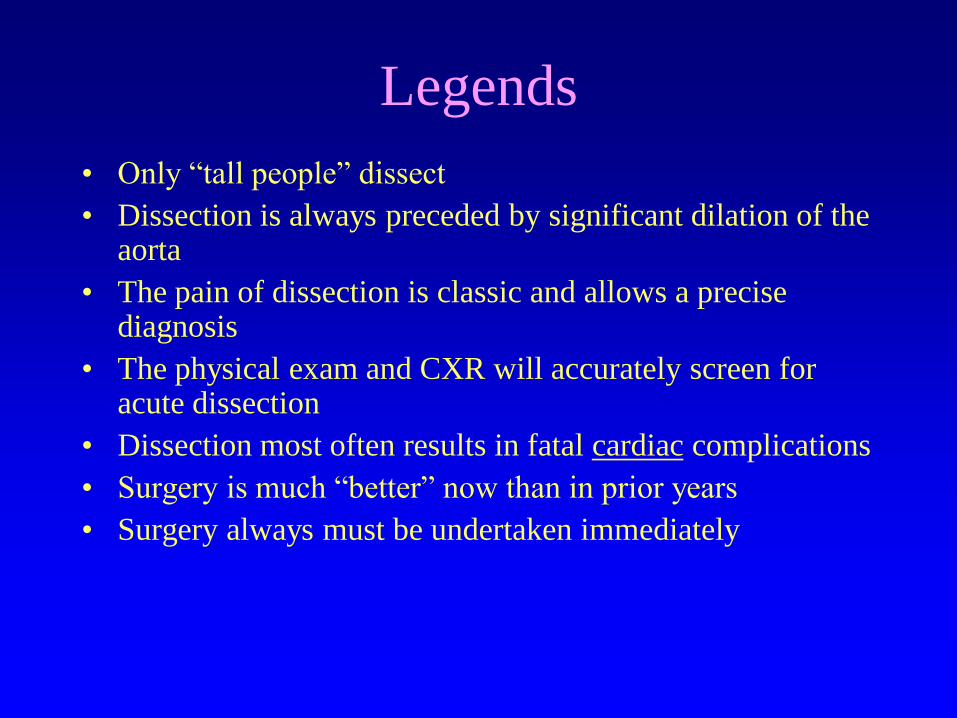
Legends
• Only “tall people” dissect
• Dissection is always preceded by significant dilation of the aorta
• The pain of dissection is classic and allows a precise diagnosis
• The physical exam and CXR will accurately screen for acute dissection
• Dissection most often results in fatal cardiac complications
• Surgery is much “better” now than in prior years
• Surgery always must be undertaken immediately
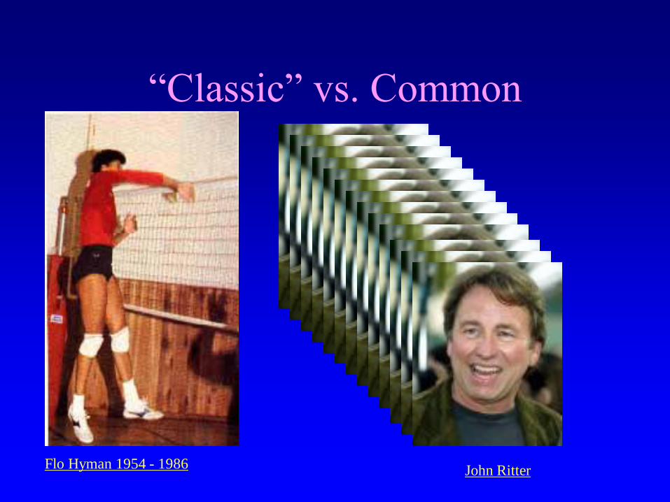
“Classic” vs. Common
Flo Hyman 1954 - 1986 John Ritter
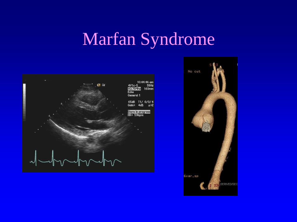
Marfan Syndrome
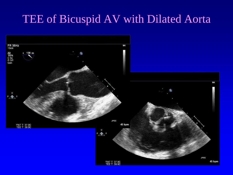
TEE of Bicuspid AV with Dilated Aorta

Aortic Dilation in Bicuspid
Aortic Valve

Younger vs.. Older Patients with
Acute Aortic Dissection - IRAD
Variables
Age <40
n = 68
Age > 40
n = 883
p Value
Age, yrs (mean + SD) 30.7 +6.6 63.9 + 11.5 NA
Type A 46 (68) 574 (65)
Hypertension 23 (34) 635 (72) < 0.001
Marfan Syndrome 34 (50) 19 (2) < 0.001
Bicuspid aortic valve 6 (9) 12 (1) < 0.001
Hypertension (SBP > 150
mm Hg)
17 (25) 394 (45) 0.003
Januzzi et al JACC 2004
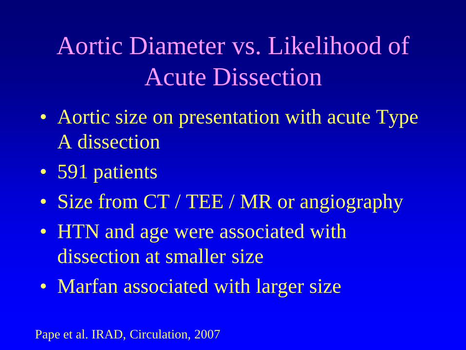
Aortic Diameter vs. Likelihood of
Acute Dissection
• Aortic size on presentation with acute Type
A dissection
• 591 patients
• Size from CT / TEE / MR or angiography
• HTN and age were associated with
dissection at smaller size
• Marfan associated with larger size
Pape et al. IRAD, Circulation, 2007
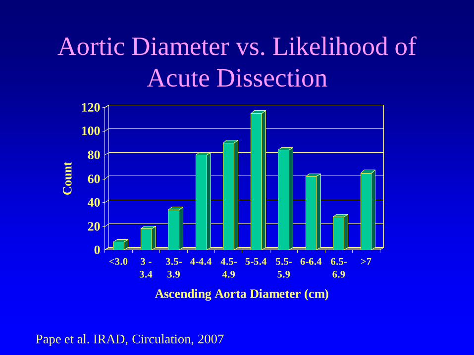
Aortic Diameter vs. Likelihood of
Acute Dissection
0
20
40
60
80
100
120
Co
un
t
<3.0 3 -
3.4
3.5-
3.9
4-4.4 4.5-
4.9
5-5.4 5.5-
5.9
6-6.4 6.5-
6.9
>7
Ascending Aorta Diameter (cm)
Pape et al. IRAD, Circulation, 2007
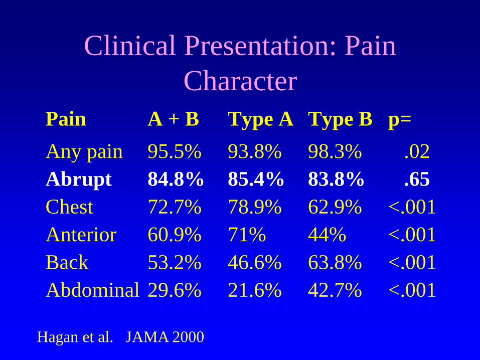
Clinical Presentation: Pain
Character
Pain A + B Type A Type B p=
Any pain 95.5% 93.8% 98.3% .02
Abrupt 84.8% 85.4% 83.8% .65
Chest 72.7% 78.9% 62.9% <.001
Anterior 60.9% 71% 44% <.001
Back 53.2% 46.6% 63.8% <.001
Abdominal 29.6% 21.6% 42.7% <.001
Hagan et al. JAMA 2000
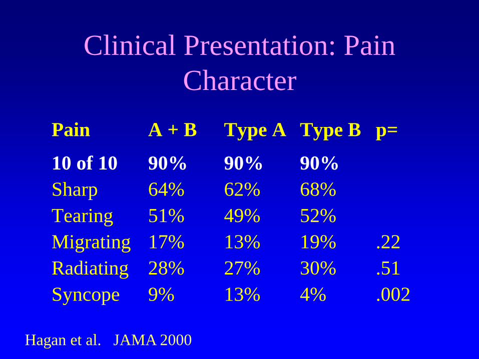
Clinical Presentation: Pain
Character
Pain A + B Type A Type B p=
10 of 10 90% 90% 90%
Sharp 64% 62% 68%
Tearing 51% 49% 52%
Migrating 17% 13% 19% .22
Radiating 28% 27% 30% .51
Syncope 9% 13% 4% .002
Hagan et al. JAMA 2000

The Pain of Dissection:
Practical Clues
• Classic, abrupt, tearing chest & back pain does occur – but it represents a minority of cases
• Other clues:
- Pain with multiple migratory areas
- Recurrent pain - stable EKG
- Pain not responsive to NTG
- Minimal troponin leak
-Abrupt onset, no prodrome
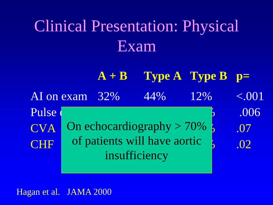
Clinical Presentation: Physical
Exam
A + B Type A Type B p=
AI on exam 32% 44% 12% <.001
Pulse deficit 15% 19% 9.2% .006
CVA 4.7% 6.1% 2.3% .07
CHF 6.6% 8.8% 3.0% .02
Hagan et al. JAMA 2000
On echocardiography > 70%
of patients will have aortic
insufficiency
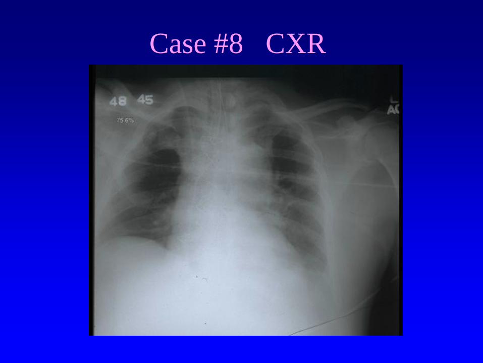
Case #8 CXR

Clinical Presentation: CXR
N= 427 A + B A B p=
No Abnormality 12% 11% 16% .08
Mediastinum Nl 21% 17% 27% .01
Wide Mediastinum 62% 63% 56% .17
Aorta Abnormal 50% 47% 53% .20
Heart Abnormal 26% 27% 24% .49
Pleural Effusion 19% 17% 22% .24Hagan et al. JAMA 2000
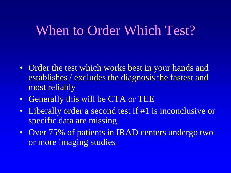
When to Order Which Test?
• Order the test which works best in your hands and establishes / excludes the diagnosis the fastest and most reliably
• Generally this will be CTA or TEE
• Liberally order a second test if #1 is inconclusive or specific data are missing
• Over 75% of patients in IRAD centers undergo two or more imaging studies
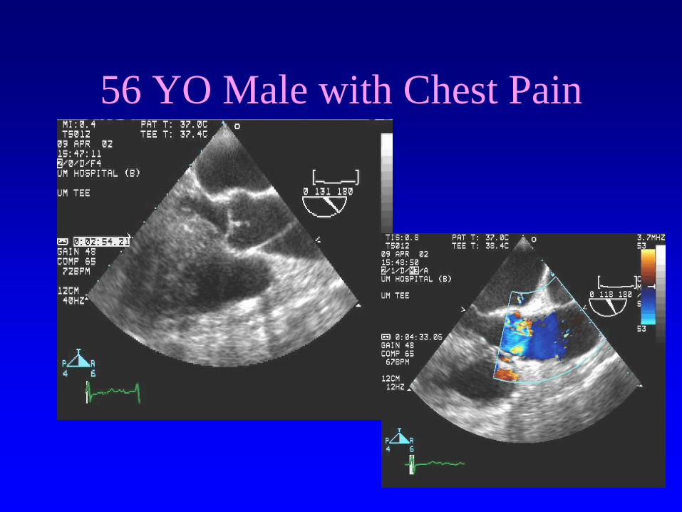
56 YO Male with Chest Pain
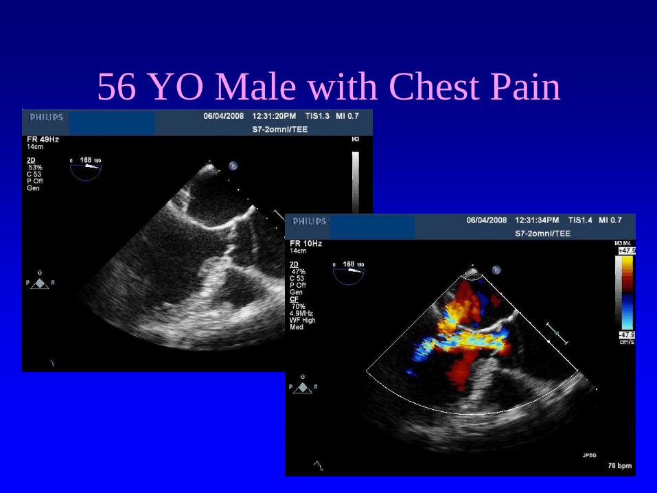
56 YO Male with Chest Pain
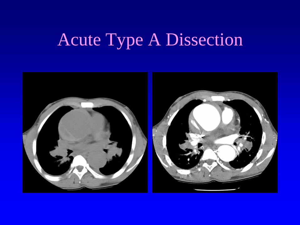
Acute Type A Dissection
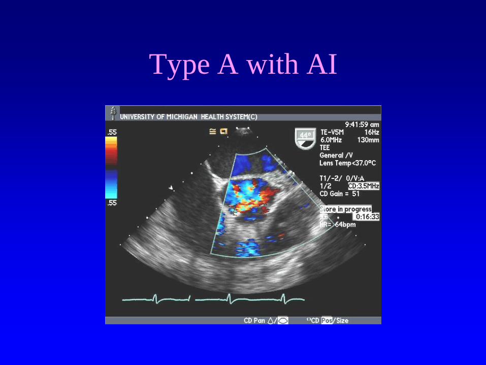
Type A with AI
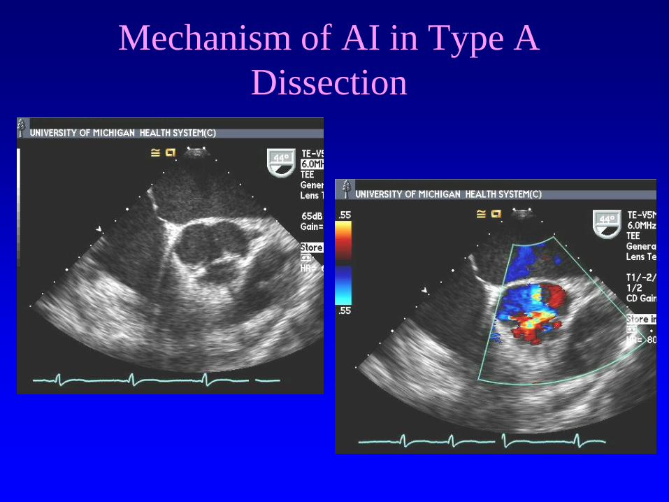
Mechanism of AI in Type A
Dissection
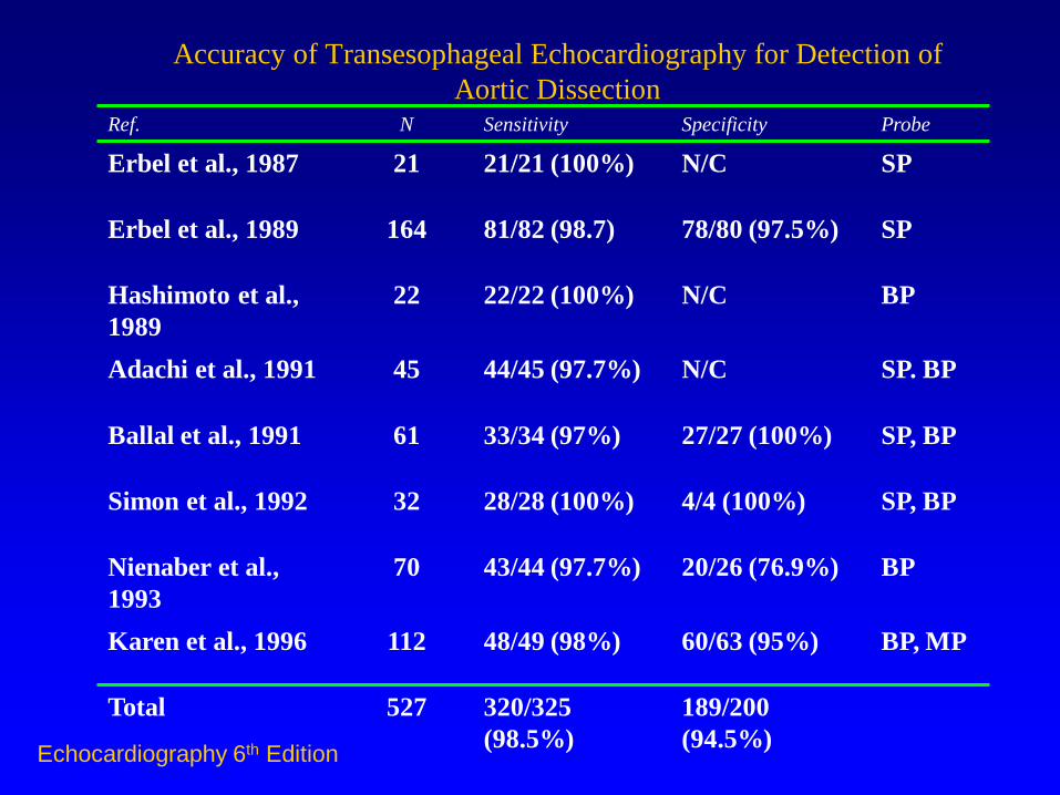
Accuracy of Transesophageal Echocardiography for Detection of
Aortic Dissection Ref. N Sensitivity Specificity Probe
Erbel et al., 1987 21 21/21 (100%) N/C SP
Erbel et al., 1989 164 81/82 (98.7) 78/80 (97.5%) SP
Hashimoto et al.,
1989
22 22/22 (100%) N/C BP
Adachi et al., 1991 45 44/45 (97.7%) N/C SP. BP
Ballal et al., 1991 61 33/34 (97%) 27/27 (100%) SP, BP
Simon et al., 1992 32 28/28 (100%) 4/4 (100%) SP, BP
Nienaber et al.,
1993
70 43/44 (97.7%) 20/26 (76.9%) BP
Karen et al., 1996 112 48/49 (98%) 60/63 (95%) BP, MP
Total 527 320/325
(98.5%)
189/200
(94.5%) Echocardiography 6th Edition
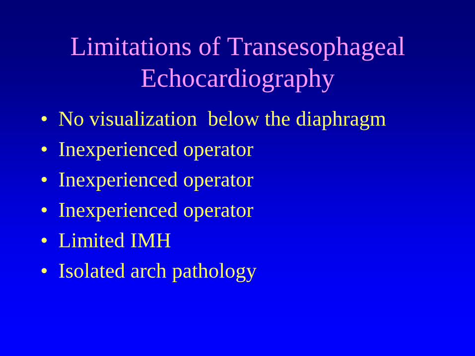
Limitations of Transesophageal
Echocardiography
• No visualization below the diaphragm
• Inexperienced operator
• Inexperienced operator
• Inexperienced operator
• Limited IMH
• Isolated arch pathology
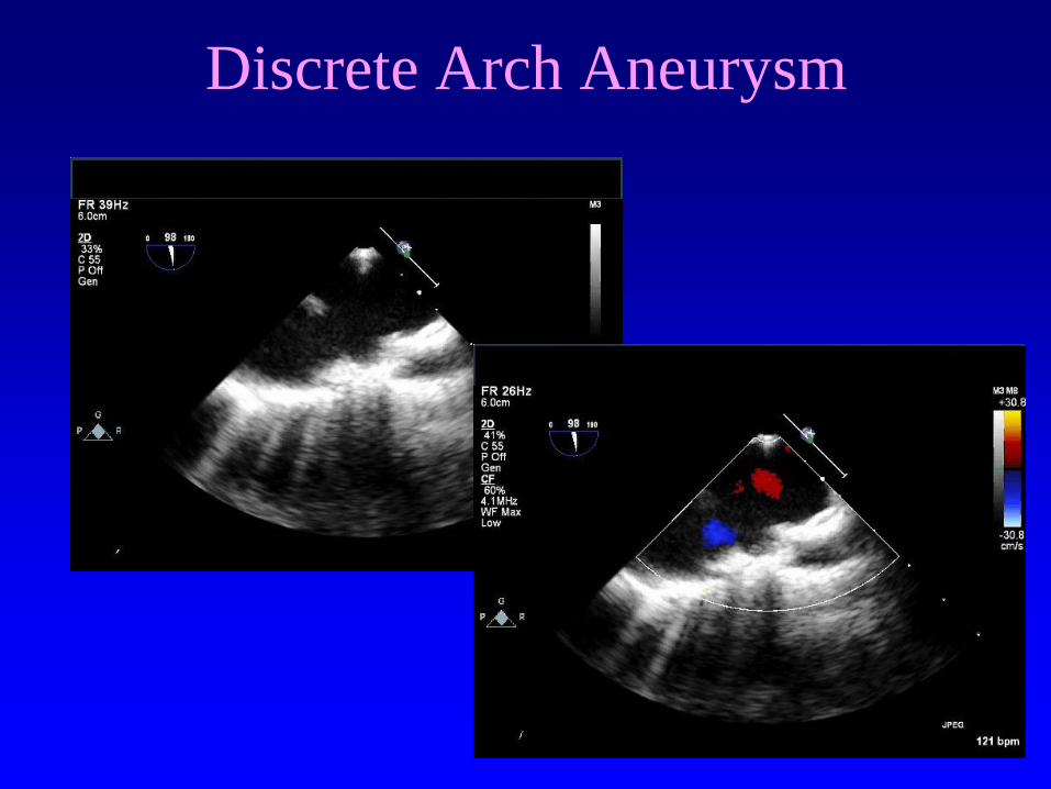
Discrete Arch Aneurysm
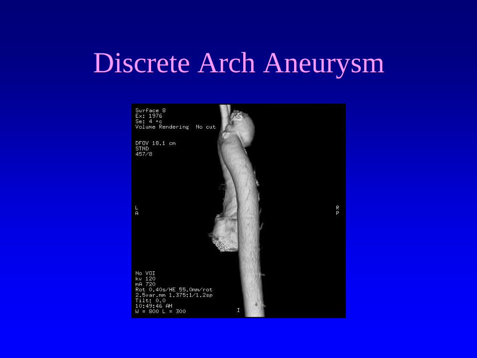
Discrete Arch Aneurysm
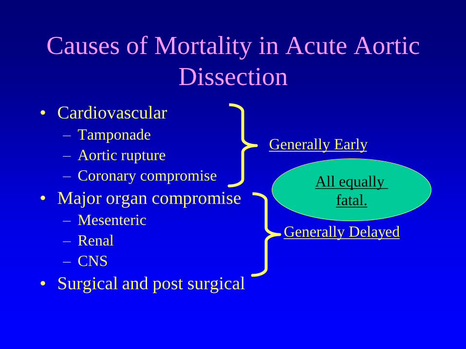
Causes of Mortality in Acute Aortic
Dissection
• Cardiovascular
– Tamponade
– Aortic rupture
– Coronary compromise
• Major organ compromise
– Mesenteric
– Renal
– CNS
• Surgical and post surgical
Generally Early
Generally Delayed
All equally
fatal.
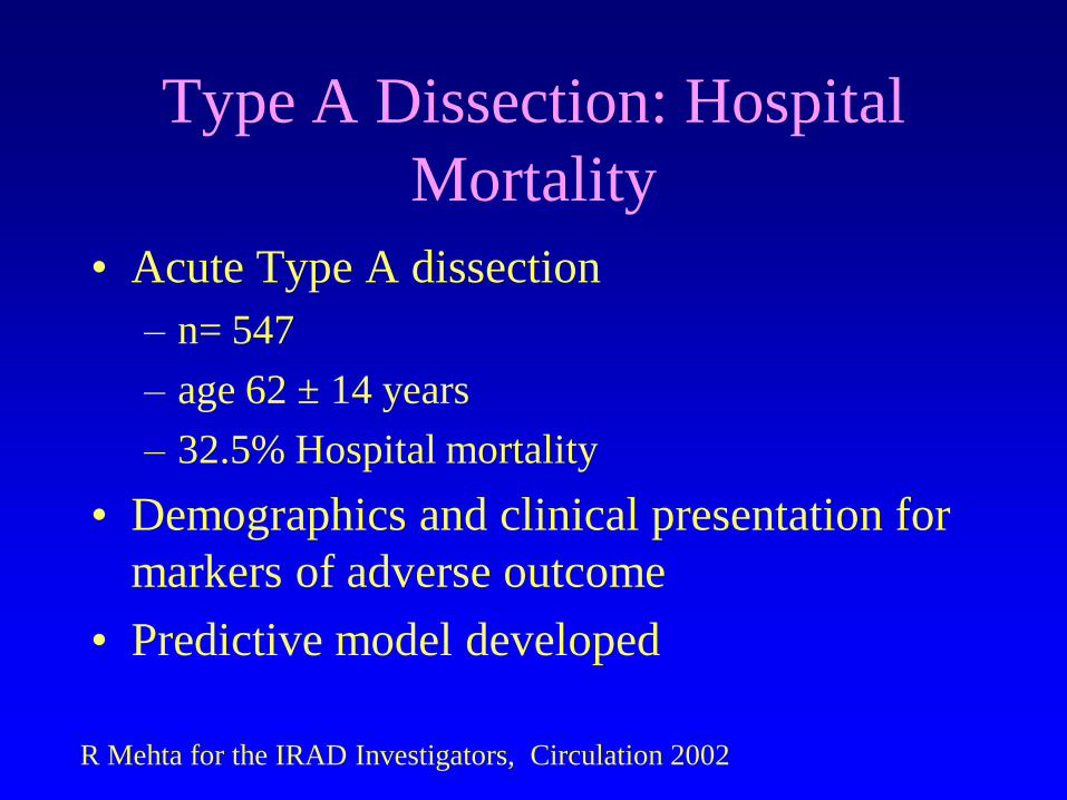
Type A Dissection: Hospital
Mortality
• Acute Type A dissection
– n= 547
– age 62 ± 14 years
– 32.5% Hospital mortality
• Demographics and clinical presentation for
markers of adverse outcome
• Predictive model developed
R Mehta for the IRAD Investigators, Circulation 2002

63 YO Male with Syncope

63 YO Male with Syncope: Two
Minutes Later

63 YO Male with Syncope: Three
Minutes Later
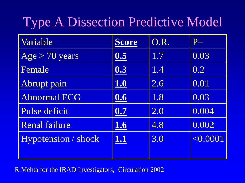
Type A Dissection Predictive Model
Variable Score O.R. P=
Age > 70 years 0.5 1.7 0.03
Female 0.3 1.4 0.2
Abrupt pain 1.0 2.6 0.01
Abnormal ECG 0.6 1.8 0.03
Pulse deficit 0.7 2.0 0.004
Renal failure 1.6 4.8 0.002
Hypotension / shock
1.1 3.0 <0.0001
R Mehta for the IRAD Investigators, Circulation 2002
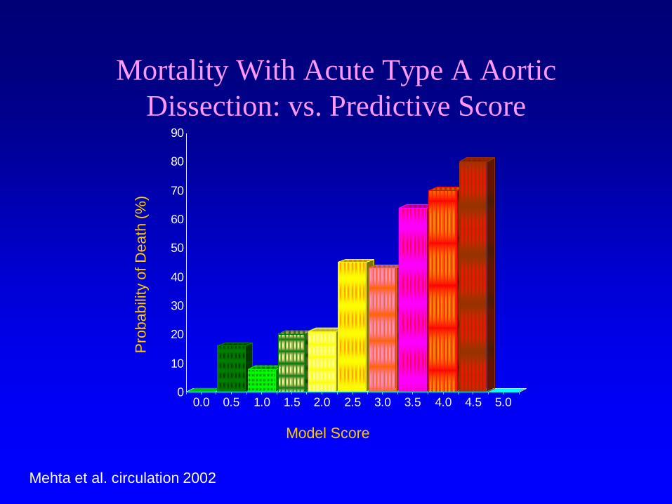
Mortality With Acute Type A Aortic
Dissection: vs. Predictive Score
0
10
20
30
40
50
60
70
80
90
0.0 0.5 1.0 1.5 2.0 2.5 3.0 3.5 4.0 4.5 5.0
Model Score
Pro
babili
ty o
f D
eath
(%
)
Mehta et al. circulation 2002
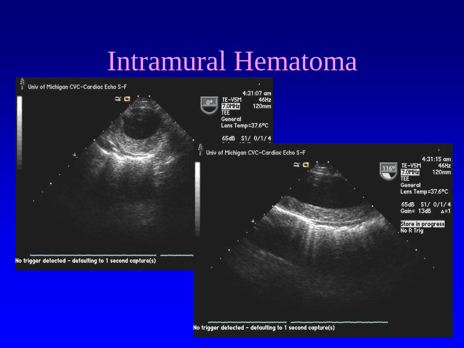
Intramural Hematoma
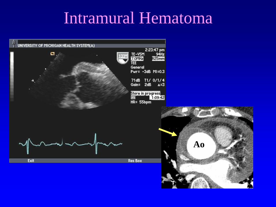
Intramural Hematoma
Ao
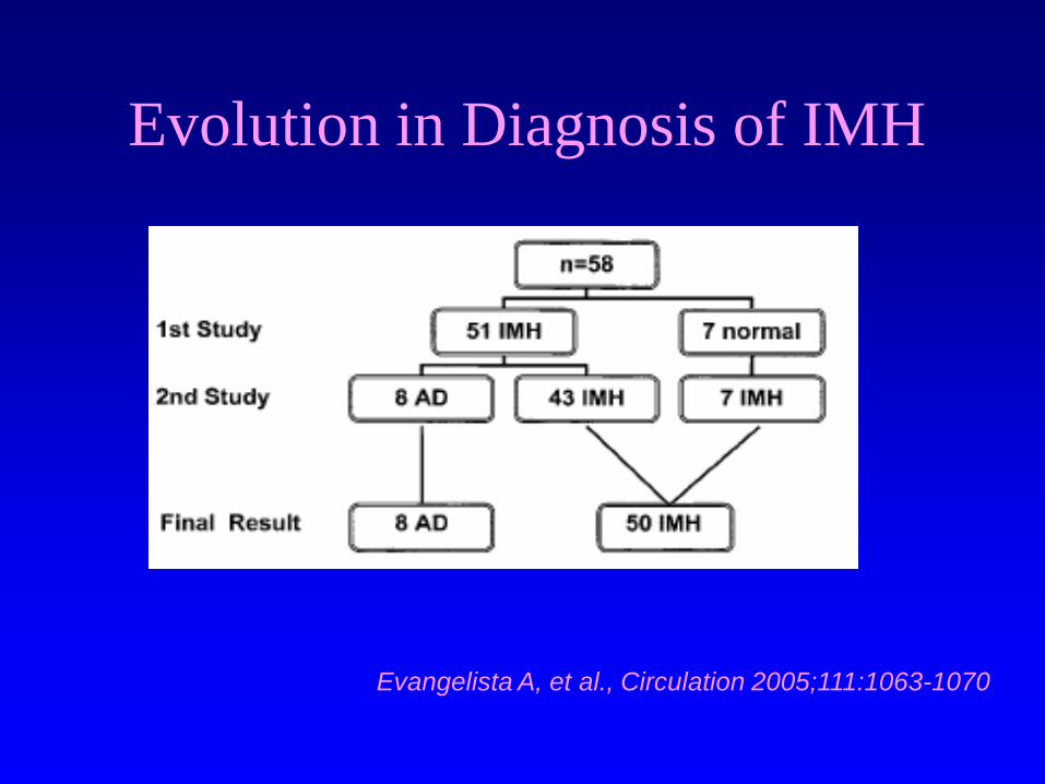
Evolution in Diagnosis of IMH
Evangelista A, et al., Circulation 2005;111:1063-1070

In-hospital Mortality for IMH
• 1010 patients with AAD in IRAD
• IMH in 58 (5.7%)
• Less likely to have AI or pulse deficits
• More difficult to diagnose
• Less often surgically treated
Evangelista A, et al., Circulation 2005;111:1063-1070
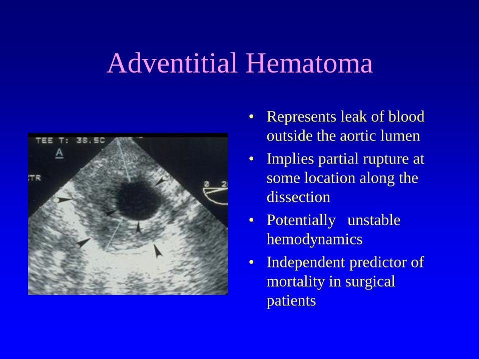
Adventitial Hematoma
• Represents leak of blood
outside the aortic lumen
• Implies partial rupture at
some location along the
dissection
• Potentially unstable
hemodynamics
• Independent predictor of
mortality in surgical
patients

TEE: Patent False Lumen
• Acutely allows further
propagation of
dissection (type A)
• Subsequent additional
organ compromise
• Chronically in type B
appears protective

False Lumen Thrombosis
• Implies a completed
event
• No hydrodynamic
drive for further
propagation
• Presumed stable wall
structure
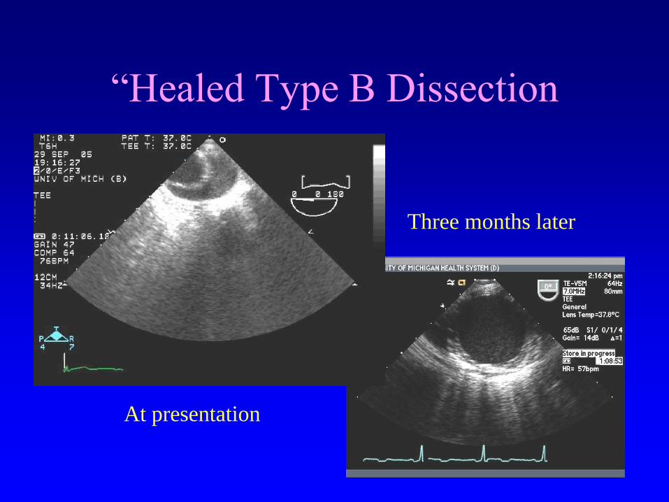
“Healed Type B Dissection
At presentation
Three months later
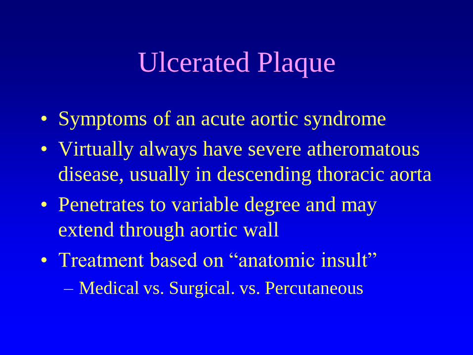
Ulcerated Plaque
• Symptoms of an acute aortic syndrome
• Virtually always have severe atheromatous
disease, usually in descending thoracic aorta
• Penetrates to variable degree and may
extend through aortic wall
• Treatment based on “anatomic insult”
– Medical vs. Surgical. vs. Percutaneous
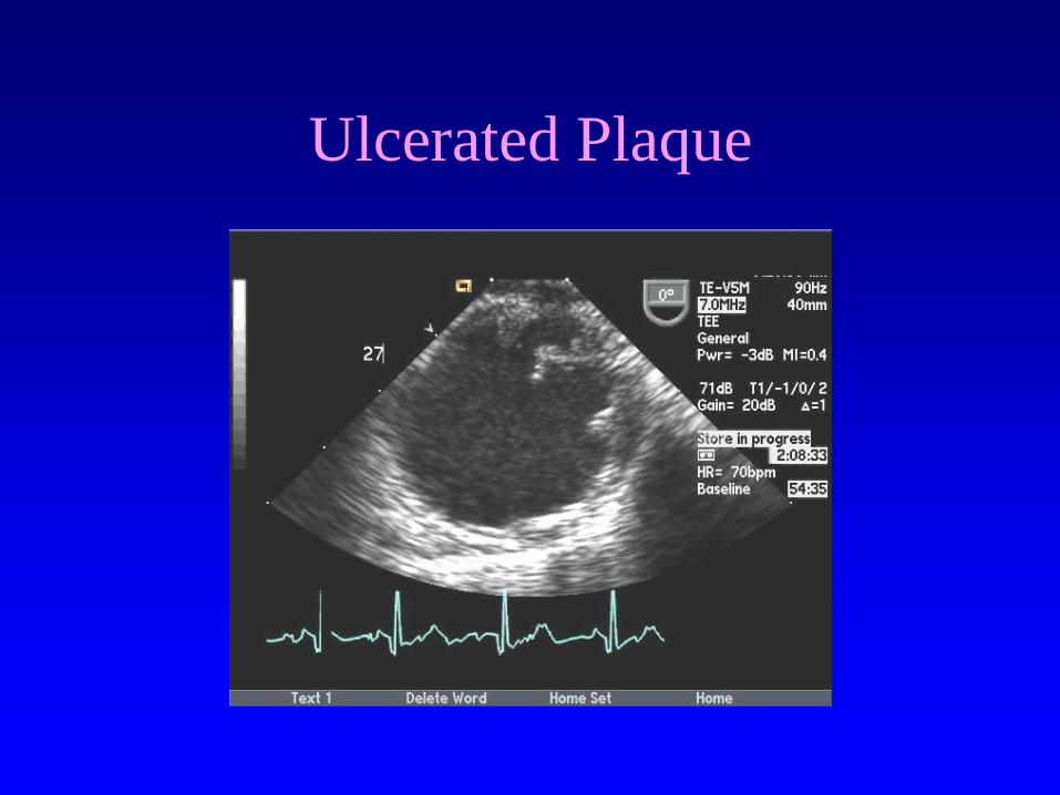
Ulcerated Plaque

Ulcerated Plaque
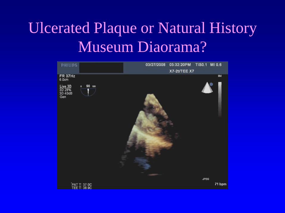
Ulcerated Plaque or Natural History
Museum Diaorama?

2009: Where are We with Surgery
for Acute Aortic Dissection?
There’s good news,
and there’s
bad news.
First the good news!!
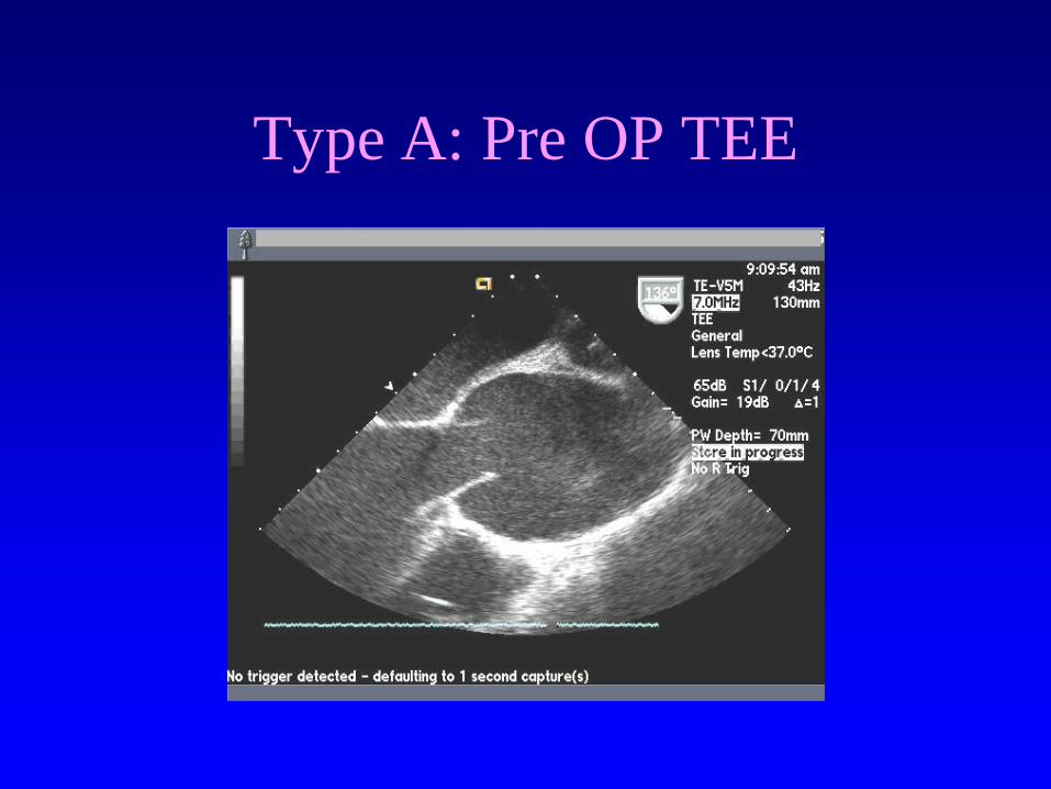
Type A: Pre OP TEE
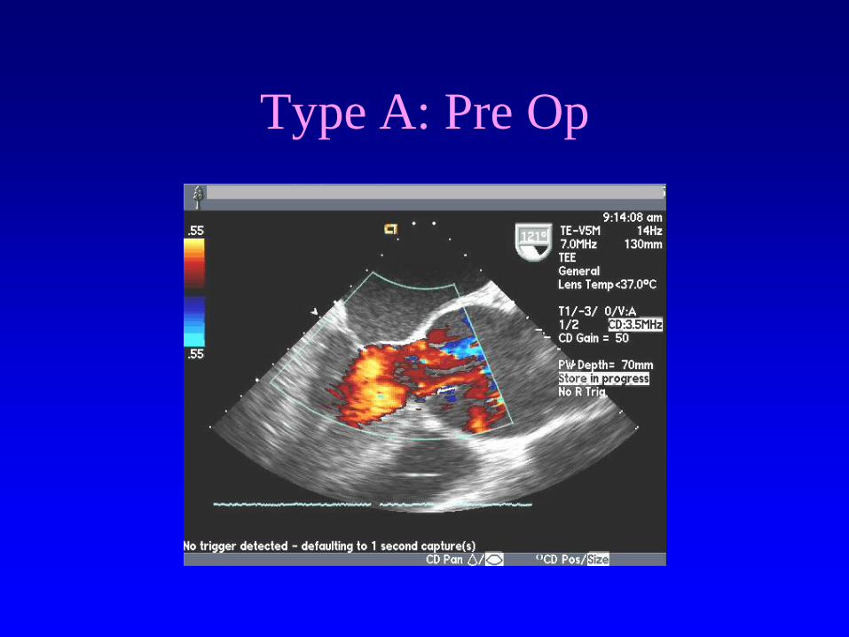
Type A: Pre Op
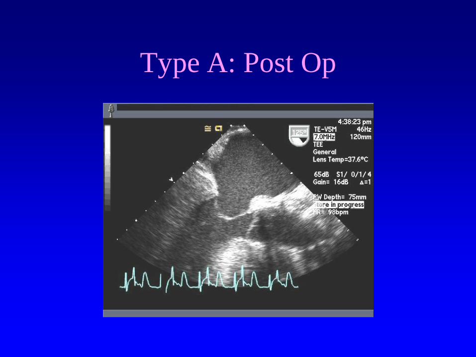
Type A: Post Op
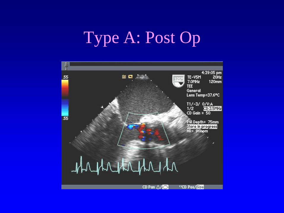
Type A: Post Op
2009: Where are We with Surgery
for Acute Aortic Dissection?
Now the bad news.

Surgical Management
• 30 year experience
• 360 patients, 256 male, age 57 + 14
• 174 acute type A (48%)
• Operative mortality 24%
Fann et al, Stanford; Circulation 1995

Aortic Dissection: Surgical
Mortality
0
10
20
30
40
50
60
63-72 73-77 78-82 83-87 88-92
- Complications
+Complications
*
*
*
*
Fann et al. Circulation 1995
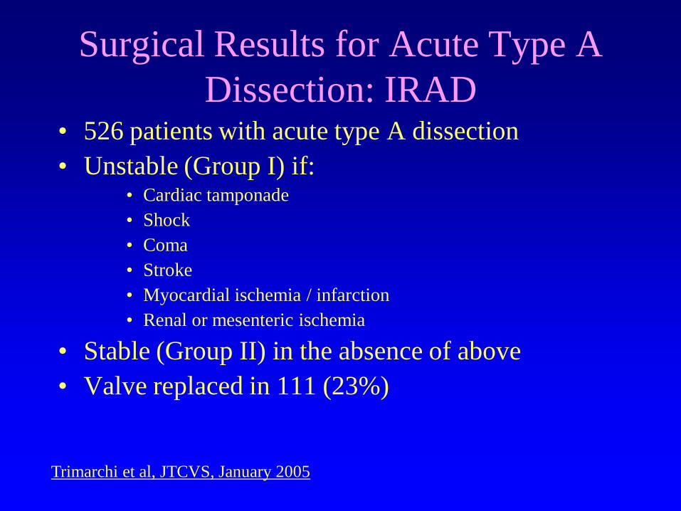
Surgical Results for Acute Type A
Dissection: IRAD • 526 patients with acute type A dissection
• Unstable (Group I) if: • Cardiac tamponade
• Shock
• Coma
• Stroke
• Myocardial ischemia / infarction
• Renal or mesenteric ischemia
• Stable (Group II) in the absence of above
• Valve replaced in 111 (23%)
Trimarchi et al, JTCVS, January 2005
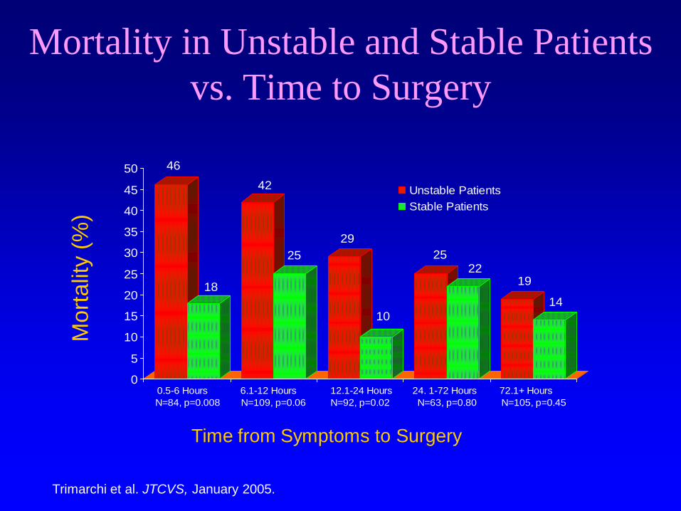
Mortality in Unstable and Stable Patients
vs. Time to Surgery
46
18
42
25
29
10
2522
19
14
0
5
10
15
20
25
30
35
40
45
50
0.5-6 Hours
N=84, p=0.008
6.1-12 Hours
N=109, p=0.06
12.1-24 Hours
N=92, p=0.02
24. 1-72 Hours
N=63, p=0.80
72.1+ Hours
N=105, p=0.45
Unstable Patients
Stable Patients
Trimarchi et al. JTCVS, January 2005.
Time from Symptoms to Surgery
Mo
rta
lity (
%)
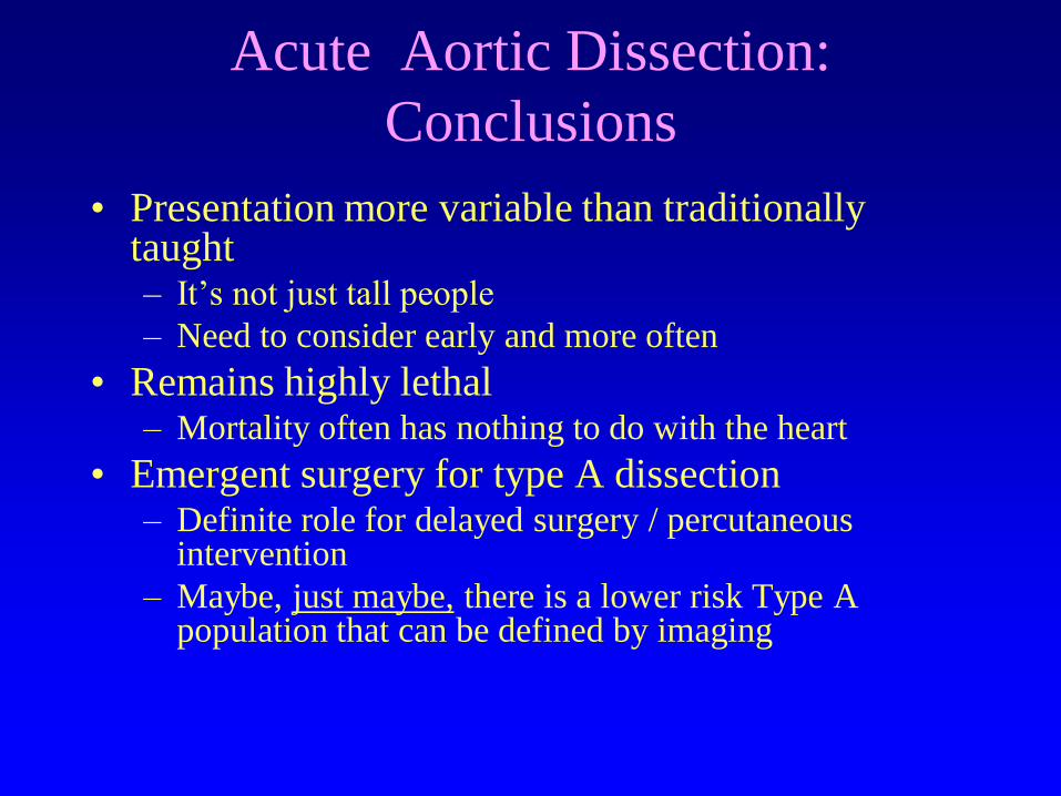
Acute Aortic Dissection:
Conclusions
• Presentation more variable than traditionally taught – It’s not just tall people
– Need to consider early and more often
• Remains highly lethal – Mortality often has nothing to do with the heart
• Emergent surgery for type A dissection – Definite role for delayed surgery / percutaneous
intervention
– Maybe, just maybe, there is a lower risk Type A population that can be defined by imaging