
E-cadherin is crucial for embryonic stem cell...
7
E-cadherin is crucial for embryonic stem cell pluripotency and can replace OCT4 during somatic cell reprogramming Torben Redmer 1,2 *, Sebastian Diecke 1,2 *, Tamara Grigoryan 1,3 , Angel Quiroga-Negreira 1,3 , Walter Birchmeier 1 & Daniel Besser 1+ 1 Max Delbrueck Center, 2 Freie Universita ¨t Berlin, Department of Biology, Chemistry and Pharmacy, and 3 Humboldt Universita ¨t zu Berlin, Faculty of Mathematics and Natural Sciences I, Berlin, Germany We report new functions of the cell-adhesion molecule E-cadherin in murine pluripotent cells. E-cadherin is highly expressed in mouse embryonic stem cells, and interference with E-cadherin causes differentiation. During cellular reprogramming of mouse fibroblasts by OCT4, SOX2, KLF4 and c-MYC, fully repro- grammed cells were exclusively observed in the E-cadherin- positive cell population and could not be obtained in the absence of E-cadherin. Moreover, reprogrammed cells could be established by viral E-cadherin in the absence of exogenous OCT4. Thus, reprogramming requires spatial cues that cross-talk with essential transcription factors. The cell-adhesion molecule E-cadherin has important functions in pluripotency and reprogramming. Keywords: pluripotency; somatic cell reprogramming; E-cadherin; OCT4 EMBO reports (2011) 12, 720–726. doi:10.1038/embor.2011.88 INTRODUCTION The regulation of pluripotency in mouse embryonic stem cells (mESCs) is a fascinating biological question with important implications for both basic science and the therapeutic potential of such cells. The same is true for induced pluripotent stem cells (iPSCs) of somatic origin. Both pluripotency and reprogramming are influenced by several cellular processes such as regulation of transcription, signal transduction, regulation of microRNAs and epigenetic changes (reviewed in Jaenisch & Young, 2008). The concept of somatic cell reprogramming began with somatic cell nuclear transfer (Wilmut et al, 1997) and was further developed by the finding that mouse embryonic fibroblasts (MEFs) can be converted into iPSCs by retroviral expression of four transcription factors: OCT4, SOX2, KLF4 and c-MYC (Takahashi & Yamanaka, 2006). The cadherins are membrane-spanning proteins of adherens junctions that have crucial roles in cell–cell contact formation. A switch from epithelial components, such as E-cadherin (Cadherin 1, Ecad) expression, to mesenchymal components can occur, termed epithelial–mesenchymal transition, which promotes the cell migration in the development of many organs and in the metastasis of tumours (Heuberger & Birchmeier, 2010). Opposite mechanisms—mesenchymal–epithelial transitions (METs)—are im- portant in strengthening tissue integrity following migratory processes in embryonic development. The function of E-cadherin in cell–cell adhesion is also connected to various signalling pathways that relay information regarding cell interactions to the cytoplasm and the nucleus (Stepniak et al, 2009). In early mammalian development, E- cadherin-based cell–cell contacts are required for the integrity of the embryonic blastocyst, and deletion of the E-cadherin gene results in failure of cell compaction at this stage (Larue et al, 1994). It has also been shown that E-cadherin controls processes of early differentiation (Larue et al, 1996) and has an important role in regulating pluripotency of different stem-cell compartments (Chou et al, 2008; Soncin et al, 2009). In this study, we show that E-cadherin is essential for the maintenance of pluripotency of mESCs, and that disturbance or loss of E-cadherin induces epithelial–mesenchymal transitions. During reprogramming of MEFs to iPSCs, E-cadherin production is associated with the reprogramming process and, remarkably, exogenous expression of E-cadherin can replace the requirement for OCT4 during reprogramming. RESULTS AND DISCUSSION E-cadherin is induced during reprogramming We observe that undifferentiated mESCs show a high level of E-cadherin and OCT4 messenger RNA (mRNA) and protein expression, that is lost when cells differentiate (supplementary Fig S1A,B online). Moreover, by using short-hairpin RNA against E-cadherin, we showed that high expression of E-cadherin in pluripotent mESCs is crucial for maintenance of the undifferentiated Received 17 September 2010; revised 23 March 2011; accepted 8 April 2011; published online 27 May 2011 *These authors contributed equally to this work + Corresponding author. Tel: þ 49 30 9406 2488; Fax: þ 49 30 9406 2656; E-mail: [email protected] 1 Max Delbrueck Center, Robert-Roessle-Strasse 10, 13125 Berlin, Germany 2 Freie Universita ¨t Berlin, Department of Biology, Chemistry and Pharmacy, Takustrasse 3, 14195 Berlin, Germany 3 Humboldt Universita ¨t zu Berlin, Faculty of Mathematics and Natural Sciences I, Unter den Linden 6, 10099 Berlin, Germany EMBO reports VOL 12 | NO 7 | 2011 &2011 EUROPEAN MOLECULAR BIOLOGY ORGANIZATION scientificreport scientific report 720
Transcript of E-cadherin is crucial for embryonic stem cell...

E-cadherin is crucial for embryonic stem cellpluripotency and can replace OCT4 duringsomatic cell reprogrammingTorben Redmer1,2*, Sebastian Diecke1,2*, Tamara Grigoryan1,3, Angel Quiroga-Negreira1,3, Walter Birchmeier1
& Daniel Besser1+
1Max Delbrueck Center, 2Freie Universitat Berlin, Department of Biology, Chemistry and Pharmacy, and 3Humboldt Universitat
zu Berlin, Faculty of Mathematics and Natural Sciences I, Berlin, Germany
We report new functions of the cell-adhesion molecule E-cadherinin murine pluripotent cells. E-cadherin is highly expressed inmouse embryonic stem cells, and interference with E-cadherincauses differentiation. During cellular reprogramming of mousefibroblasts by OCT4, SOX2, KLF4 and c-MYC, fully repro-grammed cells were exclusively observed in the E-cadherin-positive cell population and could not be obtained in the absenceof E-cadherin. Moreover, reprogrammed cells could be establishedby viral E-cadherin in the absence of exogenous OCT4. Thus,reprogramming requires spatial cues that cross-talk with essentialtranscription factors. The cell-adhesion molecule E-cadherin hasimportant functions in pluripotency and reprogramming.Keywords: pluripotency; somatic cell reprogramming; E-cadherin;OCT4EMBO reports (2011) 12, 720–726. doi:10.1038/embor.2011.88
INTRODUCTIONThe regulation of pluripotency in mouse embryonic stem cells(mESCs) is a fascinating biological question with importantimplications for both basic science and the therapeutic potentialof such cells. The same is true for induced pluripotent stem cells(iPSCs) of somatic origin. Both pluripotency and reprogrammingare influenced by several cellular processes such as regulation oftranscription, signal transduction, regulation of microRNAs andepigenetic changes (reviewed in Jaenisch & Young, 2008). Theconcept of somatic cell reprogramming began with somatic cellnuclear transfer (Wilmut et al, 1997) and was further developed bythe finding that mouse embryonic fibroblasts (MEFs) can be converted
into iPSCs by retroviral expression of four transcription factors:OCT4, SOX2, KLF4 and c-MYC (Takahashi & Yamanaka, 2006).
The cadherins are membrane-spanning proteins of adherensjunctions that have crucial roles in cell–cell contact formation.A switch from epithelial components, such as E-cadherin(Cadherin 1, Ecad) expression, to mesenchymal components canoccur, termed epithelial–mesenchymal transition, which promotesthe cell migration in the development of many organs and in themetastasis of tumours (Heuberger & Birchmeier, 2010). Oppositemechanisms—mesenchymal–epithelial transitions (METs)—are im-portant in strengthening tissue integrity following migratory processesin embryonic development. The function of E-cadherin in cell–celladhesion is also connected to various signalling pathways that relayinformation regarding cell interactions to the cytoplasm and thenucleus (Stepniak et al, 2009). In early mammalian development, E-cadherin-based cell–cell contacts are required for the integrity of theembryonic blastocyst, and deletion of the E-cadherin gene results infailure of cell compaction at this stage (Larue et al, 1994). It has alsobeen shown that E-cadherin controls processes of early differentiation(Larue et al, 1996) and has an important role in regulatingpluripotency of different stem-cell compartments (Chou et al,2008; Soncin et al, 2009).
In this study, we show that E-cadherin is essential for themaintenance of pluripotency of mESCs, and that disturbance orloss of E-cadherin induces epithelial–mesenchymal transitions.During reprogramming of MEFs to iPSCs, E-cadherin production isassociated with the reprogramming process and, remarkably,exogenous expression of E-cadherin can replace the requirementfor OCT4 during reprogramming.
RESULTS AND DISCUSSIONE-cadherin is induced during reprogrammingWe observe that undifferentiated mESCs show a high level ofE-cadherin and OCT4 messenger RNA (mRNA) and proteinexpression, that is lost when cells differentiate (supplementaryFig S1A,B online). Moreover, by using short-hairpin RNA againstE-cadherin, we showed that high expression of E-cadherin inpluripotent mESCs is crucial for maintenance of the undifferentiated
Received 17 September 2010; revised 23 March 2011; accepted 8 April 2011;published online 27 May 2011
*These authors contributed equally to this work+Corresponding author. Tel: þ 49 30 9406 2488; Fax: þ 49 30 9406 2656;E-mail: [email protected]
1Max Delbrueck Center, Robert-Roessle-Strasse 10, 13125 Berlin, Germany2Freie Universitat Berlin, Department of Biology, Chemistry and Pharmacy,Takustrasse 3, 14195 Berlin, Germany3Humboldt Universitat zu Berlin, Faculty of Mathematics and Natural Sciences I,Unter den Linden 6, 10099 Berlin, Germany
EMBO reports VOL 12 | NO 7 | 2011 &2011 EUROPEAN MOLECULAR BIOLOGY ORGANIZATION
scientificreportscientific report
720

state (supplementary Fig S1C–E online). Furthermore, mESCs thatwere treated either with a neutralizing antibody, DECMA-1, orshort-interfering RNA against Ecad, or maintained in the absenceof leukaemia inhibitory factor (LIF) for 7 days (�LIF, 7d), startedto differentiate while changing their morphology from compactcolonies to scattered cells (supplementary Fig S1F online).Differentiating cells underwent a cadherin switch to N-cadherin(Ncad) expression, whereas the expression of E-cadherin andOCT4 were lost. Our results show that E-cadherin is requiredto maintain the undifferentiated state of mESCs.
Reprogramming of MEFs through viral expression of OCT4,SOX2, KLF4 and c-MYC (OSKM) proceeds in a stepwise mannerthat gives rise to not only iPSCs, but also intermediate cell states.We used this procedure to generate iPSCs and sorted for cells thatexpressed the stem-cell marker stage-specific embryonic antigen 1(SSEA1) by FACS analysis (Fig 1A). A total of 12.5% SSEA1-positive cells were obtained 8 days after retroviral transduction(supplementary Fig S2A online), and these cells maintained SSEA1expression (supplementary Fig S2B online). Intriguingly, SSEA1-positive cell clones expressed E-cadherin but not N-cadherin,as detected by immunofluorescence analysis, whereas SSEA1-negative cell clones were low in E-cadherin but high in N-cadherin(Fig 1B). NANOG was only expressed in the SSEA1-positive cells,suggesting that these cells had undergone complete reprogram-ming. In line with the immunofluorescence analysis, reversetranscription–polymerase chain reaction (RT–PCR) analysisshowed that SSEA1-positive cell clones expressed high levelsof E-cadherin (Ecadhigh), as well as the pluripotency factorsNANOG and DPPA5, and low levels of N-cadherin (Fig 1C).The Ecadlow cell clones showed residual expression of viralOCT4, SOX2, KLF4 and c-MYC and low levels of the endogenousgenes, showing that retroviral expression was not silenced (Fig 1C,supplementary Fig S2C online).
We tested the differentiation capacity of Ecadhigh and Ecadlow
clones. E-cadherin, OCT4 and NANOG mRNA levels were similarlydownregulated in Ecadhigh clones and 129/Sv3 mESCs on LIFwithdrawal (Fig 1D), and the mRNAs for N-cadherin andMSX2 were induced (Fig 1E). Protein levels of cadherins andpluripotency factors and cell morphologies in the presence andabsence of LIF were confirmed (supplementary Fig S2D,E online).Interestingly, Ecadlow cells that still contained high N-cadherinlevels resulting from an inefficient MET process were unable toundergo spontaneous differentiation. We then examined theability of our cells to undergo more stringent differentiations.When cells of both Ecadhigh and Ecadlow clones were usedfor embryoid-body formation, only Ecadhigh clones could formembryoid bodies (supplementary Fig S2E online). In furtheranalyses, we used both cell clones for in vivo differentiation,such as teratoma formation (supplementary Fig S2F,G online)and chimera formation after blastocyst injection (supplementaryFig S2H online). We confirmed that Ecadhigh cells fulfilled allcriteria of pluripotent cells, whereas Ecadlow cells were not ableto form differentiated tumours or to integrate in blastocysts.
Deletion of E-cadherin in Ecadflox prevents reprogrammingWe isolated MEFs from mouse embryos that harboured twofloxed E-cadherin alleles, Ecadflox/flox (Boussadia et al, 2002).Cre-mediated deletion of E-cadherin was achieved by thetreatment of cultured cells with His-tat-NLS (HTN)-Cre, a
membrane-penetrable Cre recombinase, which resulted in areduction of E-cadherin-positive cell levels after viral infectionwith OSKM (Fig 2A). However, the ablation of E-cadherin byCre-mediated recombination was not observed in all cells.We followed colonies by morphological inspection and observedan 80% reduction of colony numbers (Fig 2B). These MEFs wereanalysed for expression of pluripotency genes (Fig 2C). NANOG,DPPA5 and NR5A2 were expressed at reduced levels (25%, 45%and 25%, respectively) in HTNCre-treated MEFs. We concludethat reprogramming is impaired in the absence of E-cadherin,and that an MET with an induced expression of E-cadherinis required. We further analysed whether cell colonies thatformed after Cre-mediated deletion of E-cadherin were E-cadherinnegative or escaped the Cre-mediated deletion events. We foundby immunofluorescence analysis of colonies in the Cre-treatedpool that cell clones that converted to mESC-like morphologywere still positive for E-cadherin, as well as for NANOG, showingthat these clones are derived from cells that did not undergodeletion of E-cadherin (Fig 2D).
E-cadherin can overcome the requirement of OCT4We systematically examined whether the expression of exo-genous E-cadherin influences the overall efficiency of reprogram-ming, and whether it might replace one of the OSKM factorsduring cellular reprogramming. Transduction of MEFs withthe E-cadherin-expressing retrovirus, pMXs-Ecad, led to a highexpression of E-cadherin (supplementary Fig S3A,B online). Theexpression of the viral constructs was confirmed by quantitativeRT–PCR. The analysis of the reprogramming efficiency withdifferent combinations of pluripotency factors and E-cadherinshowed that E-cadherin did not induce a general increase inreprogramming efficiency, as measured by SSEA1 expression oralkaline phosphatase activity (supplementary Fig S3C,D online).
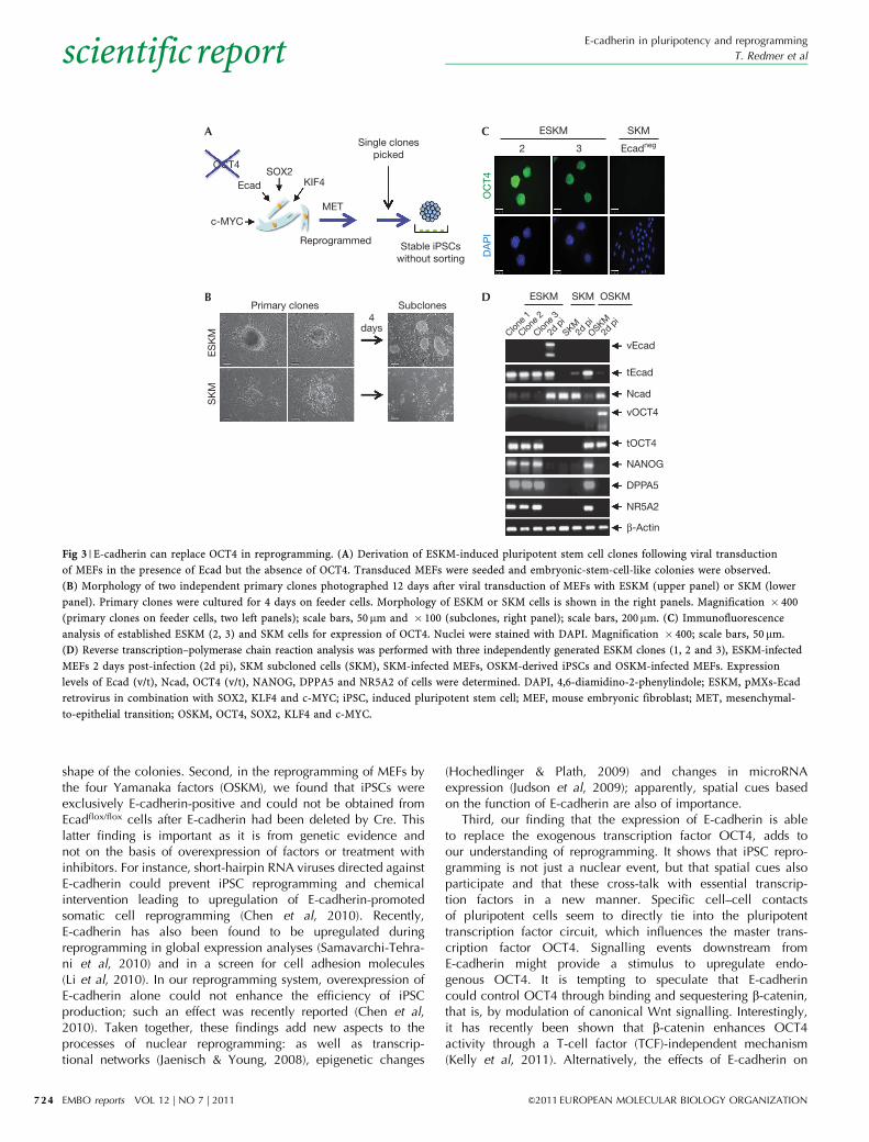
We also tested single cell clones that were produced bydifferent factor combinations and scored them for the generationof those with iPSC-like morphology (Fig 3A). Intriguingly, thepMXs-Ecad retrovirus in combination with SOX2, KLF4 andc-MYC (ESKM)—that is, without OCT4—produced cells with thetypical morphology of pluripotent iPSCs (Fig 3B), although at alower frequency than with OSKM (supplementary Fig S4A online).By using Southern blot analysis, we confirmed that the ESKMclones showed no viral integration sites for OCT4 DNA sequencesin comparison with an OSKM clone (supplementary Fig S3Eonline). When E-cadherin was omitted in the SKM combination,cell clones of iPSC-like morphology were rarely found (Fig 3B).Cell clones from ESKM and SKM combinations were cultivatedfor several passages and characterized. Remarkably, ESKM cellclones were positive for endogenous OCT4 (Fig 3C), NANOG andSSEA1 (supplementary Fig S4B online).
Independent ESKM clones (1,2,3) showed strong mRNAexpression for E-cadherin and for the pluripotency genes OCT4,NANOG, DPPA5, and NR5A2, whereas expression of N-cadherinwas not observed (Fig 3D). E-cadherin and OCT4 proteins wereproduced in ESKM clones, whereas N-cadherin protein was absent(supplementary Fig S4C online). ESKM-derived cell clones wereable to form embryoid bodies (supplementary Fig S4D online) andshowed expression of genes for endodermal (FoxA2), neuronal(Nestin) and mesodermal (a-SMA) lineages (supplementary FigS4E online). The analysis of the methylation pattern of NANOG
E-cadherin in pluripotency and reprogramming
T. Redmer et al
&2011 EUROPEAN MOLECULAR BIOLOGY ORGANIZATION EMBO reports VOL 12 | NO 7 | 2011
scientificreport
721

B C
D E
Rel
ativ
e ex
pre
ssio
n(A
U)
25
20
15
10
5
0
Ncad
MSX2
1.4
1.2
1.0
0.8
0.6
Rel
ativ
e ex
pre
ssio
n(A
U)
0.4
0.2
0.0
Ecad
OCT4
NANOG
SSEA1 sorted
Eca
dN
cad
SS
EA
1N
AN
OG
DA
PI
129/Sv3+ –
Ecadhigh Ecadlow
Ecad
Ncad
vOCT4
tOCT4
NANOG
DPPA5
β-Actin
SSEA1+ + + +– –
Ecadhigh
Cl.4 Cl.6 Cl.11
MEF
129/
Sv3
Cl.1,E
cadlow
ASOX2
OCT4
c-MYC
KLF4SSEA1 positive
SSEA1 negative
Fully reprogrammed
Partly reprogrammed
Ecadhigh/Ncadlow
Ecadlow/NcadhighNon-sorted
iPSCs
ReprogrammedMET
129/Sv3+ LIF + LIF
– LIF – LIF
+ LIF
– LIFEcadhigh Ecadlow
Fig 1 | SSEA1-positive induced pluripotent stem cells express E-cadherin and pluripotency genes. (A) Mouse embryonic fibroblasts were retrovirally
transduced with four factors, OCT4, SOX2, KLF4 and c-MYC, and sorted for SSEA1 expression after 8 days. (B) Characterization of established
cell clones derived from SSEA1-positive (Ecadhigh) cells and SSEA1-negative (Ecadlow) cells, in comparison with 129/Sv3 mouse embryonic stem cells.
Immunofluorescence for Ecad (cyan), SSEA1 (red) and NANOG (green) are shown. Nuclei were stained with DAPI (blue). Ncad (red) was analysed in
independent clones. Magnification � 400; scale bars, 50mm. (C) Agarose gel electrophoresis of reverse transcription–polymerase chain reaction products
for marker expression of three stable independent Ecadhigh iPSC clones (clones 4, 6 and 11) in comparison with cells of an Ecadlow clone (clone 1, Ecadlow),
129/Sv3 and MEFs. SSEA1 expression was determined by flow cytometry and is indicated as (þ /�). Expression of Ecad, Ncad, viral (v) and total (t) OCT4,
NANOG and DPPA5 was analysed and related to b-actin. (D) Quantitative real-time RT–PCR analysis of spontaneously differentiated Ecadhigh (Ecadhigh)
iPSCs, Ecadlow cells and 129/Sv3 cells grown for 4 days in the presence or absence of LIF. Messenger RNA levels for Ecad, OCT4 and NANOG, and
(E) Ncad and MSX2. Expression levels are related to Ecadhigh iPSCs in LIF set to 1. Median values from biological duplicates are presented as bars,
one representative experiment out of four independent experiments is shown. AU, arbitrary units; Cl., clone; DAPI, 4,6-diamidino-2-phenylindole;
iPSC, induced pluripotent stem cell; MET, mesenchymal-to-epithelial transition; SSEA1, stage-specific embryonic antigen 1.
E-cadherin in pluripotency and reprogramming
T. Redmer et al
EMBO reports VOL 12 | NO 7 | 2011 &2011 EUROPEAN MOLECULAR BIOLOGY ORGANIZATION
scientificreport
722

and OCT4 promoters showed a demethylated state in ESKM-derived and OSKM-derived iPSCs and mESCs, whereas in MEFsboth promoters showed strong methylation marks (Fig 4A). Wedetermined the methylation pattern of the E-cadherin promoterand observed no difference in the methylation statuses of iPSCs,mESCs and MEFs. Furthermore, we determined the global geneexpression profiles of ESKM2 iPSCs, 129/Sv3 mESCs and MEFs(Fig 4B). Although the profile of ESKM iPSCs differed significantlyfrom that of somatic MEFs (Pearson’s coefficient: 0.7263), asignificant similarity between ESKM iPSCs and mESCs (Pearson’scoefficient: 0.9764) was observed.
In vivo differentiation showed that ESKM-reprogrammediPSCs formed teratomas in non-obese diabetic/severe combinedimmunodeficiency (NOD/SCID) mice (Fig 4C, supplementaryFig S4F online). SKM clones showed only small cell lumps withoutsignificant growth. When stably green fluorescent protein (GFP)-transduced ESKM-iPSCs (ESKM2) were injected into blastocysts,
these cells contributed to organ formation as indicated by GFP-positive areas, which were observed in different organs (Fig 4D).These data show that iPSCs can be derived in the presence ofE-cadherin instead of OCT4 and show all the characteristicsof pluripotent, reprogrammed cells.
Our analysis is focused on understanding the function ofE-cadherin, both in ESCs during differentiation and in fibroblastsduring iPSC reprogramming. First, we demonstrate that inter-ference with E-cadherin in mESCs cause morphology changesand differentiation. It has been shown that E-cadherin is crucial tothe conversion of Fgf2-, Activin-, BIO-derived (FAB, similar toEpiSC) mouse stem cells to pluripotent mESCs (Chou et al, 2008),and the loss of E-cadherin allows the maintenance of mESCs insuspension cultures (Mohamet et al, 2010), suggesting that, underthese conditions, they might convert to intermediated cell-state-like mEpiSCs. These data indicate that E-cadherin has a crucialrole in the maintenance of ESC pluripotency and the compact
A C
DB140
120
100
80
60
40
20Col
ony
num
ber
(% o
f Ctl)
0
Ctl
wt MEFs
Ecadflo
x/flo
x
+HTNCre
1.4
Rel
ativ
e m
RN
A le
vel
(AU
)
1.2
1.0
0.8
0.6
0.4
0.2
0.0
Ecad
Ncad
NANOG
DPPA5
NR5A2
Ctl HTNCre
Ctl
Non-recombined Recombined
Eca
dN
AN
OG
DA
PI
HTNCre
Ecadflo
x/flo
x
wt MEFs
24 h HTNCre+– +–
Ecad
OSKM
α-Tubulin
Fig 2 | Loss of E-cadherin expression shows its necessity during reprogramming. (A) Ecad protein expression in OCT4, SOX2, KLF4 and c-MYC
(OSKM)-transduced non-Cre-treated (�) and His-tat-NLS (HTN)-Cre-treated (þ ) Ecadflox/flox MEFs and equally treated wild-type MEFs, respectively.
(B) Determination of colony number by counting of mouse embryonic stem cell-like colonies in pools of non-Cre-treated, transduced Ecadflox/flox
MEFs or wild-type MEFs (black) or Cre-treated Ecadflox/flox MEFs or wild-type MEFs (grey), respectively. Colony numbers were as follows: MEFs
Ecadflox/flox without HTNCre (Ctl), 319 colonies (set to 100%); with HTNCre (þHTNCre), 88 colonies (28%); wild-type MEFs Ctl, 72 colonies
(set to 100%); and þHTNCre, 93 colonies (129%). The difference in absolute colony numbers between Ecadvflox/flox and wild-type MEFs is due
to the different viability of cells isolated from different animals at different times. The experiment was repeated four times and the values from
one representative experiment are shown. (C) Relative mRNA expression level of Ecad, NANOG, DPPA5 and NR5A2 of a pool of OSKM-induced
Cre-treated and non-treated Ecadflox/flox MEFs 10 days after infection. Median values from biological duplicates are presented as bars; one
representative experiment is shown of four independent experiments. (D) Immunofluorescence analysis of cell colonies from OSKM-induced
Ecadflox/flox MEFs for Ecad and NANOG either untreated (Ctl) or Cre-treated (HTNCre) showing no recombination (non-recombined first and
second column) or recombination (recombined). DAPI was used for nuclear staining. Magnification � 400; scale bars, 50 mm. AU, arbitrary units;
Ctl, control; DAPI, 4,6-diamidino-2-phenylindole; MEF, mouse embryonic fibroblast; mRNA, messenger RNA; wt, wild type.
E-cadherin in pluripotency and reprogramming
T. Redmer et al
&2011 EUROPEAN MOLECULAR BIOLOGY ORGANIZATION EMBO reports VOL 12 | NO 7 | 2011
scientificreport
723
shape of the colonies. Second, in the reprogramming of MEFs bythe four Yamanaka factors (OSKM), we found that iPSCs wereexclusively E-cadherin-positive and could not be obtained fromEcadflox/flox cells after E-cadherin had been deleted by Cre. Thislatter finding is important as it is from genetic evidence andnot on the basis of overexpression of factors or treatment withinhibitors. For instance, short-hairpin RNA viruses directed againstE-cadherin could prevent iPSC reprogramming and chemicalintervention leading to upregulation of E-cadherin-promotedsomatic cell reprogramming (Chen et al, 2010). Recently,E-cadherin has also been found to be upregulated duringreprogramming in global expression analyses (Samavarchi-Tehra-ni et al, 2010) and in a screen for cell adhesion molecules(Li et al, 2010). In our reprogramming system, overexpression ofE-cadherin alone could not enhance the efficiency of iPSCproduction; such an effect was recently reported (Chen et al,2010). Taken together, these findings add new aspects to theprocesses of nuclear reprogramming: as well as transcrip-tional networks (Jaenisch & Young, 2008), epigenetic changes
(Hochedlinger & Plath, 2009) and changes in microRNAexpression (Judson et al, 2009); apparently, spatial cues basedon the function of E-cadherin are also of importance.
Third, our finding that the expression of E-cadherin is ableto replace the exogenous transcription factor OCT4, adds toour understanding of reprogramming. It shows that iPSC repro-gramming is not just a nuclear event, but that spatial cues alsoparticipate and that these cross-talk with essential transcrip-tion factors in a new manner. Specific cell–cell contactsof pluripotent cells seem to directly tie into the pluripotenttranscription factor circuit, which influences the master trans-cription factor OCT4. Signalling events downstream fromE-cadherin might provide a stimulus to upregulate endo-genous OCT4. It is tempting to speculate that E-cadherincould control OCT4 through binding and sequestering b-catenin,that is, by modulation of canonical Wnt signalling. Interestingly,it has recently been shown that b-catenin enhances OCT4activity through a T-cell factor (TCF)-independent mechanism(Kelly et al, 2011). Alternatively, the effects of E-cadherin on
OCT4
A C
DB
SOX2
ESKM
OC
T4D
AP
I
2 3 Ecadneg
SKM
Ecad
c-MYC
Reprogrammed
Primary clones
ES
KM
ESKM
Clone 1
Clone 2
Clone 3
SKM
2d p
i2d
pi
2d p
i
SKM
OSKM
vEcad
tEcad
Ncad
vOCT4
tOCT4
NANOG
DPPA5
NR5A2
β-Actin
OSKM
SK
M
Subclones
KIF4
MET
4days
Stable iPSCswithout sorting
Single clonespicked
Fig 3 | E-cadherin can replace OCT4 in reprogramming. (A) Derivation of ESKM-induced pluripotent stem cell clones following viral transduction
of MEFs in the presence of Ecad but the absence of OCT4. Transduced MEFs were seeded and embryonic-stem-cell-like colonies were observed.
(B) Morphology of two independent primary clones photographed 12 days after viral transduction of MEFs with ESKM (upper panel) or SKM (lower
panel). Primary clones were cultured for 4 days on feeder cells. Morphology of ESKM or SKM cells is shown in the right panels. Magnification � 400
(primary clones on feeder cells, two left panels); scale bars, 50 mm and � 100 (subclones, right panel); scale bars, 200mm. (C) Immunofluorescence
analysis of established ESKM (2, 3) and SKM cells for expression of OCT4. Nuclei were stained with DAPI. Magnification � 400; scale bars, 50 mm.
(D) Reverse transcription–polymerase chain reaction analysis was performed with three independently generated ESKM clones (1, 2 and 3), ESKM-infected
MEFs 2 days post-infection (2d pi), SKM subcloned cells (SKM), SKM-infected MEFs, OSKM-derived iPSCs and OSKM-infected MEFs. Expression
levels of Ecad (v/t), Ncad, OCT4 (v/t), NANOG, DPPA5 and NR5A2 of cells were determined. DAPI, 4,6-diamidino-2-phenylindole; ESKM, pMXs-Ecad
retrovirus in combination with SOX2, KLF4 and c-MYC; iPSC, induced pluripotent stem cell; MEF, mouse embryonic fibroblast; MET, mesenchymal-
to-epithelial transition; OSKM, OCT4, SOX2, KLF4 and c-MYC.
E-cadherin in pluripotency and reprogramming
T. Redmer et al
EMBO reports VOL 12 | NO 7 | 2011 &2011 EUROPEAN MOLECULAR BIOLOGY ORGANIZATION
scientificreport
724

induced pluripotency might also require the involvement ofRho-family GTPases (Fukata & Kaibuchi, 2001). The elucidationof the molecular mechanisms that establish the cross-talkbetween the cell surface and nuclear machinery, especiallythe regulation of OCT4, awaits further analysis. With these threeparts of our investigation, we have discovered new functions
for the cell-adhesion molecule E-cadherin in pluripotencyand reprogramming.
METHODSCell culture. 129/Sv3-derived mESCs (from S. Noggle, New York,USA) and established stable iPSC lines generated were cultured on
i
iv
ii
Epithelium
MuscleNeuronal
v vi
ESKM2
A B
DC
ES
KM
2E
SK
M2
ES
KM
2S
KM
16R 2=0.7263
R 2=0.9764
14
12
10
8
6
16
14
12
10
8
6
6 8 10
MEF
12 14 16
6E
SK
M2
GFP
8 10 12 14 16
ESKM3
OSKM1
129/Sv3
129/Sv3
MEF
NANOG
iii
OCT4 Ecad
Fig 4 | ESKM induced pluripotent stem cells fulfil all criteria of pluripotent stem cells. (A) Bisulphite sequencing of promoter regions of NANOG,
OCT4 and Ecad, of ESKM cells (clone 2 and 3), OSKM cells (clone 1), mouse embryonic stem cells (mESCs; 129/Sv3) and mouse embryonic
fibroblasts (MEFs). Shown are 5–6 reactions for each region. (B) Global gene expression profiling of ESKM-iPSCs (clone 2) in comparison with MEFs
and mESCs. Similarity is shown by the R2 value. Three biological replicates of each cell line were included for analysis. (C) Whole morphology of
tumours derived from ESKM-derived iPSCs (ESKM 2; 1) or SKM-derived cells (v). Histological analysis of tissue sections by haematoxylin and eosin
staining of differentiated tumours (ESKM2) comprising epithelial (ii), neuronal (iii) and muscle-derived (iv) structures, or of a undifferentiated tumour
(SKM; vi). Representative tumours are shown out of n¼ 3 injections/cell clone, compare supplementary Table 3 for further details. Magnification
of haematoxylin and eosin-stained sections � 25; scale bars, 500mm. (D) Localization of green fluorescent protein (GFP)-positive areas in offspring
following injection of stable GFP-expressing ESKM-iPSCs (clone 2) in blastocysts. ESKM, pMXs-Ecad retrovirus in combination with SOX2, KLF4
and c-MYC; OSKM, OCT4, SOX2, KLF4 and c-MYC.
E-cadherin in pluripotency and reprogramming
T. Redmer et al
&2011 EUROPEAN MOLECULAR BIOLOGY ORGANIZATION EMBO reports VOL 12 | NO 7 | 2011
scientificreport
725

mitomycin-C-treated (2 mg/ml) MEFs or on gelatin-coated plates.MEFs were isolated according to standard procedures eitherfrom embryos of the mouse strain B6.129/Sv3-Cdh1tm2Kem/J(Ecadflox/flox) or from the outbred strain Cf1.Generation of iPSCs. Generation of iPSCs was performed asdescribed previously (Takahashi & Yamanaka, 2006). In brief,5� 106 Plat-E cells were seeded 1 day before transfection withpMXs-based retroviral vectors (Ecad or OCT4, SOX2, KLF4 andc-MYC; from Addgene) using calcium phosphate precipitation inthe presence of 25 mM chloroquine. For expression of E-cadherin,the coding sequence of mouse E-cadherin was introduced into thepMXs vector. For the derivation of ESKM-iPSC clones, 2� 105
MEFs/10 cm dish were seeded on Matrigel (BD) 4 days after viralinfection in mESC medium. OSKM-induced cells were seeded to5� 104 cells/10 cm dish on mitomycin-C-treated MEFs.Cell-based assays. Quantitative RT–PCR and western blot analysiswere performed as described earlier (Diecke et al, 2008).
Cre-recombinase treatment: cells were treated with 3 mMpurified, membrane-penetrable HTNCre for 24 h, as describedpreviously (Peitz et al, 2002).
Bisulphite sequencing: genomic DNA was isolated fromdifferent cell lines. Bisulphite conversion of 2 mg DNA was donewith the EpiTect Kit (Qiagen) and converted DNA was used forPCR using specific primers recognizing regions in the promoters ofNANOG, OCT4 and E-cadherin. PCR products were cloned in thepGEM-Vector (Promega) and sequenced using SP6 primer.Global gene expression analysis. Expression analysis was done intriplicates using the MouseWG-6 v2.0 Expression BeadChip Kits(Illumina), using standard protocols. The microarray analysis onthe Illumina MouseWG-6 v2.0 expression beadchips (Fig 4B) hasnow been deposited at the National Center for BiotechnologyInformation (NCBI)/Gene Expression Omnibus (GEO). The datacan be found at http://www.ncbi.nlm.nih.gov/geo/query/acc.cgi?acc¼GSE28594.Animal-based assays. Teratoma formation and histological ana-lysis: 1� 106 cells were subcutaneously injected in NOD/SCIDmice, as described previously (Takahashi & Yamanaka, 2006).Tumours were grown for 21 days and sections of surgicallydissected tumours were stained with haematoxylin and eosin andbiopsies were taken for RT–PCR analysis.
Blastocyst injection: stably GFP-expressing iPSCs transducedwith the plasmid MP71-GFP were injected at the blastocyst stage.Vehicles were transferred in uteri of surrogate mothers. Pups wereanalysed for GFP-positive spots 2 days after birth.Supplementary information is available at EMBO reports online(http://www.emboreports.org).
ACKNOWLEDGEMENTSWe thank A. Kirschner, M. Muhlbauer and Anne Schafer for technicalassistance, and A. Klaus, H.-P. Rahn, B. von Eyss, K. Eckert, I. Fichtner,B. Jerchow and K. Becker for technical help. We express our appreciationto R. Hodge, U. Ziebold, I. Ibanez-Tallon and M. Gossen for criticalreading of the manuscript. A Bundersministerium fur Bildung undForschung (BMBF) grant in the joint project START-MSC (standardisationfor regenerative therapy–mesenchymal stem cells; 01GN0941) and
a European Commission Marie Curie International Reintigration Grant(FP6-021407) supported this work.
CONFLICT OF INTERESTThe authors declare that they have no conflict of interest.
REFERENCESBoussadia O, Kutsch S, Hierholzer A, Delmas V, Kemler R (2002) E-cadherin
is a survival factor for the lactating mouse mammary gland. Mech Dev115: 53–62
Chen T, Yuan D, Wei B, Jiang J, Kang J, Ling K, Gu Y, Li J, Xiao L, Pei G (2010)E-cadherin-mediated cell–cell contact is critical for induced pluripotentstem-cell generation. Stem Cells 28: 1315–1325
Chou YF, Chen HH, Eijpe M, Yabuuchi A, Chenoweth JG, Tesar P, Lu J,McKay RD, Geijsen N (2008) The growth factor environment definesdistinct pluripotent ground states in novel blastocyst-derived stem cells.Cell 135: 449–461
Diecke S, Quiroga-Negreira A, Redmer T, Besser D (2008) FGF2 signalingin mouse embryonic fibroblasts is crucial for self-renewal of embryonicstem cells. Cells Tissues Organs 188: 52–61
Fukata M, Kaibuchi K (2001) Rho-family GTPases in cadherin-mediatedcell–cell adhesion. Nat Rev Mol Cell Biol 2: 887–897
Heuberger J, Birchmeier W (2010) Interplay of cadherin-mediated celladhesion and canonical Wnt signaling. Cold Spring Harb Perspect Biol2: a002915
Hochedlinger K, Plath K (2009) Epigenetic reprogramming and inducedpluripotency. Development 136: 509–523
Jaenisch R, Young R (2008) Stem cells, the molecular circuitry of pluripotencyand nuclear reprogramming. Cell 132: 567–582
Judson RL, Babiarz JE, Venere M, Blelloch R (2009) Embryonic stem cell-specific microRNAs promote induced pluripotency. Nat Biotechnol 27:459–461
Kelly KF, Ng DY, Jayakumaran G, Wood GA, Koide H, Doble BW (2011)b-Catenin enhances Oct-4 activity and reinforces pluripotency througha TCF-independent mechanism. Cell Stem Cell 8: 214–227
Larue L, Ohsugi M, Hirchenhain J, Kemler R (1994) E-cadherin null mutantembryos fail to form a trophectoderm epithelium. Proc Natl Acad SciUSA 91: 8263–8267
Larue L, Antos C, Butz S, Huber O, Delmas V, Dominis M, Kemler R (1996)A role for cadherins in tissue formation. Development 122: 3185–3194
Li R et al (2010) A mesenchymal-to-epithelial transition initiates and isrequired for the nuclear reprogramming of mouse fibroblasts. Cell StemCell 7: 51–63
Mohamet L, Lea ML, Ward CM (2010) Abrogation of E-cadherin-mediatedcellular aggregation allows proliferation of pluripotent mouse embryonicstem cells in shake flask bioreactors. PLoS ONE 5: e12921
Peitz M, Pfannkuche K, Rajewsky K, Edenhofer F (2002) Ability of thehydrophobic FGF and basic TAT peptides to promote cellular uptakeof recombinant Cre recombinase: a tool for efficient genetic engineeringof mammalian genomes. Proc Natl Acad Sci USA 99: 4489–4494
Samavarchi-Tehrani P, Golipour A, David L, Sung HK, Beyer TA, Datti A,Woltjen K, Nagy A, Wrana JL (2010) Functional genomics reveals a BMP-driven mesenchymal-to-epithelial transition in the initiation of somaticcell reprogramming. Cell Stem Cell 7: 64–77
Soncin F et al (2009) Abrogation of E-cadherin-mediated cell–cell contact inmouse embryonic stem cells results in reversible LIF-independent self-renewal. Stem Cells 27: 2069–2080
Stepniak E, Radice GL, Vasioukhin V (2009) Adhesive and signaling functionsof cadherins and catenins in vertebrate development. Cold Spring HarbPerspect Biol 1: a002949
Takahashi K, Yamanaka S (2006) Induction of pluripotent stem cells frommouse embryonic and adult fibroblast cultures by defined factors. Cell126: 663–676
Wilmut I, Schnieke AE, McWhir J, Kind AJ, Campbell KH (1997) Viable offspringderived from fetal and adult mammalian cells. Nature 385: 810–813
E-cadherin in pluripotency and reprogramming
T. Redmer et al
EMBO reports VOL 12 | NO 7 | 2011 &2011 EUROPEAN MOLECULAR BIOLOGY ORGANIZATION
scientificreport
726


![Genomewide screen identifies a novel p97CDC48dependent …embor.embopress.org/content/embor/16/3/332.full.pdf · Because p97/CDC-48 is involved in protein degradation [5], we reasoned](https://static.fdocuments.net/doc/165x107/5ce7c58388c9932d758ce93e/genomewide-screen-identifies-a-novel-p97cdc48dependent-embor-because-p97cdc-48.jpg)


![Molecular mechanisms of asymmetric divisions in mammary ...embor.embopress.org/content/embor/early/2016/11/21/embr.201643021... · H3-Thr10 by the mitotic kinase Haspin [15], whereas](https://static.fdocuments.net/doc/165x107/5b0116547f8b9a84338da31c/molecular-mechanisms-of-asymmetric-divisions-in-mammary-embor-by-the-mitotic.jpg)
