
A Salmonella Typhi homologue of bacteriophage muramidases...
8
A Salmonella Typhi homologue of bacteriophage muramidases controls typhoid toxin secretion He´le`ne Hodak & Jorge E. Gala´n + Department of Microbial Pathogenesis, Yale University School of Medicine, New Haven, Connecticut, USA Unlike other Salmonella, which can infect a broad range of hosts causing self-limiting infection, Salmonella Typhi is an exclusively human pathogen that causes typhoid fever, a life-threatening systemic disease. Typhoid toxin is a unique virulence factor of Salmonella Typhi, which is expressed when the bacteria are within mammalian cells. Here, we report that an N-acetyl-b-D- muramidase similar to phage endolysins encoded within the same pathogenicity islet as the toxin is required for typhoid toxin secretion. Genetic and functional analysis of TtsA revealed unique amino acids at its predicted peptidoglycan-binding domain that are essential for protein secretion and that distinguishes this protein from other homologues. We propose that TtsA defines a new protein secretion mechanism recently evolved from the machine that mediates phage release. Keywords: bacterial toxins; protein secretion; typhoid fever; pathogen evolution; holins EMBO reports (2013) 14, 95–102. doi:10.1038/embor.2012.186 INTRODUCTION Salmonella enterica serovar Typhi (S. Typhi) is the cause of typhoid fever, which kills an estimated 200,000 people every year [1–3]. In contrast to other S. enterica serovars, S. Typhi can only infect humans causing a life-threatening systemic illness [1,4]. The molecular bases for these remarkable differences in the disease presentation and host range are poorly understood although it is believed that they are the result of genome reduction and the acquisition of unique genes [5,6]. One of the few virulence factors uniquely present in S. Typhi is typhoid toxin, a remarkable AB 5 toxin with similarities to pertussis and cytolethal distending toxins [7–9]. Typhoid toxin has a unique biology in that it is only expressed when S. Typhi is within mammalian host cells [8,10]. After its synthesis and secretion from the bacteria, the toxin is transported by carrier intermediates from the Salmonella-containing vacuole to the extracellular space, from where it gains access to target cells via autocrine or paracrine routes [7,11]. The mechanisms by which the toxin is secreted from the bacteria are unknown. The three components of typhoid toxin have canonical secretion signals, implying that, like other AB 5 toxins, the Sec machinery exports the toxin components to the periplasm where they assemble into a holotoxin. However, there is no information on how the holotoxin gets out of the bacteria before its packaging into transport carriers. Here, we report that an N-acetyl-b-D-muramidase similar to phage endolysins encoded within the same pathogenicity islet as the toxin is required for typhoid toxin secretion. We identified a critical region at the carboxy-terminal peptidoglycan (PG)-binding region of this enzyme that is necessary to carry out protein secretion functions. We propose that this muramidase is the result of the evolutionary adaptation of a phage enzyme to conform a new protein secretion mechanism. Finally, we present bioinformatics evidence that suggests that the protein secretion mechanism described here is widespread in bacteria. RESULTS AND DISCUSSION ttsA is required for typhoid toxin secretion The genomic islet that encodes typhoid toxin also contains two other uncharacterized genes, sty1887 and sty1889 (Fig 1A). As functionally related genes are often encoded in close proximity to one another, we investigated the potential involvement of these genes in typhoid toxin secretion. Typhoid toxin secretion cannot be studied in vitro because its components are not expressed [7–9]. Therefore, we investigated toxin secretion by immunofluorescence staining of CdtB, a typhoid toxin subunit, in S. Typhi-infected cultured epithelial cells. Previous studies have shown that several hours after infection, typhoid toxin can be seen in puncta radiating from the S. Typhi-containing vacuole and spreading throughout the entire cell [7]. Therefore, we used the appearance of these puncta as an assay to measure typhoid toxin secretion from the bacterial cell. Cells infected with a S. Typhi mutant strain carrying a deletion of sty1887 showed a staining pattern that was indistinguishable from that of cells infected with wild type (supplementary Fig S1 online). In contrast, cells infected with an S. Typhi strain carrying a deletion in sty1889 showed no CdtB puncta staining (Fig 1B and supplementary Figs S2 and S3 online), although the CdtB expression levels in the mutant were indistinguishable from those of wild type (Fig 1C). Introduction of a plasmid encoding sty1889 effectively restored puncta staining Department of Microbial Pathogenesis, Yale University School of Medicine, New Haven, Connecticut 06536, USA + Corresponding author. Tel: +203 737 2404; Fax: +203 737 2630; E-mail: [email protected] Received 8 August 2012; revised 15 October 2012; accepted 25 October 2012; pubished online 23 November 2012 scientificreport scientific report 95 &2013 EUROPEAN MOLECULAR BIOLOGY ORGANIZATION EMBO reports VOL 14 | NO 1 | 2013
Transcript of A Salmonella Typhi homologue of bacteriophage muramidases...

A Salmonella Typhi homologue of bacteriophagemuramidases controls typhoid toxin secretionHelene Hodak & Jorge E. Galan+
Department of Microbial Pathogenesis, Yale University School of Medicine, New Haven, Connecticut, USA
Unlike other Salmonella, which can infect a broad range of hostscausing self-limiting infection, Salmonella Typhi is an exclusivelyhuman pathogen that causes typhoid fever, a life-threateningsystemic disease. Typhoid toxin is a unique virulence factor ofSalmonella Typhi, which is expressed when the bacteria arewithin mammalian cells. Here, we report that an N-acetyl-b-D-muramidase similar to phage endolysins encoded within the samepathogenicity islet as the toxin is required for typhoid toxinsecretion. Genetic and functional analysis of TtsA revealedunique amino acids at its predicted peptidoglycan-bindingdomain that are essential for protein secretion and thatdistinguishes this protein from other homologues. We proposethat TtsA defines a new protein secretion mechanism recentlyevolved from the machine that mediates phage release.Keywords: bacterial toxins; protein secretion; typhoidfever; pathogen evolution; holinsEMBO reports (2013) 14, 95–102. doi:10.1038/embor.2012.186
INTRODUCTIONSalmonella enterica serovar Typhi (S. Typhi) is the cause oftyphoid fever, which kills an estimated 200,000 people everyyear [1–3]. In contrast to other S. enterica serovars, S. Typhican only infect humans causing a life-threatening systemicillness [1,4]. The molecular bases for these remarkabledifferences in the disease presentation and host range are poorlyunderstood although it is believed that they are the result ofgenome reduction and the acquisition of unique genes [5,6]. Oneof the few virulence factors uniquely present in S. Typhi is typhoidtoxin, a remarkable AB5 toxin with similarities to pertussis andcytolethal distending toxins [7–9]. Typhoid toxin has a uniquebiology in that it is only expressed when S. Typhi is withinmammalian host cells [8,10]. After its synthesis and secretion fromthe bacteria, the toxin is transported by carrier intermediatesfrom the Salmonella-containing vacuole to the extracellular space,from where it gains access to target cells via autocrine or paracrine
routes [7,11]. The mechanisms by which the toxin is secreted fromthe bacteria are unknown. The three components of typhoid toxinhave canonical secretion signals, implying that, like other AB5
toxins, the Sec machinery exports the toxin components to theperiplasm where they assemble into a holotoxin. However, thereis no information on how the holotoxin gets out of the bacteriabefore its packaging into transport carriers. Here, we report that anN-acetyl-b-D-muramidase similar to phage endolysins encodedwithin the same pathogenicity islet as the toxin is required fortyphoid toxin secretion. We identified a critical region at thecarboxy-terminal peptidoglycan (PG)-binding region of thisenzyme that is necessary to carry out protein secretionfunctions. We propose that this muramidase is the result of theevolutionary adaptation of a phage enzyme to conform a newprotein secretion mechanism. Finally, we present bioinformaticsevidence that suggests that the protein secretion mechanismdescribed here is widespread in bacteria.
RESULTS AND DISCUSSIONttsA is required for typhoid toxin secretionThe genomic islet that encodes typhoid toxin also contains twoother uncharacterized genes, sty1887 and sty1889 (Fig 1A). Asfunctionally related genes are often encoded in close proximity toone another, we investigated the potential involvement of thesegenes in typhoid toxin secretion. Typhoid toxin secretioncannot be studied in vitro because its components are notexpressed [7–9]. Therefore, we investigated toxin secretion byimmunofluorescence staining of CdtB, a typhoid toxin subunit, inS. Typhi-infected cultured epithelial cells. Previous studies haveshown that several hours after infection, typhoid toxin can be seenin puncta radiating from the S. Typhi-containing vacuole andspreading throughout the entire cell [7]. Therefore, we used theappearance of these puncta as an assay to measure typhoid toxinsecretion from the bacterial cell. Cells infected with a S. Typhimutant strain carrying a deletion of sty1887 showed a stainingpattern that was indistinguishable from that of cells infected withwild type (supplementary Fig S1 online). In contrast, cells infectedwith an S. Typhi strain carrying a deletion in sty1889 showed noCdtB puncta staining (Fig 1B and supplementary Figs S2 and S3online), although the CdtB expression levels in the mutant wereindistinguishable from those of wild type (Fig 1C). Introduction ofa plasmid encoding sty1889 effectively restored puncta staining
Department of Microbial Pathogenesis, Yale University School of Medicine,New Haven, Connecticut 06536, USA+Corresponding author. Tel: +203 737 2404; Fax: +203 737 2630;E-mail: [email protected]
Received 8 August 2012; revised 15 October 2012; accepted 25 October 2012;pubished online 23 November 2012
scientificreportscientific report
95&2013 EUROPEAN MOLECULAR BIOLOGY ORGANIZATION EMBO reports VOL 14 | NO 1 | 2013

(Fig 1B,C and supplementary Fig S3 online). Expression of theperiplasmic protein MalE in intracellular S. Typhi did not result inits detection on either the bacterial surface or in puncta afterequally processing the samples for staining, indicating that thedetection of CdtB in wild-type intracellular bacteria was notowing to nonspecific leakage of periplasmic proteins or bacteriallysis (supplementary Fig S4 online). These results indicate thatSty1889 is required for toxin secretion from the bacterial cell, andtherefore we renamed it ttsA (for typhoid toxin secretion A).To examine whether ttsA was co-regulated with the typhoid toxingenes, we tested its pattern of expression during infection. Wefound that, like typhoid toxin [7,8], TtsA was not detected whenS. Typhi was grown in L-broth. However, TtsA was detectedwithin cultured epithelial cells (Fig 1D) although it requiredslightly longer infection times than those required to detecttyphoid toxin (Fig 1D). Taken together, these results indicate thatTtsA is required for toxin secretion, and that, consistent with thisfunction, its expression correlated with that of typhoid toxin.
TtsA belongs to a family of bacteriophage muramidasesStojkovic and Rothman–Denes have previously reported thatthe coliphage N4 Gp61 N-acetylmuramidase defines a new familyof bacteriophage muramidases and noticed the presence ofhomologues of this protein family in S. enterica includingsty1889 (which we now have renamed ttsA) [12]. This proteinfamily is composed of a amino-terminal lysozyme-like domainbelonging to glycoside hydrolase family 108, and a C-terminalputative PG-binding domain (Fig 2A,B) [13]. Most members of thisfamily, including ttsA, lack an identifiable N-terminal sec-dependent secretion signal and are delivered to the periplasmby holins, a diverse group of small membrane proteins that
are most often encoded immediately adjacent to themuramidases [14]. In a regulated fashion, holins form a pore inthe inner membrane that allows the passage of the murein-lyticenzymes. Interestingly, in the vicinity of ttsA there are no genesthat could encode a protein with features similar to those expectedin a holin (a small protein with one or two transmembranedomains). Despite repeated efforts, we were not able todemonstrate in vitro muramidase activity with purified TtsAusing standard zymography. Similar observations have beenpreviously made for other members of this protein family [12].Therefore, we investigated its putative muramidase activity usingalternative indirect approaches. It has been observed that in theabsence of holins (or in the absence of an activating signal thatwould trigger their activity), this family of muramidases is stillcapable of inducing bacterial lysis when overexpressed and afteraddition of low concentrations of chloroform [12]. It is thoughtthat chloroform destabilizes the inner membrane allowing the‘leakage’ of the cytoplasmic muramidases into the periplasmicspace. Consistent with its putative muramidase activity, TtsAwas capable of inducing bacterial lysis under these conditions(Fig 2C,D). Although each muramidase exerts its function togetherwith a holin encoded in its immediate vicinity, some holins showlittle specificity and are indeed capable of transporting unrelatedmuramidases [15]. Therefore to further test TtsA’s putativemuramidase activity, we co-expressed it with Sty0015, a holinfor the adjacently encoded muramidase Sty0016, both encodeddistantly from the typhoid toxin locus [12]. We found thatco-expression of TtsA and Sty0015 led to rapid bacteriallysis (Fig 2E,F). In contrast, expression of TtsA alone did not. Theseresults further demonstrate that TtsA exhibits muramidase activityand suggest that TtsA is likely to exert its activity in conjunction with
D
BWT ΔttsA
CdtBS. TyphiDNA
ΔttsA + TtsA
ApltA
sty1889(ttsA)
sty1887 cdtB
C
CdtB
TtsA
25 –
1 3 4 5 7 10 220
Time of infection (h)
WT
25 –
kDa
kDa
CdtB
ΔttsA +
Tts
A
ΔttsA
TtsA
pltB
Fig 1 | TtsA is required for typhoid toxin secretion. (A) Schematic representation of the S. Typhi pathogenicity islet encoding typhoid toxin.
(B) Sty1889 (TtsA) is required for typhoid toxin secretion. Henle-407 cells were infected with S. Typhi expressing chromosomally encoded 3� FLAG-
epitope-tagged CdtB, a DttsA isogenic mutant, or the complemented DttsA mutant. Twenty-two hours after infection, cells were stained with an
antibody directed to the FLAG epitope (green) (to visualize CdtB), a rabbit antibody directed to S. Typhi LPS (red) and DAPI for DNA detection
(blue). The bar represents 10 mm. (C) Western blot analysis of the strains used in panel (B). (D) TtsA expression profile during bacterial infection of
cultured cells. At the indicated times after infection, cells were lysed and the levels of CdtB and TtsA were examined in CFU standardized samples by
western blot analysis. CFU, colony-forming units; DAPI, 4,6-diamidino-2-phenylindole; LPS, lipopolysaccharide; WT, wild-type.
A muramidase controls typhoid toxin secretion
H. Hodak & J.E. Galanscientificreport
96 EMBO reports VOL 14 | NO 1 | 2013 &2013 EUROPEAN MOLECULAR BIOLOGY ORGANIZATION

a co-regulated holin encoded elsewhere in the S. Typhi genome.Such holin is not Sty0015 as we found wild-type toxin secretionin a Dsty0015 S. Typhi mutant (supplementary Figure S5 online).
It has been previously reported that the muramidase activity ofthe TtsA protein family is associated with a catalytic glutamic acidlocated within a conserved EGGY motif located close to theirN terminus [12]. Mutation in TtsA of a glutamic acid residuewithin this motif (TtsAE14A) (Fig 2A,B) abolished its ability toinduce bacterial lysis either on addition of chloroform (Fig 2C,D)or after its co-expression with the Sty0015 holin (Fig 2E,F). These
results further support the notion that TtsA is a muramidase.Inducible expression of a mutant TtsA containing a sec signal fromDsbA at its N terminus also led to bacterial lysis. However, wewere not able to establish experimental conditions with whichwe could consistently observe differences between wild-type andthe catalytic mutant. We then tested the requirement of thepredicted TtsA muramidase activity for typhoid toxin secretion.Consistent with the requirement of its predicted enzymatic activityfor toxin secretion, no typhoid toxin puncta were detected in cellsinfected with a S. Typhi strain expressing TtsAE14A (Fig 2G,H).
0
0.5
1
1.5
OD
60
0 n
m 2
2.5
3
0 100 200 300
40
60
100120
159180
180
N CGH PG binding
0.15
0.2
0.25
0.3
0.35
0.4
0.45
0.5
0 60 120
TtsAE14A
TtsA
TtsAGp61
TtsAGp61
TtsAGp61
TtsAGp61 208
GH
1 180
TtsA
TtsA
TtsA
E14A
20
25
OD
600 n
m
Time (min)
+ CHCl3
ΔttsA +
Tts
AE14
A
ΔttsA +
Tts
AΔttsA + TtsAE14AΔttsA + TtsA
CdtB
S. Typhi
DNA
TtsASty0016
TtsAE14A
TtsAE14A
20
25
25
CdtB
TtsA
EGGY
Time (min)
+ Arabinose
TtsAE14A+ Sty0015
Sty0016 + Sty0015
TtsA + Sty0015
TtsAE14A
Sty0016
TtsA
kDa
kDa
kDa
TtsA
E14A
TtsA
E14A +
Sty
0016
Sty00
16 +
Sty
0016
Sty00
16
TtsA
+ S
ty00
16
TtsA
C
BA
D
G
E
F
H
Fig 2 | The TtsA enzymatic activity is required for toxin secretion. (A) TtsA domain organization. Indicated are the locations of the GH and
PG-binding domains, as well as the catalytic site (EGGY). (B) Amino-acid sequence alignment of TtsA and coliphage N4 Gp61 muramidase. The
conserved catalytic glutamate is highlighted. (C) Bacteriolytic activity of TtsA. FLAG-epitope tagged TtsA or TtsAE14A were overexpressed in S. Typhi
DttsA mutant and at the indicated time 0.3% chloroform (CHCl3). Bacterial lysis was monitored by measuring the OD600 nm of the bacterial cultures.
The graph shows the average and s.d.’s of six independent assays. (D) Western blot analysis of the expression levels of the indicated proteins in the
strains used in panel (C) in aliquots obtained immediately before adding CHCl3. (E) Holin-assisted bacteriolysis by TtsA. FLAG-tagged TtsA, TtsAE14A
or Sty0016 were expressed in a DttsA S. Typhi mutant from an arabinose-inducible promoter either by themselves, or along with the Sty0015 holin.
At the indicated time, arabinose was added to induce holin/endolysin expression and bacterial lysis monitored by measuring the OD600 nm of the
different cultures. The graph shows the average and s.d.’s of six independent assays. (F) Western blot analysis of the expression levels of the different
endolysins in the strains used in panel (E) 20 min following the induction with arabinose. (G) The TtsA catalytic activity is required for toxin secretion.
Henle-407 cells were infected with a DttsA S. Typhi mutant expressing chromosomally encoded FLAG-tagged cdtB complemented with either wild-type
TtsA or the catalytic mutant TtsAE14A. Twenty-two hours post infection, infected cells were fixed and stained with a mouse monoclonal antibody
directed to the FLAG epitope (to visualize CdtB), a rabbit antibody directed to S. Typhi LPS and DAPI for DNA detection. The bar represents 10mm.
(H) Protein levels in CFU standardized lysates of strains used in panel (G) 22 h after infection of Henle-407 cells. CFU, colony forming units;
DAPI, 4,6-diamidino-2-phenylindole; GH, glycosyl hydrolase; LPS, lipopolysaccharide; PG, peptidoglycan; s.d., standard deviation.
A muramidase controls typhoid toxin secretion
H. Hodak & J.E. Galan scientificreport
97&2013 EUROPEAN MOLECULAR BIOLOGY ORGANIZATION EMBO reports VOL 14 | NO 1 | 2013

Taken together, these results indicate that TtsA is a muramidaseand that this enzymatic activity is required for typhoid toxinsecretion or release.
Functional specificity of the TtsA protein familyTo investigate whether the amino-acid sequence of TtsA could becorrelated with its specific adaptation to mediate typhoid toxinsecretion, we generated a phylogenetic tree of TtsA homologuespresenting at least 50% identity (supplementary Fig S6 online). Inaddition, we use the PHAge Search Tool (PHAST) to analyse theloci of all the homologues represented in the tree to identifypossible prophages. The different muramidases clustered intothree large groups in the phylogenetic tree, which we arbitrarilyrefer to as group 1 (which harbours TtsA), 2 and 3. As expected forthis family of muramidases, many of the TtsA homologues wereencoded within the context of complete or incomplete phagegenomes (supplementary Fig S6 online). However, no phage-related genes were detected in the vicinity of a significant numberof family members suggesting a much more diverse set of functionfor this protein family. Although muramidases associated withphage genomes were found in the three phylogenetic groups,there were fewer phage-associated muramidases within group 2.Whether this observation suggests a specific function associatedwith this subgroup of muramidases is unknown. Nevertheless,TtsA, which most likely does not have a phage-associatedfunction, did not cluster in this group, although Zymomonas
mobilis ZlyS, which has been proposed to activate the secretion ofan extracellular levansucrase and invertase [16], did.
Consistent with the most common mechanism associatedwith the transport of these cytoplasmic muramidases through theinner membrane, we were able to identify holins encodedimmediately adjacent to most of the muramidases weanalysed (supplementary Table S1 online). Interestingly, anexception was ttsA, as we were not able to identify in its vicinityany protein that shows the expected features of a putative holin,suggesting that TtsA might be transported by a holin encodedelsewhere in the chromosome that is co-expressed with typhoidtoxin. Of note, in several instances, we found toxins or predictedextracellular enzymes encoded immediately adjacent to murami-dase/holin pairs, including a chitinase encoded in the vicinity ofthe S. Typhi sty0015/sty0016 holin/endolysin pair (Fig 3 andsupplementary Table S1 online). This finding suggests that themechanism of secretion described here might not be unique totyphoid toxin but, rather, widespread in bacteria. In most of thesecases, no phage sequences were detected in these loci suggestingthat, like in the case of TtsA, the holin/muramidase pairshave been coopted from their phage origins to carry outprotein secretion functions.
To gain insight into possible functional specificity determinantswithin this protein family, we tested whether members of otherphylogenetic groups could complement a DttsA S. Typhi mutant.We chose a representative member of the phylogenetic groups
sty1886 CdtB hmpref9694_00801 Immunity protein
hmpref9694_00802 Endolysin
Klebsiella oxytoca 10-5250
erjg_04114 Endolysinerjg_04113 Holin
erjg_04112 Holinerjg_04111 PtxB erjg_04116 Heat-labile enterotoxin ll
erjg_04115 Heat-labile enterotoxin l
Escherichia coli M863- Escherichia phage HK639 (Incomplete)
sty0018 Chitinasesty0015 Holin
sty0016 Endolysin
S. Enterica enterica serovar Typhi
zz6_0874 β-fructofuranosidase (Invertase)zz6_0875 β-D-fructofuranosyl transferase (levansucrase)
erjg_04110 PtxA
Colicin/pyosin nuclease family
Holin
sty1887 DUF1353sty1889 Endolysin (TtsA)
eccg_01862 Heat-labile enterotoxin llBeccg_01861 Endolysin
rahaq_3626 Holinrahaq_3630 Chitinase rahaq_3628 Endolysin
zz6_0369 Holinzz6_0368 Endolysin
Rahnella sp. Y9602
(Endolysin/holin locus) (Substrate locus)Zymomonas mobilis subso. mobilis ATCC 29191
rahaq_3625 Chitinase
eccg_01860 Holineccg_01859 Holin
sty1890 PltAsty1891 PltB
S.enterica enterica serovar Typhi
Heat-labile enterotoxin llA
Escherichia coli B088 - Escherichia phage HK639 (incomplete)
Exported enzyme/toxin
Transposable element
Bacterial gene
tRNA, rRNA
Gene of phage origin
Endolysin/TtsA homologue
Holin
Gene identified in silico in this study
Fig 3 | Genomic organization of loci showing the presence of toxin or extracellular enzymes in the vicinity endolysin/holin pairs. rRNA, ribosomal
RNA; tRNA, transfer RNA.
A muramidase controls typhoid toxin secretion
H. Hodak & J.E. Galanscientificreport
98 EMBO reports VOL 14 | NO 1 | 2013 &2013 EUROPEAN MOLECULAR BIOLOGY ORGANIZATION

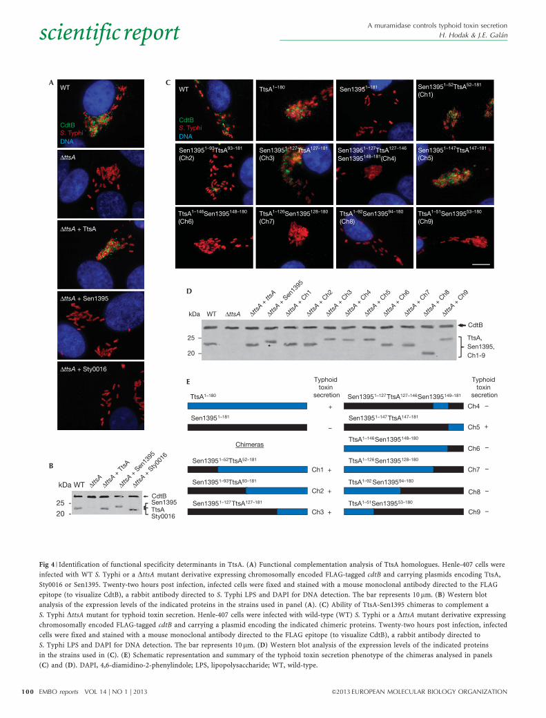
encoded by S. enterica to avoid potential expression and/or codonusage issues. We found that neither S. Typhi Sty0016 norS. Enteritidis Sen1395 belonging to groups 2 and 3, respectively,were able to complement a DttsA S. Typhi mutant for toxinsecretion (Fig 4A,B), despite the fact that both proteins are likelyto be enzymatically active as, like TtsA, their overexpressionresulted in bacterial lysis (supplementary Fig S7 online). Theseresults indicate that despite the high structural similarity,these muramidases are adapted to carry out specific functions.
The C-terminus of TtsA determines secretion functionTo identify determinants within TtsA that are important to carry outprotein secretion functions, we examined the ability of a series ofchimeras between TtsA and the non-complementing muramidaseSen1395 to complement a S. Typhi DttsA mutant for typhoidtoxin secretion (Fig 4C,D). This protein family is composedof a glycosyl hydrolase (GH) domain at the N terminus and aPG-binding domain at the C terminus (supplementary Fig S8online). We replaced the GH domain of TtsA for an equivalentdomain from Sen1395. The resulting chimera, as well as otherchimeras carrying smaller segments of this domain, was able tocomplement the S. Typhi DttsA mutant (Fig 4C–E). These resultsindicate that the specificity determinants that allow TtsA to carryout its specialized function must lie within the PG-binding domain.Consistent with this hypothesis, a chimera consisting of the GHdomain from TtsA and the PG-binding domain from Sen1395 didnot complement the S. Typhi DttsA mutant despite the fact that itwas enzymatically active (supplementary Fig S9 online) andexhibited wild-type levels of chimeric protein expression(Fig 4C–E and supplementary Fig S9 online). To narrow downthe specificity region, we constructed more chimeras with anincreasing proportion of the N terminus of Sen1395 includingportions of its PG-binding domain. We found that a chimeraconsisting of all but the last 32 amino acids of Sen1395 was ableto complement the S. Typhi DttsA mutant (Fig 4C–E andsupplementary Fig S9 online), indicating that the specificitydeterminants that confer TtsA its ability to carry out typhoid toxinsecretion function must lie within its last 32 amino acids.
We compared the amino-acid sequence of this region of TtsAwith the equivalent region of Sen1395 and identified severalamino acids that were not conserved between thesetwo muramidases and therefore potentially important for TtsAfunction (Fig 5A). We tested this hypothesis by changing theseresidues in TtsA into the equivalent residues present in Sen1395.We found that changing Q154, Q164, I174 and R179 into thecorresponding amino acids present in Sen1395 did not affect TtsAfunction (Fig 5B,C). However, a TtsA mutant, in which asparagine166 was changed into aspartic acid (present in Sen1395), wasunable to complement the S. Typhi DttsA mutant for typhoid toxinsecretion (Fig 5B,C). Furthermore, we found that, remarkably,simply changing the aspartic acid residue in Sen1395 intoasparagine (present in TtsA at that position) allowed thismuramidase to restore typhoid toxin secretion to a S. Typhi DttsAmutant (Fig 4D,E). Overexpression of TtsAN166D (unable tocomplement the DttsA mutant for typhoid toxin secretion) ledto S. Typhi lysis after chloroform treatment (supplementary Fig S10online), indicating that introduction of these mutations did notaffect its enzymatic activity. These results not only indicate thatN166 is critical for TtsA function but suggest that remarkably
minor changes in the PG-binding domain of members of thismuramidase family can result in significant changes in theirfunction and specificity, presumably by altering their targetingmechanisms. We examined other muramidases for the presence ofN166 and found that this amino acid is present in many membersof the family. We expressed one of these members, the Yersiniaenterocolitica Ye1815, in the S. Typhi DttsA mutant strain andfound that it was able to complement this strain for typhoid toxinsecretion (Fig 5D,E), consistent with the importance of N166 inconferring these muramidases the ability to function in proteinsecretion. However, Sty0016, which has the N166 residue, wasunable to complement the S. Typhi DttsA mutant strain for typhoidtoxin secretion (Fig 4A) despite the fact that its overexpression inS. Typhi indicated that it is enzymatically active (supplementaryFig S10 online). This finding indicates that in addition to N166,other residues must contribute to the TtsA specificity. Indeed,structure modelling of this region predicts that N166 is locatedwithin a loop bounded by two well-organized helixes(supplementary Fig S11 online). It is therefore possible that theoverall configuration of the loop might be important for targeting.Interestingly, phylogenetic analysis of the last 36 amino acidsof TtsA family members clustered all the muramidases capable ofcomplementing typhoid toxin within the same group, indicatingthe presence of common features within this domain in theseprotein homologues (supplementary Fig S12 online). Takentogether, these results indicate that information located withinthe C terminus of this enzyme family determines functionalspecificity, perhaps by targeting the enzymatic activity so as todisrupt the PG layer in close proximity to a periplasmic poolof the protein or complex of proteins (for example, typhoid toxin)destined to be secreted by this pathway.
Concluding remarksWe have identified a muramidase belonging to a widelydistributed protein family that is required for S. Typhi’s typhoidtoxin secretion. We propose that these muramidases representa recent adaptation of phage-associated enzymes to carry out adifferent function. Consistent with this hypothesis, we note thatSty1887, located immediately adjacent to ttsA, is predicted toencode a homologue of phage tail proteins. In addition, wepropose that these enzymes define a new protein secretionsystem (Fig 5F) that is likely to be widespread in bacteriaas we found several instances in which toxins or predictedextracellular enzymes are encoded immediately adjacent toholin/muramidase pairs.
METHODSBacterial strains were constructed as previously described [17].Immunofluorescence microscopy and cultured cell infectionassays, and western blot analysis were performed as previouslydescribed [7]. Bacteriolytic assays after addition of 0.3% CHCl3were carried out as previously described [12] with somemodifications. Briefly, overnight grown bacteria were diluted toan OD600 nm of 0.1 in fresh LB medium containing 0.2%arabinose. After 60 min of induction, 0.3% CHCl3 was added tothe cultures and lysis was monitored for additional 60 min bymeasuring their OD600 nm. For holin-assisted bacteriolysis,overnight cultures were diluted in fresh LB medium to anOD600 nm of 0.1 and further grown to an OD600 nm of 0.5. At
A muramidase controls typhoid toxin secretion
H. Hodak & J.E. Galan scientificreport
99&2013 EUROPEAN MOLECULAR BIOLOGY ORGANIZATION EMBO reports VOL 14 | NO 1 | 2013
AWT
ΔttsA
ΔttsA + TtsA
ΔttsA + Sty0016
CdtB
S. Typhi
DNA
ΔttsA + Sen1395
B
20 -
25 -
WT
CdtB
TtsASty0016
Sen1395
E
C
D
Sen13951–93TtsA93–181
(Ch2)
TtsA1–146Sen1395148–180
(Ch6)
TtsA1–180 Sen13951–181 Sen13951–52TtsA52–181
(Ch1)
Sen13951–127TtsA127–181
(Ch3)
Sen13951–127TtsA127–146
Sen1395148–181(Ch4)
Sen13951–147TtsA147–181
(Ch5)
TtsA1–126Sen1395128–180
(Ch7)
TtsA1–92Sen139594–180
(Ch8)
TtsA1–51Sen139553–180
(Ch9)
WT
CdtB
WT ΔttsA
25 –
20 –
TtsA,
Sen1395,
Ch1-9
TtsA1–180
+
Sen13951–181
–
TtsA1–51Sen139553–180
–Sen13951–127TtsA127–181
+
Sen13951–52TtsA52–181
+
Sen13951–93TtsA93–181
+
+
TtsA1–146Sen1395148–180
Sen13951–147TtsA147–181
–
TtsA1–126Sen1395128–180
–
–
TtsA1–92 Sen139594–180
–
Sen13951–127TtsA127–146Sen1395149–181
Typhoid
toxin
secretion
Typhoid
toxin
secretion
Chimeras
Ch1
Ch2
Ch3
Ch4
Ch5
Ch6
Ch7
Ch8
Ch9
kDa
kDa
CdtB
S. Typhi
DNA
ΔttsA +
ttsA
ΔttsA +
TtsA
ΔttsA +
Sen
1395
ΔttsA +
Sty
0016
ΔttsA
ΔttsA +
Sen
1395
ΔttsA +
Ch1
ΔttsA +
Ch2
ΔttsA +
Ch3
ΔttsA +
Ch4
ΔttsA +
Ch5
ΔttsA +
Ch6
ΔttsA +
Ch7
ΔttsA +
Ch8
ΔttsA +
Ch9
Fig 4 | Identification of functional specificity determinants in TtsA. (A) Functional complementation analysis of TtsA homologues. Henle-407 cells were
infected with WT S. Typhi or a DttsA mutant derivative expressing chromosomally encoded FLAG-tagged cdtB and carrying plasmids encoding TtsA,
Sty0016 or Sen1395. Twenty-two hours post infection, infected cells were fixed and stained with a mouse monoclonal antibody directed to the FLAG
epitope (to visualize CdtB), a rabbit antibody directed to S. Typhi LPS and DAPI for DNA detection. The bar represents 10 mm. (B) Western blot
analysis of the expression levels of the indicated proteins in the strains used in panel (A). (C) Ability of TtsA-Sen1395 chimeras to complement a
S. Typhi DttsA mutant for typhoid toxin secretion. Henle-407 cells were infected with wild-type (WT) S. Typhi or a DttsA mutant derivative expressing
chromosomally encoded FLAG-tagged cdtB and carrying a plasmid encoding the indicated chimeric proteins. Twenty-two hours post infection, infected
cells were fixed and stained with a mouse monoclonal antibody directed to the FLAG epitope (to visualize CdtB), a rabbit antibody directed to
S. Typhi LPS and DAPI for DNA detection. The bar represents 10 mm. (D) Western blot analysis of the expression levels of the indicated proteins
in the strains used in (C). (E) Schematic representation and summary of the typhoid toxin secretion phenotype of the chimeras analysed in panels
(C) and (D). DAPI, 4,6-diamidino-2-phenylindole; LPS, lipopolysaccharide; WT, wild-type.
A muramidase controls typhoid toxin secretion
H. Hodak & J.E. Galanscientificreport
100 EMBO reports VOL 14 | NO 1 | 2013 &2013 EUROPEAN MOLECULAR BIOLOGY ORGANIZATION

that time, 0.002% arabinose was added to induce holin and/orendolysin expression, and bacterial lysis was monitored foradditional 280 min by measuring the OD600 nm of the bacterialcultures. Phylogenetic trees were generated using ClustalW2,homologues of Sty1889 were identified using BLASTP and phagegenes were detected using the PHAST. A more detaileddescription of the Methods appears in supplementary materials.
Supplementary information is available at EMBO reports online(http://www.emboreports.org).
ACKNOWLEDGEMENTSWe thank members of the Galan laboratory for critical readingof this manuscript. This work was supported by NIAID Grant AI079022to J.E.G.
D
F
A
180181
147148
N166Q164Q154 I174 R179
TtsASen1395
ΔttsA
ΔttsA + Sen1395
ΔttsA + Sen1395D167N
WT
ΔttsA + Ye1815
E
ΔttsA + TtsA
ΔttsA + TtsAQ154E
ΔttsA + TtsAQ164E
ΔttsA + TtsAN166D
ΔttsA + TtsAI174M
ΔttsA + TtsAR179L
CdtB25 -
20 -TtsAYe1815
Sen1395
B
ΔttsA + TtsA
ΔttsA + TtsA
ΔttsA
ΔttsA + TtsAQ154E
ΔttsA + TtsAQ164E ΔttsA + TtsAN166D
ΔttsA + TtsAI174M
WT
CdtBS.Typhi DNA
ΔttsA + TtsAR197L
CdtBS. TyphiDNA
C
WT ΔttsA
25 -CdtB
TtsA
kDa
TtsA
Holin?SecYEG
PltA
PltBCdtB
Typhoid toxin
IM
OM
Periplasm
PG
Cytoplasm
Sec SP
?
WT ΔttsA + TtsA
ΔttsA + Sen1395
ΔttsA + Sen1395D167N
ΔttsA + Ye1815
kDa ΔttsA
Fig 5 | A C-terminal region of TtsA determines its ability to secrete typhoid toxin. (A) Amino-acid sequence comparison of the C terminus of TtsA and
Sen1395. The residues mutated in this analysis are indicated. (B) Mutagenesis analysis of the C-terminal region of TtsA. Henle-407 cells were infected
with WT S. Typhi or a DttsA mutant derivative expressing chromosomally encoded FLAG-tagged cdtB and carrying plasmids encoding the indicated
TtsA mutant proteins. Twenty-two hours post infection, infected cells were fixed and stained with a mouse monoclonal antibody directed to the FLAG
epitope (to visualize CdtB), a rabbit antibody directed to S. Typhi LPS and DAPI for DNA detection. The bar represents 10 mm. (C) Western blot
analysis of the expression levels of the indicated proteins in the strains used in panel (B). (D) The N166 residue of TtsA is crucial for typhoid toxin
secretion. Henle-407 cells were infected with WT S. Typhi or a DttsA mutant derivative expressing chromosomally encoded FLAG-tagged cdtB and
carrying plasmids encoding the indicated proteins. Twenty-two hours post infection, infected cells were fixed and stained with a mouse monoclonal
antibody directed to the FLAG epitope (to visualize CdtB), a rabbit antibody directed to S. Typhi LPS and DAPI for DNA detection. The bar represents
10 mm. (E) Western blot analysis of the expression levels of the indicated proteins in the strains used in panel (D). (F) Typhoid toxin secretion model.
Typhoid toxin components PltA, PltB and CdtB have an N-terminal Sec signal peptide, which mediates their export to the periplasm through the Sec
machinery. Further protein export requires enzymatically active TtsA, which lacks a canonical N-terminal signal peptide and therefore must be
exported to the periplasm by a yet unidentified holin. Sec signal peptides (Sec SP), PG, IM and OM are indicated. DAPI, 4,6-diamidino-2-phenylindole;
IM, inner membrane; LPS, lipopolysaccharide; OM, outer membrane; PG, peptidoglycan; WT, wild-type.
A muramidase controls typhoid toxin secretion
H. Hodak & J.E. Galan scientificreport
101&2013 EUROPEAN MOLECULAR BIOLOGY ORGANIZATION EMBO reports VOL 14 | NO 1 | 2013

Author contributions: H.H. designed, interpreted and conductedexperiments; wrote the paper. J.E.G. designed and interpretedexperiments; wrote the paper.
CONFLICT OF INTERESTThe authors declare that they have no conflict of interest.
REFERENCES1. Parry C, Hien TT, Dougan G, White N, Farrar J (2002) Typhoid fever.
N Engl J Med 347: 1770–17822. Crump J, Mintz E (2010) Global trends in typhoid and paratyphoid fever.
Clin Infect Dis 50: 241–2463. Raffatellu M, Wilson R, Winter S, Baumler A (2008) Clinical pathogenesis
of typhoid fever. J Infect Dev Ctries 2: 260–2664. House D, Bishop A, Parry C, Dougan G, Wain J (2001) Typhoid fever:
pathogenesis and disease. Curr Opin Infect Dis 14: 573–5785. Baker S, Dougan G (2007) The genome of Salmonella enterica serovar
Typhi. Clin Infect Dis 45(Suppl 1): S29–S336. Parkhill J et al (2001) Complete genome sequence of a multiple drug
resistant Salmonella enterica serovar Typhi CT18. Nature 413: 848–8527. Spano S, Ugalde JE, Galan JE (2008) Delivery of a Salmonella Typhi
exotoxin from a host intracellular compartment. Cell Host Microbe 3: 30–388. Haghjoo E, Galan JE (2004) Salmonella Typhi encodes a functional
cytolethal distending toxin that is delivered into host cells by abacterial-internalization pathway. Proc Natl Acad Sci USA 101:4614–4619
9. Spano S, Galan JE (2008) A novel pathway for exotoxin delivery by anintracellular pathogen. Curr Opin Microbiol 11: 15–20
10. Haghjoo E, Galan JE (2007) Identification of a transcriptional regulatorthat controls intracellular gene expression in Salmonella Typhi. MolMicrobiol 64: 1549–1561
11. Spano S, Liu X, Galan JE (2011) Proteolytic targeting of Rab29by an effector protein distinguishes the intracellular compartments ofhuman-adapted and broad-host Salmonella. Proc Natl Acad Sci USA108: 18418–18423
12. Stojkovic E, Rothman-Denes L (2007) Coliphage N4N-acetylmuramidase defines a new family of murein hydrolases. J MolBiol 366: 406–419
13. Pei J, Grishin NV (2005) COG3926 and COG5526: a tale of two newlysozyme-like protein families. Protein Sci 14: 2574–2581
14. Young R (2002) Bacteriophage holins: deadly diversity. J Mol MicrobiolBiotechnol 4: 21–36
15. Wang I, Smith D, Young R (2000) Holins: the proteinclocks of bacteriophage infections. Annu Rev Microbiol 54:799–825
16. Kondo Y, Toyoda A, Fukushi H, Yanase H, Tonomura K, Kawasaki H,Sakai T (1994) Cloning and characterization of a pair of genes thatstimulate the production and secretion of Zymomonas mobilisextracellular levansucrase and invertase. Biosci Biotechnol Biochem 58:526–530
17. Kaniga K, Bossio JC, Galan JE (1994) The Salmonella typhimuriuminvasion genes invF and invG encode homologues of the AraC and PulDfamily of proteins. Mol Microbiol 13: 555–568
A muramidase controls typhoid toxin secretion
H. Hodak & J.E. Galanscientificreport
102 EMBO reports VOL 14 | NO 1 | 2013 &2013 EUROPEAN MOLECULAR BIOLOGY ORGANIZATION


