Dynamics and interplay of nuclear architecture, genome...
18
REVIEW Dynamics and interplay of nuclear architecture, genome organization, and gene expression Robert Schneider and Rudolf Grosschedl 1 Max Planck Institute of Immunobiology, 79108 Freiburg, Germany The organization of the genome in the nucleus of a eu- karyotic cell is fairly complex and dynamic. Various fea- tures of the nuclear architecture, including compart- mentalization of molecular machines and the spatial arrangement of genomic sequences, help to carry out and regulate nuclear processes, such as DNA replication, DNA repair, gene transcription, RNA processing, and mRNA transport. Compartmentalized multiprotein complexes undergo extensive modifications or exchange of protein subunits, allowing for an exquisite dynamics of structural components and functional processes of the nucleus. The architecture of the interphase nucleus is linked to the spatial arrangement of genes and gene clus- ters, the structure of chromatin, and the accessibility of regulatory DNA elements. In this review, we discuss re- cent studies that have provided exciting insight into the interplay between nuclear architecture, genome organi- zation, and gene expression. One of the striking features of the eukaryotic cell nucleus, which carries and reads the genetic informa- tion, is its functional and structural complexity. The hu- man genome, containing some 35,000 genes and 3.2 bil- lion base pairs of DNA, is compacted 400,000-fold to fit within a nuclear volume of ∼1000 μm 3 . The packing of DNA into chromatin is an extremely efficient way of storing the DNA within the nucleus. However, the ge- netic information has to become accessible for DNA- dependent processes such as transcription, DNA repair, and replication. Most importantly, this accessibility is regulated, as not all regions of the genome are active at a given time during the cell cycle. In recent years, increas- ing evidence has been accumulated suggesting that cis- and trans-acting regulatory DNA sequences may not be the only determinants of gene expression, but that DNA transactions also depend on the genomic position of genes, the subnuclear localization of DNA sequences, and a complex interplay of the genome with specific fea- tures of nuclear architecture. The nucleus is organized in specific compartments that include proteinaceous nuclear bodies, eukaryotic and heterochromatic chromatin domains, compartmen- talized multiprotein complexes, and nuclear pores that allow for nucleocytoplasmic transport. The dynamic, temporal, and spatial organization of the eukaryotic cell nucleus emerges as a central determinant of genome function, and it is important to understand the relation- ship between nuclear architecture, genome organization, and gene expression. Recently, high-resolution tech- niques have permitted new insights into the nuclear ar- chitecture and its relationship to gene expression. In this review, we focus on the dynamic organization of the genome into chromosome territories and chroma- tin domains. We also discuss the link of subnuclear gene positioning with gene expression and how intra- and interchromosomal interactions may contribute to gene regulation. Within the nucleus many proteinaceous nuclear bod- ies, such as PML bodies or Cajal bodies, have been de- scribed (for detailed review, see Spector 2003). These bodies have a nonrandom positioning relative to spe- cific nuclear regions. Distinct functions have been at- tributed to the various nuclear bodies, but we are just beginning to understand the role of nuclear bodies in gene transcription, RNA processing, and DNA repair. An interesting feature of nuclear bodies is the involvement of post-transcriptional protein modifications, such as SUMOylation for PML bodies or arginine methylation for Cajal bodies, in their generation and/or maintenance (for review, see Seeler and Dejean 2003; Heun 2007). Chromatin domains In addition to the proteinaceous nuclear bodies, large- scale chromatin domains exist that are mainly cytologi- cally defined. In 1928, Heitz subdivided chromatin— based on its microscopic appearance—into heterochro- matin and euchromatin (Heitz 1928). Euchromatin was correlated with the bulk of transcribed chromatin, which is in an “open” chromatin conformation. In contrast, heterochromatin was correlated with more condensed chromatin, which is enriched in inactive and silenced chromatin regions. Heterochromatin was considered to [Keywords: Nuclear organization; chromatin; gene localization; chromo- somal interactions] 1 Corresponding author. E-MAIL [email protected]; FAX 49-761-5108798. Article is online at http://www.genesdev.org/cgi/doi/10.1101/gad.1604607. GENES & DEVELOPMENT 21:3027–3043 © 2007 by Cold Spring Harbor Laboratory Press ISSN 0890-9369/07; www.genesdev.org 3027 Cold Spring Harbor Laboratory Press on May 20, 2018 - Published by genesdev.cshlp.org Downloaded from
Transcript of Dynamics and interplay of nuclear architecture, genome...
REVIEW
Dynamics and interplay of nucleararchitecture, genome organization,and gene expressionRobert Schneider and Rudolf Grosschedl1
Max Planck Institute of Immunobiology, 79108 Freiburg, Germany
The organization of the genome in the nucleus of a eu-karyotic cell is fairly complex and dynamic. Various fea-tures of the nuclear architecture, including compart-mentalization of molecular machines and the spatialarrangement of genomic sequences, help to carry out andregulate nuclear processes, such as DNA replication,DNA repair, gene transcription, RNA processing, andmRNA transport. Compartmentalized multiproteincomplexes undergo extensive modifications or exchangeof protein subunits, allowing for an exquisite dynamicsof structural components and functional processes of thenucleus. The architecture of the interphase nucleus islinked to the spatial arrangement of genes and gene clus-ters, the structure of chromatin, and the accessibility ofregulatory DNA elements. In this review, we discuss re-cent studies that have provided exciting insight into theinterplay between nuclear architecture, genome organi-zation, and gene expression.
One of the striking features of the eukaryotic cellnucleus, which carries and reads the genetic informa-tion, is its functional and structural complexity. The hu-man genome, containing some 35,000 genes and 3.2 bil-lion base pairs of DNA, is compacted 400,000-fold to fitwithin a nuclear volume of ∼1000 µm3. The packing ofDNA into chromatin is an extremely efficient way ofstoring the DNA within the nucleus. However, the ge-netic information has to become accessible for DNA-dependent processes such as transcription, DNA repair,and replication. Most importantly, this accessibility isregulated, as not all regions of the genome are active at agiven time during the cell cycle. In recent years, increas-ing evidence has been accumulated suggesting that cis-and trans-acting regulatory DNA sequences may not bethe only determinants of gene expression, but that DNAtransactions also depend on the genomic position ofgenes, the subnuclear localization of DNA sequences,and a complex interplay of the genome with specific fea-tures of nuclear architecture.
The nucleus is organized in specific compartmentsthat include proteinaceous nuclear bodies, eukaryoticand heterochromatic chromatin domains, compartmen-talized multiprotein complexes, and nuclear pores thatallow for nucleocytoplasmic transport. The dynamic,temporal, and spatial organization of the eukaryotic cellnucleus emerges as a central determinant of genomefunction, and it is important to understand the relation-ship between nuclear architecture, genome organization,and gene expression. Recently, high-resolution tech-niques have permitted new insights into the nuclear ar-chitecture and its relationship to gene expression.
In this review, we focus on the dynamic organizationof the genome into chromosome territories and chroma-tin domains. We also discuss the link of subnucleargene positioning with gene expression and how intra-and interchromosomal interactions may contribute togene regulation.
Within the nucleus many proteinaceous nuclear bod-ies, such as PML bodies or Cajal bodies, have been de-scribed (for detailed review, see Spector 2003). Thesebodies have a nonrandom positioning relative to spe-cific nuclear regions. Distinct functions have been at-tributed to the various nuclear bodies, but we are justbeginning to understand the role of nuclear bodies ingene transcription, RNA processing, and DNA repair. Aninteresting feature of nuclear bodies is the involvementof post-transcriptional protein modifications, such asSUMOylation for PML bodies or arginine methylationfor Cajal bodies, in their generation and/or maintenance(for review, see Seeler and Dejean 2003; Heun 2007).
Chromatin domains
In addition to the proteinaceous nuclear bodies, large-scale chromatin domains exist that are mainly cytologi-cally defined. In 1928, Heitz subdivided chromatin—based on its microscopic appearance—into heterochro-matin and euchromatin (Heitz 1928). Euchromatin wascorrelated with the bulk of transcribed chromatin, whichis in an “open” chromatin conformation. In contrast,heterochromatin was correlated with more condensedchromatin, which is enriched in inactive and silencedchromatin regions. Heterochromatin was considered to
[Keywords: Nuclear organization; chromatin; gene localization; chromo-somal interactions]1Corresponding author.E-MAIL [email protected]; FAX 49-761-5108798.Article is online at http://www.genesdev.org/cgi/doi/10.1101/gad.1604607.
GENES & DEVELOPMENT 21:3027–3043 © 2007 by Cold Spring Harbor Laboratory Press ISSN 0890-9369/07; www.genesdev.org 3027
Cold Spring Harbor Laboratory Press on May 20, 2018 - Published by genesdev.cshlp.orgDownloaded from
be transcriptionally inert. However, in the last few yearsit has become clear that this correlation of chromatinstructure and gene function is limited. Recently, theBickmore group (Gilbert et al. 2004) analyzed chromatinstructure at a more global level. They fractionated chro-matin into open, bulk, and closed chromatin fibers andassayed the distribution of gene density and activity inthese chromatin fractions. Surprisingly, they found alink between chromatin structure and gene density, in-dependent of the status of gene activity. Open chromatinfibers correlate with highest gene density, but not geneexpression levels, whereas compact chromatin fibersgenerally have a low gene density, but can also containactive genes (Gilbert et al. 2004). The ability of genes tobe activated is not necessarily lost when chromatin ispacked into more compact fibers. Conversely, inactivegenes close to active genes in an open chromatin envi-ronment can stay inactive. In these cases, it is not thelocation of a gene in heterochromatic or euchromaticchromatin domains that regulates gene activity, butrather, other factors such as covalent modifications ofhistones or DNA (Spector 2004).
Transcription factories
Microscopic analysis of sites of active transcription inHeLa cell nuclei, using Br-UTP incorporation, revealed anonhomogeneous clustering of ∼104 transcription sites,with 8000 sites representing RNA polymerase II clustersand the rest RNA polymerase III transcription sites(Pombo et al. 1999). The number of transcription sitesvaries in different cells. These sites of active transcrip-tion measure ∼80 nm in diameter, and they have beentermed “transcription factories” (Jackson et al. 1998).
The textbook concept for a long time was that activegenes recruit the transcription machinery, and it is thetranscription machinery that relocates to the active genes.However, three-dimensional fluorescence in situ hybrid-ization (3D-FISH), immunofluorescence, and chromo-some conformation capture (3C) analysis, which detectsclose physical proximity between remote chromatin seg-ments (Dekker 2006), provided new insights into the or-ganization of transcription sites and raised questionsabout this dogma. Using a combination of these meth-ods, the Fraser group (Osborne et al. 2004) showed thatwidely separated active genes can colocalize to thesesites of active transcription. They demonstrated thatgenes are dynamically recruited to these sites of activetranscription (not vice versa), and that most genes canmove in and out of these sites, resulting in activation orabatement of their transcription (Osborne et al. 2004).Genes that are capable of high expression levels, such as�-globin, �-globin, and immunoglobulin genes, seem tobe constantly associated with “transcription factories”in cells that express these genes, whereas temporarilyquiescent alleles are located away from the factories (Os-borne et al. 2004). Transcriptional activation of highlyexpressed genes such as immediate early genes involvestheir relocalization to preassembled transcription sites(Osborne et al. 2007). These transcription sites may be
maintained by flanking, ubiquitously expressed house-keeping genes (Zhou et al. 2006), or by locus controlregions (Ragoczy et al. 2006).
According to the “transcription factory” model, RNApolymerase complexes, and transcription factors clusterand form a “cloud” of up to 20 DNA loops around thetranscription factory (Cook 1999; Faro-Trindade andCook 2006). The polymerase would be an immobilecomponent of the factory, and DNA loops would appearand disappear as polymerases initiate, elongate, and ter-minate transcription. Each factory contains only onetype of RNA polymerase, and factories may be enrichedin specific transcription factors involved in the transcrip-tion of specific groups of genes (Bartlett et al. 2006). Thegenes near the factory would be more likely to be tran-scribed. When released after termination, a gene wouldstill be near a factory and still carry the active histonemodifications that could keep it in an open state, leadingto efficient reinitiation (Bartlett et al. 2006). In agree-ment with this concept, domains of decondensed andrecently transcribed chromatin carry histone marks thatare associated with active transcription (Muller et al.2007).
What is the driving force to move the DNA templatetoward the transcription sites? One possibility is thatactive polymerase can function as a motor that pulls inits template. Optical tweezers experiments showed thatthe force produced by a single polymerase molecule dur-ing transcription can be substantially larger than thoseproduced by the cytoskeletal motors kinesin and myosin(Yin et al. 1995). Alternatively, other molecular motorssuch as nuclear actin and myosin could be involved inthe relocalization of the DNA template. Another unre-solved issue concerns the anchoring of transcriptionsites. Many active genes and transcription factors areassociated with the “nuclear matrix,” an ill-defined andcontroversial nuclear substructure (Jackson and Cook1985; Davie 1995; Iborra et al. 1996; Kumar et al.2007).
Although the model of “transcription factories” fitswith the observation that active genes can transientlycluster (Simonis et al. 2006), one has to keep in mindthat biochemical evidence for these “transcription fac-tories,” including their isolation and functional charac-terization, is still missing. Future experiments areneeded to validate this model and to prove the functionalrelevance of transcription factories.
Organization of chromosomes in interphase nuclei
Via its association with histones and other nonhistoneproteins, the DNA is packed into a higher-order chroma-tin structure. More than 120 years ago, Rabl suggestedthat chromatin is not randomly organized, but occupiesdiscrete territories. Almost 100 years later, Cremer et al.(1982) showed that UV irradiation of specific interphasenuclear areas damaged discrete chromosomal regions,suggesting that chromosomes occupy distinct positionsin the nucleus, the so-called interphase chromosome ter-ritories (for review, see Cremer et al. 2006; Heard and
Schneider and Grosschedl
3028 GENES & DEVELOPMENT
Cold Spring Harbor Laboratory Press on May 20, 2018 - Published by genesdev.cshlp.orgDownloaded from
Bickmore 2007). By using specific probes for individualchromosomes for in situ hybridization, several groups con-firmed that each chromosome occupies a specific territoryin the nucleus (Borden and Manuelidis 1988; Cremer et al.1988; Pinkel et al. 1988). These territories are nonover-lapping, and—at least in mammalian cells—homologouschromosome territories are usually not adjacent (Cremeret al. 2001). The most gene-rich chromosome territoriestend to concentrate in the nuclear interior, whereasgene-poor chromosomes tend to localize toward thenuclear periphery (Croft et al. 1999; Boyle et al. 2001).The organization of chromosomes in specific territorieshas been proposed to facilitate interactions of chromo-somes with other chromosomes and, therefore, chromo-somal territories may have major implications in DNA-dependent processes, such as transcription.
The evidence for a relationship between physical chro-mosome size and nuclear position is still conflicting(Croft et al. 1999; Cremer et al. 2001). The chromosometerritories are dynamic and plastic structures. Individualchromosomal regions can be dynamically repositioned.Upon cell cycle exit, a gene-poor human chromosomecan move from the nuclear periphery to a more internalsite and after re-entry into the cell cycle, the chromo-some moves back to the periphery (Wilmut and Camp-bell 1998; Bridger et al. 2000).
Localization within the chromosome territories
Initial studies proposed a model of a distinct boundary atthe surface of the chromosome territories, with the ter-ritory on one side and the interchromosomal space con-taining the transcriptional machinery on the other side.Generally, inactive genes tend to be located in interiorregions of chromosome territories, whereas active genestend to concentrate along the periphery close to the in-terchromosomal space (Cremer et al. 2001). However,this general rule seems to be an oversimplification. Incontradiction to this rule, studies of the 11p13 locus,containing ubiquitously expressed genes and tissue-spe-cifically expressed genes, showed that this locus is lo-cated within a discrete chromosome territory, and thatafter activation of tissue-specific genes, these geneswere not relocated to the periphery of the territory(Mahy et al. 2002a,b).
The question arises as to how the borders of territoriesare defined and as to whether different chromosome ter-ritories are completely separated or whether chromo-somes can intermingle. Volpi et al. (2000) showed thatthe gene-rich major histocompatibilty complex (MHC)can be found on large chromatin loops, containing sev-eral megabases of DNA that extend outward from thechromosome territory (see Fig. 1B). Interestingly, tran-scriptional up-regulation of the MHC genes led to anincrease in the frequency with which this cluster wasfound on a chromatin loop extending out of the chromo-some territory (Volpi et al. 2000). The HoxB gene clusteris another interesting example for looping out of thechromosome territory. Activation of the HoxB gene clus-ter during differentiation correlates with its relocaliza-
tion away from its chromosome territory (Chambeyronand Bickmore 2004). Thus, the surface of the chromo-some territories can be increased by looping out of DNAinto the interchromosomal compartment and by infold-ings of the interchromosomal compartment into the ter-ritories (Cremer et al. 2006; Heard and Bickmore 2007.This revised model of chromosomal territories also takesinto account that active genes in the interior of chromo-some territories (such as active genes in the 11p13 locus)are accessible for the transcription machinery throughDNA-free channels (Cremer et al. 2006). In several stud-ied examples, such as the HoxB gene cluster, looping outof the chromosome territories correlates with “decon-densation” of the chromatin, which is required for tran-scription and for the flexibility of the looped-out chro-matin fiber (Chambeyron and Bickmore 2004). However,this looping and the chromatin “decondensation” arenot always linked. For example, in the Hoxd locus, de-condensed alleles can be found within the chromosometerritories and, conversely, looped-out gene loci can stillbe condensed (Morey et al. 2007). These findings suggestthat looping out and decondensation are not causallycoupled and could constitute two separate modes ofregulation. Therefore, it will be interesting to investigatehow histone modifications (see below) are involved inthe looping out and decondensation of specific gene locisuch as HoxB and HoxD.
Recent studies applying high resolution in situ hybrid-ization and chromosome conformation-capture assays
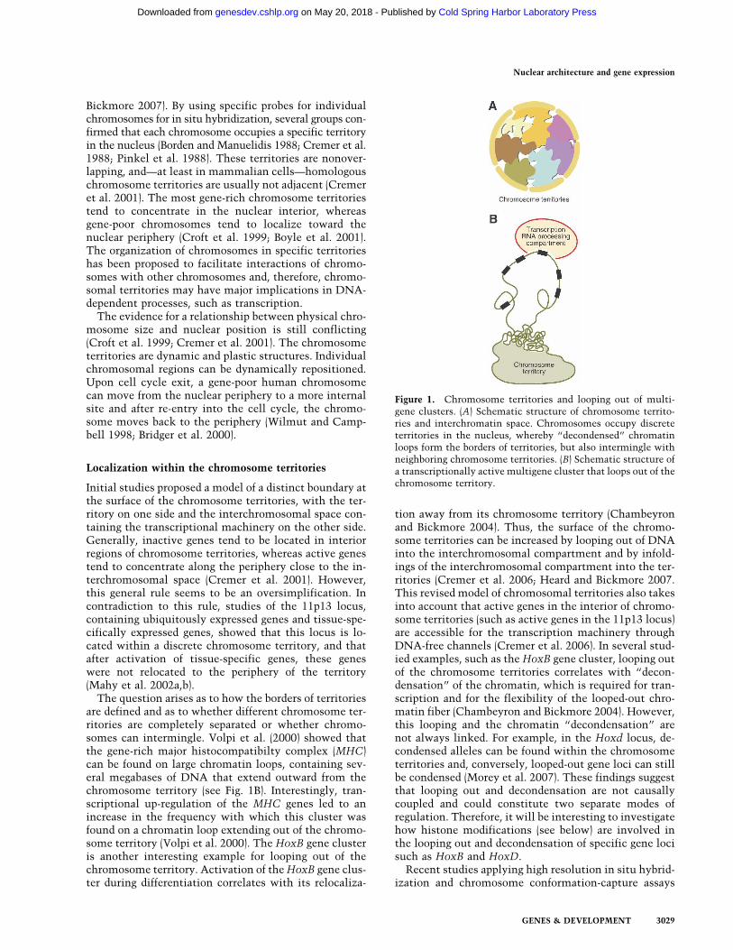
Figure 1. Chromosome territories and looping out of multi-gene clusters. (A) Schematic structure of chromosome territo-ries and interchromatin space. Chromosomes occupy discreteterritories in the nucleus, whereby “decondensed” chromatinloops form the borders of territories, but also intermingle withneighboring chromosome territories. (B) Schematic structure ofa transcriptionally active multigene cluster that loops out of thechromosome territory.
Nuclear architecture and gene expression
GENES & DEVELOPMENT 3029
Cold Spring Harbor Laboratory Press on May 20, 2018 - Published by genesdev.cshlp.orgDownloaded from

revealed that chromatin fibers from the periphery ofchromosome territories are intermingled in interphasenuclei (Branco and Pombo 2006; Simonis et al. 2006).Interestingly, blocking transcription can change the pat-terns of intermingling of territories without changingthe general properties of the chromosome territories(Branco and Pombo 2006). The looped-out, activatedHoxB gene locus shows increased interchromosomal in-teractions, as compared with the inactive locus, whichinteracts preferentially with other loci on the same chro-mosome (Wurtele and Chartrand 2006). The currentview is that the chromosome territories have a sponge-like architecture with the interchromatin compartmentmeandering into the territories through the infoldings,whereby some gene loci loop out of the territories, re-sulting in intermingling of different territories (Cremeret al. 2006). Thus, intrachromosomal interactions wouldfavor compactness of the chromosome territories,whereas interchromosomal interactions would favor in-termingling. The balance between these two interac-tions appears to depend on transcription, chromosomestructure, and chromatin modifications.
Chromatin modifications
Inactive and active chromatin domains can also be de-fined molecularly by the presence of specific post-trans-lational histone modifications. To date, numerous his-tone modifications have been described (for review, seeKouzarides 2007). Histones can be methylated at ar-ginines or lysines, phosphorylated on serines and lysines,acetylated on lysines, sumolyated and ubiquitinated onlysines, and ADP-ribosylated. Moreover, the number ofmethyl groups added to a single lysine or arginineresidue can vary. Lysine residues can be mono-, di-, ortrimethylated, and arginine residues can be monometh-ylated and symmetrically or asymmetrically dimethylated.Importantly, the precise methylation status (mono-, di-,or trimethylation) can influence the transcriptional sta-tus of genes (Schneider et al. 2004). These modificationscould directly affect the structure of chromatin; e.g., byneutralizing the positive charge of histones. In addition,the enormous combinatorial potential of these modifi-cations can be read out by proteins that bind to specificmodifications, which has provided the basis for the so-called histone “code” hypothesis (Turner 1993; Strahland Allis 2000). Although it is still unproven whetherthese modifications form a true “code,” it is now wellestablished that they are involved in the regulation ofgene expression (Turner 2007).
In the last few years, many of the enzymes that addthese modifications, such as histone acetyltransferases,lysine and arginine methyltransferases, and enzymesthat can remove these modifications, such as histonedeacetylases, was described (for review, see Couture andTrievel 2006). In contrast to histone acetylation, the ly-sine methylation had been considered to be a very stablemodification, but the recent discovery of lysine demeth-ylases (Shi et al. 2004) revealed that lysine methylationcan also be dynamic. Interestingly, many of the genes
encoding enzymes modifying histones have been foundto be rearranged, amplified, or mutated in various typesof cancer (Schneider et al. 2002). Several studies focusingon specific genes or loci showed that active chromatin isgenerally enriched in acetylated histones H3, H4, H2A(Davie and Candido 1978), and histone H3 that is meth-ylated at Lys 4 (H3/K4) (Litt et al. 2001). H3/K4 di- andtrimethylation and H3 acetylation correlate globallywith open chromatin (The ENCODE Project Consor-tium 2007). In contrast to this, inactive chromatin ischaracterized by histone hypoacetylation and methyl-ation of histone H3 Lys 9 (H3/K9) (Litt et al. 2001). Themethylation of H3/K9 has been thought to be a mark forheterochromatin. Methylated H3/K9 can be “read out”by the Heterochromatin Protein 1 (HP1), a structuralcomponent of condensed chromatin that specifically rec-ognizes and binds to the methylated form of H3/K9 (Ban-nister et al. 2001; Lachner et al. 2001). The loss of K9methylation in heterochromatin can affect the hetero-chromatin organization (Peters et al. 2001). Interestingly,HP1 itself interacts with the enzyme that methylatesH3/K9, forming a positive feedback loop that would al-low heterochromatin to spread over large chromosomalregions until further spreading is prevented by a bound-ary element (Bannister et al. 2001; Lachner et al. 2001).
Changes in chromatin structure that include modifi-cations of histones may also have a role in the position-ing of chromosomes. Long-term treatment of cells withtrichostatin A, an inhibitor of histone deacetylases thatincreases the acetylation level of histones, results inlarge-scale movement of centromeric and pericentro-meric chromatin to the nuclear periphery. After drug re-moval, these changes in localization are rapidly reversed(Taddei et al. 2001).
Recent genome-wide mapping studies of histonemodifications that combined chromatin immunopre-cipitation (ChIP) with microarrays or direct sequencingallowed for high-resolution mapping of many histonemarks (Barski et al. 2007). So far, these studies have re-vealed that actively transcribed chromatin regions areenriched in H3/K4 mono-, di-, or trimethylation, H3/K36trimethylation, and monomethylation of H3/K9, H3/K27, and H4/K20. The distribution of these marks overthe transcribed region of genes is not equal; trimethyl-ation of H3/K4 is enriched at the 5� end of transcribedregions and can serve as an indicator for transcriptionstart sites, whereas H3/K36 methylation is enriched atthe 3� end and may be linked to processing of the tran-scripts (Bannister et al. 2005). In contrast to this, tri-methylation of H3/K9, H3/K27, and H3/K79 is linkedwith repression (Barski et al. 2007). Interestingly, activepromoters and enhancers are found to be associated withH3/K4 methylation and H3/K9 monomethylation. Addi-tionally, active promoters can be identified by the pres-ence of further activating marks that are linked withtranscriptional elongation (such as H3/K36 methylation)downstream in the transcribed region of the gene (seeFig. 2; Barski et al. 2007). These criteria can be used toidentify active promoters. Interestingly, at genes that arenot regulated at the level of transcription initiation
Schneider and Grosschedl
3030 GENES & DEVELOPMENT
Cold Spring Harbor Laboratory Press on May 20, 2018 - Published by genesdev.cshlp.orgDownloaded from
(polymerase II can be found at these genes) but ratherelongation, H3/K4 methylation is enriched at the pro-moter. Consistent with the post-initiation regulation ofthese genes, marks linked with transcriptional elonga-tion are absent (Guenther et al. 2007). However, this“black-and-white” picture of active and repressivemarks may be an oversimplification. Some activatingmarks are not only found on genes that are transcribed,but also on genes that are poised for transcription. A niceexample is provided by genes that are stimulated by en-dotoxins in macrophages. A subset of these genes main-tains some activating marks after initial induction,priming them for efficient reactivation (Foster et al.2007). Additionally, modifications considered to be re-pressing, such as H3/K9 dimethylation, can be found notonly in heterochromatin but also on certain active genes.Furthermore, in so-called “bivalent chromatin domains”repressive marks (H3/K27 trimethylation) and activatingmarks (H3/K4 trimethylation) can coexist (see below;Azuara et al. 2006; Bernstein et al. 2006; Lee et al. 2006).These findings suggest that the histone “code” is morecomplex than initially expected and that not single
modifications, but rather the combination of modifica-tions is an indicator for the transcriptional state.
In the rapidly developing field of chromatin modifica-tions, many exciting questions are waiting to be ad-dressed. Currently, little is known about how thesemodifications can regulate the localization of specificgenes. Are histone modifications involved in tetheringthe genes to a particular nuclear region or compartment?Are histone modifications the cause or the consequenceof active/inactive chromatin domains? What is their rolein establishing or maintaining nuclear domains? How dothey change when genes loop out of their chromatin ter-ritory? It will be exciting to understand precisely howhistone modifications regulate the functional status notonly of specific genes but also of chromatin domains andto link the modification status with specific gene local-izations or relocalizations of genomic regions.
Mobility and movements of gene loci
The mobility and movements of gene loci have beenstudied extensively by live cell imaging of transgenic
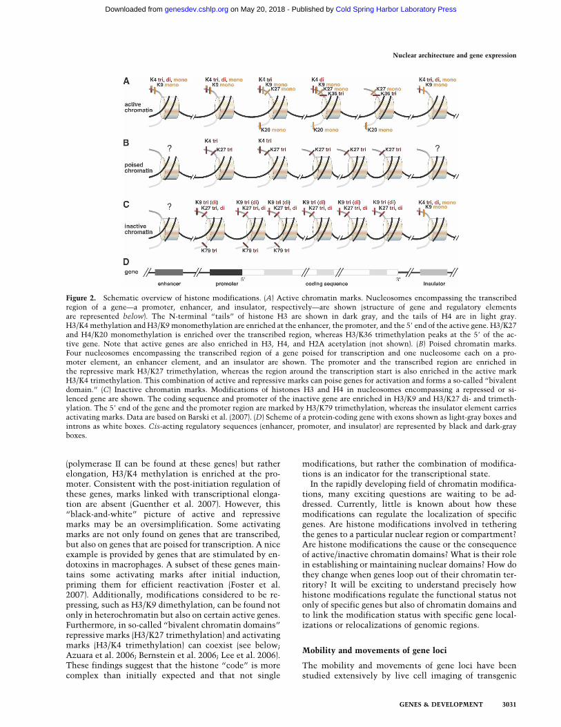
Figure 2. Schematic overview of histone modifications. (A) Active chromatin marks. Nucleosomes encompassing the transcribedregion of a gene—a promoter, enhancer, and insulator, respectively—are shown (structure of gene and regulatory elementsare represented below). The N-terminal “tails” of histone H3 are shown in dark gray, and the tails of H4 are in light gray.H3/K4 methylation and H3/K9 monomethylation are enriched at the enhancer, the promoter, and the 5� end of the active gene. H3/K27and H4/K20 monomethylation is enriched over the transcribed region, whereas H3/K36 trimethylation peaks at the 5� of the ac-tive gene. Note that active genes are also enriched in H3, H4, and H2A acetylation (not shown). (B) Poised chromatin marks.Four nucleosomes encompassing the transcribed region of a gene poised for transcription and one nucleosome each on a pro-moter element, an enhancer element, and an insulator are shown. The promoter and the transcribed region are enriched inthe repressive mark H3/K27 trimethylation, whereas the region around the transcription start is also enriched in the active markH3/K4 trimethylation. This combination of active and repressive marks can poise genes for activation and forms a so-called “bivalentdomain.” (C) Inactive chromatin marks. Modifications of histones H3 and H4 in nucleosomes encompassing a repressed or si-lenced gene are shown. The coding sequence and promoter of the inactive gene are enriched in H3/K9 and H3/K27 di- and trimeth-ylation. The 5� end of the gene and the promoter region are marked by H3/K79 trimethylation, whereas the insulator element carriesactivating marks. Data are based on Barski et al. (2007). (D) Scheme of a protein-coding gene with exons shown as light-gray boxes andintrons as white boxes. Cis-acting regulatory sequences (enhancer, promoter, and insulator) are represented by black and dark-grayboxes.
Nuclear architecture and gene expression
GENES & DEVELOPMENT 3031
Cold Spring Harbor Laboratory Press on May 20, 2018 - Published by genesdev.cshlp.orgDownloaded from

arrays of fluorescently labeled lac operators that havebeen stably integrated in various chromosomal loca-tions. These studies indicated that the mobility of suchtransgenes is predominantly confined to the radius ofchromosomal territories and depends on their relativenuclear localization (Vazquez et al. 2001). Moreover,transgenes that are localized near the nuclear peripheryor are associated with the nucleolus were found to beless mobile than transgenes residing in the nucleoplasm(Chubb et al. 2002). Finally, chromatin repositioning wasdetected predominantly in the G1 phase of the cell cycle(Thomson et al. 2004). Although the molecular basis forchromatin repositioning is fairly obscure, the spatial dis-tribution in the nucleus has been implicated in the es-tablishment and maintenance of active, poised, or re-pressed chromatin states.
Association of genes with the nuclear peripheryand nuclear pores
Early electron microscopic analysis of calf thymus nu-clei revealed a peripheral localization of condensed het-erochromatic regions (Mirsky and Allfrey 1960). In yeast,a role of the nuclear periphery in gene silencing wasshown by the artificial tethering of a reporter gene to thenuclear envelope via a membrane-spanning anchor (An-drulis et al. 1998). In these experiments, the reportergene had been linked to silencer sites that mediate thebinding of silent information regulators (Sir proteins).Immunolocalization studies indicated that Sir proteinsaccumulate near the nuclear periphery in telomere foci,thus apparently contributing to the silencing of genes
localized near the nuclear periphery (Maillet et al. 1996).However, silencing can persist in the absence of anchor-age to the nuclear envelope (Gartenberg et al. 2004). Themolecular basis for anchoring the Sir protein complexto the nuclear periphery has been elucidated by demon-strating that Sir4 interacts with the membrane-associ-ated protein Esc1, which is localized at the nuclearperiphery, but does not colocalize with nuclear pores(Andrulis et al. 2002).
In addition to the role of the nuclear periphery in genesilencing (see Fig. 3), nuclear pores have been implicatedin the protection of accessible chromatin regions fromspreading repression by heterochromatin. A geneticscreen in yeast aimed at identifying components thatconfer chromatin boundary activity upon a syntheticboundary element led to the identification of severalgenes that had been implicated previously in nuclear-cytoplasmic trafficking of tRNA and mRNA (Ishii et al.2002). Proteins involved in trafficking of tRNA andmRNA are known to interact during transit with recep-tors of the inner basket of the nuclear pore complex. Inparticular, the Nup2p component of the nuclear porecomplex has been shown to mediate the boundary activ-ity by the physical tethering of boundary elements to thenuclear pore basket (Ishii et al. 2002). The potential in-teraction of transcribed genes with the nuclear pore com-plex has also been studied by time-lapse studies of livecells and by chromatin endogenous cleavage (ChEC) ex-periments. In genome-wide ChEC experiments, in whichthe Nup2p protein has been fused to micrococcal nucle-ase to cleave DNA that interacts with the nuclear pore,numerous transcribed genes were found to interact with
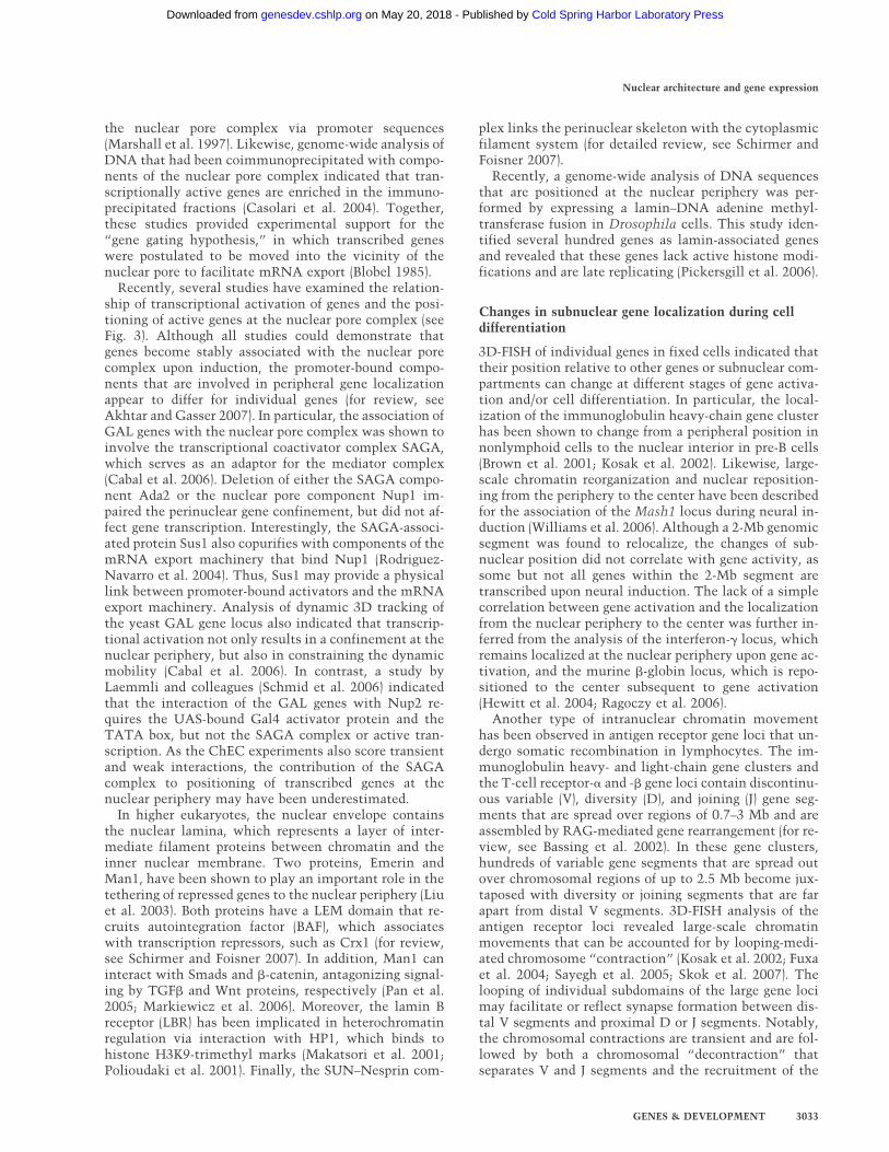
Figure 3. Structural and functional hallmarks of the nuclear periphery in yeast and metazoans. The membrane bilayer of the nucleusis perforated by nuclear pores, which consist of an ∼30-subunit-containing nuclear pore complex (NPC). (Left) In yeast, the Nup1 andNup2 components of the nuclear pore complex have been found to interact with components of the SAGA coactivator complex viaSus1, which is also a component of the TREX complex, involved in mRNA export. The nuclear periphery, excluding the pores, is alsoimplicated in gene silencing, whereby the yeast Sir4 protein is tethered to the periphery via the enhancer of silent chromatin (Esc1)or Ku protein. (Right) In metazoans, the inner nuclear membrane is associated with lamins and lamin-associated proteins. The laminB receptor (LBR) has been implicated in heterochromatin regulation via interaction with HP1, which binds to histone H3K9 trimethylmarks. Emerin and Man1 recruit, via their LEM domain, the barrier to autointegration factor (BAF), which associates with transcrip-tion repressors such as Crx1. In addition they bind and inhibit the function of the signaling effectors Smad and �-catenin. TheSUN–Nesprin complex, which is formed by an interaction of the KASH domain of Nesprin with SUN proteins, links the perinuclearskeleton with the cytoplasmic filament system. (IF) Intermediate filament.
Schneider and Grosschedl
3032 GENES & DEVELOPMENT
Cold Spring Harbor Laboratory Press on May 20, 2018 - Published by genesdev.cshlp.orgDownloaded from
the nuclear pore complex via promoter sequences(Marshall et al. 1997). Likewise, genome-wide analysis ofDNA that had been coimmunoprecipitated with compo-nents of the nuclear pore complex indicated that tran-scriptionally active genes are enriched in the immuno-precipitated fractions (Casolari et al. 2004). Together,these studies provided experimental support for the“gene gating hypothesis,” in which transcribed geneswere postulated to be moved into the vicinity of thenuclear pore to facilitate mRNA export (Blobel 1985).
Recently, several studies have examined the relation-ship of transcriptional activation of genes and the posi-tioning of active genes at the nuclear pore complex (seeFig. 3). Although all studies could demonstrate thatgenes become stably associated with the nuclear porecomplex upon induction, the promoter-bound compo-nents that are involved in peripheral gene localizationappear to differ for individual genes (for review, seeAkhtar and Gasser 2007). In particular, the association ofGAL genes with the nuclear pore complex was shown toinvolve the transcriptional coactivator complex SAGA,which serves as an adaptor for the mediator complex(Cabal et al. 2006). Deletion of either the SAGA compo-nent Ada2 or the nuclear pore component Nup1 im-paired the perinuclear gene confinement, but did not af-fect gene transcription. Interestingly, the SAGA-associ-ated protein Sus1 also copurifies with components of themRNA export machinery that bind Nup1 (Rodriguez-Navarro et al. 2004). Thus, Sus1 may provide a physicallink between promoter-bound activators and the mRNAexport machinery. Analysis of dynamic 3D tracking ofthe yeast GAL gene locus also indicated that transcrip-tional activation not only results in a confinement at thenuclear periphery, but also in constraining the dynamicmobility (Cabal et al. 2006). In contrast, a study byLaemmli and colleagues (Schmid et al. 2006) indicatedthat the interaction of the GAL genes with Nup2 re-quires the UAS-bound Gal4 activator protein and theTATA box, but not the SAGA complex or active tran-scription. As the ChEC experiments also score transientand weak interactions, the contribution of the SAGAcomplex to positioning of transcribed genes at thenuclear periphery may have been underestimated.
In higher eukaryotes, the nuclear envelope containsthe nuclear lamina, which represents a layer of inter-mediate filament proteins between chromatin and theinner nuclear membrane. Two proteins, Emerin andMan1, have been shown to play an important role in thetethering of repressed genes to the nuclear periphery (Liuet al. 2003). Both proteins have a LEM domain that re-cruits autointegration factor (BAF), which associateswith transcription repressors, such as Crx1 (for review,see Schirmer and Foisner 2007). In addition, Man1 caninteract with Smads and �-catenin, antagonizing signal-ing by TGF� and Wnt proteins, respectively (Pan et al.2005; Markiewicz et al. 2006). Moreover, the lamin Breceptor (LBR) has been implicated in heterochromatinregulation via interaction with HP1, which binds tohistone H3K9-trimethyl marks (Makatsori et al. 2001;Polioudaki et al. 2001). Finally, the SUN–Nesprin com-
plex links the perinuclear skeleton with the cytoplasmicfilament system (for detailed review, see Schirmer andFoisner 2007).
Recently, a genome-wide analysis of DNA sequencesthat are positioned at the nuclear periphery was per-formed by expressing a lamin–DNA adenine methyl-transferase fusion in Drosophila cells. This study iden-tified several hundred genes as lamin-associated genesand revealed that these genes lack active histone modi-fications and are late replicating (Pickersgill et al. 2006).
Changes in subnuclear gene localization during celldifferentiation
3D-FISH of individual genes in fixed cells indicated thattheir position relative to other genes or subnuclear com-partments can change at different stages of gene activa-tion and/or cell differentiation. In particular, the local-ization of the immunoglobulin heavy-chain gene clusterhas been shown to change from a peripheral position innonlymphoid cells to the nuclear interior in pre-B cells(Brown et al. 2001; Kosak et al. 2002). Likewise, large-scale chromatin reorganization and nuclear reposition-ing from the periphery to the center have been describedfor the association of the Mash1 locus during neural in-duction (Williams et al. 2006). Although a 2-Mb genomicsegment was found to relocalize, the changes of sub-nuclear position did not correlate with gene activity, assome but not all genes within the 2-Mb segment aretranscribed upon neural induction. The lack of a simplecorrelation between gene activation and the localizationfrom the nuclear periphery to the center was further in-ferred from the analysis of the interferon-� locus, whichremains localized at the nuclear periphery upon gene ac-tivation, and the murine �-globin locus, which is repo-sitioned to the center subsequent to gene activation(Hewitt et al. 2004; Ragoczy et al. 2006).
Another type of intranuclear chromatin movementhas been observed in antigen receptor gene loci that un-dergo somatic recombination in lymphocytes. The im-munoglobulin heavy- and light-chain gene clusters andthe T-cell receptor-� and -� gene loci contain discontinu-ous variable (V), diversity (D), and joining (J) gene seg-ments that are spread over regions of 0.7–3 Mb and areassembled by RAG-mediated gene rearrangement (for re-view, see Bassing et al. 2002). In these gene clusters,hundreds of variable gene segments that are spread outover chromosomal regions of up to 2.5 Mb become jux-taposed with diversity or joining segments that are farapart from distal V segments. 3D-FISH analysis of theantigen receptor loci revealed large-scale chromatinmovements that can be accounted for by looping-medi-ated chromosome “contraction” (Kosak et al. 2002; Fuxaet al. 2004; Sayegh et al. 2005; Skok et al. 2007). Thelooping of individual subdomains of the large gene locimay facilitate or reflect synapse formation between dis-tal V segments and proximal D or J segments. Notably,the chromosomal contractions are transient and are fol-lowed by both a chromosomal “decontraction” thatseparates V and J segments and the recruitment of the
Nuclear architecture and gene expression
GENES & DEVELOPMENT 3033
Cold Spring Harbor Laboratory Press on May 20, 2018 - Published by genesdev.cshlp.orgDownloaded from
locus to pericentromeric heterochromatin (Roldan et al.2005; Skok et al. 2007). For the immunoglobulin heavy-chain locus, the decontraction has been shown to occurat a developmental stage in which the productive generearrangement of one allele produces an active pre-B-cellreceptor that generates a negative feedback signal, result-ing in decontraction of the other allele and “allelic ex-clusion” (Roldan et al. 2005). Similar transient chromo-somal contractions have been described for the T-cellreceptor � and � loci (Skok et al. 2007). The decontrac-tion that separates V and J segments appears to representa general principle underlying allelic exclusion, which isestablished as an allelic asynchrony in early stages ofmouse embryogenesis (Mostoslavsky et al. 2001; Gold-mit et al. 2005). Although the molecular basis for theestablishment of allelic asynchrony is still unclear, dif-ferential demethylation and replication timing of thetwo alleles may represent an important mechanism(Mostoslavsky et al. 2001).
The molecular mechanisms underlying the changes ofgene localization from perinuclear or heterochromaticregions to transcription factories or euchromatic regionsare still obscure, but seem to correlate with histoneacetylation (Chowdhury and Sen 2001; Hawwari andKrangel 2005) In contrast, some insight into the molecu-lar basis for the intrachromosomal “contraction” of theimmunoglobulin heavy-chain cluster has been providedby the analysis of knockout mice. Both Pax5-deficientmice and mice defective in the Ezh2 component of thePRC2 polycomb repressive complex (an H3/K27-specifichistone methyltransferase) have been shown to fail toundergo intrachromosomal contractions (Fuxa et al.2004). This was found to be dependent on the function ofthe transcription factor Pax5 and on the Ezh2 componentof the Polycomb repressive complex PRC2 (Su et al.2003). The recruitment of PRC2 and the accompanyingmethylation of Lys 27 of histone H3 at the immuno-globulin heavy-chain locus may attract the PRC1 com-plex and result in contraction of the locus (Francis et al.2004; Su et al. 2005), suggesting a role of this histonemark in the locus contraction.
Interchromosomal interactions
The positioning of multiple genome regions on differentchromosomes relative to each other is a rapidly emergingnew and important determinant for their function. Thelocal clustering of genes can have regulatory function;e.g., by regulating their expression or allowing inter-actions with key regulatory elements (for review, seeMisteli 2007). Here we will discuss examples for homo-log and nonhomolog interchromosomal interactions of-ten also termed “chromosome-kissing.”
Homologous interchromosomal interactions
Physical pairing of chromosomes is particularly visiblein dipteran insects such as Drosophila. Such pairing mayenable one allele to support the function of the secondallele (for reviews, see Pirrotta 1999; Duncan 2002). E.B.
Lewis termed this process “transvection.” He found thatthe phenotypes of mutant Ubx (Ultrabithorax) alleleswere stronger when pairing with a wild allele was pre-vented by chromosomal rearrangements. The pairing canincrease Ubx expression and allows the alleles to par-tially complement each other. This transvection,whereby a mutation can be complemented in trans bythe other allele, has now been shown for a number of lociin Drosophila (Duncan 2002). Another well-studied ex-ample is the Hox gene Abd-B (Abdominal-B). Geneticstudies have demonstrated that transvection of Abd-Bdepends on a 3�-flanking region termed the TMR (trans-vection-mediating region) (Hopmann et al. 1995). UsingRNA FISH, M. Levine’s group (Ronshaugen and Levine2004) was able to visualize transvection events. Theyshowed that Abd-B enhancers located on one chromo-some can frequently pair with the Abd-B gene located onthe other homolog chromosome. The TMR sequencefunctions as a pairing element. Interestingly, this TMRcontains a Polycomb-responsive element (Zhou andLevine 1999; Zhou et al. 1999; Bantignies et al. 2003).The colocalization allows the enhancers that normallyloop in cis to target promoters at the same chromosometo loop in trans and activate the Abd-B gene in trans.The tighter the pairing is, the more frequent are thetrans-interactions between the enhancer and the Abd-Bgene (Ronshaugen and Levine 2004).
Another well-studied example for interchromosomalinteractions of homolog chromsomes is the mammalianX chromosome (for a review, see Carrel 2006). X-chro-mosome inactivation involves the counting of the chro-mosomes and subsequent choosing of which of the twoX chromosomes becomes inactivated. This requires“communication” between the two X chromosomesthat could occur through homolog chromosome interac-tions (Marahrens 1999). In the last few years significantinsight into the pairing of the two X chromosomes wasobtained. Critical for X inactivation is the Xic (X inacti-vation center) region (Brown et al. 1991; Lee 2005). Intwo elegant studies, the groups of Lee and Heard (Bacheret al. 2006; Xu et al. 2006) used FISH in mouse femaleembryonic stem (ES) cells to demonstrate that during theonset of X inactivation, the X chromosomes move fromthe nuclear periphery, where they have random positionsrelative to each other, and come into close proximitywith their Xic regions juxtaposed. The two X chromo-somes are only found together at the time during earlyES cell differentiation when X inactivation is triggered,but not at later or earlier time points (Fig. 4). Interest-ingly, deletion mutants in the Xic region that disruptedrandom X inactivation also strongly affected the pairingbetween the X chromosomes (Bacher et al. 2006; Xu et al.2006). During the pairing, the two X chromosomes arephysically interacting, directly or via their DNA-boundproteins (Xu et al. 2006), allowing cross-talk and mutu-ally exclusive designation of the active or inactive state.After dissociation of the two homologous partners, theinactive X (Xi) is targeted to the perinucleolar compart-ment (Fig. 4; Zhang et al. 2007). The Xi is organized as arepeat-rich core with the gene-rich regions around
Schneider and Grosschedl
3034 GENES & DEVELOPMENT
Cold Spring Harbor Laboratory Press on May 20, 2018 - Published by genesdev.cshlp.orgDownloaded from
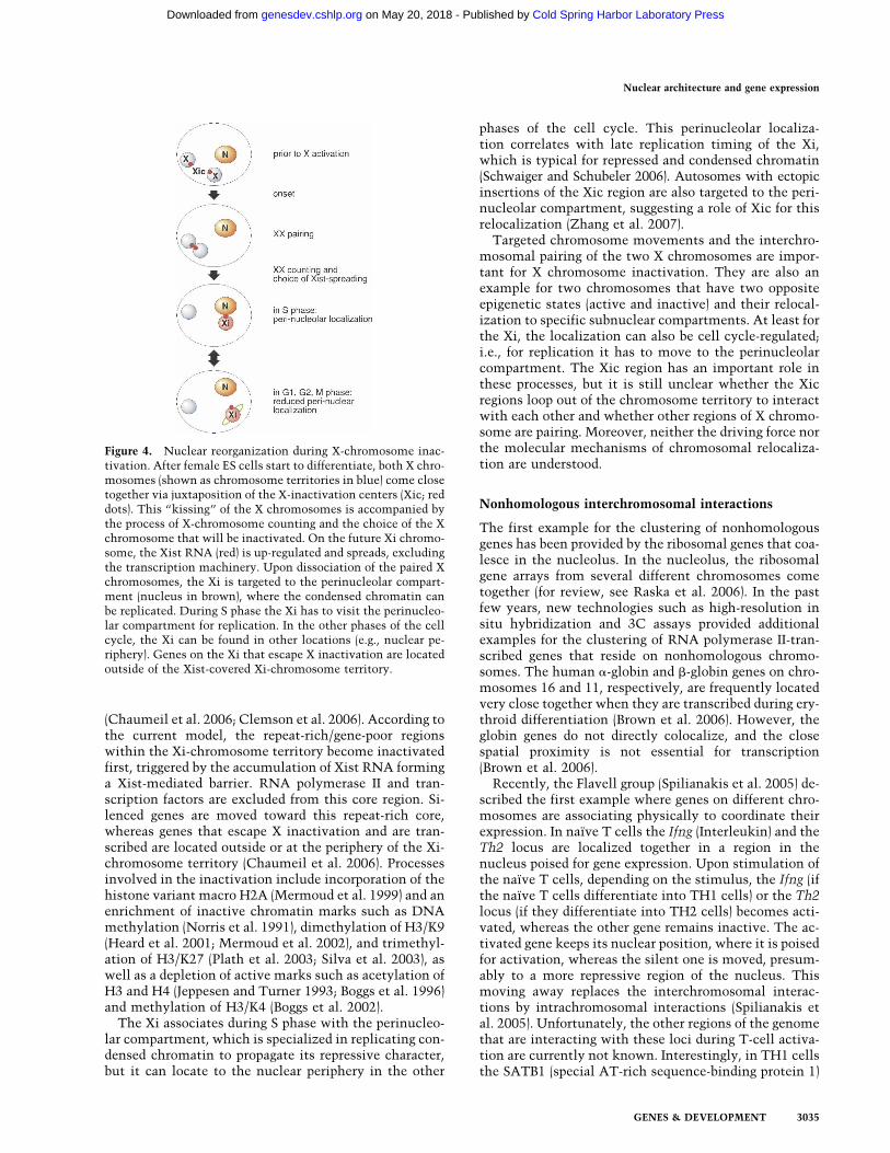
(Chaumeil et al. 2006; Clemson et al. 2006). According tothe current model, the repeat-rich/gene-poor regionswithin the Xi-chromosome territory become inactivatedfirst, triggered by the accumulation of Xist RNA forminga Xist-mediated barrier. RNA polymerase II and tran-scription factors are excluded from this core region. Si-lenced genes are moved toward this repeat-rich core,whereas genes that escape X inactivation and are tran-scribed are located outside or at the periphery of the Xi-chromosome territory (Chaumeil et al. 2006). Processesinvolved in the inactivation include incorporation of thehistone variant macro H2A (Mermoud et al. 1999) and anenrichment of inactive chromatin marks such as DNAmethylation (Norris et al. 1991), dimethylation of H3/K9(Heard et al. 2001; Mermoud et al. 2002), and trimethyl-ation of H3/K27 (Plath et al. 2003; Silva et al. 2003), aswell as a depletion of active marks such as acetylation ofH3 and H4 (Jeppesen and Turner 1993; Boggs et al. 1996)and methylation of H3/K4 (Boggs et al. 2002).
The Xi associates during S phase with the perinucleo-lar compartment, which is specialized in replicating con-densed chromatin to propagate its repressive character,but it can locate to the nuclear periphery in the other
phases of the cell cycle. This perinucleolar localiza-tion correlates with late replication timing of the Xi,which is typical for repressed and condensed chromatin(Schwaiger and Schubeler 2006). Autosomes with ectopicinsertions of the Xic region are also targeted to the peri-nucleolar compartment, suggesting a role of Xic for thisrelocalization (Zhang et al. 2007).
Targeted chromosome movements and the interchro-mosomal pairing of the two X chromosomes are impor-tant for X chromosome inactivation. They are also anexample for two chromosomes that have two oppositeepigenetic states (active and inactive) and their relocal-ization to specific subnuclear compartments. At least forthe Xi, the localization can also be cell cycle-regulated;i.e., for replication it has to move to the perinucleolarcompartment. The Xic region has an important role inthese processes, but it is still unclear whether the Xicregions loop out of the chromosome territory to interactwith each other and whether other regions of X chromo-some are pairing. Moreover, neither the driving force northe molecular mechanisms of chromosomal relocaliza-tion are understood.
Nonhomologous interchromosomal interactions
The first example for the clustering of nonhomologousgenes has been provided by the ribosomal genes that coa-lesce in the nucleolus. In the nucleolus, the ribosomalgene arrays from several different chromosomes cometogether (for review, see Raska et al. 2006). In the pastfew years, new technologies such as high-resolution insitu hybridization and 3C assays provided additionalexamples for the clustering of RNA polymerase II-tran-scribed genes that reside on nonhomologous chromo-somes. The human �-globin and �-globin genes on chro-mosomes 16 and 11, respectively, are frequently locatedvery close together when they are transcribed during ery-throid differentiation (Brown et al. 2006). However, theglobin genes do not directly colocalize, and the closespatial proximity is not essential for transcription(Brown et al. 2006).
Recently, the Flavell group (Spilianakis et al. 2005) de-scribed the first example where genes on different chro-mosomes are associating physically to coordinate theirexpression. In naïve T cells the Ifng (Interleukin) and theTh2 locus are localized together in a region in thenucleus poised for gene expression. Upon stimulation ofthe naïve T cells, depending on the stimulus, the Ifng (ifthe naïve T cells differentiate into TH1 cells) or the Th2locus (if they differentiate into TH2 cells) becomes acti-vated, whereas the other gene remains inactive. The ac-tivated gene keeps its nuclear position, where it is poisedfor activation, whereas the silent one is moved, presum-ably to a more repressive region of the nucleus. Thismoving away replaces the interchromosomal interac-tions by intrachromosomal interactions (Spilianakis etal. 2005). Unfortunately, the other regions of the genomethat are interacting with these loci during T-cell activa-tion are currently not known. Interestingly, in TH1 cellsthe SATB1 (special AT-rich sequence-binding protein 1)
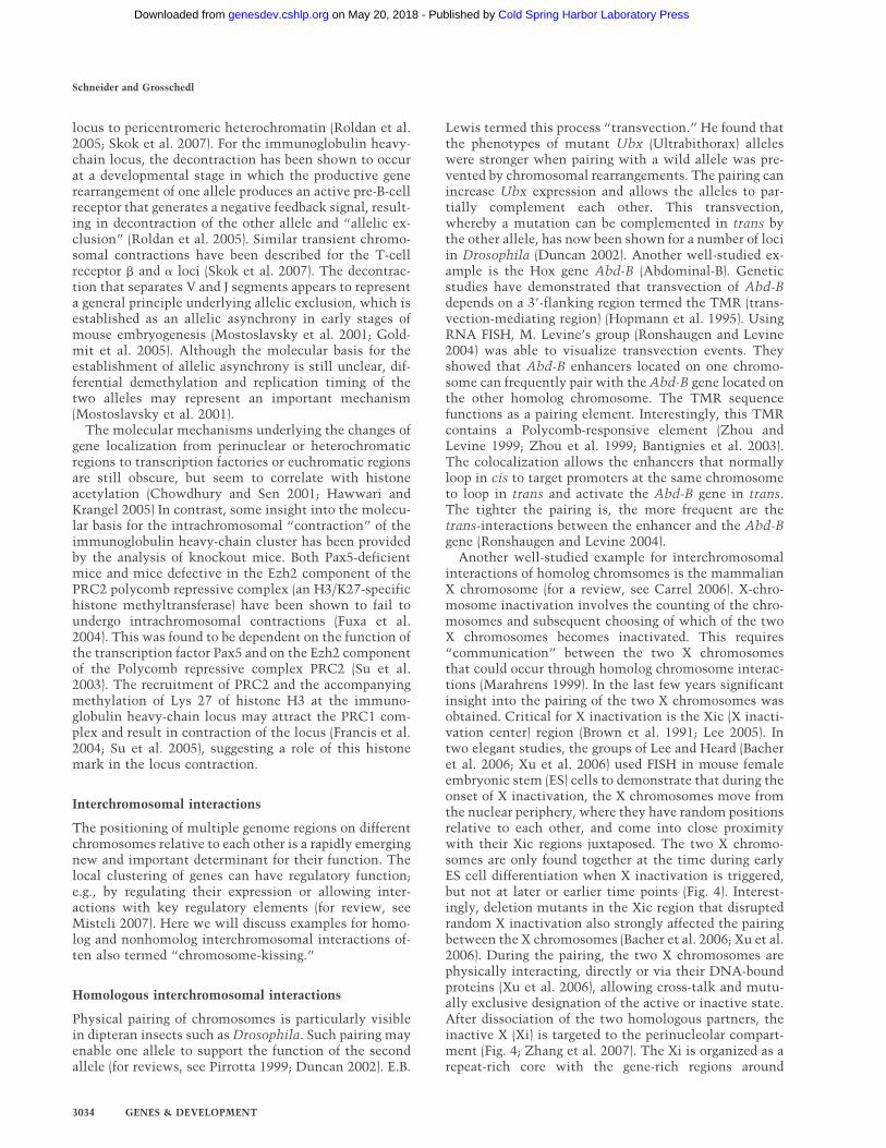
Figure 4. Nuclear reorganization during X-chromosome inac-tivation. After female ES cells start to differentiate, both X chro-mosomes (shown as chromosome territories in blue) come closetogether via juxtaposition of the X-inactivation centers (Xic; reddots). This “kissing” of the X chromosomes is accompanied bythe process of X-chromosome counting and the choice of the Xchromosome that will be inactivated. On the future Xi chromo-some, the Xist RNA (red) is up-regulated and spreads, excludingthe transcription machinery. Upon dissociation of the paired Xchromosomes, the Xi is targeted to the perinucleolar compart-ment (nucleus in brown), where the condensed chromatin canbe replicated. During S phase the Xi has to visit the perinucleo-lar compartment for replication. In the other phases of the cellcycle, the Xi can be found in other locations (e.g., nuclear pe-riphery). Genes on the Xi that escape X inactivation are locatedoutside of the Xist-covered Xi-chromosome territory.
Nuclear architecture and gene expression
GENES & DEVELOPMENT 3035
Cold Spring Harbor Laboratory Press on May 20, 2018 - Published by genesdev.cshlp.orgDownloaded from
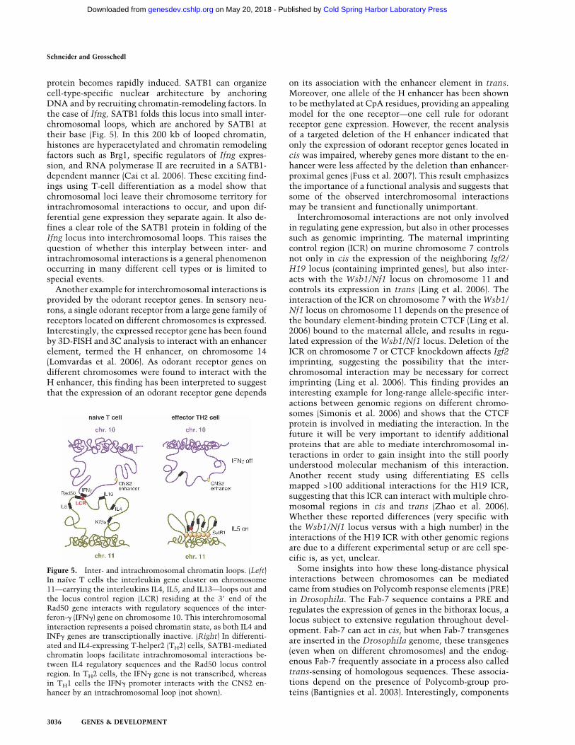
protein becomes rapidly induced. SATB1 can organizecell-type-specific nuclear architecture by anchoringDNA and by recruiting chromatin-remodeling factors. Inthe case of Ifng, SATB1 folds this locus into small inter-chromosomal loops, which are anchored by SATB1 attheir base (Fig. 5). In this 200 kb of looped chromatin,histones are hyperacetylated and chromatin remodelingfactors such as Brg1, specific regulators of Ifng expres-sion, and RNA polymerase II are recruited in a SATB1-dependent manner (Cai et al. 2006). These exciting find-ings using T-cell differentiation as a model show thatchromosomal loci leave their chromosome territory forintrachromosomal interactions to occur, and upon dif-ferential gene expression they separate again. It also de-fines a clear role of the SATB1 protein in folding of theIfng locus into interchromosomal loops. This raises thequestion of whether this interplay between inter- andintrachromosomal interactions is a general phenomenonoccurring in many different cell types or is limited tospecial events.
Another example for interchromosomal interactions isprovided by the odorant receptor genes. In sensory neu-rons, a single odorant receptor from a large gene family ofreceptors located on different chromosomes is expressed.Interestingly, the expressed receptor gene has been foundby 3D-FISH and 3C analysis to interact with an enhancerelement, termed the H enhancer, on chromosome 14(Lomvardas et al. 2006). As odorant receptor genes ondifferent chromosomes were found to interact with theH enhancer, this finding has been interpreted to suggestthat the expression of an odorant receptor gene depends
on its association with the enhancer element in trans.Moreover, one allele of the H enhancer has been shownto be methylated at CpA residues, providing an appealingmodel for the one receptor—one cell rule for odorantreceptor gene expression. However, the recent analysisof a targeted deletion of the H enhancer indicated thatonly the expression of odorant receptor genes located incis was impaired, whereby genes more distant to the en-hancer were less affected by the deletion than enhancer-proximal genes (Fuss et al. 2007). This result emphasizesthe importance of a functional analysis and suggests thatsome of the observed interchromosomal interactionsmay be transient and functionally unimportant.
Interchromosomal interactions are not only involvedin regulating gene expression, but also in other processessuch as genomic imprinting. The maternal imprintingcontrol region (ICR) on murine chromosome 7 controlsnot only in cis the expression of the neighboring Igf2/H19 locus (containing imprinted genes), but also inter-acts with the Wsb1/Nf1 locus on chromosome 11 andcontrols its expression in trans (Ling et al. 2006). Theinteraction of the ICR on chromosome 7 with the Wsb1/Nf1 locus on chromosome 11 depends on the presence ofthe boundary element-binding protein CTCF (Ling et al.2006) bound to the maternal allele, and results in regu-lated expression of the Wsb1/Nf1 locus. Deletion of theICR on chromosome 7 or CTCF knockdown affects Igf2imprinting, suggesting the possibility that the inter-chromosomal interaction may be necessary for correctimprinting (Ling et al. 2006). This finding provides aninteresting example for long-range allele-specific inter-actions between genomic regions on different chromo-somes (Simonis et al. 2006) and shows that the CTCFprotein is involved in mediating the interaction. In thefuture it will be very important to identify additionalproteins that are able to mediate interchromosomal in-teractions in order to gain insight into the still poorlyunderstood molecular mechanism of this interaction.Another recent study using differentiating ES cellsmapped >100 additional interactions for the H19 ICR,suggesting that this ICR can interact with multiple chro-mosomal regions in cis and trans (Zhao et al. 2006).Whether these reported differences (very specific withthe Wsb1/Nf1 locus versus with a high number) in theinteractions of the H19 ICR with other genomic regionsare due to a different experimental setup or are cell spe-cific is, as yet, unclear.
Some insights into how these long-distance physicalinteractions between chromosomes can be mediatedcame from studies on Polycomb response elements (PRE)in Drosophila. The Fab-7 sequence contains a PRE andregulates the expression of genes in the bithorax locus, alocus subject to extensive regulation throughout devel-opment. Fab-7 can act in cis, but when Fab-7 transgenesare inserted in the Drosophila genome, these transgenes(even when on different chromosomes) and the endog-enous Fab-7 frequently associate in a process also calledtrans-sensing of homologous sequences. These associa-tions depend on the presence of Polycomb-group pro-teins (Bantignies et al. 2003). Interestingly, components
Figure 5. Inter- and intrachromosomal chromatin loops. (Left)In naïve T cells the interleukin gene cluster on chromosome11—carrying the interleukins IL4, IL5, and IL13—loops out andthe locus control region (LCR) residing at the 3� end of theRad50 gene interacts with regulatory sequences of the inter-feron-� (IFN�) gene on chromosome 10. This interchromosomalinteraction represents a poised chromatin state, as both IL4 andINF� genes are transcriptionally inactive. (Right) In differenti-ated and IL4-expressing T-helper2 (TH2) cells, SATB1-mediatedchromatin loops facilitate intrachromosomal interactions be-tween IL4 regulatory sequences and the Rad50 locus controlregion. In TH2 cells, the IFN� gene is not transcribed, whereasin TH1 cells the IFN� promoter interacts with the CNS2 en-hancer by an intrachromosomal loop (not shown).
Schneider and Grosschedl
3036 GENES & DEVELOPMENT
Cold Spring Harbor Laboratory Press on May 20, 2018 - Published by genesdev.cshlp.orgDownloaded from
of the RNA interference (RNAi) machinery, such asdicer-2, as well as Argonaute genes are required for thelong-distance interactions. The RNAi machinery is notrequired for establishment of the interchromosomal in-teractions, but for its maintenance (Grimaud et al. 2006).The following model had been put forward: Once thepairing is established, sense and antisense transcriptionin the vicinity is stimulated, and the RNAi machineryproduces small interfering RNA (siRNA). The siRNAcan then be bound by Polycomb-group proteins to act asmolecular glue that stabilizes the chromosomal interac-tion and the silencing of both loci (Grimaud et al. 2006;Lei and Corces 2006). While providing some first cluesabout a role of RNAi in the physical interaction betweendifferent chromosomes, many mechanistic questions ofhow these interactions are mediated remain open. In-tense work and new techniques will be required toinvestigate the role of RNA in interchromosomal inter-actions and to fully understand how these interactionsare mediated.
Recent findings again raise the question of how gen-eral the interchromosomal cross-talk is. A recent studyof the mouse Hox gene clusters demonstrated that thecoregulation of the four clusters localized on differentchromosomes is not associated with colocalization ofthe loci (Lanctot et al. 2007). These results demonstratethat coregulation of Hox genes can be independent ofcolocalization. Additionally, the active �- and �-globingenes colocalize only in human but not in mouse eryth-roblasts. This difference may be due to different chromo-somal context; the mouse locus lies in a gene-poor regionnear the centromere, whereas the human locus is locatedin a gene-rich subtelomeric region (Brown et al. 2006).This suggests that not only transcriptional regulationbut also additional factors such as chromosomal struc-ture, location, repeat, or gene density may influence theinterchromosomal cross-talk.
Altogether, the examples discussed above stronglysupport the concept of trans-regulation via interchromo-somal contacts and communication—at least at specificloci—and implicate interchromosomal contacts in theregulation of gene expression states that can be inherit-able. In addition to the so far better understood cis-in-teractions on the same chromosome, this interchromo-some cross-talk adds an additional layer of complexity tothe regulation of DNA-dependent processes. We are cur-rently only at the beginning of understanding the inter-chromosomal cross-talk, and it will be of central impor-tance to study these interactions in more detail. Liveimaging will tell us what the dynamics of these interac-tions are and whether they are transient or prolonged.The identification and manipulation of DNA elementsrequired for the interaction will allow us to study thefunctional relevance of the interactions. Many examplesof chromosomal interactions are currently just observa-tions of proximity, and many questions are still open.Are the chromosomes indeed in direct physical contactor only in close proximity? Which proteins and proteincomplexes are mediating these interactions? Are thesame proteins involved in loop formation and interchro-
mosomal contacts? How are these factors recruited?What is the role of RNA in this process? Is there a feed-back mechanism that allows DNA elements to interactwith just one other DNA element?
In the future, we will have to search intensively fortrans-regulatory elements in the genome and considerthat long-range interactions may be targets for gene regu-lation. Only new high-throughput techniques such as 3Cassays coupled with microarrays (Simonis et al. 2006) ordirect sequencing will allow an unbiased screen of thegenome for DNA loci that contact each other. However,one pitfall of these techniques is that they also capturevery transient interactions. A combination with high-throughput FISH could be used to verify these interac-tions.
Nuclear organization and rearrangementsin pluripotent cells
Pluripotent cells have the potential to self-renew indefi-nitely and to differentiate into any other cell type. Thisrequires that the pluripotent cells have a nuclear archi-tecture that allows maintenance of the pluripotent state,but which is at the same time so plastic that they canenter any differentiation pathway. As soon as the differ-entiation pathway is started, lineage specification ini-tiates and a cell-type-specific transcription programstarts. Here we discuss the dynamics and plasticity ofnuclear organization in ES cells and early mouse em-bryos compared with lineage-committed cells.
ES cells
ES cells are enriched in less-compact euchromatin andshow a more diffuse heterochromatin structure. Upondifferentiation, the heterochromatin rearranges and thenumber of heterochromatin foci increases (for reviews,see Arney and Fisher 2004; Meshorer and Misteli 2006).The large-scale organization of chromosome territoriesis similar in ES cells compared with differentiated cells(Wiblin et al. 2005). However, ES cell-specific geneschange their position during differentiation—e.g., inhumans the NANOG gene relocates from a more periph-eral positioning in ES cells to a more central position inB cells—and the OCT4 gene loops out from its chromo-some territory in ES cells (Wiblin et al. 2005). This alsocorrelates with an early replication timing of these genesin ES cells compared with a later one in differentiatedcells (Perry et al. 2004). In mouse ES cells “bivalent chro-matin domains” of large regions of active and repressivehistone modifications exist. These domains containchromatin enriched in H3/K27 methylation, a mark con-sidered specific for repressed, condensed chromatin;within it smaller regions enriched for H3/K4 methyl-ation, a mark for active chromatin, were found (Azuaraet al. 2006; Bernstein et al. 2006; Lee et al. 2006). Thesedomains are thought to function, via opposing histonemodifications, to silence developmental genes in ES cellswhile keeping them poised for activation later on. AfterES cell differentiation, when genes in these bivalent do-
Nuclear architecture and gene expression
GENES & DEVELOPMENT 3037
Cold Spring Harbor Laboratory Press on May 20, 2018 - Published by genesdev.cshlp.orgDownloaded from
mains become turned on, the repressive H3/K27 meth-ylation decreases, whereas the H3/K4 methylation stays(Bernstein et al. 2006; for a recent review about histonemodifications in ES cells, see Spivakov and Fisher 2007).
Interestingly, a recent study from the Misteli group(Meshoer et al. 2006) showed that chromatin in ES cellsis globally decondensed and contains a high fraction ofonly loosely bound architectural chromatin proteins, in-cluding linker histone H1 and the HP1 protein. The na-ture of this pool is still under discussion (Gilbert et al.2007); it has been shown that this hyperdynamic proteinpopulation is a hallmark for truly pluripotent cells, sincelineage-committed cells do not have it (Meshorer et al.2006). Consistent with this, pluripotent cells are en-riched in acetylated histones and repressive heterochro-matic marks are reduced (Kimura et al. 2004). This open,hyperdynamic chromatin is necessary for ES cell dif-ferentiation and was termed “breathing” chromatin(Meshorer et al. 2006). This pool of hyperdynamic chro-matin proteins may be needed to establish chromatindomains once cells start lineage-specific gene expressionprograms by the sequestration of genes into specific ac-tive or repressive chromatin domains or proteinaceousbodies (Meshorer et al. 2006). The hyperdynamic, openchromatin in ES cells could allow regulatory factors ac-cess to their binding sites and also the rapid activation oflineage-specific gene expression programs.
Early mouse embryo
Similar to the ES cells, in the early mouse embryo theconcept of a “dynamic” chromatin structure, allowingthe development of pluripotent cells, has also been de-scribed. A recent study linked histone modificationswith cell fate in the early embryo. Increasing the level ofarginine methylation of histones, generally considered asan activating mark, results in up-regulation of nanog andthe “biased” cell fate decision toward the inner cell mass(Torres-Padilla et al. 2007). However, it remains to bestudied whether there is any direct link between the ac-tivation of particular lineage restriction genes during de-velopment and this chromatin enriched in active marks,and what the exact role of the nuclear organization inthese embryonic cells is.
The nuclear organization in the mouse embryo duringpreimplantation development (in the zygote and duringthe first rounds of cleavage divisions) displays particularfeatures that are dynamic and dramatically distinctivefrom somatic cells (Morgan et al. 2005); e.g., a particularorganization and relocalization of the centromeric re-peats within the nucleus. It is likely that these featuresare necessary for the reprogramming of the chromatinthat has to occur in the embryo after fertilization. Adetailed analysis of the localization of the centromericmajor and minor satellite repeats (forming the so-calledchromocenter) in the paternal and maternal genomes af-ter fertilization has revealed that the nuclear organiza-tion of the centromeric satellite regions becomes remod-eled in the space of a few hours (Probst et al. 2007). The
major satellites move toward the center of the pronucleiand adopt a special ring-like structure around the nucleo-lar-like bodies, which are spherical structures consideredas the precursors of the nucleoli. This relocation of therepeats is accompanied by a switch in replication timing(Martin et al. 2006) and occurs at the same time as thetranscriptional activation of the silenced embryonic ge-nome. The consequence of this organization is that thepericentromeric regions of different chromosomes comein physical proximity, potentially allowing some kind ofcross-talk. It is interesting to note that not all of thechromosomes adopt this particular configuration of thecentromeres, as a prochromocenter-like structure pre-vails specifically in the female pronucleus (Probst et al.2007). Upon cell differentiation during the formation ofthe blastocyst, the centromeric repeats move and adopt asomatic-like organization (Martin et al. 2006). The sig-nificance for normal development and the molecularmechanism behind this particular relocalization of thecentromeres remains to be determined. However, incloned embryos derived from somatic nuclear transfer,the somatic chromocenter configuration of the donornucleus is rapidly reverted (Martin et al. 2006). This sug-gests that the relocalization of huge genomic regions andthe acquisition of this specific embryonic chromocenterconfiguration are required for reprogramming.
Interestingly, pluripotent cells seem to require a dy-namic, active nuclear organization enabling mainte-nance of the pluripotent state and the potential to differ-entiate into any lineage. The intense chromatin repro-gramming in early mouse embryos that is necessary forthe plasticity of the embryo involves histone modifica-tions, nuclear repositioning, and in particular, reorgani-zation on a more global level compared with the chro-matin movements involved in the activation of specificgenes. Currently, our knowledge about the contributionof nuclear architecture to pluripotency is still limited.To gain insight into this, it will be important to comparethe nuclear organization and dynamics of pluripotentand lineage-committed cells in more detail.
Perspectives
The understanding of the relationship between nucleararchitecture, genome organization, and gene expressionwill be aided by the application of new imaging tech-niques that include improvements of the resolution,time-lapse, and live cell imaging. The advent of high-throughput analysis of histone modifications has pro-vided us with a genome-wide map for many chromatinmodifications. Although these data allow for a detailedknowledge of chromatin states, we still have very lim-ited insight into the causal relationship between chro-matin modification, higher-order chromatin organiza-tion, subnuclear gene localization, and gene expression.In particular, the clarification of the cause–effect rela-tionship of nuclear organization and the function of thegenome represents one of the most important futurechallenges. Further experiments are needed to determinewhether the spatial organization of the nucleus is a con-
Schneider and Grosschedl
3038 GENES & DEVELOPMENT
Cold Spring Harbor Laboratory Press on May 20, 2018 - Published by genesdev.cshlp.orgDownloaded from

sequence of genome organization, chromatin modifica-tions, and DNA-based processes, or whether nuclear ar-chitecture is an important determinant of the function ofthe genome. For example, the concept of self-organiza-tion of nuclear structures and subcompartments by spe-cific DNA sequences is supported by the extensiveanalysis of the ribosomal genes and their role in the gen-esis of the nucleolus. However, this model needs furthervalidation, and its generality has to be addressed, includ-ing whether and to what extent such genome-drivennuclear self-organization plays a role at other gene loci.Another important future avenue will be the identifica-tion of proteins that, like CTCF, mediate intra- and/orinterchromosomal interactions. A molecular character-ization of these proteins and their multiprotein com-plexes will help to understand the coordinate expressionof genes that do not reside in gene clusters. Finally, thequestion of the molecular forces in the nucleus that me-diate chromatin mobility will be of utmost importancefor our understanding of the dynamics of the genome.With the advent of innovative techniques and interdis-ciplinary approaches, combined with the enthusiasm ofthis field of research, we can expect fascinating futureinsight into a central biological problem.
Acknowledgments
We thank Dr. Patrick Heun for critical reading of the manu-script. Research in the R.S. laboratory is supported by a HFSPcareer development award, the Epigenome Network of Excel-lence, the DFG, and the MPG. Research in the R.G. laboratoryis supported by the MPG and the DFG.
References
Akhtar, A. and Gasser, S.M. 2007. The nuclear envelope andtranscriptional control. Nat. Rev. Genet. 8: 507–517.
Andrulis, E.D., Neiman, A.M., Zappulla, D.C., and Sternglanz,R. 1998. Perinuclear localization of chromatin facilitatestranscriptional silencing. Nature 394: 592–595.
Andrulis, E.D., Zappulla, D.C., Ansari, A., Perrod, S., Laiosa,C.V., Gartenberg, M.R., and Sternglanz, R. 2002. Esc1, anuclear periphery protein required for Sir4-based plasmid an-choring and partitioning. Mol. Cell. Biol. 22: 8292–8301.
Arney, K.L. and Fisher, A.G. 2004. Epigenetic aspects of differ-entiation. J. Cell Sci. 117: 4355–4363.
Azuara, V., Perry, P., Sauer, S., Spivakov, M., Jorgensen, H.F.,John, R.M., Gouti, M., Casanova, M., Warnes, G., Merken-schlager, M., et al. 2006. Chromatin signatures of pluripo-tent cell lines. Nat. Cell Biol. 8: 532–538.
Bacher, C.P., Guggiari, M., Brors, B., Augui, S., Clerc, P., Avner,P., Eils, R., and Heard, E. 2006. Transient colocalization ofX-inactivation centres accompanies the initiation of X inac-tivation. Nat. Cell Biol. 8: 293–299.
Bannister, A.J., Zegerman, P., Partridge, J.F., Miska, E.A.,Thomas, J.O., Allshire, R.C., and Kouzarides, T. 2001. Selec-tive recognition of methylated lysine 9 on histone H3 by theHP1 chromo domain. Nature 410: 120–124.
Bannister, A.J., Schneider, R., Myers, F.A., Thorne, A.W., Crane-Robinson, C., and Kouzarides, T. 2005. Spatial distributionof di- and tri-methyl lysine 36 of histone H3 at active genes.J. Biol. Chem. 280: 17732–17736.
Bantignies, F., Grimaud, C., Lavrov, S., Gabut, M., and Cavalli,G. 2003. Inheritance of Polycomb-dependent chromo-somal interactions in Drosophila. Genes & Dev. 17: 2406–2420.
Barski, A., Cuddapah, S., Cui, K., Roh, T.Y., Schones, D.E.,Wang, Z., Wei, G., Chepelev, I., and Zhao, K. 2007. High-resolution profiling of histone methylations in the humangenome. Cell 129: 823–837.
Bartlett, J., Blagojevic, J., Carter, D., Eskiw, C., Fromaget, M.,Job, C., Shamsher, M., Trindade, I.F., Xu, M., and Cook, P.R.2006. Specialized transcription factories. Biochem. Soc.Symp. 2006: 67–75.
Bassing, C.H., Swat, W., and Alt, F.W. 2002. The mechanismand regulation of chromosomal V(D)J recombination. Cell109 (Suppl.): S45–S55. doi: 10.1016/S0092-8674(02)0067-X.
Bernstein, B.E., Mikkelsen, T.S., Xie, X., Kamal, M., Huebert,D.J., Cuff, J., Fry, B., Meissner, A., Wernig, M., Plath, K., etal. 2006. A bivalent chromatin structure marks key devel-opmental genes in embryonic stem cells. Cell 125: 315–326.
Blobel, G. 1985. Gene gating: A hypothesis. Proc. Natl. Acad.Sci. 82: 8527–8529.
Boggs, B.A., Connors, B., Sobel, R.E., Chinault, A.C., and Allis,C.D. 1996. Reduced levels of histone H3 acetylation on theinactive X chromosome in human females. Chromosoma105: 303–309.
Boggs, B.A., Cheung, P., Heard, E., Spector, D.L., Chinault, A.C.,and Allis, C.D. 2002. Differentially methylated forms of his-tone H3 show unique association patterns with inactive hu-man X chromosomes. Nat. Genet. 30: 73–76.
Borden, J. and Manuelidis, L. 1988. Movement of the X chro-mosome in epilepsy. Science 242: 1687–1691.
Boyle, S., Gilchrist, S., Bridger, J.M., Mahy, N.L., Ellis, J.A., andBickmore, W.A. 2001. The spatial organization of humanchromosomes within the nuclei of normal and emerin-mu-tant cells. Hum. Mol. Genet. 10: 211–219.
Branco, M.R. and Pombo, A. 2006. Intermingling of chromo-some territories in interphase suggests role in translocationsand transcription-dependent associations. PLoS Biol. 4:e138. doi: 10.1371/journal.pbio.0040138.
Bridger, J.M., Boyle, S., Kill, I.R., and Bickmore, W.A. 2000.Re-modelling of nuclear architecture in quiescent and senes-cent human fibroblasts. Curr. Biol. 10: 149–152.
Brown, C.J., Lafreniere, R.G., Powers, V.E., Sebastio, G., Balla-bio, A., Pettigrew, A.L., Ledbetter, D.H., Levy, E., Craig,I.W., and Willard, H.F. 1991. Localization of the X inactiva-tion centre on the human X chromosome in Xq13. Nature349: 82–84.
Brown, K.E., Amoils, S., Horn, J.M., Buckle, V.J., Higgs, D.R.,Merkenschlager, M., and Fisher, A.G. 2001. Expression of �-and �-globin genes occurs within different nuclear domainsin haemopoietic cells. Nat. Cell Biol. 3: 602–606.
Brown, J.M., Leach, J., Reittie, J.E., Atzberger, A., Lee-Prudhoe,J., Wood, W.G., Higgs, D.R., Iborra, F.J., and Buckle, V.J.2006. Coregulated human globin genes are frequently in spa-tial proximity when active. J. Cell Biol. 172: 177–187.
Cabal, G.G., Genovesio, A., Rodriguez-Navarro, S., Zimmer, C.,Gadal, O., Lesne, A., Buc, H., Feuerbach-Fournier, F., Olivo-Marin, J.C., Hurt, E.C., et al. 2006. SAGA interacting factorsconfine sub-diffusion of transcribed genes to the nuclear en-velope. Nature 441: 770–773.
Cai, S., Lee, C.C., and Kohwi-Shigematsu, T. 2006. SATB1 pack-ages densely looped, transcriptionally active chromatin forcoordinated expression of cytokine genes. Nat. Genet. 38:1278–1288.
Carrel, L. 2006. Molecular biology. ‘X’-rated chromosomal ren-dezvous. Science 311: 1107–1109.
Nuclear architecture and gene expression
GENES & DEVELOPMENT 3039
Cold Spring Harbor Laboratory Press on May 20, 2018 - Published by genesdev.cshlp.orgDownloaded from
Casolari, J.M., Brown, C.R., Komili, S., West, J., Hieronymus,H., and Silver, P.A. 2004. Genome-wide localization of thenuclear transport machinery couples transcriptional statusand nuclear organization. Cell 117: 427–439.
Chambeyron, S. and Bickmore, W.A. 2004. Chromatin decon-densation and nuclear reorganization of the HoxB locus uponinduction of transcription. Genes & Dev. 18: 1119–1130.
Chaumeil, J., Le Baccon, P., Wutz, A., and Heard, E. 2006. Anovel role for Xist RNA in the formation of a repressivenuclear compartment into which genes are recruited whensilenced. Genes & Dev. 20: 2223–2237.
Chowdhury, D. and Sen, R. 2001. Stepwise activation of theimmunoglobulin µ heavy chain gene locus. EMBO J. 20:6394–6403.
Chubb, J.R., Boyle, S., Perry, P., and Bickmore, W.A. 2002. Chro-matin motion is constrained by association with nuclearcompartments in human cells. Curr. Biol. 12: 439–445.
Clemson, C.M., Hall, L.L., Byron, M., McNeil, J., and Lawrence,J.B. 2006. The X chromosome is organized into a gene-richouter rim and an internal core containing silenced nongenicsequences. Proc. Natl. Acad. Sci. 103: 7688–7693.
Cook, P.R. 1999. The organization of replication and transcrip-tion. Science 284: 1790–1795.
Couture, J.F. and Trievel, R.C. 2006. Histone-modifying en-zymes: Encrypting an enigmatic epigenetic code. Curr.Opin. Struct. Biol. 16: 753–760.
Cremer, T., Cremer, C., Baumann, H., Luedtke, E.K., Sperling,K., Teuber, V., and Zorn, C. 1982. Rabl’s model of the inter-phase chromosome arrangement tested in Chinese hamstercells by premature chromosome condensation and laser–UV-microbeam experiments. Hum. Genet. 60: 46–56.
Cremer, T., Lichter, P., Borden, J., Ward, D.C., and Manuelidis,L. 1988. Detection of chromosome aberrations in metaphaseand interphase tumor cells by in situ hybridization usingchromosome-specific library probes. Hum. Genet. 80: 235–246.
Cremer, M., von Hase, J., Volm, T., Brero, A., Kreth, G., Walter,J., Fischer, C., Solovei, I., Cremer, C., and Cremer, T. 2001.Non-random radial higher-order chromatin arrangements innuclei of diploid human cells. Chromosome Res. 9: 541–567.
Cremer, T., Cremer, M., Dietzel, S., Muller, S., Solovei, I., andFakan, S. 2006. Chromosome territories—A functionalnuclear landscape. Curr. Opin. Cell Biol. 18: 307–316.
Croft, J.A., Bridger, J.M., Boyle, S., Perry, P., Teague, P., andBickmore, W.A. 1999. Differences in the localization andmorphology of chromosomes in the human nucleus. J. CellBiol. 145: 1119–1131.
Davie, J.R. 1995. The nuclear matrix and the regulation of chro-matin organization and function. Int. Rev. Cytol. 162A: 191–250.
Davie, J.R. and Candido, E.P. 1978. Acetylated histone H4 ispreferentially associated with template-active chromatin.Proc. Natl. Acad. Sci. 75: 3574–3577.
Dekker, J. 2006. The three ‘C’s of chromosome conformationcapture: Controls, controls, controls. Nat. Methods 3: 17–21.
Duncan, I.W. 2002. Transvection effects in Drosophila. Annu.Rev. Genet. 36: 521–556.
The ENCODE Project Consortium. 2007. Identification andanalysis of functional elements in 1% of the human genomeby the ENCODE pilot project. Nature 447: 799–816.
Faro-Trindade, I. and Cook, P.R. 2006. Transcription factories:Structures conserved during differentiation and evolution.Biochem. Soc. Trans. 34: 1133–1137.
Foster, S.L., Hargreaves, D.C., and Medzhitov, R. 2007. Gene-specific control of inflammation by TLR-induced chromatinmodifications. Nature 447: 972–978.
Francis, N.J., Kingston, R.E., and Woodcock, C.L. 2004. Chro-matin compaction by a polycomb group protein complex.Science 306: 1574–1577.
Fuss, S.H., Omura, M., and Mombaerts, P. 2007. Local and ciseffects of the H element on expression of odorant receptorgenes in mouse. Cell 130: 373–384.
Fuxa, M., Skok, J., Souabni, A., Salvagiotto, G., Roldan, E., andBusslinger, M. 2004. Pax5 induces V-to-DJ rearrangementsand locus contraction of the immunoglobulin heavy-chaingene. Genes & Dev. 18: 411–422.
Gartenberg, M.R., Neumann, F.R., Laroche, T., Blaszczyk, M.,and Gasser, S.M. 2004. Sir-mediated repression can occurindependently of chromosomal and subnuclear contexts.Cell 119: 955–967.
Gilbert, N., Boyle, S., Fiegler, H., Woodfine, K., Carter, N.P., andBickmore, W.A. 2004. Chromatin architecture of the humangenome: Gene-rich domains are enriched in open chromatinfibers. Cell 118: 555–566.
Gilbert, N., Thomson, I., Boyle, S., Allan, J., Ramsahoye, B., andBickmore, W.A. 2007. DNA methylation affects nuclear or-ganization, histone modifications, and linker histone bind-ing but not chromatin compaction. J. Cell Biol. 177: 401–411.
Goldmit, M., Ji, Y., Skok, J., Roldan, E., Jung, S., Cedar, H., andBergman, Y. 2005. Epigenetic ontogeny of the Igk locus dur-ing B cell development. Nat. Immunol. 6: 198–203.
Grimaud, C., Bantignies, F., Pal-Bhadra, M., Ghana, P., Bhadra,U., and Cavalli, G. 2006. RNAi components are required fornuclear clustering of Polycomb group response elements.Cell 124: 957–971.
Guenther, M.G., Levine, S.S., Boyer, L.A., Jaenisch, R., andYoung, R.A. 2007. A chromatin landmark and transcriptioninitiation at most promoters in human cells. Cell 130: 77–88.
Hawwari, A. and Krangel, M.S. 2005. Regulation of TCR � and� repertoires by local and long-distance control of variablegene segment chromatin structure. J. Exp. Med. 202: 467–472.
Heard, E. and Bickmore, W. 2007. The ins and outs of generegulation and chromosome territory organisation. Curr.Opin. Cell. Biol. 19: 311–316.
Heard, E., Rougeulle, C., Arnaud, D., Avner, P., Allis, C.D., andSpector, D.L. 2001. Methylation of histone H3 at Lys-9 is anearly mark on the X chromosome during X inactivation. Cell107: 727–738.
Heitz, E. 1928. Das Heterochromatin der Moose. Jarbuch Wiss.Bot. 69: 762–780.
Heun, P. 2007. SUMOrganization of the nucleus. Curr. Opin.Cell Biol. 19: 350–355.
Hewitt, S.L., High, F.A., Reiner, S.L., Fisher, A.G., and Merken-schlager, M. 2004. Nuclear repositioning marks the selectiveexclusion of lineage-inappropriate transcription factor lociduring T helper cell differentiation. Eur. J. Immunol. 34:3604–3613.
Hopmann, R., Duncan, D., and Duncan, I. 1995. Transvection inthe iab-5,6,7 region of the bithorax complex of Drosophila:Homology independent interactions in trans. Genetics 139:815–833.
Iborra, F.J., Pombo, A., McManus, J., Jackson, D.A., and Cook,P.R. 1996. The topology of transcription by immobilizedpolymerases. Exp. Cell Res. 229: 167–173.
Ishii, K., Arib, G., Lin, C., Van Houwe, G., and Laemmli, U.K.2002. Chromatin boundaries in budding yeast: The nuclearpore connection. Cell 109: 551–562.
Jackson, D.A. and Cook, P.R. 1985. Transcription occurs at anucleoskeleton. EMBO J. 4: 919–925.
Schneider and Grosschedl
3040 GENES & DEVELOPMENT
Cold Spring Harbor Laboratory Press on May 20, 2018 - Published by genesdev.cshlp.orgDownloaded from
Jackson, D.A., Iborra, F.J., Manders, E.M., and Cook, P.R. 1998.Numbers and organization of RNA polymerases, nascenttranscripts, and transcription units in HeLa nuclei. Mol.Biol. Cell 9: 1523–1536.
Jeppesen, P. and Turner, B.M. 1993. The inactive X chromosomein female mammals is distinguished by a lack of histone H4acetylation, a cytogenetic marker for gene expression. Cell74: 281–289.
Kimura, H., Tada, M., Nakatsuji, N., and Tada, T. 2004. Histonecode modifications on pluripotential nuclei of repro-grammed somatic cells. Mol. Cell. Biol. 24: 5710–5720.
Kosak, S.T., Skok, J.A., Medina, K.L., Riblet, R., Le Beau, M.M.,Fisher, A.G., and Singh, H. 2002. Subnuclear compartmen-talization of immunoglobulin loci during lymphocyte devel-opment. Science 296: 158–162.
Kouzarides, T. 2007. Chromatin modifications and their func-tion. Cell 128: 693–705.
Kumar, P.P., Bischof, O., Purbey, P.K., Notani, D., Urlaub, H.,Dejean, A., and Galande, S. 2007. Functional interaction be-tween PML and SATB1 regulates chromatin-loop architec-ture and transcription of the MHC class I locus. Nat. CellBiol. 9: 45–56.
Lachner, M., O’Carroll, D., Rea, S., Mechtler, K., and Jenuwein,T. 2001. Methylation of histone H3 lysine 9 creates a bindingsite for HP1 proteins. Nature 410: 116–120.
Lanctot, C., Kaspar, C., and Cremer, T. 2007. Positioning of themouse Hox gene clusters in the nuclei of developing em-bryos and differentiating embryoid bodies. Exp. Cell Res.313: 1449–1459.
Lee, J.T. 2005. Regulation of X-chromosome counting by Tsixand Xite sequences. Science 309: 768–771.
Lee, T.I., Jenner, R.G., Boyer, L.A., Guenther, M.G., Levine, S.S.,Kumar, R.M., Chevalier, B., Johnstone, S.E., Cole, M.F.,Isono, K., et al. 2006. Control of developmental regulators byPolycomb in human embryonic stem cells. Cell 125: 301–313.
Lei, E.P. and Corces, V.G. 2006. A long-distance relationshipbetween RNAi and Polycomb. Cell 124: 886–888.
Lewis, E.B. 1954. The theory and application of a new method ofdetecting chromosomal rearrangements in Drosophila me-lanogaster. Am. Nat. 88: 225–239.
Ling, J.Q., Li, T., Hu, J.F., Vu, T.H., Chen, H.L., Qiu, X.W.,Cherry, A.M., and Hoffman, A.R. 2006. CTCF mediates in-terchromosomal colocalization between Igf2/H19 andWsb1/Nf1. Science 312: 269–272.
Litt, M.D., Simpson, M., Gaszner, M., Allis, C.D., and Felsen-feld, G. 2001. Correlation between histone lysine methyl-ation and developmental changes at the chicken �-globinlocus. Science 293: 2453–2455.
Liu, J., Lee, K.K., Segura-Totten, M., Neufeld, E., Wilson, K.L.,and Gruenbaum, Y. 2003. MAN1 and emerin have overlap-ping function(s) essential for chromosome segregation andcell division in Caenorhabditis elegans. Proc. Natl. Acad.Sci. 100: 4598–4603.
Lomvardas, S., Barnea, G., Pisapia, D.J., Mendelsohn, M.,Kirkland, J., and Axel, R. 2006. Interchromosomal inter-actions and olfactory receptor choice. Cell 126: 403–413.
Mahy, N.L., Perry, P.E., and Bickmore, W.A. 2002a. Gene den-sity and transcription influence the localization of chroma-tin outside of chromosome territories detectable by FISH.J. Cell Biol. 159: 753–763.
Mahy, N.L., Perry, P.E., Gilchrist, S., Baldock, R.A., and Bick-more, W.A. 2002b. Spatial organization of active and inac-tive genes and noncoding DNA within chromosome territo-ries. J. Cell Biol. 157: 579–589.
Maillet, L., Boscheron, C., Gotta, M., Marcand, S., Gilson, E.,
and Gasser, S.M. 1996. Evidence for silencing compartmentswithin the yeast nucleus: A role for telomere proximity andSir protein concentration in silencer-mediated repression.Genes & Dev. 10: 1796–1811.
Makatsori, D., Kourmouli, N., Polioudaki, H., Shultz, L.D.,McLean, K., Theodoropoulos, P.A., Singh, P.B., and Georga-tos, S.D. 2001. The inner nuclear membrane protein lamin Breceptor forms distinct microdomains and links epigeneti-cally marked chromatin to the nuclear envelope. J. Biol.Chem. 279: 25567–25573.
Marahrens, Y. 1999. X-inactivation by chromosomal pairingevents. Genes & Dev. 13: 2624–2632.
Markiewicz, E., Tilgner, K., Barker, N., van de Wetering, M.,Clevers, H., Dorobek, M., Hausmanowa-Petrusewicz, I.,Ramaekers, F.C., Broers, J.L., Blankesteijn, W.M., et al. 2006.The inner nuclear membrane protein emerin regulates �-catenin activity by restricting its accumulation in thenucleus. EMBO J. 25: 3275–3285.
Marshall, W.F., Straight, A., Marko, J.F., Swedlow, J., Dernburg,A., Belmont, A., Murray, A.W., Agard, D.A., and Sedat, J.W.1997. Interphase chromosomes undergo constrained diffu-sional motion in living cells. Curr. Biol. 7: 930–939.
Martin, C., Beaujean, N., Brochard, V., Audouard, C., Zink, D.,and Debey, P. 2006. Genome restructuring in mouse em-bryos during reprogramming and early development. Dev.Biol. 292: 317–332.
Mermoud, J.E., Costanzi, C., Pehrson, J.R., and Brockdorff, N.1999. Histone macroH2A1.2 relocates to the inactive Xchromosome after initiation and propagation of X-inactiva-tion. J. Cell Biol. 147: 1399–1408.
Mermoud, J.E., Popova, B., Peters, A.H., Jenuwein, T., andBrockdorff, N. 2002. Histone H3 lysine 9 methylation occursrapidly at the onset of random X chromosome inactivation.Curr. Biol. 12: 247–251.
Meshorer, E. and Misteli, T. 2006. Chromatin in pluripotentembryonic stem cells and differentiation. Nat. Rev. Mol.Cell Biol. 7: 540–546.
Meshorer, E., Yellajoshula, D., George, E., Scambler, P.J.,Brown, D.T., and Misteli, T. 2006. Hyperdynamic plasticityof chromatin proteins in pluripotent embryonic stem cells.Dev. Cell 10: 105–116.
Mirsky, A.E. and Allfrey, V. 1960. Biochemical activities of thecell nucleus. Dis. Nerv. Syst. 21 (Suppl.): 23–28.
Misteli, T. 2007. Beyond the sequence: Cellular organization ofgenome function. Cell 128: 787–800.
Morey, C., Da Silva, N.R., Perry, P., and Bickmore, W.A. 2007.Nuclear reorganisation and chromatin decondensation areconserved, but distinct, mechanisms linked to Hox gene ac-tivation. Development 134: 909–919.
Morgan, H.D., Santos, F., Green, K., Dean, W., and Reik, W.2005. Epigenetic reprogramming in mammals. Hum. Mol.Genet. 14: R47–R58. doi: 10.1093/hmg/ddi114.
Mostoslavsky, R., Singh, N., Tenzen, T., Goldmit, M., Gabay,C., Elizur, S., Qi, P., Reubinoff, B.E., Chess, A., Cedar, H., etal. 2001. Asynchronous replication and allelic exclusion inthe immune system. Nature 414: 221–225.
Muller, W.G., Rieder, D., Karpova, T.S., John, S., Trajanoski, Z.,and McNally, J.G. 2007. Organization of chromatin and his-tone modifications at a transcription site. J. Cell Biol. 177:957–967.
Norris, D.P., Brockdorff, N., and Rastan, S. 1991. Methylationstatus of CpG-rich islands on active and inactive mouse Xchromosomes. Mamm. Genome 1: 78–83.
Osborne, C.S., Chakalova, L., Brown, K.E., Carter, D., Horton,A., Debrand, E., Goyenechea, B., Mitchell, J.A., Lopes, S.,Reik, W., et al. 2004. Active genes dynamically colocalize to
Nuclear architecture and gene expression
GENES & DEVELOPMENT 3041
Cold Spring Harbor Laboratory Press on May 20, 2018 - Published by genesdev.cshlp.orgDownloaded from
shared sites of ongoing transcription. Nat. Genet. 36: 1065–1071.
Osborne, C.S., Chakalova, L., Mitchell, J.A., Horton, A., Wood,A.L., Bolland, D.J., Corcoran, A.E., and Fraser, P. 2007. Mycdynamically and preferentially relocates to a transcriptionfactory occupied by Igh. PLoS Biol. 5: e192. doi: 10.1371/journal.pbio.0050192.
Pan, D., Estevez-Salmeron, L.D., Stroschein, S.L., Zhu, X., He,J., Zhou, S., and Luo, K. 2005. The integral inner nuclearmembrane protein MAN1 physically interacts with theR-Smad proteins to repress signaling by the transforminggrowth factor-� superfamily of cytokines. J. Biol. Chem. 280:15992–16001.
Perry, P., Sauer, S., Billon, N., Richardson, W.D., Spivakov, M.,Warnes, G., Livesey, F.J., Merkenschlager, M., Fisher, A.G.,and Azuara, V. 2004. A dynamic switch in the replicationtiming of key regulator genes in embryonic stem cells uponneural induction. Cell Cycle 3: 1645–1650.
Peters, A.H., O’Carroll, D., Scherthan, H., Mechtler, K., Sauer,S., Schofer, C., Weipoltshammer, K., Pagani, M., Lachner,M., Kohlmaier, A., et al. 2001. Loss of the Suv39h histonemethyltransferases impairs mammalian heterochromatinand genome stability. Cell 107: 323–337.
Pickersgill, H., Kalverda, B., de Wit, E., Talhout, W., Fornerod,M., and van Steensel, B. 2006. Characterization of the Dro-sophila melanogaster genome at the nuclear lamina. Nat.Genet. 38: 1005–1014.
Pinkel, D., Landegent, J., Collins, C., Fuscoe, J., Segraves, R.,Lucas, J., and Gray, J. 1988. Fluorescence in situ hybridiza-tion with human chromosome-specific libraries: Detectionof trisomy 21 and translocations of chromosome 4. Proc.Natl. Acad. Sci. 85: 9138–9142.
Pirrotta, V. 1999. Transvection and chromosomal trans-interac-tion effects. Biochim. Biophys. Acta 1424: M1–M8. doi:10.1016/S0304-419(99)00019-0.
Plath, K., Fang, J., Mlynarczyk-Evans, S.K., Cao, R., Worringer,K.A., Wang, H., de la Cruz, C.C., Otte, A.P., Panning, B., andZhang, Y. 2003. Role of histone H3 lysine 27 methylation inX inactivation. Science 300: 131–135.
Polioudaki, H., Kourmouli, N., Drosou, V., Bakou, A., Theodo-ropoulos, P.A., Singh, P.B, Giannakouros, T., and Georgatos,S.D. 2001. Histones H3/H4 form a tight complex with theinner nuclear membrane protein LBR and heterochromatinprotein 1. EMBO Rep. 2: 920–925.
Pombo, A., Jackson, D.A., Hollinshead, M., Wang, Z., Roeder,R.G., and Cook, P.R. 1999. Regional specialization in humannuclei: Visualization of discrete sites of transcription byRNA polymerase III. EMBO J. 18: 2241–2253.
Probst, A.V., Santos, F., Reik, W., Almouzni, G., and Dean, W.2007. Structural differences in centromeric heterochromatinare spatially reconciled on fertilisation in the mouse zygote.Chromosoma 116: 403–415.
Ragoczy, T., Bender, M.A., Telling, A., Byron, R., and Groudine,M. 2006. The locus control region is required for associationof the murine �-globin locus with engaged transcription fac-tories during erythroid maturation. Genes & Dev. 20: 1447–1457.
Raska, I., Shaw, P.J., and Cmarko, D. 2006. Structure and func-tion of the nucleolus in the spotlight. Curr. Opin. Cell Biol.18: 325–334.
Rodriguez-Navarro, S., Fischer, T., Luo, M.J., Antunez, O.,Brettschneider, S., Lechner, J., Perez-Ortin, J.E., Reed, R., andHurt, E. 2004. Sus1, a functional component of the SAGAhistone acetylase complex and the nuclear pore-associatedmRNA export machinery. Cell 116: 75–86.
Roldan, E., Fuxa, M., Chong, W., Martinez, D., Novatchkova,
M., Busslinger, M., and Skok, J.A. 2005. Locus ‘decontrac-tion’ and centromeric recruitment contribute to allelic ex-clusion of the immunoglobulin heavy-chain gene. Nat. Im-munol. 6: 31–41.
Ronshaugen, M. and Levine, M. 2004. Visualization of trans-homolog enhancer-promoter interactions at the Abd-B Hoxlocus in the Drosophila embryo. Dev. Cell 7: 925–932.
Sayegh, C., Jhunjhunwala, S., Riblet, R., and Murre, C. 2005.Visualization of looping involving the immunoglobulinheavy-chain locus in developing B cells. Genes & Dev. 19:322–327.
Schirmer, E.C. and Foisner, R. 2007. Proteins that associatewith lamins: Many faces, many functions. Exp. Cell Res.313: 2167–2179.
Schmid, M., Arib, G., Laemmli, C., Nishikawa, J., Durussel, T.,and Laemmli, U.K. 2006. Nup-PI: The nucleopore–promoterinteraction of genes in yeast. Mol. Cell 21: 379–391.
Schneider, R., Bannister, A.J., and Kouzarides, T. 2002. UnsafeSETs: Histone lysine methyltransferases and cancer. TrendsBiochem. Sci. 27: 396–402.
Schneider, R., Bannister, A.J., Myers, F.A., Thorne, A.W., Crane-Robinson, C., and Kouzarides, T. 2004. Histone H3 lysine 4methylation patterns in higher eukaryotic genes. Nat. CellBiol. 6: 73–77.
Schwaiger, M. and Schubeler, D. 2006. A question of timing:Emerging links between transcription and replication. Curr.Opin. Genet. Dev. 16: 177–183.
Seeler, J.S. and Dejean, A. 2003. Nuclear and unclear functionsof SUMO. Nat. Rev. Mol. Cell Biol. 4: 690–699.
Shi, Y., Lan, F., Matson, C., Mulligan, P., Whetstine, J.R., Cole,P.A., Casero, R.A., and Shi, Y. 2004. Histone demethylationmediated by the nuclear amine oxidase homolog LSD1. Cell119: 941–953.
Silva, J., Mak, W., Zvetkova, I., Appanah, R., Nesterova, T.B.,Webster, Z., Peters, A.H., Jenuwein, T., Otte, A.P., andBrockdorff, N. 2003. Establishment of histone H3 methyl-ation on the inactive X chromosome requires transient re-cruitment of Eed–Enx1 polycomb group complexes. Dev.Cell 4: 481–495.
Simonis, M., Klous, P., Splinter, E., Moshkin, Y., Willemsen, R.,de Wit, E., van Steensel, B., and de Laat, W. 2006. Nuclearorganization of active and inactive chromatin domains un-covered by chromosome conformation capture-on-chip (4C).Nat. Genet. 38: 1348–1354.
Skok, J.A., Gisler, R., Novatchkova, M., Farmer, D., de Laat, W.,and Busslinger, M. 2007. Reversible contraction by loopingof the Tcra and Tcrb loci in rearranging thymocytes. Nat.Immunol. 8: 378–387.
Spector, D.L. 2003. The dynamics of chromosome organizationand gene regulation. Annu. Rev. Biochem. 2: 573–608.
Spector, D.L. 2004. Stopping for FISH and chips along the chro-matin fiber superhighway. Mol. Cell 15: 844–846.
Spilianakis, C.G., Lalioti, M.D., Town, T., Lee, G.R., andFlavell, R.A. 2005. Interchromosomal associations betweenalternatively expressed loci. Nature 435: 637–645.
Spivakov, M. and Fisher, A.G. 2007. Epigenetic signatures ofstem-cell identity. Nat. Rev. Genet. 8: 263–271.
Strahl, B.D. and Allis, C.D. 2000. The language of covalent his-tone modifications. Nature 403: 41–45.
Su, I.H., Basavaraj, A., Krutchinsky, A.N., Hobert, O., Ullrich,A., Chait, B.T., and Tarakhovsky, A. 2003. Ezh2 controls Bcell development through histone H3 methylation and Ighrearrangement. Nat. Immunol. 4: 124–131.
Su, I.H., Dobenecker, M.W., Dickinson, E., Oser, M., Basavaraj,A., Marqueron, R., Viale, A., Reinberg, D., Wulfing, C., andTarakhovsky, A. 2005. Polycomb group protein ezh2 con-
Schneider and Grosschedl
3042 GENES & DEVELOPMENT
Cold Spring Harbor Laboratory Press on May 20, 2018 - Published by genesdev.cshlp.orgDownloaded from

trols actin polymerization and cell signaling. Cell 121: 425–436.
Taddei, A., Maison, C., Roche, D., and Almouzni, G. 2001. Re-versible disruption of pericentric heterochromatin and cen-tromere function by inhibiting deacetylases. Nat. Cell Biol.3: 114–120.
Thomson, I., Gilchrist, S., Bickmore, W.A., and Chubb, J.R.2004. The radial positioning of chromatin is not inheritedthrough mitosis but is established de novo in early G1. Curr.Biol. 14: 166–172.
Torres-Padilla, M.E., Parfitt, D.E., Kouzarides, T., and Zernicka-Goetz, M. 2007. Histone arginine methylation regulates plu-ripotency in the early mouse embryo. Nature 445: 214–218.
Turner, B.M. 1993. Decoding the nucleosome. Cell 75: 5–8.Turner, B.M. 2007. Defining an epigenetic code. Nat. Cell Biol.
9: 2–6.Vazquez, J., Belmont, A.S., and Sedat, J.W. 2001. Multiple re-
gimes of constrained chromosome motion are regulated inthe interphase Drosophila nucleus. Curr. Biol. 11: 1227–1239.
Volpi, E.V., Chevret, E., Jones, T., Vatcheva, R., Williamson, J.,Beck, S., Campbell, R.D., Goldsworthy, M., Powis, S.H., Ra-goussis, J., et al. 2000. Large-scale chromatin organization ofthe major histocompatibility complex and other regions ofhuman chromosome 6 and its response to interferon in in-terphase nuclei. J. Cell Sci. 113: 1565–1576.
Wiblin, A.E., Cui, W., Clark, A.J., and Bickmore, W.A. 2005.Distinctive nuclear organisation of centromeres and regionsinvolved in pluripotency in human embryonic stem cells. J.Cell Sci. 118: 3861–3868.
Williams, R.R., Azuara, V., Perry, P., Sauer, S., Dvorkina, M.,Jorgensen, H., Roix, J., McQueen, P., Misteli, T., Merken-schlager, M., et al. 2006. Neural induction promotes large-scale chromatin reorganisation of the Mash1 locus. J. CellSci. 119: 132–140.
Wilmut, I. and Campbell, K.H. 1998. Quiescence in nucleartransfer. Science 281: 1611.
Wurtele, H. and Chartrand, P. 2006. Genome-wide scanning ofHoxB1-associated loci in mouse ES cells using an open-ended chromosome conformation capture methodology.Chromosome Res. 14: 477–495.
Xu, N., Tsai, C.L., and Lee, J.T. 2006. Transient homologouschromosome pairing marks the onset of X inactivation. Sci-ence 311: 1149–1152.
Yin, H., Wang, M.D., Svoboda, K., Landick, R., Block, S.M., andGelles, J. 1995. Transcription against an applied force. Sci-ence 270: 1653–1657.
Zhang, L.F., Huynh, K.D., and Lee, J.T. 2007. Perinucleolar tar-geting of the inactive X during S phase: Evidence for a role inthe maintenance of silencing. Cell 129: 693–706.
Zhao, Z., Tavoosidana, G., Sjolinder, M., Gondor, A., Mariano,P., Wang, S., Kanduri, C., Lezcano, M., Sandhu, K.S., Singh,U., et al. 2006. Circular chromosome conformation capture(4C) uncovers extensive networks of epigenetically regulatedintra- and interchromosomal interactions. Nat. Genet. 38:1341–1347.
Zhou, J. and Levine, M. 1999. A novel cis-regulatory element,the PTS, mediates an anti-insulator activity in the Dro-sophila embryo. Cell 99: 567–575.
Zhou, J., Ashe, H., Burks, C., and Levine, M. 1999. Character-ization of the transvection mediating region of the abdomi-nal-B locus in Drosophila. Development 126: 3057–3065.
Zhou, G.L., Xin, L., Song, W., Di, L.J., Liu, G., Wu, X.S., Liu,D.P., and Liang, C.C. 2006. Active chromatin hub of themouse �-globin locus forms in a transcription factory of clus-tered housekeeping genes. Mol. Cell. Biol. 26: 5096–5105.
Nuclear architecture and gene expression
GENES & DEVELOPMENT 3043
Cold Spring Harbor Laboratory Press on May 20, 2018 - Published by genesdev.cshlp.orgDownloaded from
10.1101/gad.1604607Access the most recent version at doi: 21:2007, Genes Dev.
Robert Schneider and Rudolf Grosschedl and gene expressionDynamics and interplay of nuclear architecture, genome organization,
References
http://genesdev.cshlp.org/content/21/23/3027.full.html#ref-list-1
This article cites 153 articles, 49 of which can be accessed free at:
License
ServiceEmail Alerting
click here.right corner of the article or
Receive free email alerts when new articles cite this article - sign up in the box at the top
Copyright © 2007, Cold Spring Harbor Laboratory Press
Cold Spring Harbor Laboratory Press on May 20, 2018 - Published by genesdev.cshlp.orgDownloaded from