Dynamic bioengineered hydrogels as scaffolds for advanced ...
15
Biomaterials for 3D Cell Biology Prospective Article Dynamic bioengineered hydrogels as scaffolds for advanced stem cell and organoid culture Laura C. Bahlmann†, Institute of Biomaterials and Biomedical Engineering, University of Toronto, 164 College Street, Toronto, Ontario M5S3E1, Canada Ana Fokina†, Department of Chemical Engineering and Applied Chemistry, University of Toronto, 200 College Street, Toronto, Ontario M5S 3E5, Canada Molly S. Shoichet, Institute of Biomaterials and Biomedical Engineering, University of Toronto, 164 College Street, Toronto, Ontario M5S3E1, Canada; Department of Chemical Engineering and Applied Chemistry, University of Toronto, 200 College Street, Toronto, Ontario M5S 3E5, Canada; Department of Chemistry, University of Toronto, 80 St. George Street, Toronto, Ontario M5S 3H6, Canada Address all correspondence to Molly S. Shoichet at [email protected] (Received 11 May 2017; accepted 10 August 2017) Abstract Bioengineered hydrogels enable systematic variation of mechanical and biochemical properties, resulting in the identification of optimal in vitro three-dimensional culture conditions for individual cell types. As the scientific community attempts to mimic and study more complex biologic processes, hydrogel design has become multi-faceted. To mimic organ and tissue heterogeneity in terms of spatial arrangement and temporal changes, hydrogels with spatiotemporal control over mechanical and biochemical properties are needed. In this prospective article, we present studies that focus on the development of hydrogels with dynamic mechanical and biochemical properties, highlighting the discoveries made using these scaffolds. Introduction Organoids, multi-cellular aggregates which recapitulate multi- ple aspects of a single organ, present promising models for in vitro organ development, disease modeling, and drug screen- ing. During organoid formation, cells differentiate and self- organize according to the biochemical and mechanical cues encountered. Every organ in the human body has a very specific set of these cues that result in precise control over cell fate. To create organoids, which resemble the organ of specific interest, be it liver or heart, scientists must learn what these cues are and how to provide them in vitro in order to recapitulate the natural microenvironment. Matrigel is a naturally derived material that has been shown to promote organoid growth and self- assembly; however, its xenogeneic source and batch-to-batch variation, limit its use for in vivo applications and systematic studies of tumor and organ development. [1] Moreover, since Matrigel contains a variety of extracellular matrix (ECM) pro- teins, cytokines, and growth factors, it is difficult to determine which components are essential for a specific developmental event. Bioengineered hydrogels allow researchers to mimic specific in vivo conditions in a controlled manner, where bio- chemical and mechanical properties can be varied systemati- cally and independently from each other. Recently, hydrogels have been used to investigate the role of the ECM on the forma- tion of self-organized multi-cellular aggregates. [2] By tuning matrix stiffness, degradability, and type and amount of biolog- ically active ligand, the optimal conditions for proliferation, differentiation, and self-organization can be identified. Interestingly, the optimal hydrogel compositions vary signifi- cantly for different cell types, reflecting the advantage of spe- cifically bioengineered matrices over Matrigel. Currently, researchers have a good grasp on the tools avail- able to develop hydrogels with static mechanical and biochem- ical properties. However, in the natural tissue environment, mechanical and biochemical signals are presented in a spatio- temporal manner. Elasticity of tissues and organs change dur- ing organ development, disease progression, and recovery after injury. [3] During embryonic development cells perceive spatiotemporally resolved mechanical forces, which affect their differentiation and morphogenesis giving rise to the for- mation of individual organs. These mechanical forces are com- municated to cells/tissues on various scales, starting with force generation on individual cells through force transmission to neighboring cells and eventual force integration within tissue to promote collective events and activate large-scale changes. Detailed explanations about the role of mechanical forces on organ development and their mechanisms of action can be found in recent reviews. [4,5] Even within a fully developed, uninjured organ, mechanical properties are not uniform. Using an advanced atomic force microscopy mapping tech- nique, Bouchonville et al. revealed that brain tissue rigidity changes as steeply as 12 kPa/μm. [6] While the mean elasticity of the human-derived pituitary gland tissue was 9.5 kPa, † These authors contributed equally to this work. MRS Communications (2017), 7, 472–486 © Materials Research Society, 2017 doi:10.1557/mrc.2017.72 472 ▪ MRS COMMUNICATIONS • VOLUME 7 • ISSUE 3 • www.mrs.org/mrc https://doi.org/10.1557/mrc.2017.72 Downloaded from https://www.cambridge.org/core. University of Toronto, on 09 Mar 2018 at 19:10:14, subject to the Cambridge Core terms of use, available at https://www.cambridge.org/core/terms.
Transcript of Dynamic bioengineered hydrogels as scaffolds for advanced ...

Biomaterials for 3D Cell Biology Prospective Article
Dynamic bioengineered hydrogels as scaffolds for advanced stem cell andorganoid culture
Laura C. Bahlmann†, Institute of Biomaterials and Biomedical Engineering, University of Toronto, 164 College Street, Toronto, Ontario M5S3E1, CanadaAna Fokina†, Department of Chemical Engineering and Applied Chemistry, University of Toronto, 200 College Street, Toronto, Ontario M5S 3E5, CanadaMolly S. Shoichet, Institute of Biomaterials and Biomedical Engineering, University of Toronto, 164 College Street, Toronto, Ontario M5S3E1, Canada;Department of Chemical Engineering and Applied Chemistry, University of Toronto, 200 College Street, Toronto, Ontario M5S 3E5, Canada; Department ofChemistry, University of Toronto, 80 St. George Street, Toronto, Ontario M5S 3H6, Canada
Address all correspondence to Molly S. Shoichet at [email protected]
(Received 11 May 2017; accepted 10 August 2017)
AbstractBioengineered hydrogels enable systematic variation of mechanical and biochemical properties, resulting in the identification of optimal invitro three-dimensional culture conditions for individual cell types. As the scientific community attempts to mimic and study more complexbiologic processes, hydrogel design has become multi-faceted. To mimic organ and tissue heterogeneity in terms of spatial arrangement andtemporal changes, hydrogels with spatiotemporal control over mechanical and biochemical properties are needed. In this prospective article,we present studies that focus on the development of hydrogels with dynamic mechanical and biochemical properties, highlighting thediscoveries made using these scaffolds.
IntroductionOrganoids, multi-cellular aggregates which recapitulate multi-ple aspects of a single organ, present promising models for invitro organ development, disease modeling, and drug screen-ing. During organoid formation, cells differentiate and self-organize according to the biochemical and mechanical cuesencountered. Every organ in the human body has a very specificset of these cues that result in precise control over cell fate. Tocreate organoids, which resemble the organ of specific interest,be it liver or heart, scientists must learn what these cues are andhow to provide them in vitro in order to recapitulate the naturalmicroenvironment. Matrigel is a naturally derived material thathas been shown to promote organoid growth and self-assembly; however, its xenogeneic source and batch-to-batchvariation, limit its use for in vivo applications and systematicstudies of tumor and organ development.[1] Moreover, sinceMatrigel contains a variety of extracellular matrix (ECM) pro-teins, cytokines, and growth factors, it is difficult to determinewhich components are essential for a specific developmentalevent. Bioengineered hydrogels allow researchers to mimicspecific in vivo conditions in a controlled manner, where bio-chemical and mechanical properties can be varied systemati-cally and independently from each other. Recently, hydrogelshave been used to investigate the role of the ECM on the forma-tion of self-organized multi-cellular aggregates.[2] By tuning
matrix stiffness, degradability, and type and amount of biolog-ically active ligand, the optimal conditions for proliferation,differentiation, and self-organization can be identified.Interestingly, the optimal hydrogel compositions vary signifi-cantly for different cell types, reflecting the advantage of spe-cifically bioengineered matrices over Matrigel.
Currently, researchers have a good grasp on the tools avail-able to develop hydrogels with static mechanical and biochem-ical properties. However, in the natural tissue environment,mechanical and biochemical signals are presented in a spatio-temporal manner. Elasticity of tissues and organs change dur-ing organ development, disease progression, and recoveryafter injury.[3] During embryonic development cells perceivespatiotemporally resolved mechanical forces, which affecttheir differentiation and morphogenesis giving rise to the for-mation of individual organs. These mechanical forces are com-municated to cells/tissues on various scales, starting with forcegeneration on individual cells through force transmission toneighboring cells and eventual force integration within tissueto promote collective events and activate large-scale changes.Detailed explanations about the role of mechanical forces onorgan development and their mechanisms of action can befound in recent reviews.[4,5] Even within a fully developed,uninjured organ, mechanical properties are not uniform.Using an advanced atomic force microscopy mapping tech-nique, Bouchonville et al. revealed that brain tissue rigiditychanges as steeply as 12 kPa/μm.[6] While the mean elasticityof the human-derived pituitary gland tissue was 9.5 kPa,† These authors contributed equally to this work.
MRS Communications (2017), 7, 472–486© Materials Research Society, 2017doi:10.1557/mrc.2017.72
472▪ MRS COMMUNICATIONS • VOLUME 7 • ISSUE 3 • www.mrs.org/mrchttps://doi.org/10.1557/mrc.2017.72Downloaded from https://www.cambridge.org/core. University of Toronto, on 09 Mar 2018 at 19:10:14, subject to the Cambridge Core terms of use, available at https://www.cambridge.org/core/terms.

areas with moduli as high as 25.9 kPa and as low as 3.5 kPawere detected. Organs and tissues also receive and interpret arange of biochemical signals that vary in space and time; pre-sentation and removal of biologically relevant molecules affectcell differentiation and morphology. To study cell response tospatial and temporal changes in the cellular microenvironment,dynamic hydrogels with universal chemistries that can beadapted for multiple cell types must be developed. In this per-spective, we showcase hydrogels with tailored mechanical andbiochemical properties, with an emphasis on hydrogels thatenable spatiotemporal control over the aforementioned proper-ties, and their effects on cell behavior.
Influence of mechanical properties inbioengineered hydrogelsTissue elasticity varies from organ to organ, increasing from∼1 kPa for soft tissues like the brain to ∼500 kPa for cartilageand ∼20 GPa for hard tissues such as cortical bone.[7–10]
Hydrogels are particularly useful to mimic soft tissues. Theyenable in vitro culture of multiple cell types by mimicking invivo matrix elasticity of various tissues and organs. Hydrogelstiffness can be tuned independently of biochemical properties,which enable the analysis of cell responses to specific mechan-ical changes. It is widely accepted that stem cell differentiation,maturation, and morphogenesis are influenced by matrix elas-ticity. Using collagen-modified polyacrylamide hydrogels,Engler et al. revealed that lineage preference of naive mesen-chymal stem cells (MSCs) could be modulated by the stiffnessof the culture matrix.[11] When cultured on soft, medium, andstiff hydrogels, MSCs exhibited increased expression of neuro-genic, myogenic, and osteogenic lineage markers, respectively.It was later identified that stem cells sense the mechanical prop-erties of their environment by adhering to and pulling on theECM components of the scaffold.[12] In synthetic hydrogelscomprised of the components which cannot support celladhesion on their own, ECM proteins or cell adhesive peptidesequences are covalently integrated into the matrix.Consequentially, forces exerted by cells on the proteins/peptides are extended toward the entire scaffold and thuscells are able to detect the bulk scaffold elasticity.[13] Duringthe past decade, the importance of the substrate stiffness forsuccessful directed stem cell differentiation has been demon-strated for different kinds of stem cells. For example, Leipzigand Shoichet demonstrated that differentiation of NSPC (neuralstem/progenitor cells) into three central nervous system line-ages: neurons, oligodendrocytes, and astrocytes depended onthe stiffness of the hydrogels used for their culture.[14]
Substrate stiffness not only affects stem cell differentiation,but also affects cell maturation. Cells typically differentiate andmature more effectively when cultured on substrates thatresemble the mechanical properties of the natural tissue.[15]
Yu and co-workers showed that the hydrogel that best resem-bled the mechanical properties of the adult liver led to the for-mation of the most adult-like hepatocytes from humanpluripotent stem cell-derived hepatocytes (hpst-Hep).[16]
Hydrogels with elastic moduli of 20, 45, and 140 kPa were pre-pared, with the 20 kPa hydrogel having the most similar stiff-ness to that of the liver.[17,18] Albumin production, ameasurement that correlates well with hepatocyte maturity,was the highest in cells cultured on the softest hydrogel anddeclined with increasing scaffold stiffness. The expression ofkey enzymes involved in drug metabolism, CYP1A2, andCYP3A4, also correlates to hepatocyte maturity and is higherin the adult liver compared with that of the fetus. When theexpression of these enzymes was investigated in hpst-Hepcells, it was found that expression levels were the highest forcells cultured on the softest scaffold.
During organoid formation multi-cellular self-organizationis as important as cell differentiation and maturation.Therefore, it is essential to create scaffolds that enable cellmigration. It has been shown that the elasticity of the scaffoldaffects the progression of vascular morphogenesis. Gerechtet al. studied the influence of substrate stiffness on the tubulo-genesis of endothelial progenitor cells (EPCs) cultured on poly(ethylene glycol)-diacrylate (PEGDA) crosslinked hyaluronicacid (HA)-gelatin hydrogels.[19] The number, length and thick-ness of the formed tubes increased with decreasing scaffoldstiffness. EPCs cultured on softer substrates readily assembledinto chains and formed the longest tubes with the largest openlumen spaces. However, tube formation on all scaffolds wasonly possible in the presence of high vascular endothelialgrowth factor (VEGF) concentrations. VEGF activated the pro-duction of matrix metalloproteinases (MMPs), which enabledcell-mediated scaffold remodeling necessary for cell migration.The use of hydrogels with MMP-cleavable crosslinkers will befurther explored in the following sections.
In the studies described, cells were cultured on top of thehydrogels. Burdick et al. recently identified that stem cellresponse to the mechanical properties of the scaffold dependson culture dimensionality.[20] Cell spreading increased whenMSCs were cultured on top of stiffer HA-based hydrogels [two-dimensional (2D)], but the opposite trend was observed for thecells encapsulated within the hydrogels [three-dimensional(3D)]. Cells cultured within stiff, highly crosslinked hydrogelsdid not spread and displayed predominantly rounded morphol-ogy. In another study, Burdick and coworkers showed, how-ever, that cell spreading within covalently crosslinkedhydrogels can be induced by the incorporation of proteolyti-cally cleavable crosslinks.[21] Furthermore, Mooney andcoworkers showed, using murine MSCs, that during 3D encap-sulation lineage fate did not correlate with cell morphology as itdid in previous studies on 2D surfaces.[22] Together, these stud-ies emphasize that influence of scaffold properties on cellbehavior must be considered with respect to the culturedimensionality.
Temporal control of mechanicalpropertiesOrganoid formation from stem cells involves multiple phases:cell proliferation, differentiation, migration, and self-assembly,
Biomaterials for 3D Cell Biology Prospective Article
MRS COMMUNICATIONS • VOLUME 7 • ISSUE 3 • www.mrs.org/mrc ▪ 473https://doi.org/10.1557/mrc.2017.72Downloaded from https://www.cambridge.org/core. University of Toronto, on 09 Mar 2018 at 19:10:14, subject to the Cambridge Core terms of use, available at https://www.cambridge.org/core/terms.

all of which occur over different time scales. For optimal orga-noid formation within a single scaffold, its mechanical proper-ties must be tunable in order to match (or adapt to) each of thebiologic phases. Lutolf and co-workers showed that changes inmatrix stiffness were required for organoid formation fromintestinal stem cell spheroids.[23] Initially when cells weregrown in soft hydrogels (shear modulus of 0.2 kPa), their pro-liferation was impeded and optimal cell expansion wasachieved by using stiffer hydrogels (shear modulus of approx-imately 1.3 kPa). However, these stiffer hydrogels did not sup-port cell differentiation or organoid formation. To achievespheroid growth and morphogenesis, hydrogels had to be soft-ened following cell expansion from 1.3 kPa to approximately0.2 kPa. This was achieved by using mechanically dynamichydrogels with hydrolytically degradable components. Thedegree of hydrolysis, and therefore the final modulus, was con-trolled by the ratio between the two hydrogel precursors:mechanically static vinylsulfone-functionalized poly(ethyleneglycol) (PEG) and hydrolytically degradable acrylate-functionalized PEG. In the aqueous media, the ester functional-ity of the latter is hydrolyzed, which results in hydrogelsoftening.
There are various methods to achieve temporal control overmechanical properties of hydrogels. In the following sections,we present studies in which temporal mechanical changes inhydrogels are achieved by chemical, light, magnetic, or thermalstimuli.
Chemically induced gradual mechanicalchangeIncorporation of certain functional groups within precursormaterials or crosslinkers provides hydrogels with dynamicmechanical properties. The most commonly used strategy toinduce temporal changes of mechanical properties is hydroly-sis. During hydrolysis, water labile functionalities (e.g. esters)undergo substitution reactions with water molecules, leadingto bond dissociation and softening of the hydrogel due to adecrease in crosslink density. The rate and degree of hydrogeldissociation can be adjusted by controlling the concentration ofhydrolytically labile bonds. Burdick and co-workers studiedhepatic stellate cell behavior during fibrosis regression usingwater-labile hydrogels.[24] To mimic the tissue softening duringfibrosis regression, an HA hydrogel system with a hydrolyti-cally degradable PETMA [pentaerythritol tetrakis(mercaptoa-cetate)] as crosslinker was synthesized; the elastic modulusgradually decreased over 14 days from ∼17 to 3 kPa [Fig. 1(a)]. To understand the role of elastic modulus on hepatic stel-late cell spreading, cells were cultured on both static (stiff andsoft) and dynamic (gradually softening) hydrogels. Hepaticstellate cells differentiated toward the myofibroblast phenotype,with the characteristic spread morphology, when cultured onhard tissue culture polystyrene (TCPS) for 7 days [Fig. 1(b)].The pre-differentiated cells transferred onto the stiff, statichydrogel retained their elongated morphology, whereas cellscultured on soft, static hydrogel became more rounded. Cells
transferred onto dynamically softening hydrogels graduallyaltered their morphology from elongated to rounded concomi-tant with the changing mechanical environment [Fig. 1(c)].
In addition to the carboxylic ester functionality, hydrolysissusceptible hydrazine bonds can be used to create graduallysoftening hydrogels. Recently, Maynard and co-workers com-bined non-reversible oxime and reversible hydrazine chemis-tries to form hydrogels with tunable degradability.[25]
Interestingly, the authors noted that their hydrogels degradedmore rapidly in the cell culture media than in the buffer solutionand that the degradation time was decreased even further in thepresence of cells. This observation underscores the importanceof testing materials in physiologically relevant conditions asmechanical testing is usually done in buffer solutions, whichignores the influence of enzymes, proteins, and hormones typ-ically found in the culture media. In principle, due to similarchemistry, gradually degrading materials developed for con-trolled cargo delivery can be adapted for the use as graduallysoftening scaffolds in cell culture.[26]
In other cases, such as simulating the transition from meso-derm to adult myocardium, gradual hydrogel stiffening issought. Young and Engler showed that the chicken heart under-goes a ninefold increase in the elastic modulus, from ∼0.9 kPaat 36 h post-fertilization to ∼8.2 kPa at 408 h, as a result of themesoderm to adult myocardium transition.[27] They were ableto synthesize a hydrogel that had a similar mechanical changethrough the time-dependent Michael-type addition reactionbetween thiol-modified HA and acrylate-functionalized PEGcrosslinker. Alternatively, hydrogel stiffening can be achievedby light-triggered secondary crosslinking; however, it leads tomore abrupt changes in the mechanical properties.
Light-induced externally controlledmechanical changePhoto-activated reactions enable externally controlled mechan-ical changes, which are useful for studying the mechanicalmemory of cells. Depending on the chemical structure of thehydrogel, light irradiation can induce softening or stiffeningof the scaffold. Anseth and co-workers decreased theYoung’s modulus of the hydrogel from 10 to 2 kPa upon ultra-violet (UV) irradiation by using hydrogel precursors with pho-tolabile o-nitrobenzylether groups.[28] The authors used thissystem to investigate the mechanical memory of human mesen-chymal stem cells (hMSCs) based on expression of the tran-scriptional activator Yes-associated protein (YAP). WhenhMSCs are cultured on stiff substrates, YAP is activated inthe nucleus. In hMSC cultured on soft substrates, YAP is deac-tivated and relocates to the cytoplasm. By inducing hydrogelsoftening at different time points of hMSCs culture, authorsshowed that YAP remained in the nucleus when hMSCs werecultured on soft hydrogels after being cultured on stiff sub-strates for an extended period of time; however, when cellswere cultured on the stiff substrate for a shortened period oftime below a certain threshold, cells were able to adapt to thenew softer environment and YAP was deactivated.
474▪ MRS COMMUNICATIONS • VOLUME 7 • ISSUE 3 • www.mrs.org/mrchttps://doi.org/10.1557/mrc.2017.72Downloaded from https://www.cambridge.org/core. University of Toronto, on 09 Mar 2018 at 19:10:14, subject to the Cambridge Core terms of use, available at https://www.cambridge.org/core/terms.

Alternatively, Guvendiren and Burdick performed a delayedhydrogel stiffening using a sequential crosslinking technique,where initial gelation was obtained by an addition reactionand the delayed secondary crosslinking was induced by light-triggered radical polymerization.[29] Hydrogel stiffening from∼3 to 30 kPa was performed during hMSC culture and wasused to study their differentiation. Substrate stiffening at earlierstages of hMSCs culture led to predominantly osteogenic celldifferentiation, while stiffening at later time points led to thedevelopment of nearly equally mixed osteogenic/adipogeniccell populations. When the mechanical change was introducedat the later time point, some cells already exhibited an adipo-genic phenotype and the subsequent change in hydrogel stiff-ness did not influence their phenotype. Cells, which were
undifferentiated prior to hydrogel stiffening, adapted to thenew mechanical environment and differentiated toward theosteogenic lineage.
Irradiation can also be used to release compounds that pro-mote mechanical changes of hydrogels. For example, Suggsand co-workers developed liposomes, which released calcium-chelating agents upon irradiation with near-infrared light,reducing the initial crosslink density and softening alginate-based hydrogels, which are typically crosslinked with diva-lent ions such as calcium.[30] By incorporating calcium ionsinstead of the chelating agent, authors used the same methodto dynamically stiffen the gels. With this approach, hydrogelstorage modulus was varied between 10 and 5000 Pa. Thedynamic tuning of hydrogel stiffness depended on the initial
Figure 1. (a) Schematic representation of the experimental set-up to study fibrosis regression using gradually softening hydrogels. (b) During 7-day mechanicalprime on glass/TCPS, hepatic stellate cells differentiate toward the myofibroblast phenotype with characteristic spreading. (c) Morphological and phenotypicalvariations in hepatic stellate cells cultured on soft, gradually softening (stiff-to-soft), and stiff hydrogels. Scale bars: 100 µm. Reproduced from Ref. 24 withpermission from The Royal Society of Chemistry.
Biomaterials for 3D Cell Biology Prospective Article
MRS COMMUNICATIONS • VOLUME 7 • ISSUE 3 • www.mrs.org/mrc ▪ 475https://doi.org/10.1557/mrc.2017.72Downloaded from https://www.cambridge.org/core. University of Toronto, on 09 Mar 2018 at 19:10:14, subject to the Cambridge Core terms of use, available at https://www.cambridge.org/core/terms.
calcium concentration, liposome concentration, and irradia-tion time.
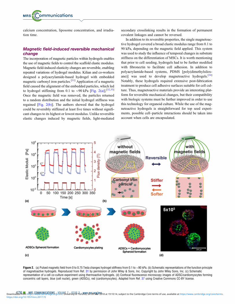
Magnetic field-induced reversible mechanicalchangeThe incorporation of magnetic particles within hydrogels enablesthe use of magnetic fields to control the scaffold elastic modulus.Magnetic field-induced elasticity changes are reversible, enablingrepeated variations of hydrogel modulus. Kilian and co-workersdesigned a polyacrylamide-based hydrogel with embeddedmagnetic carbonyl iron particles.[31] Application of a magneticfield caused the alignment of the embedded particles, which ledto hydrogel stiffening from 0.1 to ∼90 kPa [Fig. 2(a)].[32,33]
Once the magnetic field was removed, the particles returnedto a random distribution and the initial hydrogel stiffness wasregained [Fig. 2(b)]. The authors showed that the hydrogelcould be reversibly stiffened at least five times without signifi-cant changes to its highest or lowest modulus. Unlike reversibleelastic changes induced by magnetic fields, light-mediated
secondary crosslinking results in the formation of permanentcovalent linkages and cannot be reversed.
In addition to its reversible properties, the single magnetoac-tive hydrogel covered a broad elastic modulus range from 0.1 to90 kPa, depending on the magnetic field applied. This systemwas used to study the influence of temporal changes in substratestiffness on the differentiation of MSCs. It is worth mentioningthat prior to cell seeding, hydrogels had to be further modifiedwith fibronectin to facilitate cell adhesion. In addition topolyacrylamide-based systems, PDMS [poly(dimethylsilox-ane)] was used to develop magnetoactive hydrogels.[34]
Notably, these hydrogels required extensive post-fabricationtreatment to produce cell adhesive surfaces suitable for cell cul-ture. Thus, magnetoactive materials provide an interesting plat-form for reversible mechanical changes, but their compatibilitywith biologic systems must be further improved in order to usethis technology for organoid culture. While the use of the mag-netoactive hydrogels is straightforward for top seed experi-ments, possible cell–particle interactions should be taken intoaccount when cells are encapsulated.
Figure 2. (a) Pulsed magnetic field from 0 to 0.75 Tesla changes hydrogel stiffness from 0.1 to ∼90 kPa. (b) Schematic representations of the function principleof magnetoactive hydrogels. Reproduced from Ref. 31 by permission of John Wiley & Sons, Inc. Copyright by John Wiley Sons, Inc. (c) Schematicrepresentation of a cell co-culture experiment using thermoactive hydrogels. (d) Confocal fluorescence microscopy images of ADSC/cardiomyocytes formingconcentric cell layers, blue (cell nuclei), green (ADSCs), red (cardiomyocytes). Adapted from Ref. 37 using Creative Commons CC-BY license.
476▪ MRS COMMUNICATIONS • VOLUME 7 • ISSUE 3 • www.mrs.org/mrchttps://doi.org/10.1557/mrc.2017.72Downloaded from https://www.cambridge.org/core. University of Toronto, on 09 Mar 2018 at 19:10:14, subject to the Cambridge Core terms of use, available at https://www.cambridge.org/core/terms.

Temperature-controlled reversiblemechanical changeThermoresponsive hydrogels that undergo temperature-inducedgel–liquid transition provide an elegant method for long-term3D cell culture and subsequent cell extraction. Lei and Schafferused a thermoresponsive hydrogel composed of commerciallyavailable poly(N-isopropylacrylamide) (PNIPAAm)–PEG forhuman pluripotent stem cell (hPSC) expansion and differentia-tion.[35] The polymer solution is a liquid at 4 °C but solidifiesinto a hydrogel at 37 °C. Thus, cells can be seeded at low tem-perature, grown suspended in a solid gel at 37 °C, and subse-quently harvested or passaged by cooling the gel. hPSCscultured in this scaffold formed dense multi-cellular spheroidsand expanded by tenfold at 4 days after seeding, while in con-ventional static-suspension culture only a threefold expansionwas achieved. The hPSCs cultured in this scaffold could becontinuously propagated for 280 days. Notably, the highexpansion rate, pluripotency marker expression, and spheroidsize distribution remained consistent over these numerous pas-sages, indicating the suitability of this system for long-termexpansion. Later the PNIPAAm–PEG system was successfullyadapted to the scalable production of glioblastoma tumor-initiating cells.[36] The ability to easily harvest intact multi-cellular spheroids from thermoresponsive hydrogels by justlowering the temperature was used by Huh and co-workers tostudy cell co-culture.[37] After adipose-derived stromal cell(ADSC) spheroid formation, the hydrogel was cooled downand neonatal cardiomyocytes were added to the liquefied sub-strate [Fig. 2(c)]. Subsequently, the temperature was increasedto 37 °C resulting in both cells being encapsulated within thehydrogel. By adjusting the ratio between the two cell types,spheroids with concentric ADSCs/cardiomyocytes layerscould be formed [Fig. 2(d)]. Co-culture can recapitulate thecell–cell interactions in natural tissue, and thermoresponsivehydrogels may offer a feasible method to systematically inte-grate additional cell types for co-culture experiments.
Spatial control of mechanicalpropertiesCells respond to the mechanical properties of their environmenton the micron scale and hence sense the mechanical heteroge-neity of organs.[38,39] To further bridge the gap between com-plex in vivo microenvironments and engineered hydrogels fororganoid culture, researchers are beginning to design scaffoldsthat mimic the mechanical heterogeneity of tissues using exog-enous and endogenous methods.
Light-induced mechanical changesOver the past decade a variety of techniques were developed tocreate scaffolds with user-defined mechanical gradients asdetailed in recent reviews.[40,41] Here we highlight an examplewhere mechanical gradients are used to study stem cell behav-ior. Tse and Engler studied MSC behavior along a physiologi-cally relevant gradient of 1 kPa/mm (from 1 to 14 kPa) in an
attempt to mimic the migration of MSCs from bone marrowto injured tissue.[42] A gradient in elastic modulus was achievedby using a gradient photomask, which controlled the amount ofirradiation applied to crosslink acrylamide-based hydrogel pre-cursor materials. MSCs seeded on top of the hydrogelsmigrated toward the stiffer regions of the scaffold with fewcells remaining on regions where the stiffness fell below 6kPa. Although after 21 days of culture the majority of cellswere located in regions stiffer than 10 kPa, their phenotype dif-fered from that of cells continuously cultured on the homoge-neously stiff (11 kPa) hydrogel. While MSCs cultured onthe homogeneously stiff hydrogels differentiated toward amyogenic phenotype, those cultured on the gradually stiffen-ing substrate expressed both myogenic and neural pheno-types. This suggests that cells initially seeded on the softerregions possessed memory of the softer mechanical environ-ment where they typically differentiated to neural cells.Differentiation toward a mixed phenotype was also possibleby temporally changing the bulk mechanical properties ofthe hydrogel[29]; however, this technique did not promotecell migration.
Stiffness gradients spanning multiple millimeters provide agreat platform to study cell migration in response to gradualmechanical changes. In addition to cell sensitivity to mechani-cal changes on the macroscopic scale, recent studies proposedthat cells are able to sense their mechanical environment withmicron-scale precision.[38,39] Cells sense micron-scale changesin elasticity and even respond to the spatially oriented mechan-ical cues. Yang et al. designed hydrogels with either regularlyalternating [Figs. 3(a) and 3(b)] or randomly placed [Fig. 3(c)]stiff and soft 2 × 2 µm2 squares to determine whether hMSCsrespond to sub-cellular differences in scaffold elasticity or ifthey simply sense average substrate stiffness.[43] Increasingthe number of stiff squares on the regularly patterned hydrogelsincreased cell spreading, led to more elongated cells, andpromoted YAP activation similar to the hMSCs cultured onthe homogeneously stiff substrate [Figs. 3(d) and 3(e)]. ForhMSCs cultured on the random patterns, the increase in thenumber of stiff regions did not affect the cells. They exhibiteddecreased spreading and deactivated YAP in the cytoplasm,resembling cells cultured on the homogenously soft substrate[Fig. 3(f)]. Furthermore, while hMSCs cultured on regularlypattered gels differentiated toward the osteogenic lineage,hMSCs cultured on the randomly pattered hydrogel remainedlargely undifferentiated and continued to exhibit the MSCmarker, CD105.
Enzymatically induced mechanical changesBy developing hydrogels susceptible to cell-mediated changes,scientists can study how cells influence and remodel their ownmicroenvironment, thus gaining deeper insights into native cellbehavior. Incorporation of MMP cleavable crosslinkers intohydrogels enables cell-mediated hydrogel degradation, migra-tion, and self-assembly.[44] Proteolytically driven cell invasioninto HA-based hydrogels was shown by Fisher et al.[45] In this
Biomaterials for 3D Cell Biology Prospective Article
MRS COMMUNICATIONS • VOLUME 7 • ISSUE 3 • www.mrs.org/mrc ▪ 477https://doi.org/10.1557/mrc.2017.72Downloaded from https://www.cambridge.org/core. University of Toronto, on 09 Mar 2018 at 19:10:14, subject to the Cambridge Core terms of use, available at https://www.cambridge.org/core/terms.

study, invasive MDA-MB-231 breast cancer cells migratedtwice as far into the gels crosslinked with an MMP degrad-able peptide sequence (GPQG-IWGQ) compared with thegels with a non-degradable (GAGGAG) crosslinker. Thisdemonstrated that these cells actively remodel and degradethe hydrogel scaffold by MMP secretion. Such dynamic cel-lular remodeling by MMPs was quantitatively characterizedby Schultz et al.[46] The migration of hMSCs within PEGhydrogels containing MMP-degradable GPQG-IWGQ pep-tide sequences was analyzed by a multiple particle trackingmicrorheology technique. In a short time frame after cellseeding, a degradation gradient was measured in the pericel-lular region, with areas of increased degradation observedfarther away from the cell. The identified gradient indicatedthat cell-secreted enzymes diffused away from the cell fasterthan they could cleave the peptides. For the longer timescalestudy, after initial cell attachment and spreading, the pericel-lular regions changed from an elastic gel into a viscoelasticfluid in which cells could rapidly migrate, reaching a speedof up to 140 µm/h. Using a fluorescently labeled peptide,authors showed that, with time, hMSCs irreversibly remod-eled their environment leaving permanent migration pathseroded into the substrate.
Physically induced mechanical changesTo study endogenous matrix remodeling, scaffolds should besusceptible to cell-induced changes. Chemically crosslinkedhydrogels that have MMP-degradable crosslinkers enable enzy-matically induced bond cleavage; however, if the rest of thescaffold is non-degradable, there is an inherent limitation.Physically crosslinked hydrogels are in dynamic equilibriumwith their soluble components and thus are easily remodeled;however, they often suffer from poor stability. An alternativeis provided by hydrogels with mobile crosslinkers that areboth stable and susceptible to mechanical remodeling. Tongand Yang used modified hydrophilic α-cyclodextrins contain-ing polyrotaxane (SCPR-VS) to develop the first sliding hydro-gel with encapsulated cells.[47] Chemically crosslinked bonds,located on cyclic α-cyclodextrins, can rotate and slide alongthe PEG backbone, thus providing the hydrogel with mechan-ical freedom. The incorporation of α-cyclodextrins did notchange the bulk mechanical properties or stability (over 30days) from the covalent equivalent, where PEG chains weredirectly bonded to each other. This mobility enabled encapsu-lated hMSCs cells to extend protrusions through the rearrange-ment of crosslinking molecules and adhesive ligands. Cellscultured within the hydrogel with permanent crosslinks did
Figure 3. (a) Schematic representations of the patterning of hydrogels on the subcellular scale; (b) regularly alternating stiffness pattern, scale bar 2 µm; (c)randomly alternating stiffness pattern; (d) rounded cell on the hydrogel with 11% of regularly patterned stiff areas; (e) cell with a spread morphology on thehydrogel with 75% of regularly patterned stiff areas; (f) cell with decreased spreading on the hydrogel with 75% of randomly patterned stiff areas. Scale bars 20µm. Reproduced from Ref. 43.
478▪ MRS COMMUNICATIONS • VOLUME 7 • ISSUE 3 • www.mrs.org/mrchttps://doi.org/10.1557/mrc.2017.72Downloaded from https://www.cambridge.org/core. University of Toronto, on 09 Mar 2018 at 19:10:14, subject to the Cambridge Core terms of use, available at https://www.cambridge.org/core/terms.

not form protrusions, indicating that the flexible network wasessential to induce cell spreading. Although sliding hydrogelsdo not promote cell morphogenesis to the same extent asMMP-degradable or hydrolytically susceptible hydrogels,combining these approaches might lead to the developmentof materials that provide greater opportunity for cellularremodeling.
Naturally derived ECM components and native tissues,unlike chemically crosslinked synthetic hydrogels, are visco-elastic and exhibit partial stress relaxation—that is, matrixdeformation upon applied strain.[48–50] Materials with fastand slow rates of stress relaxation require short and long straindurations, respectively. In viscoelastic scaffolds, cells canmechanically remodel their local environment by applying trac-tion forces.[51] Furthermore, recent studies showed that the vis-coelastic properties of scaffolds influence cell behaviorindependent of bulk scaffold stiffness. By varying the molecu-lar weight of polysaccharides, Mooney et al. developedalginate-based hydrogels with variable rates of stress relaxa-tion.[52] Relaxation rate-dependent differences in spreadingand proliferation of 3T3 fibroblasts encapsulated within thehydrogels were observed; cells encapsulated within hydrogelswith long relaxation rates remained rounded and showed sup-pressed proliferation compared to cells cultured in hydrogelswith short relaxation rates, which spread and proliferated.Notably, the initial elastic modulus of all hydrogels was 9kPa, indicating that differences in cell shape and proliferationwere induced by matrix susceptibly to mechanical remodeling.
Modulation of biochemical propertiesin bioengineered hydrogelsIt has been demonstrated that the incorporation of biochemicalfeatures can control cell fate in 3D culture when combined withoptimized mechanics. These include: adhesive peptides,growth factors, and co-culture of multiple cell types. PEG iscommonly used as a scaffold because it is non-adsorptive toproteins and non-adhesive to cells, allowing researchers toevaluate how different components of the microenvironmentaffect cell fate.[53] HA, heparin, and PNIPAAm are also usedin engineered hydrogels due to their role in biologic signaling,affinity-mediated growth factor release, and thermoresponsiveproperties, respectively.[54–57] Cell-adhesive peptide ligandsand matrix metalloprotease-degradable crosslinking moleculesare ubiquitous features across 3D scaffold design. Notwithst-anding the advances in 3D cell culture, it remains somewhatof an art. Slight variation in peptide ligand sequence, rate ofdegradation, growth factor presentation, and co-culture condi-tions can all impact cell fate.
Heparin-based hydrogels for growth factorreleaseCovalently functionalizing proteins to hydrogel scaffolds canresult in a loss of protein bioactivity. Heparin is a sulfatedglycosaminoglycan (GAG) that binds proteins reversibly viaelectrostatic interactions, and researchers have harnessed this
natural affinity for growth factor release within hydrogels.For example, Chwalek et al. developed a heterocellular angio-genesis model by designing a hydrogel system composed ofstar-PEG and heparin to support endothelial cell (EC) morpho-genesis into capillary structures.[58] The incorporation ofVEGF, basic fibroblast growth factor (bFGF), and stromal-derived growth factor (SDF-1α) was investigated. The simulta-neous introduction of all three growth factors (5 µg/mL each ofVEGF, bFGF, and SDF-1α) resulted in capillary networks withsignificantly increased branch length, density, and stability (at10 days versus 4 days) when compared with VEGF alone[Fig. 4(a)]. The effects of co-culture were also examined.Using 10% MSCs, ECs formed stable capillary networks forover 4 weeks, whereas with more than 10% MSCs, the forma-tion of EC tube structures was inhibited. This long-term cultureperiod of ECs and MSCs (at 10%) resulted in lumens within theEC tubes, with diameters matching those of human capillariesin vivo. Co-culture with different cell types caused dramaticdifferences in capillary density, suggesting that functional dif-ferences in vivo are determined by proximity and recruitmentof specific mural cells.
While heparin-based hydrogels are advantageous for preser-vation of growth factor bioactivity and in vivo compatibility,inconsistent release profiles and batch-to-batch variability com-plicate their use for 3D cell culture applications. Hettiaratchiet al. demonstrated that the release rate of bone morphogeneticprotein-2 (BMP-2) from heparin-based microparticles increasedin the presence of bovine serum due to competitive proteinbinding.[59] In another study by Hettiaratchi et al., it wasshown that <25% of bound BMP-2 was released from heparinmicroparticles over 28 days.[60] In this case, BMP-2 bioactivitywas conserved. However, the xenogenic source of heparin, por-cine intestinal mucosa, has inherent batch-to-batch variability,and provides an incentive to engineer synthetic heparin-likematerials.[61,62] Furthermore, varying concentrations of heparinmay have detrimental effects on cell survival. Kottke-Marchantand co-workers demonstrated that increasing heparin concen-tration in PEGMA/PEGDA hydrogels decreased the spreadingand proliferation of smooth muscle cells (SMC) in vitro.[63]
Integrin–peptide interactions affect cell fateWhile hydrogels are often functionalized with peptide ligands,integrin expression is not always characterized. Evaluatingintegrin expression has the potential to guide hydrogel designand explain organoid phenotype. For example, Levental andco-workers discussed the role of integrin binding in culturingmammary epithelial cells (MECs).[1] Polarized acini formedin soft formulations of star-PEG and heparin hydrogels, butonly when an MMP-degradable crosslinker was used. InMMP-degradable gels, laminin-332 (LN-332) was secretedby cells and distributed around the basal surface after 4 daysof culture. When a blocking antibody was used against α6β4,an integrin involved in LN-332 binding, cell growth was arrested.The authors hypothesized two explanations for MEC mor-phogenesis that are not mutually exclusive: (1) secreted
Biomaterials for 3D Cell Biology Prospective Article
MRS COMMUNICATIONS • VOLUME 7 • ISSUE 3 • www.mrs.org/mrc ▪ 479https://doi.org/10.1557/mrc.2017.72Downloaded from https://www.cambridge.org/core. University of Toronto, on 09 Mar 2018 at 19:10:14, subject to the Cambridge Core terms of use, available at https://www.cambridge.org/core/terms.

LN-332 binds to heparin and activates integrins on MECs; and(2) heparin promotes adhesion and survival by binding cellintegrins.
Studying integrin expression levels can guide peptide selec-tion. The role of integrin-matrix interactions on cell aggregationand proliferation was investigated in the context of non-Hodgkin’s lymphoma by Singh and co-workers[64] A co-culturesystem with lymphoma and follicular dendritic cells (FDCs) wasdesigned using maleimide-functionalized star-PEG, MMP2/9-degradable peptide crosslinkers (GCRDVPMS↓MRGGDRCG),and adhesive peptides containing fibronectin-derived RGD orREDV sequences which bind to integrins αvβ3 and α4β1,respectively. It was found that RGD peptides increased the aggre-gation of B-cell lymphoma cell line, HBL-1. Although RGD pep-tides increased the clustering of HBL-1 cells, REDV peptidesincreased proliferation [Fig. 4(b)]. This was further demonstratedthrough cell cycle analysis, which revealed a 28% reduction in theDNA synthesis phase when RGD peptides were used instead ofREDV peptides.
Studies investigating the effects of different peptide ligandsmay benefit from considering integrin-peptide binding affini-ties. Gould and Anseth investigated the effects of peptideligand sequence and MMP activity on valvular interstitial cell(VIC) fate in an eight-arm-PEG hydrogel.[65] VICs culturedwith either RGDS or P15 (collagen-1-derived) peptidesexhibited significantly higher MMP activity when comparedwith those cultured with VGVAPG (elastin-derived). The
percentage of cells that stained positive for α-SMA (α-smoothmuscle actin) was the highest when VGVAPG was used, sug-gesting that the myofibroblast phenotype is associated withlow MMP activity and elongation. However, the authors ofthis study note that binding strengths between peptides andintegrins are not evaluated. Significant differences in bindingaffinities may weaken conclusions made about an MMPactivity-dependent phenotype, and should be considered forfuture studies of this nature.
Biochemical cue concentration depicts cellphenotypeAlthough adhesive peptides and MMP-degradable crosslinkersare typically incorporated into 3D cell culture, achieving theappropriate concentrations for desirable phenotypes is non-trivial. Enemchukwu et al. investigated the effects of alteringbiochemical cue concentration on epithelial morphogenesis ina star-PEG hydrogel with MMP-degradable crosslinkers andRGD peptide ligands.[66] Polarity and lumen formation ofMDCK (Madin–Darby canine kidney) epithelial cells dependedon RGD concentration, whereas proliferation and self-assemblyinto multi-cellular structures were not affected. High concentra-tions of RGD (>250 µM) gave rise to cysts that containedlumen and internal apical polarity, and at 2000 µM RGD, hydro-gels exhibited a phenotype that closely matched that observedwith collagen, a common matrix material for epithelial cell cul-ture. A study by Lin et al. demonstrated that there is an optimal
Figure 4. (a) The influence of VEGF, bFGF, and SDF incorporation into hydrogels with heparin and star-PEG on branch length, density, and stability ofendothelial cells. Adapted from Ref. 58 with permission from Nature Publishing Group. (b) Effect of RGD and REDV ligand presentation on B cell proliferation.Adapted from Ref. 64 with permission from Elsevier. (c) Influence of immobilized RGD ligand concentration on the proliferation of smooth muscle cells inPEG-based hydrogels. Adapted from Ref. 63 with permission from Elsevier.
480▪ MRS COMMUNICATIONS • VOLUME 7 • ISSUE 3 • www.mrs.org/mrchttps://doi.org/10.1557/mrc.2017.72Downloaded from https://www.cambridge.org/core. University of Toronto, on 09 Mar 2018 at 19:10:14, subject to the Cambridge Core terms of use, available at https://www.cambridge.org/core/terms.

concentration of immobilized RGD ligand that increases the pro-liferation of SMC in vitro.[63] Concentrations between 0.625 and1.25 mM maximized proliferation in PEG, while >1.25 mMresulted in decreased proliferation [Fig. 4(c)]. Furthermore,MMP-degradable crosslinker concentration had implications oncell proliferation and spreading. Limited cell spreading wasobserved when slowly MMP-degradable crosslinkers were usedwhen compared with rapid MMP-degradable crosslinkers; yet,increasing the concentration of rapid MMP-degradable cross-linkers decreased cell proliferation and spreading. Thus, a balanceof rate and concentration of MMP-degradable crosslinkers wasrequired.
The types and concentrations of growth factors, peptides,crosslinkers, and co-cultured cells are all factors that can beoptimized in bioengineered hydrogels to achieve a desirablephenotype; however, control over bulk biochemical cues isinsufficient for engineering more sophisticated tissue-specificorganoids. Organs have multiple cell types and intricate archi-tectures. Spatiotemporal control over biochemical cues inhydrogel systems will be necessary to study dynamic processesthat occur during organogenesis, disease, and injury.
Temporal control of biochemicalpropertiesTemporal control over biochemical cues is required for dynamiccontrol of cell adhesion and differentiation. Photocaging is a tech-nique where bioligands are capped with photocleavable moieties,rendering them inactive. Focused illumination activates bioli-gands and promotes cell interactions. Photocaging and photo-cleavable molecules have been engineered in hydrogels tocontrol the attachment and removal of bioactive ligands aswell as to temporally control cell phenotype. In vivo activationof biochemical ligands also impacts cell adhesion, inflamma-tion, fibrous encapsulation, and vascularization.[67]
Temporal control of bioactive ligands topromote differentiationControlled presentation and removal of biochemical cuesimpact cell differentiation. For example, dynamically tuningthe presence and release of adhesive ligands is required formodeling in vivo differentiation of hMSCs, as demonstratedby Anseth and co-workers.[68] A nitrobenzyl ether-derived pho-tolabile moiety bound to an acrylate monomer (photodegrad-able acrylate, PDA) was bound to an RGDS peptide. Thiscompound was copolymerized with PEG-diacrylate (PEGDA)to form a hydrogel with photolabile RGDS. The hMSC viabil-ity and chondrogenic differentiation was influenced by RGDSpresentation. When hMSCs differentiate into chondrocytes invivo, fibronectin is downregulated between 7 and 10 daysafter which cells upregulate GAG and type II collagen(COLII), indicators of chondrogenic differentiation. This pat-tern of fibronectin downregulation was mimicked throughtimely controlled RGDS removal. PEG-only hydrogels (with-out RGDS functionalization) resulted in significantly lowerhMSC viability compared with hydrogels with RGDS. When
RGDS was photocleaved on day 10 in culture, hMSC viabilitywas not affected; however, GAG production increased fourfoldwith statistical significance compared to hydrogels with cons-tant RGDS or no RGDS. A decrease in CD105 expression(hMSC marker) and increase in COLII production wasobserved, indicating chondrocyte differentiation [Fig. 5(a)].
Orthogonal photocaging enables light-mediated attachmentand subsequent removal of biologically active molecules topromote differentiation. DeForest and Tirrell designed a PEGhydrogel system with spatiotemporal control over full-lengthprotein attachment and subsequent removal.[69] The chemistriesfor bioconjugation and detachment are orthogonal and cyto-compatible, and the reactions can be performed in the presenceof live cells. To yield the initial scaffold, azide-functionalizedpeptide crosslinkers and azide-functionalized 2-(2-nitro-phenyl)propoxycarbonyl (NPPOC)-photocaged alkoxyaminemoieties underwent strain-promoted azide-alkyne cycloaddi-tion (SPAAC) with cyclooctyne functionalized star-PEG.Upon exposure to UV light, uncaged alkoxyamines reactedwith aldehyde-functionalized proteins resulting in the protein-modified scaffold. The incorporation of a second photocleav-able moiety, o-nitrobenzyl ester, between the aldehyde func-tionality and the protein, enabled protein detachment withUV exposure. Vitronectin (VTN) was patterned to investigatehMSC differentiation into osteocytes; osteocalcin (OC) immu-nostaining and alkaline phosphatase (ALP) activity were eval-uated as indicators of osteogenic differentiation. VTN wasintroduced on day 1 and cells showed an increase in OC stain-ing and ALP activity by day 4. Upon VTN removal on day 4,OC staining and ALP activity decreased and returned to origi-nal levels by day 10 [Fig. 5(b)].
Spatial control of biochemicalpropertiesThe complex architecture of organ systems gives researchersincentive to develop techniques for spatial tuning of biochem-ical cues in 3D culture systems. Spatial manipulation of biolog-ically relevant molecules enables studies of cell adhesion,aggregation, spreading, and invasion within user-defined andintricate geometries.[70–74] Concentration gradients of bioactivemolecules enable studies of cell migration, development, andgrowth.[75,76] Ultimately, simplified and adaptable systemswill be required for widespread implementation whether forresearch or clinical application.
Light-induced patterning of biochemical cuesPhotocaging can be used for biochemical ligand presentation;upon irradiation, active proteins are exposed to cells to elicita biochemical response. However, proteins can be damagedupon irradiation, which impacts their bioactivity and stability.A unique technique was developed by Lutolf and co-workersto immobilize proteins on hydrogels with minimal effect onprotein stability and bioactivity whereby uncaged hydrogel-bound peptides were bound to proteins through enzyme-mediated conjugation.[74] Specifically, transglutaminase factor
Biomaterials for 3D Cell Biology Prospective Article
MRS COMMUNICATIONS • VOLUME 7 • ISSUE 3 • www.mrs.org/mrc ▪ 481https://doi.org/10.1557/mrc.2017.72Downloaded from https://www.cambridge.org/core. University of Toronto, on 09 Mar 2018 at 19:10:14, subject to the Cambridge Core terms of use, available at https://www.cambridge.org/core/terms.

Figure 5. (a) hMSC expression of integrin αvβ3 (green) when cultured in RGDS (yellow gel) or cleaved RGDS (white gel) after 4 or 21 days (panel A). CleavingRGDS on day 10 showed a decrease in CD105 (green) and an increase in COLII (red) production by day 21, indicating chondrocyte differentiation (panel B).Adapted from Ref. 68 with permission from Science Publishing Group. (b) hMSCs show markers of osteocyte differentiation after vitronectin is patterned into aPEG-based hydrogel. Osteocalcin (OC) (green) and ALP activity increase in areas where VTN is photopatterned. Adapted from Ref. 69 with permission fromNature Publishing Group.
482▪ MRS COMMUNICATIONS • VOLUME 7 • ISSUE 3 • www.mrs.org/mrchttps://doi.org/10.1557/mrc.2017.72Downloaded from https://www.cambridge.org/core. University of Toronto, on 09 Mar 2018 at 19:10:14, subject to the Cambridge Core terms of use, available at https://www.cambridge.org/core/terms.

XII (FXIIIa) catalyzed the reaction between an amine-donorFXIIIa substrate AcFKG (K-peptide) bound to the PEG hydro-gel and an amine-acceptor peptide NQEQVSPL (Q-peptide)bound to the desired protein. K-peptide was caged with nitro-veratryloxycarbonyl (Nvoc) to enable spatially controlledenzyme-mediated photopatterning [Fig. 6(a)]. VEGF, ProteinA (for tethering to Fc-tagged proteins), RGD peptide, recombi-nant fibronectin fragment FN9–10, and platelet-derived growthfactor B (PDGF-BB) were all immobilized to the PEG hydrogelusing this method. Multicellular clusters of MSCs were incor-porated into MMP-cleavable PEG hydrogels, and a definedarea of the hydrogel was patterned with RGD, FN9–10, orPDGF-BB. Cell invasion into the RGD, FN9–10 andPDGF-BB patterned regions was significantly greater
compared with unpatterned regions, demonstrating bioactivityafter immobilization and photo-illumination.
Two-photon irradiation is advantageous for fine-tuning thespatial control and concentration of biologically relevant mole-cules in 3D culture systems. Complex patterns incorporatingone or more biologically relevant molecules can be achievedwith two-photon irradiation.[77,78] Recent studies demonstratethe advantages of two-photon irradiation for fine-tuning thespatial control of biologically relevant molecules in 3D culturesystems. Wosnick and Shoichet functionalized agarose with6-bromo-7-hydroxy coumarin (Bhc)-protected thiol moietiesto spatially control bioconjugation.[77] Aizawa et al. demon-strated that, with a two-photon microscope, exposed thiols inan agarose hydrogel could be reacted with maleimide-modified
Figure 6. (a) Enzyme-mediated conjugation scheme of biochemical cues. FXIIIa catalyzes the reaction between K- and Q-peptides following photo-activation ofthe K-peptide. Adapted from Ref. 74 with permission from Nature Publishing Group. (b) Results of simultaneously patterning Barstar–SHH–488 (green) andbiotin–CNTF–633 (red) into an agarose-based hydrogel using two-photo patterning. Adapted from Ref. 78 with permission from Nature Publishing Group. (c)Glioblastoma and human astrocyte clusters magnetically combined into a single spheroid in a hydrogel containing gold nanoparticles, iron oxide nanoparticles,and M13-derived phage particles. Adapted from Ref. 81 with permission from Nature Publishing Group.
Biomaterials for 3D Cell Biology Prospective Article
MRS COMMUNICATIONS • VOLUME 7 • ISSUE 3 • www.mrs.org/mrc ▪ 483https://doi.org/10.1557/mrc.2017.72Downloaded from https://www.cambridge.org/core. University of Toronto, on 09 Mar 2018 at 19:10:14, subject to the Cambridge Core terms of use, available at https://www.cambridge.org/core/terms.

biomolecules such as vascular-endothelial growth factor-A(VEGF-A).[79]
Orthogonal immobilization chemistries are advantageousfor precise spatial control of multiple bioligands. Wylie et al.took advantage of two high-binding, orthogonal reactions—streptavidin–biotin and barnase–barstar—to immobilize twoproteins simultaneously in spatially defined volumes with dis-tinct protein concentrations.[78] Agarose, a transparent hydro-gel, was modified with Bhc-protected thiol moieties that weremodified after exposure to multi-photon irradiation with malei-mide–streptavidin and maleimide–barnase, which have highaffinities to biotin and barstar, respectively. Ciliary neurotro-phic factor (CNTF) expressed recombinantly with biotin, andsonic hedgehog (SHH) with barstar, were simultaneouslyimmobilized to agarose [Fig. 6(b)]. These recombinant proteinsremained bioactive and when immobilized as a gradient guidedretinal stem cell progenitors into the gels. While two-photonirradiation has the advantages of improved resolution and pen-etration depth, molecules with low uncaging efficiencies limitsubsequent bioconjugation. For example, Bhc protectinggroups can undergo photoisomerization reactions followingirradiation. Mahmoodi et al. developed 6-bromo-7-hydroxy-3-methylcoumarin-4-ylmethyl (mBhc) to cage thiol moie-ties.[80] HA-based hydrogels modified with mBhc-protectedthiols had a fourfold greater uncaging efficiency thanBhc-protected thiols, which impacts the success of biochemicalphotopatterning.
Magnetic fields for spatial control over celldistributionWhile spatially defined aggregates with multiple cell types arenot common in current tissue engineering and regenerativemedicine approaches, strategic co-culture of multiple celltypes will be required for complex organogenesis models andintricate organoid culture. Magnetically controlled systemsenable user-defined control of cell distribution. For example,with magnetic levitation, a magnet is used to guide cell distri-bution of a system comprised of magnetic particles intercalatedwith cells. Souza et al. designed a hydrogel containing goldnanoparticles, iron oxide nanoparticles, and M13-derivedphage particles in order to form human glioblastoma multi-cellular aggregates with defined geometries and cell–cell con-tacts.[81] The shape of the magnet determined the geometryof the cell cluster. Moreover, separate glioblastoma andhuman astrocyte clusters were magnetically combined into asingle spheroid [Fig. 6(c)]. In another study by Bratt-Lealet al., magnetic nanoparticles were incorporated into the extra-cellular space of embryoid bodies (EBs).[82] Cell response tomagnetic fields was dose dependent, and EBs labeled with dif-ferent fluorescent tags could form aggregates with user-definedpatterns.
ConclusionsWe have highlighted the importance of spatiotemporal controlover mechanical and biochemical properties in engineered
hydrogels. While controlling the bulk properties of the hydro-gel has implications on cell viability, aggregation, proliferation,and differentiation, spatiotemporal control enables guided cellgrowth and helps to recapitulate intricate cell networks thatare required for studying biologically relevant processes. Weexpect future efforts to be focused on translating this knowl-edge to organoid development, for the applications of diseasemodeling and drug screening.
Research efforts that focus on developing hydrogel systemswith stable and orthogonal chemistries for controlling mechan-ical and biochemical cues separately are required to understandthe key factors of the cellular microenvironment that guide cellfate and organ formation. “Plug and play” chemistries thatenable modular design with multiple combinations of biologi-cally relevant components will enable biologists to answer fun-damental questions pertaining to organ development anddisease modeling. For hydrogel platforms to have widespreaduse in the research community, they must be versatile, easyto work with, and affordable. In the coming years, we expecta push toward the development of high-throughput screeningplatforms for drug development and personalized medicine,where patient-derived iPSCs could be used for individualdrug screening. There are already platforms for screeningwith 3D culture conditions, such as microfluidic devices andassays developed in a conventional 96-well plate format.[83,84].We expect hydrogel systems to not only advance the under-standing of fundamental biologic processes, but advance thedevelopment of novel therapeutics and in vivo regenerativemedicine technologies.
AcknowledgmentsWe are grateful for funding from the Canadian Institutes ofHealth Research (Foundation grant to M.S.S.), the NaturalSciences and Engineering Research Council (Discovery grantto M.S.S.), the QEII-GSST graduate student scholarship (toL.C.B.) and the Canada First Research Excellence Fund,Medicine by Design (to M.S.S.).
References1. M. Nowak, U. Freudenberg, M.V. Tsurkan, C. Werner, and K.R. Levental:
Modular GAG-matrices to promote mammary epithelial morphogenesis invitro. Biomaterials 112, 20 (2017).
2. A. Ranga, M. Girgin, A. Meinhardt, D. Eberle, M. Caiazzo, E.M. Tanaka,and M.P. Lutolf: Neural tube morphogenesis in synthetic 3D microenvi-ronments. Proc. Natl. Acad. Sci. U.S.A. 113, E6831 (2016).
3. E. Moeendarbary, I.P. Weber, G.K. Sheridan, D.E. Koser, S. Soleman,B. Haenzi, E.J. Bradbury, J. Fawcett, and K. Franze: The soft mechanicalsignature of glial scars in the central nervous system. Nat. Commun. 8,14787 (2017).
4. T. Mammoto, A. Mammoto, and D.E. Ingber: Mechanobiology and devel-opmental control. Annu. Rev. Cell Dev. Biol. 29, 27 (2013).
5. C.-P. Heisenberg and Y. Bellaïche: Forces in tissue morphogenesis andpatterning. Cell 153, 948 (2013).
6. N. Bouchonville, M. Meyer, C. Gaude, E. Gay, D. Ratel, A. Nicolas,C.A. Reinhart-King, S.S. Margulies, M. Dembo, D. Boettiger,D.A. Hammer, V.M. Weaver, Y. Shi, and S.P. Robinson: AFM mappingof the elastic properties of brain tissue reveals kPa μm−1 gradients ofrigidity. Soft Mat. 12, 6232 (2016).
484▪ MRS COMMUNICATIONS • VOLUME 7 • ISSUE 3 • www.mrs.org/mrchttps://doi.org/10.1557/mrc.2017.72Downloaded from https://www.cambridge.org/core. University of Toronto, on 09 Mar 2018 at 19:10:14, subject to the Cambridge Core terms of use, available at https://www.cambridge.org/core/terms.

7. S. Budday, G. Sommer, C. Birkl, C. Langkammer, J. Haybaeck, J. Kohnert,M. Bauer, F. Paulsen, P. Steinmann, E. Kuhl, and G.A. Holzapfel:Mechanical characterization of human brain tissue. Acta Biomater. 48,319 (2017).
8. J.Y. Rho, R.B. Ashman, and C.H. Turner: Young’s modulus of trabecularand cortical bone material: Ultrasonic and microtensile measurements.J. Biomech. 26, 111 (1993).
9. E. Hoenig, U. Leicht, T. Winkler, G. Mielke, K. Beck, F. Peters,A.F. Schilling, and M.M. Morlock: Mechanical properties of native andtissue-engineered cartilage depend on carrier permeability: a bioreactorstudy. Tissue Eng. A 19, 1534 (2013).
10.L.E. Bilston: Brain Tissue Mechanical Properties in Biological and MedicalPhysics, Biomedical Engineering (Springer, New York, USA, 2011, vol. 69).
11.A.J. Engler, S. Sen, H.L. Sweeney, and D.E. Discher: Matrix elasticitydirects stem cell lineage specification. Cell 126, 677 (2006).
12.B. Trappmann, J.E. Gautrot, J.T. Connelly, D.G.T. Strange, Y. Li, M.L. Oyen,M.A. Cohen Stuart, H. Boehm, B. Li, V. Vogel, J.P. Spatz, F.M.Watt, andW.T.S. Huck: Extracellular-matrix tethering regulates stem-cell fate. Nat. Mater.11, 642 (2012).
13. J.H. Wen, L.G. Vincent, A. Fuhrmann, Y.S. Choi, K.C. Hribar,H. Taylor-Weiner, S. Chen, and A.J. Engler: Interplay of matrix stiffnessand protein tethering in stem cell differentiation. Nat. Mater. 13, 979(2014).
14.N.D. Leipzig and M.S. Shoichet: The effect of substrate stiffness on adultneural stem cell behavior. Biomaterials 30, 6867 (2009).
15. J. You, S.-A. Park, D.-S. Shin, D. Patel, V.K. Raghunathan, M. Kim,C.J. Murphy, G. Tae, and A. Revzin: Characterizing the effects of heparingel stiffness on function of primary hepatocytes. Tissue Eng. A 19, 2655(2013).
16.N. Mittal, F. Tasnim, C. Yue, Y. Qu, D. Phan, Y. Choudhury, M.-H. Tan, andH. Yu: Substrate stiffness modulates the maturation of human pluripo-tent stem-cell-derived hepatocytes. ACS Biomater. Sci. Eng. 2, 1649(2016).
17.A. Nava, E. Mazza, M. Furrer, P. Villiger, and W.H. Reinhart: In vivomechanical characterization of human liver. Med. Image Anal. 12, 203(2008).
18.L. Huwart, F. Peeters, R. Sinkus, L. Annet, N. Salameh, L.C. ter Beek,Y. Horsmans, and B.E. Van Beers: Liver fibrosis: non-invasive assess-ment with MR elastography. NMR Biomed.. 19, 173 (2006).
19.D. Hanjaya-Putra, J. Yee, D. Ceci, R. Truitt, D. Yee, and S. Gerecht:Vascular endothelial growth factor and substrate mechanics regulate invitro tubulogenesis of endothelial progenitor cells. J. Cell. Mol. Med.14, 2436 (2010).
20.S.R. Caliari, S.L. Vega, M. Kwon, E.M. Soulas, and J.A. Burdick:Dimensionality and spreading influence MSC YAP/TAZ signaling in hydro-gel environments. Biomaterials 103, 314 (2016).
21.S. Khetan, M. Guvendiren, W.R. Legant, D.M. Cohen, C.S. Chen, andJ.A. Burdick: Degradation-mediated cellular traction directs stem cellfate in covalently crosslinked three-dimensional hydrogels. Nat. Mater.12, 458 (2013).
22.N. Huebsch, P.R. Arany, A.S. Mao, D. Shvartsman, O.A. Ali,S.A. Bencherif, J. Rivera-feliciano, and D.J. Mooney: Harnessing traction-mediated manipulation of the cell/matrix interface to control stem-cellfate. Nat. Mater. 9, 518 (2010).
23.N. Gjorevski, N. Sachs, A. Manfrin, S. Giger, M.E. Bragina,P. Ordóñez-Morán, H. Clevers, and M.P. Lutolf: Designer matrices forintestinal stem cell and organoid culture. Nat. Publ. Gr. 539, 560 (2016).
24.S.R. Caliari, M. Perepelyuk, E.M. Soulas, G.Y. Lee, R.G. Wells, andJ.A. Burdick: Gradually softening hydrogels for modeling hepatic stellatecell behavior during fibrosis regression. Integr. Biol. 8, 720 (2016).
25.N. Boehnke, C. Cam, E. Bat, T. Segura, and H.D. Maynard: Imine hydrogelswith tunable degradability for tissue engineering. Biomacromolecules 16,2101 (2015).
26.P.M. Kharkar, A.M. Kloxin, and K.L. Kiick: Dually degradable click hydro-gels for controlled degradation and protein release. J. Mater. Chem. B,Mater. Biol. Med. 2, 5511 (2014).
27. J.L. Young and A.J. Engler: Hydrogels with time-dependent materialproperties enhance cardiomyocyte differentiation in vitro. Biomaterials32, 1002 (2011).
28.C. Yang, M.W. Tibbitt, L. Basta, and K.S. Anseth: Mechanical memory anddosing influence stem cell fate. Nat. Mater. 13, 645 (2014).
29.M. Guvendiren and J.A. Burdick: Stiffening hydrogels to probe short- andlong-term cellular responses to dynamic mechanics. Nat. Commun. 3,792 (2012).
30.R.S. Stowers, S.C. Allen, and L.J. Suggs: Dynamic phototuning of 3Dhydrogel stiffness. Proc. Natl. Acad. Sci. U.S.A. 112, 1953 (2015).
31.A.A. Abdeen, J. Lee, N.A. Bharadwaj, R.H. Ewoldt, and K.A. Kilian:Temporal modulation of stem cell activity using magnetoactive hydrogels.Adv. Healthc. Mater. 5, 2536 (2016).
32.U. Ubaidillah, J. Sutrisno, A. Purwanto, and S.A. Mazlan: Recent progresson magnetorheological solids: materials, fabrication, testing, and applica-tions. Adv. Eng. Mater. 17, 563 (2015).
33.T. Mitsumata, S. Ohori, A. Honda, and M. Kawai: Magnetism and visco-elasticity of magnetic elastomers with wide range modulation of dynamicmodulus. Soft Matter 9, 904 (2013).
34.M. Mayer, R. Rabindranath, J. Boerner, E. Hoerner, A. Bentz, J. Salgado,H. Han, H. Boese, J. Probst, M. Shamonin, G.J. Monkman, andG. Schlunck: Ultra-soft PDMS-based magnetoactive elastomers asdynamic cell culture substrata. PLoS ONE 8, e76196 (2013).
35.Y. Lei and D.V. Schaffer: A fully defined and scalable 3D culture systemfor human stem cell expansion and differentiation. Proc. Natl. Acad.Sci. U. S. A. 110, E5039 (2013).
36.Q. Li, H. Lin, O. Wang, X. Qiu, S. Kidambi, L.P. Deleyrolle, B.A. Reynolds,and Y. Lei: Scalable production of glioblastoma tumor-initiating cells in 3dimension thermoreversible hydrogels. Sci. Rep. 6, 31915 (2016).
37.M.O. Cho, Z. Li, H.-E. Shim, I.-S. Cho, M. Nurunnabi, H. Park, K.Y. Lee,S.-H. Moon, K.-S. Kim, S.-W. Kang, and K.M. Huh: Bioinspired tuning ofglycol chitosan for 3D cell culture. NPG Asia Mater. 8, e309 (2016).
38.H. Wolfenson, G. Meacci, S. Liu, M.R. Stachowiak, T. Iskratsch,S. Ghassemi, P. Roca-Cusachs, B. O’Shaughnessy, J. Hone, andM.P. Sheetz: Tropomyosin controls sarcomere-like contractions for rigid-ity sensing and suppressing growth on soft matrices. Nat. Cell Biol. 18, 33(2015).
39.G. Meacci, H. Wolfenson, S. Liu, M.R. Stachowiak, T. Iskratsch,A. Mathur, S. Ghassemi, N. Gauthier, E. Tabdanov, J. Lohner,A. Gondarenko, A.C. Chander, P. Roca-Cusachs, B. O’Shaughnessy,J. Hone, and M.P. Sheetz: α-Actinin links extracellular matrix rigidity-sensing contractile units with periodic cell-edge retractions. Mol. Biol.Cell 27, 3471 (2016).
40.M. Whang and J. Kim: Synthetic hydrogels with stiffness gradients fordurotaxis study and tissue engineering scaffolds. Tissue Eng. Regen.Med. 13, 126 (2016).
41.X. Tingting, L. Wanqian, and Y. Li: A review of gradient stiffness hydro-gels used in tissue engineering and regenerative medicine. J. Biomed.Mater. Res. A 105, 1799 (2017).
42. J.R. Tse and A.J. Engler: Stiffness gradients mimicking in vivo tissue var-iation regulate mesenchymal stem cell fate. PLoS ONE 6, e15978 (2011).
43.C. Yang, F.W. DelRio, H. Ma, A.R. Killaars, L.P. Basta, K.A. Kyburz, andK.S. Anseth: Spatially patterned matrix elasticity directs stem cell fate.Proc. Natl. Acad. Sci. U.S.A. 113, E4439 (2016).
44.M.P. Lutolf, J.L. Lauer-Fields, H.G. Schmoekel, A.T. Metters, F.E. Weber,G.B. Fields, and J.A. Hubbell: Synthetic matrix metalloproteinase-sensitive hydrogels for the conduction of tissue regeneration: engineeringcell-invasion characteristics. Proc. Natl. Acad. Sci. U.S.A. 100, 5413(2003).
45.S.A. Fisher, P.N. Anandakumaran, S.C. Owen, and M.S. Shoichet: Tuningthe microenvironment: click-crosslinked hyaluronic acid-based hydrogelsprovide a platform for studying breast cancer cell invasion. Adv. Funct.Mater. 25, 7163 (2015).
46.K.M. Schultz, K.A. Kyburz, and K.S. Anseth: Measuring dynamic cell–material interactions and remodeling during 3D human mesenchymalstem cell migration in hydrogels. Proc. Natl. Acad. Sci. U.S.A. 112,E3757 (2015).
47.X. Tong and F. Yang: Sliding hydrogels with mobile molecular ligands andcrosslinks as 3D stem cell niche. Adv. Mater. 28, 7257 (2016).
48.Z. Liu and L. Bilston: On the viscoelastic character of liver tissue: exper-iments and modelling of the linear behaviour. Biorheology 37, 191(2000).
Biomaterials for 3D Cell Biology Prospective Article
MRS COMMUNICATIONS • VOLUME 7 • ISSUE 3 • www.mrs.org/mrc ▪ 485https://doi.org/10.1557/mrc.2017.72Downloaded from https://www.cambridge.org/core. University of Toronto, on 09 Mar 2018 at 19:10:14, subject to the Cambridge Core terms of use, available at https://www.cambridge.org/core/terms.

49.M. Geerligs, G.W.M. Peters, P.A.J. Ackermans, C.W.J. Oomens, andF.P.T. Baaijens: Linear viscoelastic behavior of subcutaneous adiposetissue. Biorheology 45, 677 (2008).
50.S.J. McDonald, P.C. Dooley, A.C. McDonald, J.A. Schuijers, A.R. Ward,and B.L. Grills: Early fracture callus displays smooth muscle-like visco-elastic properties ex vivo: implications for fracture healing. J. Orthop.Res. 27, 1508 (2009).
51.O. Chaudhuri, L. Gu, M. Darnell, D. Klumpers, S.A. Bencherif,J.C. Weaver, N. Huebsch, and D.J. Mooney: Substrate stress relaxationregulates cell spreading. Nat. Commun. 6, 6365 (2015).
52.O. Chaudhuri, L. Gu, D. Klumpers, M. Darnell, S.A. Bencherif,J.C. Weaver, N. Huebsch, H.-P. Lee, E. Lippens, G.N. Duda, andD.J. Mooney: Hydrogels with tunable stress relaxation regulate stemcell fate and activity. Nat. Mater. 15, 326 (2015).
53. J. Tao, H. Wang, Q. Lin, H. Shen, and L.S. Li: Quantum-dot-based light-emitting diodes with improved brightness and stability by using sulfuricacid-treated PEDOT:PSS as efficient hole injection layer. IEEE Trans.Nanotechnol. 14, 57 (2015).
54. J.A. Burdick and G.D. Prestwich: Hyaluronic acid hydrogels for biomed-ical applications. Adv. Mater. 23, H41 (2011).
55.S.A. Fisher, R.Y. Tam, and M.S. Shoichet: Tissue mimetics: engineeredhydrogel matrices provide biomimetic environments for cell growth.Tissue Eng. A 20, 895 (2014).
56.T. Nie, R.E. Akins, and K.L. Kiick: Production of heparin-containing hydro-gels for modulating cell responses. Acta Biomater.. 5, 865 (2009).
57.H.H. Nguyen, B. Payré, J. Fitremann, N. Lauth-De Viguerie, andJ.D. Marty: Thermoresponsive properties of PNIPAM-based hydrogels:effect of molecular architecture and embedded gold nanoparticles.Langmuir 31, 4761 (2015).
58.K. Chwalek, M.V. Tsurkan, U. Freudenberg, and C. Werner:Glycosaminoglycan-based hydrogels to modulate heterocellular commu-nication in in vitro angiogenesis models. Sci. Rep. 4, 4414 (2014).
59.M.H. Hettiaratchi, C. Chou, N. Servies, J.M. Smeekens, A. Cheng,C. Esancy, R. Wu, T.C. McDevitt, R.E. Guldberg, and L. Krishnan:Competitive protein binding influences heparin-based modulation of spa-tial growth factor delivery for bone regeneration. Tissue Eng. A 23, 683(2017).
60.M.H. Hettiaratchi, T. Miller, J.S. Temenoff, R.E. Guldberg, andT.C. McDevitt: Heparin microparticle effects on presentation and bioactiv-ity of bone morphogenetic protein-2. Biomaterials 35, 7228 (2014).
61. J.M. Chalovich and E. Eisenberg: Heparin mimicking polymer promotesmyogenic differentiation of muscle progenitor cells. Biophys. Chem.257, 2432 (2005).
62.K.L. Christman, V. Vázquez-Dorbatt, E. Schopf, C.M. Kolodziej, R.C. Li, R.M. Broyer, Y. Chen, and H.D. Maynard: Nanoscale growth factor patternsby immobilization on a heparin-mimicking polymer. J. Am. Chem. Soc.130, 16585 (2008).
63.L. Lin, R.E. Marchant, J. Zhu, and K. Kottke-Marchant: Extracellularmatrix-mimetic poly(ethylene glycol) hydrogels engineered to regulatesmooth muscle cell proliferation in 3-D. Acta Biomater. 10, 5106 (2014).
64.Y.F. Tian, H. Ahn, R.S. Schneider, S.N. Yang, L. Roman-Gonzalez,A.M. Melnick, L. Cerchietti, and A. Singh: Integrin-specific hydrogels asadaptable tumor organoids for malignant B and T cells. Biomaterials73, 110 (2015).
65.S.T. Gould and K.S. Anseth: Role of cell-matrix interactions on VIC phe-notype and tissue deposition in 3D PEG hydrogels. J. Tissue Eng. Regen.Med. 10, E443 (2016).
66.N.O. Enemchukwu, R. Cruz-Acuna, T. Bongiorno, C.T. Johnson, J.R. Garcia,T. Sulchek, and A.J. Garcia: Synthetic matrices reveal contributions of ECMbiophysical and biochemical properties to epithelial morphogenesis. J. CellBiol. 212, 113 (2016).
67.T.T. Lee, J.R. García, J.I. Paez, A. Singh, E.A. Phelps, S. Weis, Z. Shafiq,A. Shekaran, A. Del Campo, and A.J. García: Light-triggered in vivo acti-vation of adhesive peptides regulates cell adhesion, inflammation andvascularization of biomaterials. Nat. Mater. 14, 352 (2015).
68.A.M. Kloxin, A.M. Kasko, C.N. Salinas, and K.S. Anseth: Photodegradablehydrogels for dynamic tuning of physical and chemical properties.Science 324, 59 (2009).
69.C.A. DeForest and D.A. Tirrell: A photoreversible protein-patterningapproach for guiding stem cell fate in three-dimensional gels. Nat.Mater. 14, 523 (2015).
70.C.A. Goubko, S. Majumdar, A. Basak, and X. Cao: Hydrogel cell patterningincorporating photocaged RGDS peptides. Biomed. Microdevices 12, 555(2010).
71.Y. Luo and M.S. Shoichet: Light-activated immobilization of biomoleculesto agarose hydrogels for controlled cellular response. Biomacromolecules 5,2315 (2004).
72.S.V. Wegner, O.I. Sentürk, and J.P. Spatz: Photocleavable linker for thepatterning of bioactive molecules. Sci. Rep. 5, 18309 (2015).
73.M.V. Tsurkan, R. Wetzel, H.R. Pérez-Hernández, K. Chwalek, A. Kozlova,U. Freudenberg, G. Kempermann, Y. Zhang, A.F. Lasagni, and C. Werner:Photopatterning of multifunctional hydrogels to direct adult neural pre-cursor cells. Adv. Healthc. Mater. 4, 516 (2015).
74.K.A. Mosiewicz, L. Kolb, A.J. van der Vlies, M.M. Martino,P.S. Lienemann, J.A. Hubbell, M. Ehrbar, and M.P. Lutolf: In situ cellmanipulation through enzymatic hydrogel photopatterning. Nat. Mater.12, 1072 (2013).
75.S.C. Owen, S.A. Fisher, R.Y. Tam, C.M. Nimmo, and M.S. Shoichet:Hyaluronic acid click hydrogels emulate the extracellular matrix.Langmuir 29, 7393 (2013).
76.B. Gao, T. Konno, and K. Ishihara: Building cell-containing multilayeredphospholipid polymer hydrogels for controlling the diffusion of a bioac-tive reagent. RSC Adv. 5, 44408 (2015).
77. J.H. Wosnick and M.S. Shoichet: Three-dimensional chemical patterningof transparent hydrogels. Chem. Mater. 20, 55 (2008).
78.R.G. Wylie, S. Ahsan, Y. Aizawa, K.L. Maxwell, C.M. Morshead, andM.S. Shoichet: Spatially controlled simultaneous patterning of multiplegrowth factors in three-dimensional hydrogels. Nat. Mater. 10, 799(2011).
79.Y. Aizawa, R. Wylie, and M. Shoichet: Endothelial cell guidance in 3D pat-terned scaffolds. Adv. Mater. 22, 4831 (2010).
80.M.M. Mahmoodi, S.A. Fisher, R.Y. Tam, P.C. Goff, R.B. Anderson,J.E. Wissinger, D.A. Blank, M.S. Shoichet, M.D. Distefano:6-Bromo-7-hydroxy-3-methylcoumarin (mBhc) is an efficient multi-photon labile protecting group for thiol caging and three-dimensionalchemical patterning. Org. Biomol. Chem. 14, 8289 (2016).
81.G.R. Souza, J.R. Molina, R.M. Raphael, M.G. Ozawa, D.J. Stark,C.S. Levin, L.F. Bronk, J.S. Ananta, J. Mandelin, M.-M. Georgescu,J.A. Bankson, J.G. Gelovani, T.C. Killian, W. Arap, and R. Pasqualini:Three-dimensional tissue culture based on magnetic cell levitation. Nat.Nanotechnol. 5, 291 (2010).
82.A.M. Bratt-Leal, K.L. Kepple, R.L. Carpenedo, M.T. Cooke, andT.C. McDevitt: Magnetic manipulation and spatial patterning of multi-cellular stem cell aggregates. Integr. Biol. 3, 1224 (2011).
83.K.M. Mabry, M.E. Schroeder, S.Z. Payne, and K.S. Anseth:Three-dimensional high-throughput cell encapsulation platform to studychanges in cell-matrix interactions. ACS Appl. Mater. Interfaces 8,21914 (2016).
84.H.F. Chan, Y. Zhang, Y.-P. Ho, Y.-L. Chiu, Y. Jung, and K.W. Leong: Rapidformation of multicellular spheroids in double-emulsion droplets withcontrollable microenvironment. Sci. Rep. 3, 3462 (2013).
486▪ MRS COMMUNICATIONS • VOLUME 7 • ISSUE 3 • www.mrs.org/mrchttps://doi.org/10.1557/mrc.2017.72Downloaded from https://www.cambridge.org/core. University of Toronto, on 09 Mar 2018 at 19:10:14, subject to the Cambridge Core terms of use, available at https://www.cambridge.org/core/terms.