Genetic Disorders During Meiosis Karyotypes Genetic Technologies Lesson 3.

PROTEIN SYNTHESIS DURING MEIOSIS*
BY YASUO HOTTA, L. G. PARCHMANt AND HERBERT STERNDEPARTMENT OF BIOLOGY, UNIVERSITY OF CALIFORNIA, SAN DIEGO (LA JOLLA)
Communicated by Clifford Grobstein, March 21, 1968
Studies on the behavior of chromosomes during the extended prophase ofmeiosis have been largely in the province of cytogenetics. Biochemical analyseshave been few in number and these have centered on the properties of deoxyribo-nucleic acid (DNA). The significance of the meiotic history of DNA to chromo-some pairing and crossing-over is patent, but the adequacy of such history inaccounting for these two major phenomena is rendered improbable by thecomplexity of chromosome organization during meiotic prophase. The pio-neering autoradiographic studies of Taylor' demonstrated the occurrence ofprotein synthesis during meiotic prophase, and his conclusions were subsequentlyconfirmed in a general way by biochemical analysis.2 Other studies of meioticproteins have been more specifically concerned with histones.3' 4 Taken as awhole, the attempts to define a relationship between proteins and the behaviorof meiotic chromosomes have fallen short of their goal. Past studies only revealthat unidentified proteins are synthesized during meiotic prophase and thatthese proteins are located in both nucleus and cytoplasm.
This report furnishes limited evidence for the selective synthesis of certainnuclear proteins during meiotic prophase and for their possible relationship tosynapsis and crossing-over. The evidence is limited in the sense that no attempthas been made to survey all of the proteins synthesized during meiotic prophasenor to purify those proteins which appear to have a distinctive functional role.The purpose of these experiments has been to establish some temporal correla-tions between the synthesis of particular nuclear proteins and the behavior ofchromosomes during meiotic prophase.Methods.-Meiotic cells obtained from two horticultural varieties of lily, "Cinnabar"
and "Bright Star," were cultured in vitro as described in an earlier publication.5 Proteinsynthesis was followed by the addition of uniformly labeled C'4-leucine or H'-leucine(labeled in 4 and 5 positions) to the culture medium at concentrations of 0.25 ,4c/ml or2.5 Mc/ml, respectively. H'-thymidine (methyl label) was used at a concentration of 10Mc/ml in studies of deoxyribonucleic acid (DNA) synthesis. The procedures used forharvesting the cells were the same as those previously described.6
Nuclei were isolated in glycerin-sucrose media.7 The nuclear fraction was homogenizedin a glass tissue grinder with 25-50 vol of 0.1 AM K-Na phosphate buffer (pH 8.0). Thepestle was held in the chuck of a '/4-in. electric drill controlled by a voltage regulator.The homogenate was allowed to stand in an ice bath for 20 min and then centrifuged for10 min at 30,000 X g. The precipitate was re-extracted once and the combined super-natant fluids were dialyzed against 0.005 Al phosphate buffer (pH 8.8) for 2 hr with twochanges of external medium. This extract will be referred to as the "alkaline" extract.The residue remaining after alkaline extraction was resuspended in 0.1 M phosphatebuffer (pH 6.0). The suspension was centrifuged at 30,000 X g for 10 min after standingin an ice bath for 20 min. The resulting supernatant fluid will be referred to as the "pH6.0" fraction. The residue was resuspended in 1.0 M NaCl containing 0.01 M phosphatebuffer (pH 7.0). The suspension was shaken overnight at 20C and then centrifuged for10 min at 10,000 X g. The supernatant fluid is designated as the "1.0 M" fraction. Theresidue remaining will be referred to as such.
575
BIOCHEMISTRY: HOTTA ET AL.
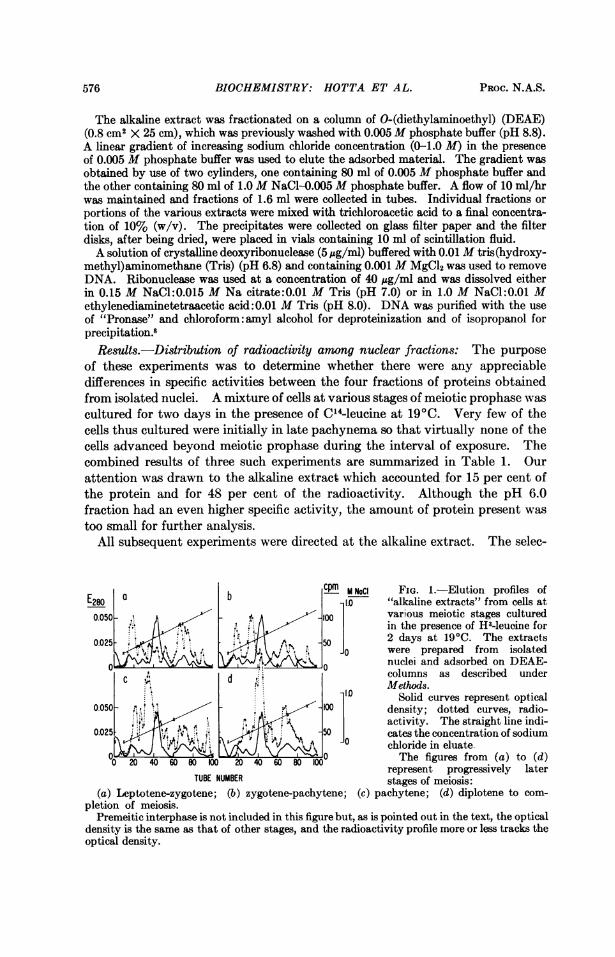
The alkaline extract was fractionated on a column of O-(diethylaminoethyl) (DEAE)(0.8 cm2 X 25 cm), which was previously washed with 0.005M phosphate buffer (pH 8.8).A linear gradient of increasing sodium chloride concentration (0-1.0 M) in the presenceof 0.005 M phosphate buffer was used to elute the adsorbed material. The gradient wasobtained by use of two cylinders, one containing 80 ml of 0.005 M phosphate buffer andthe other containing 80 ml of 1.0M NaCl-0.005 M phosphate buffer. A flow of 10 ml/hrwas maintained and fractions of 1.6 ml were collected in tubes. Individual fractions orportions of the various extracts were mixed with trichloroacetic acid to a final concentra-tion of 10% (w/v). The precipitates were collected on glass filter paper and the filterdisks, after being dried, were placed in vials containing 10 ml of scintillation fluid.A solution of crystalline deoxyribonuclease (5 ,gg/ml) buffered with 0.01M tris(hydroxy-
methyl)aminomethane (Tris) (pH 6.8) and containing 0.001 M MgCl2 was used to removeDNA. Ribonuclease was used at a concentration of 40 jug/ml and was dissolved eitherin 0.15 M NaCl:0.015 M Na citrate:0.01 M Tris (pH 7.0) or in 1.0 M NaCl:0.01 Methylenediaminetetraacetic acid:0.01 M Tris (pH 8.0). DNA was purified with the useof "Pronase" and chloroform:amyl alcohol for deproteinization and of isopropanol forprecipitation.8
Results.-Distribution of radioactivity among nuclear fractions: The purposeof these experiments was to determine whether there were any appreciabledifferences in specific activities between the four fractions of proteins obtainedfrom isolated nuclei. A mixture of cells at various stages of meiotic prophase wascultured for two days in the presence of C'4-leucine at 19'C. Very few of thecells thus cultured were initially in late pachynema so that virtually none of thecells advanced beyond meiotic prophase during the interval of exposure. Thecombined results of three such experiments are summarized in Table 1. Ourattention was drawn to the alkaline extract which accounted for 15 per cent ofthe protein and for 48 per cent of the radioactivity. Although the pH 6.0fraction had an even higher specific activity, the amount of protein present wastoo small for further analysis.
All subsequent experiments were directed at the alkaline extract. The selec-
E280a b cm M NaCI FIG. 1.-Elution profiles ofE80 - o1.0 "alkaline extracts" from cells at
0.OS0 .' . A A -100various meiotic stages cultured' il,<:; s . . [1 in the presence of H3-leuoine for
0.025 .,,.,,"' st z _ S0 2 days at 19°C. The extractsJ1-o were prepared from isolated0W9 /\ 90nuclei and adsorbed on DEAE-
0 0.d , columns as described underc d ~~~~~~~~'~~Methods.1..0 Solid curves represent optical
0.050 100 density; dotted curves, radio-4.45&g« , ,<> 50 activity. The straight line indi-
0.025- 50 catesthe concentration of sodium4.. v ~~~~~~~ chloride in eluate.
&0 The figures from (a) to (d)represent progressively later
TUBE NUMBER stages of meiosis:(a) Leptotene-zygotene; (b) zygotene-pachytene; (c) pachytene; (d) diplotene to com-
pletion of meiosis.Premeitic interphase is not included in this figure but, as is pointed out in the text, the optical
density is the same as that of other stages, and the radioactivity profile more or less tracks theoptical density.
PRoc. N.A.S.576

BIOCHEMISTRY: HOTTA ET AL.
TABLE 1. Distribution of radioactivity in nuclear proteins of cells in meiotic prophase.Protein content Specific activity Radioactivity
Nuclear fraction (% of total) (cpm/pig protein) (% of total)Alkaline (pH 8.0) 15.2 37.6 48.2pH 6.0 0.6 42.7 2.11.0 M NaCl 16.3 12.8 17.2Residual 67.9 6.8 33.4
Cells in the zygotene and pachytene stages of meiosis were cultured for 2 days at 19'C in thepresence of 0.25 Ac of C'4-leucine/ml. The nuclei were isolated and fractionated as described underMethods. Attention is drawn to the alkaline fraction that contains about half of the radioactivity.All subsequent experiments reported here are concerned with this fraction.
tion was necessarily arbitrary, since no criteria were available for assigningsignificance to any particular protein or group of proteins in relation to chromo-somal behavior during meiotic prophase. The value of this selection for anunderstanding of meiotic events will become apparent from the results.
Protein synthesis in relation to meiotic stage: A satisfactory, but by no meanscomplete, resolution of the proteins present in the alkaline extract was obtainedby the use of DEAE-columns. With a linearly increasing concentration of so-dium chloride as eluant, several peaks of protein concentration were observed(Fig. 1). The elution pattern was the same for all stages of meiosis and also forthe premeiotic cells. Thus, no gross changes in composition of the alkaline ex-tract were observed during the progression of the cells from premeiotic interphaseto completion of meiosis. The apparent constancy in protein composition wasnot reflected, however, in the profiles of radioactivity. Only in the case of cellsexposed to labeled amino acids during the interval of premeiotic mitosis did theprofile of radioactivity track that of optical density.The respective profiles of radioactivity in proteins obtained from cells at
different meiotic intervals are shown in Figure 1. A striking feature of theprofiles is the elution of a major peak of radioactivity at a salt concentration ofabout 0.2 M. This peak was found only in cells that had been exposed to isotopeduring the zygotene-pachytene intervals. This peak was pronounced in extractsfrom pachytene cells (Fig. ic). Another peak of radioactivity eluting at0.08 M salt was present in extracts from cells in early zygotene through pachytene(Fig. la-c) but not in postpachytene cells. Other differences in radioactivityprofiles may also be noted but the actual differences between successive meioticstages are probably blurred to some extent because of the overlapping of stagesbetween the groups tested. The most important inference that may be drawnfrom these results is the occurrence of a progressive change in patterns of pro-tein synthesis paralleling the progress of the cells through meiosis. The changeis marked by a distinctive pattern of synthesis during the zygotene-pachyteneinterval. The pattern is reproducible and is virtually identical in the two vari-eties of lily tested. The fact that the major peaks of radioactivity do not coincidewith those of optical density during the prophase stages (Fig. la-c) suggests thatthe principal syntheses occur in certain quantitatively minor components. It isduring these same stages that pairing and chiasma formation occur and thatquantitatively minor components of DNA are synthesized.6' 9
Relationship between DNA and protein synthesis during meiotic prophase: The
VOL. 60, 1968 5,77

BIOCHEMISTRY: HOTTA ET AL.
TABLE 2. Distribution of protein and DNA between alkaline and other nuclear fractions inzygotene-pachytene cells.
Protein DNAExtraction content content H3-DNA H3-DNA
Fraction method (%) (%) (total cpm) (cpm//sg)"Alkaline" "Complete" 15.2 5.5 3134 111
"Mild" 3.25 0.7 1983 302
Remainder "Complete" 84.8 94.5 3732"Mild" 96.75 99.3 4663
The distribution of total protein and DNA was determined in three separate experiments. For"mild" extraction, the nuclear suspension was homogenized with 5 strokes of the pestle at a settingof 50 v (see Method8). For "complete" extraction, three successive extractions were made, each with20 strokes of the pestle and at a setting of 90 v. This latter method gave similar results to passageof the nuclear suspension through a French press. The low contents of DNA and protein in the mildalkaline extract showed appreciable variations between experiments (2.5-5% for protein and 0.25-1.0% for DNA), but the average of these is recorded in the table. Radioactivities of proteins underconditions of complete extraction are shown in Table 1. In this table only the data on H3-thymidineincorporation have been included. In the actual experiment zygotene and pachytene cells wereexposed simultaneously to C14lleucine and H3-thymidine for 2 days at 191C.
coincidence of nuclear protein and DNA syntheses in zygotene-pachytene cellspoints to the possibility that the two syntheses might be functionally interre-lated. To test this possibility, zygotene and pachytene cells were incubated inthe presence of C14-leucine and H3-thymidine. Nuclei were then isolated andthe distribution of each of the labels among the nuclear fractions was determined.The results of this experiment are summarized in Table 2. Of major interest isthe observation that about half of the DNA synthesized during zygotene andpachytene stages is extracted at pH 8.0, even though about 95 per cent of thetotal DNA remains unextracted. The precise significance of the actual pro-portion of labeled DNA that is extracted in the alkaline medium is unclear.Neither labeled protein nor DNA can be extracted from the nuclei prior to theirdisruption. As illustrated in Table 2, the yield of labeled material partly de-pends upon the degree of nuclear disruption. It is reasonably clear, however,that the DNA synthesized during meiotic prophase is selectively extractedby the same procedure that selectively extracts certain nuclear proteins withhigh specific activity. Although milder disruption of nuclei yields lower amountsof labeled DNA, the specific activity of that DNA is much higher. I\Iore vig-
FIG. 2.-Elution profile of alkaline extract0.2 - -200 from zygotene-pachytene nuclei. Cells were
cultured in the presence of H3-thymidine and, C14-leucine at 19'C. and the extract ad-
,, X sorbed on 'a DEAE-column. Conditions of° 0.1-100 W elution are described under Methods. Solidcurve with solid circles represents optical
t density. Solid curve with open circles repre-> sents HI-DNA activity, and dotted curve
0 0020030 4 represents C4-leucine activity. The opticaldensity profile is different from that shown in
TUBE NUMBER Fig. 1 because readings are recorded at 260rather than 280 mju. Profile of eluate between tubes 55 and 100 has been omitted because noDNA counts have been found in that region; H3-counts do occur but they are entirely due toRNA, In this particular fractionation 1.3-ml rather than 1.6-ml samples were collected.
578 PROC. N.A.S.

BIOCHEMISTRY: HOTTA ET AL.
FIG. 3.-Distribution of la-beled DNA and protein from .4alkaline extracts of zygotene-pachytene nuclei in a CsCl;gradient following centrifugation - -for 60 hr at 73,500 X g. The LJ--dub -regression lines represent thedensity (gm/cc) of the fractionsin the collecting tubes. The solid 7-curve is the optical densityprofile. Dashed curve is pro- 4C 8,OC 40 613 VCfile of HI-DNA activity anddotted curve is profile of C"t- - uE NLSTERprotein activity. (a) Nativeextract; (b) extract adjusted to pH 13.0 and allowed to stand for 20 min at room temperatureprior to addition of CsCl solution and subsequent centrifugation. The shaded portion repre-sents the profile of native DNA that was added to the mixture just prior to centrifugation.
orous disruption of nuclei renders a higher proportion of the unlabeled DNA ex-tractable at pH 8.0.
In order to determine whether any association exists between the prophase-labeled DNA and protein, the extracts were examined by column chromatog-raphy and by centrifugation in solutions of CsCl. The elution profile of thedoubly labeled material is shown in Figure 2. Most of the labeled DNA iscollected in tubes 20-40 and the curve of tritium activity tracks that of_ C4-leucine. This is the region of the labeled protein profile which is most prominentin extracts of pachytene nuclei (Fig. lb and c). A high peak of tritium activity(not shown in the curve) occurs between tubes 50-100 but it is not due to DNA.It is unaffected by DNase and is completely removed by RNase. This latterresult is in line with other unpublished observations in our laboratory on theincorporation of label from thymidine into RNA. The nature of the associationbetween RNA and nuclear protein has not been pursued in these studies. Ofdirect relevance to the present study is the fact that those nuclear proteins thatare distinctively synthesized during the zygotene-pachytene interval behave on aDEAE-column as though they were associated with the DNA that is alsodistinctively synthesized during that same interval.6
Centrifugation of the alkaline extract in the presence of CsCl also reveals thepresence of a complex between labeled protein and DNA (Fig. 3a). However,
FIG. 4.-Profile of radioactivityof DNA purified from alkaline ex-tract obtained from zygotene- y H3 C14pachytene nuclei cultured in the 0.2 400 -10presence of H'-thymidine and C14- . ileucine. DNA was prepared as -described under Methods. Solidcurve represents the optical density 0 - 200--50 Xprofile of marker DNA added just -prior to centrifugation. Dashedcurve represents H3-activity. Thedots along the bottom line represent 30C"Lactivity and indicate the ab- 40 60 80sence of labeled protein in the puri- TUBE NUMBERfied preparation.
VOL. 60, 1968 579

BIOCHEMISTRY: HOTTA ET AL.
TABLE 3. Effect of cycloheximide (CHI) on synthesis of nuclear protein during zygotene-pachytene stages.
Specific Activity (cpm/,jg) Per centFraction - CHI +CHI inhibition
Alkaline 37.6 13.1 65.2pH 6.0 42.7 2.0 53.61.0 M NaCl 12.8 13.9 52.4Residual 6.8 52.5 23.5
Conditions of culture were the same as those described under Table 1. The columns marked"+CHI" represent the values obtained for cells that had been cultured in the presence of 0.5 usg/mlof cycloheximide and C14-leucine.
the proportion of labeled protein apparently complexed with the labeledDNA in the CsCl gradient is much less than that observed with column chro-matography. About 30 per cent of the protein count is found in the DNAregion of the gradient. Four peaks of protein radioactivity match the positionsof the four peaks of DNA radioactivity. If the extract is denatured with alkaliprior to centrifugation, the protein counts are lost from the DNA region and theDNA peaks are found in positions expected for the denatured product (Fig. 3b).The amount of protein associated with each of the native DNA peaks may beestimated by comparing Figure 3a with the profile of DNA that has been purifiedfrom the alkaline extract (Fig. 4). These estimates, however, are unlikely tohave much value except for indicating that the protein content is of the order of0-12 per cent, and that each of the peaks probably has a different protein con-tent. For present purposes it is sufficient to point out that the radioactiveprofile of the DNA prepared from the alkaline extract is the same as that reportedfor DNA prepared from whole zygotene-pachytene cells,6 and that this DNAremains associated with protein synthesized during the zygotene-pachyteneinterval. The physical association may imply a functional relationship.
Significance of protein synthesis to meiotic development: The relevance ofprotein synthesis to meiotic development was examined by culturing meioticcells in the presence of cycloheximide. At concentrations of the drug exceeding2 pig/ml, protein synthesis is virtually abolished and meiotic development iscompletely arrested. The responses of the cells to concentrations in the range of0.2-1.0 .g/ml are more interesting. At these lower concentrations of cyclohexi-mide, inhibition is incomplete and, as will be described elsewhere,'0 meiotic de-velopment is only partially suppressed. The inhibition of protein synthesisappears to be selective. Generally, the proteins in the alkaline fraction are moresensitive than those of the other fractions to 0.5 ug/ml of cycloheximide (Table3). iMioreover, a selective effect is also apparent within the alkaline fraction.The peaks of radioactivity that are characteristic of zygotene-pachytene cells areabolished by 0.5 Ag/ml of cycloheximide. The remaining radioactivity tracksthe optical density profile in a pattern that is more or less characteristic ofinterphase nuclei (Fig. 5). The cytological consequences of partially inhibitingproteinwsynthesis depend upon the particular stage at which the cells are exposedto the inhibitor and the duration of exposure. By appropriately timing theinterval of exposure it can be shown that selective inhibition of protein synthesisat the end of zygonema causes a failure of chiasma formation.'l It would thus
PROc. N.A.S.580

BIOCHEMISTRY: HOTTA ET AL.
0.
co 0.I
H3 p322 _- O 10"0-0200
50 I00o
40 50 60 70
TUBE NUMBER
FIG. 5.-Effect of cycloheximideon synthesis of nuclear protein inalkaline extract from zygotene-pachytene cells. Conditions of cul-ture are the same as those describedunder Fig. 1. Tritiated leucinewas used in these experiments.The alkaline extract from theisolated nuclei was adsorbed on aDEAE-column and eluted as de-scribed. The straight line indicatesmolarity of NaCl in eluate. Solidcurve represents optical density.Dotted curve represents radio-activity profile of control anddashed curve represents radio-activity profile of extract from cellscultured in the presence of 0.5 ,g/mlof cyclohexintide.
FIG. 6.-Effect of cycloheximide onDNA synthesis in zygotene-pachytenecells. One group of cells (dotted curve) wascultured from the zygotene stage for 5 daysin the presence of 0.2 pg/ml of cyclohexi-mide. At the end of this period themedium was replaced with standardmedium containing H3-thymidine andcultured for 2 more days. The secondgroup of cells (dashed curve) was culturedfor 7 days in the presence of P32-phosphateand 0.2 pg/ml of cycloheximide. DNAwas prepared from each of the groupsas described under Methods, and the twopreparations were combined for analysisin a CsCl gradient. Solid curve representsoptical density. Regression line repre-sents density (gm/cc) of fractions.
appear that the proteins which are distinctively synthesized during the latezygonema-early pachynema are essential to formation of chiasmata.A prominent biochemical consequence of exposure to cycloheximide is the
arrest of DNA synthesis. Data pointing to this conclusion are shown in Figure6. Concentrations of cycloheximide as low as 0.2 ,jg/ml markedly suppressDNAsynthesis. The dashed curve in Figure 6 shows the pattern of synthesis inzygotene-pachytene cells during a seven-day exposure to P32-phosphate andcycloheximide. Much the same pattern of synthesis is observed if the cells areharvested following a three-day exposure. DNA synthesis is thus brought to a
halt at least within three days of exposure to cycloheximide. The inhibition isentirely reversible as illustrated by the dotted curve. The pattern of DNAsynthesis in cells that had been exposed for five days to cycloheximide and thencultured for two days in a normal medium is the same as in untreated cells.These observations permit the conclusion that the synthesis of DNA during
the zygotene-pachytene stages requires the simultaneous synthesis of certainnuclear proteins. Whether the required proteins are those that are physicallyassociated with the DNA is open to conjecture. One obvious implication ofthis requirement is that those processes which have been shown to depend upon
E280
M NoCI1.0
10
TUBE NUMBER
VOL. 60, 1968 581

BIOCHEMISTRY: HOTTA ET AL.
prophase DNA synthesis9 must, at least indirectly, depend upon prophaseprotein synthesis.Discussion.-The experiments reported here represent no more than an
arbitrary entry into the general problem of protein functions during meioticprophase. The results are gratifying, inasmuch as they do offer convincingevidence that synthesis of certain nuclear proteins is essential to the synthesis ofDNA and that the combination of these syntheses that occur during zygonlemaand pachynema is essential to chromosome pairing and chiasma formation. Avariety of speculative schemes is made possible by these observations, but theirpursuit is probably of little value in the absence of more precise informationconcerning the nature of the proteins synthesized. The apparent physicalassociation between the protein and DNA synthesized during meiotic prophaseis no more than a pointer to a possible structural relationship between them.The principal conclusion that may be drawn from the present studies is that therespective syntheses of DNA and protein are functionally interrelated.
* This work was supported by a grant from the National Science Foundation (NSF-GB-5173x) and by supplementary assistance from the Institute for Studies in DevelopmentalBiology (USPHS-HD03015 and NSF GB 6476).
t Present address: Department of Biological Science, University of Delaware, Newark,Delaware. Holder of NIH postdoctoral fellowship.
1 Taylor, J. H., Am. J. Botany, 46, 477-484 (1959).2 Hotta, Y., and H. Stern, J. Cell Biol., 19, 45-58 (1963).3Ansley, H. R., J. Biophys. Biochem. Cytol., 4, 59-62 (1958).4Sheridan, W. F., and H. Stern, Exptl. Cell Res., 45, 323-335 (1967).5 Ito, M., and H. Stern, Devel. Biol., 16, 36-53 (1967).6 Hotta, Y., M. Ito, and H. Stern, these PROCEEDINGS, 56, 1184-1191 (1966).7Hotta, Y., and H. Stern, Protoplasma, 60, 218-232 (1965).8 Hotta, Y., A. Bassel, and H. Stern, J. Cell Biol., 27, 451-457 (1965).9 Stern, H., and Y. Hotta, in The Control of Nuclear Activity, ed. L. Goldstein (New York:
Prentice Hall, 1967), pp. 47-76.'0 Parchman, L. G., and H. Stern, manuscript in preparation.
582 PROC. N.A.S.