Durable intermediate- to long-term outcomes after ... · It is likely that pathophysiology of acute...
11
AUTHOR PROOF COPY Not for publication © 2016 Sachs et al. This work is published and licensed by Dove Medical Press Limited. The full terms of this license are available at https://www.dovepress.com/terms. php and incorporate the Creative Commons Attribution – Non Commercial (unported, v3.0) License (http://creativecommons.org/licenses/by-nc/3.0/). By accessing the work you hereby accept the Terms. Non-commercial uses of the work are permitted without any further permission from Dove Medical Press Limited, provided the work is properly attributed. For permission for commercial use of this work, please see paragraphs 4.2 and 5 of our Terms (https://www.dovepress.com/terms.php). Medical Devices: Evidence and Research 2016:9 1–11 Medical Devices: Evidence and Research Dovepress submit your manuscript | www.dovepress.com Dovepress 1 ORIGINAL RESEARCH open access to scientific and medical research Open Access Full Text Article 109276 Durable intermediate- to long-term outcomes after minimally invasive transiliac sacroiliac joint fusion using triangular titanium implants Donald Sachs 1 Don Kovalsky 2 Andy Redmond 3 Robert Limoni 4 S Craig Meyer 5 Charles Harvey 6 Dimitriy Kondrashov 7 1 Center for Spinal Stenosis and Neurologic Care, Lakeland, FL, 2 Orthopaedic Center of Southern Illinois, Mount Vernon, IL, 3 Precision Spine Care, Tyler, TX, 4 BayCare Clinic, Green Bay, WI, 5 Columbia Orthopaedic Group, Columbia, MO, 6 Riverside Medical Center, Kankakee, IL, 7 SF Spine Group at St Mary’s Spine Center, San Francisco, CA, USA Background: Sacroiliac joint (SIJ) fusion (SIJF), first performed 95 years ago, has become an increasingly accepted surgical option for chronic SIJ dysfunction. Few studies have reported intermediate- or long-term outcomes after SIJF. Objective: The objective of this study is to determine patient-based outcomes after SIJF for chronic SIJ dysfunction due to degenerative sacroiliitis or SIJ disruption at ≥3 years of follow-up. Methods: Consecutive patients who underwent SIJF prior to December 2012 were contacted over phone or through email. Participants completed questionnaires in clinic, over phone or by email, regarding SIJ pain, activities related to SIJ dysfunction, and the Oswestry Disability Index. Charts were reviewed to extract baseline parameters and the clinical course of follow-up. Results: One hundred seven patients were eligible and participated in this study. Mean (SD) preoperative SIJ pain score was 7.5 (1.7). At mean follow-up of 3.7 years, the mean SIJ pain score was 2.6 (representing a 4.8-point improvement from baseline, P<0.0001) and the mean Oswestry Disability Index was 28.2. The ability to perform activities commonly impaired by SIJ dysfunc- tion showed positive improvements in most patients. SIJ revision surgery was uncommon (five patients, 4.7%). Fourteen patients (13.1%) underwent contralateral SIJF during follow-up. Of those, 25.2% of patients had additional non-SIJ-related lumbar spine or hip surgeries during follow-up. Conclusion: In intermediate- to long-term follow-up, minimally invasive transiliac SIJF was associated with improved pain, low disability scores, and improved ability to perform activities of daily living. Keywords: sacroiliac joint fusion, chronic low back pain, multicenter study Background The sacroiliac joint (SIJ) transfers force from the spine to the pelvis. It has a dual structure, with the upper part of the joint being ligamentous and the lower part of the joint being a true synovial joint. The SIJ moves in several planes, with the largest plane of motion being sagittal. In this plane, the SIJ has been reported to have 2–4 degrees of flexion–extension (nutation–counternutation) with less motion in lateral bending and internal/external rotation. 1 The synovial part of the joint can undergo osteoarthritic changes, including joint space narrowing, osteophyte formation, subchondral sclerosis, and cyst formation. 2 Inadequate functioning of the SIJ and its associated musculature, as well as deterioration of the joint capsule and surrounding ligaments, results in increased stresses, pathologic motion, and altered biomechanics, causing chronic pain of the buttocks, lower back, as well as thigh and legs. 3 As early as the 1800s, the SIJ was thought to explain a significant proportion of all low back pain. 4 The SIJ has been Correspondence: Donald Sachs Center for Spinal Stenosis and Neurologic Care, 4310 South Florida Avenue, Lakeland, FL 33813, USA Tel +1 863 606 5937 Email drsachs@floridainlinespine.com
Transcript of Durable intermediate- to long-term outcomes after ... · It is likely that pathophysiology of acute...

AUTHOR
PROOF
COPY
Not for
publication
© 2016 Sachs et al. This work is published by Dove Medical Press Limited, and licensed under a Creative Commons Attribution License. The full terms of the License are available at http://creativecommons.org/licenses/by/4.0/. The license permits unrestricted use, distribution,
and reproduction in any medium, provided the original author and source are credited.
© 2016 Sachs et al. This work is published and licensed by Dove Medical Press Limited. The full terms of this license are available at https://www.dovepress.com/terms. php and incorporate the Creative Commons Attribution – Non Commercial (unported, v3.0) License (http://creativecommons.org/licenses/by-nc/3.0/). By accessing the work
you hereby accept the Terms. Non-commercial uses of the work are permitted without any further permission from Dove Medical Press Limited, provided the work is properly attributed. For permission for commercial use of this work, please see paragraphs 4.2 and 5 of our Terms (https://www.dovepress.com/terms.php).
Medical Devices: Evidence and Research 2016:9 1–11
Medical Devices: Evidence and Research Dovepress
submit your manuscript | www.dovepress.com
Dovepress 1
O R I G I N A L R E S E A R C H
open access to scientific and medical research
Open Access Full Text Article
109276
Durable intermediate- to long-term outcomes after minimally invasive transiliac sacroiliac joint fusion using triangular titanium implants
Donald Sachs1 Don Kovalsky2 Andy Redmond3
Robert Limoni4 S Craig Meyer5 Charles Harvey6
Dimitriy Kondrashov7
1Center for Spinal Stenosis and Neurologic Care, Lakeland, FL, 2Orthopaedic Center of Southern Illinois, Mount Vernon, IL, 3Precision Spine Care, Tyler, TX, 4BayCare Clinic, Green Bay, WI, 5Columbia Orthopaedic Group, Columbia, MO, 6Riverside Medical Center, Kankakee, IL, 7SF Spine Group at St Mary’s Spine Center, San Francisco, CA, USA
Background: Sacroiliac joint (SIJ) fusion (SIJF), first performed 95 years ago, has become
an increasingly accepted surgical option for chronic SIJ dysfunction. Few studies have reported
intermediate- or long-term outcomes after SIJF.
Objective: The objective of this study is to determine patient-based outcomes after SIJF for
chronic SIJ dysfunction due to degenerative sacroiliitis or SIJ disruption at ≥3 years of follow-up.
Methods: Consecutive patients who underwent SIJF prior to December 2012 were contacted
over phone or through email. Participants completed questionnaires in clinic, over phone or
by email, regarding SIJ pain, activities related to SIJ dysfunction, and the Oswestry Disability
Index. Charts were reviewed to extract baseline parameters and the clinical course of follow-up.
Results: One hundred seven patients were eligible and participated in this study. Mean (SD)
preoperative SIJ pain score was 7.5 (1.7). At mean follow-up of 3.7 years, the mean SIJ pain score
was 2.6 (representing a 4.8-point improvement from baseline, P<0.0001) and the mean Oswestry
Disability Index was 28.2. The ability to perform activities commonly impaired by SIJ dysfunc-
tion showed positive improvements in most patients. SIJ revision surgery was uncommon (five
patients, 4.7%). Fourteen patients (13.1%) underwent contralateral SIJF during follow-up. Of those,
25.2% of patients had additional non-SIJ-related lumbar spine or hip surgeries during follow-up.
Conclusion: In intermediate- to long-term follow-up, minimally invasive transiliac SIJF was
associated with improved pain, low disability scores, and improved ability to perform activities
of daily living.
Keywords: sacroiliac joint fusion, chronic low back pain, multicenter study
BackgroundThe sacroiliac joint (SIJ) transfers force from the spine to the pelvis. It has a dual
structure, with the upper part of the joint being ligamentous and the lower part of the
joint being a true synovial joint. The SIJ moves in several planes, with the largest plane
of motion being sagittal. In this plane, the SIJ has been reported to have 2–4 degrees
of flexion–extension (nutation–counternutation) with less motion in lateral bending
and internal/external rotation.1 The synovial part of the joint can undergo osteoarthritic
changes, including joint space narrowing, osteophyte formation, subchondral sclerosis,
and cyst formation.2 Inadequate functioning of the SIJ and its associated musculature,
as well as deterioration of the joint capsule and surrounding ligaments, results in
increased stresses, pathologic motion, and altered biomechanics, causing chronic pain
of the buttocks, lower back, as well as thigh and legs.3 As early as the 1800s, the SIJ
was thought to explain a significant proportion of all low back pain.4 The SIJ has been
Correspondence: Donald SachsCenter for Spinal Stenosis and Neurologic Care, 4310 South Florida Avenue, Lakeland, FL 33813, USA Tel +1 863 606 5937 Email [email protected]
Journal name: Medical Devices: Evidence and ResearchArticle Designation: ORIGINAL RESEARCHYear: 2016Volume: 9Running head verso: Sachs et alRunning head recto: Three-year outcomes of SIJFDOI: http://dx.doi.org/
sivasankaran
Sticky Note
Marked set by sivasankaran
sivasankaran
Sticky Note
Marked set by sivasankaran
sivasankaran
Sticky Note
Marked set by sivasankaran
sivasankaran
Sticky Note
Marked set by sivasankaran
sivasankaran
Sticky Note
Marked set by sivasankaran
sivasankaran
Sticky Note
Marked set by sivasankaran
sivasankaran
Sticky Note
Marked set by sivasankaran
sivasankaran
Sticky Note
Marked set by sivasankaran
sivasankaran
Sticky Note
Marked set by sivasankaran
djcher
Text Box
PROOF

Medical Devices: Evidence and Research 2016:9submit your manuscript | www.dovepress.com
Dovepress
Dovepress
2
Sachs et al
demonstrated to have both mechanoreceptors5 and nocicep-
tive receptors.6 It has a rather complex innervation, with
contribution from lateral branches of multiple lumbosacral
nerve roots. Pressurization of the SIJ in healthy volunteers
can elicit pain7 and anesthetics applied to the exiting dorsal
sacral nerve roots block sensation outside of the joint but not
pain elicited by joint pressurization.8
The SIJ is thought to explain 15%–23% of all chronic
low back pain.9,10 However, the exact prevalence is unknown,
at least in part due to the lack of a universally agreed-upon
diagnostic standard. The SIJ may explain an even larger
proportion of pain in patients who have had prior lumbar
spine fusion.9–11 Being immediately below the lumbosacral
junction, it falls into the spectrum of adjacent segment
disease after prior lumbar arthrodesis. Radiographic find-
ings of degeneration in the SIJ are common,12 both on CT
and MRI, and are not necessarily predictive of the presence
of SIJ pain. This is similar to the presence of radiographic
degenerative disk disease in both cervical and lumbar spines
in asymptomatic volunteers.13 Moreover, potentially due to
the nonaxial compressive forces through the SIJ, chronic
SIJ dysfunction can occur in patients with ligament and/
or capsular failure. MRI and CT scan may not show classic
articular cartilage deteriorations and/or degenerative patterns
seen in other joints.
Acute SIJ pain is fairly common and frequently tran-
sient, with most patients requiring either observation alone
or simple measures such as physical therapy, nonsteroidal
anti-inflammatory drugs, sacroiliac belts, exercise, chiropractic
treatment, and sacroiliac blocks. However, there is very little
evidence to support the effectiveness of nonsurgical interven-
tions for long-term treatment of chronic established debilitating
SIJ pain. It is likely that pathophysiology of acute and chronic
SIJ pain is quite different. Two blinded, controlled trials of RF
ablation of lateral branches of sacral nerve roots have shown
only short-term improvement in pain;14,15 a 12-month follow-
up study showed a modest long-term response rate following
this treatment.16 No high-quality study reporting long-term
outcomes has been published. Consequently, US Medicare
routinely does not reimburse for this RF ablation procedure.
SIJ fusion (SIJF) was first described in the 1920s.17 A
variety of approaches have been reported, including anterior,
posterior, and lateral transiliac. SIJF thus preceded the first
reports of lumbar discectomy for disk herniations by about
a decade.18 The original reports of SIJF included a number
of patients with infection-related SIJ pain (including TB)
and subsequent reports have included patients with arthritic
conditions as well. Several single-center retrospective reports
have suggested that open SIJF may be effective for the treat-
ment of pain in this patient population.19–24 Regardless of the
approach, open fusions of the SIJ were quite invasive and
associated with long hospital stays and recovery times, high
nonunion rates (9%–41%), 21,25,26 poor long-term results, and
low levels of satisfaction.27 They also required prolonged
periods of immobilization to achieve solid arthrodesis, mostly
due to lack of adequate internal fixation techniques.
In the past decade, there has been a resurgence of inter-
est in the SIJ as the pain generator in a substantial number
of patients requiring surgical interventions. Several device
systems are now commercially available for minimally
invasive SIJF, and the minimally invasive approach is now
used in 90% of cases.28 Most of the reported literature
describe patients treated with triangular titanium implants
(iFuse Implant System; SI-BONE, Inc., San Jose, CA,
USA) placed via a lateral transiliac approach. The current
surgical literature for this system includes single-center
retrospective cohorts,29–35 a combined multicenter analy-
sis,36 and three comparative studies of open and minimally
invasive approaches.37–39 A prospective randomized trial
of minimally invasive SIJF vs nonsurgical management
showed improved 12-month outcomes after SIJF compared
with those after nonsurgical management,40 and a single-
arm multicenter trial showed similar 12-month outcomes.41
Herein, we report intermediate- to long-term (3+ years)
outcomes after SIJF.
MethodsWe report a retrospective cohort study with a prospective
evaluation component conducted at seven centers (each with
one surgeon) in the US. The study was sponsored by the
device manufacturer (SI-BONE, Inc.). The study includes
patients at one center (DS), which has been previously
reported.36 All centers obtained institutional review board
approval prior to participation, and all participants signed a
study-specific consent form.
Eligible patients were adults (at least of age 21 years)
who underwent SIJF using the iFuse Implant System prior
to December 2012, whose charts documented preoperative
pain scores, and who provided consent to complete ques-
tionnaires. Unified criteria to diagnose SIJ dysfunction were
not used, as this study was retrospective in nature. However,
diagnosis at all sites was made on the basis of history (but-
tocks pain with optional radiation into the groin or upper
leg), typical pain reproduced on at least three physical
examination maneuvers, and a confirmatory diagnostic
anesthetic block of the SIJ producing acute pain relief.
sivasankaran
Sticky Note
Marked set by sivasankaran
sivasankaran
Sticky Note
Marked set by sivasankaran
sivasankaran
Sticky Note
Marked set by sivasankaran
sivasankaran
Sticky Note
Marked set by sivasankaran
sivasankaran
Sticky Note
Marked set by sivasankaran
djcher
Text Box
PROOF
Medical Devices: Evidence and Research 2016:9 submit your manuscript | www.dovepress.com
Dovepress
Dovepress
3
Three-year outcomes of SIJF
Physical examination signs are predictive of a positive SIJ
block,42 and diagnostic block is recommended by multiple
US specialty societies to diagnose SIJ pain.43–47 SIJF for all
patients was performed through a transiliac, muscle-sparing
approach, as described previously (Figure 1).29 The triangular
shape of the implant is designed to minimize rotation and
maximize surface area. The porous titanium plasma spray
coating allows biological fixation in bone. Patients had to
be willing to complete questionnaires and sign a consent
form allowing review of medical records by study person-
nel. Patients were paid nominal amounts for participating,
as approved by the governing IRB.
Chart review included abstraction of demographic details,
preoperative SIJ pain score, medical history focused on the
SIJ and lower back, and procedure details (procedure date,
side treated, and adverse events). Charts were also reviewed
for postoperative follow-up, including dates of visits, numeric
pain scale ratings assessed during the visit, global assess-
ments of health status, the occurrence of SIJ complications
and revisions, and the occurrence of new conditions related
to the spine and/or hip. Details about revision surgery were
not collected.
As per the study protocol, patients completed question-
naires in clinic, over phone or through email. Questionnaires
included SIJ pain rating using a numeric rating scale score
(0–10 scale), Oswestry Disability Index (ODI, Version 2.0),
satisfaction with surgery, and a customized survey consisting
of questions related to ability to perform various activities
compared with that prior to surgery. There was no interven-
tional aspect to this study, and no imaging was reviewed or
analyzed. All questionnaires were conducted by study site
staff.
Statistical analysis was generic and standard in nature.
Continuous variables were analyzed using mean and SD
and compared using Student’s t-test. Analysis of variance
was used to compare continuous variables across categories.
Ordinal and nominal values were tabulated and compared
with chi-square test or Fisher’s test.
ResultsOne hundred seven patients at seven centers had undergone
SIJF prior to the cutoff date and completed surveys. Base-
line demographic characteristics are presented in Table 1.
Patient age varied widely (18.6–87.0 years) and a history
of perisacral trauma (mostly falls, less commonly motor
vehicle accidents) resulting in SIJ pain was common (35
patients, 32.7%). Concomitant spine and hip disease were
common, and a large proportion of patients had undergone
Figure 1 Outlet view of pelvis with titanium implants.
Table 1 Characteristics of enrolled patients
Characteristics
Age, mean (range) 57.5 (18.6–87.0)Body mass index, mean (range) 29.8 (16.0–54.4)Years with SIJ pain, mean (range) 5.9 (0.3–46.0) <1, N (%) 17 (15.9) 1–5, N (%) 44 (41.1) 5–20, N (%) 31 (29.0) >20, N (%) 4 (3.7)Hispanic, N (%) 4 (3.7)Race, N (%) White 99 (92.5) Black 6 (5.6)Smoker, N (%) Current 28 (26.2) Former 16 (15.0)History of sacral trauma, N (%) 35 (32.7)History of physical therapy, N (%) 66 (61.7)RF ablation of branches of sacral nerve roots, N (%) 18 (16.8)SIJ steroid injections, N (%) 69 (64.5)
Abbreviations: SIJ, sacroiliac joint; RF, .
prior lumbar spine surgical procedures (36.4% had prior
lumbar fusion). Lumbar stenosis was more common in
older patients; otherwise age was not related to preoperative
historical factors.
Patients were highly debilitated by SIJ pain, as indicated
by high baseline pain ratings (mean 7.5). The duration of pain
prior to enrollment averaged 5.9 years (range 0.3–46.0 years).
Over half of the patients had undergone prior PT (although
it could not be determined whether PT was focused on the
SIJ); 1.9% had undergone SIJ steroid injections; and 2.8%
had undergone RF ablation of the SIJ nerve root branches.
Most patients underwent unilateral SIJF; 2.8% under-
went simultaneous bilateral SIJF. One patient underwent
sivasankaran
Sticky Note
Marked set by sivasankaran
sivasankaran
Sticky Note
Marked set by sivasankaran
sivasankaran
Sticky Note
Marked set by sivasankaran
sivasankaran
Sticky Note
Marked set by sivasankaran
sivasankaran
Sticky Note
Marked set by sivasankaran
sivasankaran
Sticky Note
Marked set by sivasankaran
sivasankaran
Sticky Note
Marked set by sivasankaran
sivasankaran
Sticky Note
Marked set by sivasankaran
djcher
Text Box
PROOF

Medical Devices: Evidence and Research 2016:9submit your manuscript | www.dovepress.com
Dovepress
Dovepress
4
Sachs et al
concomitant removal of a subcutaneous gluteus lesion in
the right buttock (pathology report showed calcinosis with
focal ossification). Adverse events related to SIJF were
uncommon: one patient had mild ileus postoperatively, one
had suture material extending from the wound at a second
postoperative visit, and one had an adhesive tape allergic
reaction. Five patients had SIJF revision surgery: one patient
had early postoperative neuropathic pain related to implant
malposition and underwent revision surgery at day 41. Sec-
ond patient had initial improvement in SIJ pain followed by
pain recurrence at month 18; CT scan showed no evidence
of bridging bone across the SIJ, possible loosening of the
uppermost implant and inadequate placement of the second
implant. Third patient had recurrent pain at month 6; CT
showed posterior placement of the third implant. The patient
underwent a revision surgery through an open approach with
placement of bone graft. Fourth patient had little postopera-
tive improvement and CT scan showed inadequate placement
of the caudal-most implant. The patient underwent revision
surgery ~3.3 years after index surgery and had also undergone
L5–S1 lumbar decompression with interbody fusion and
pedicle screw instrumentation for lumbar pain. Fifth patient
was injured in a motor vehicle accident (T-bone mechanism)
~9 months postoperatively; this patient underwent contralat-
eral SIJF (during which further implants were placed on the
original side) as well as a T9 laminotomy and placement of
a spinal cord stimulator.
Prospective follow-up assessments were done in the
clinic in 64 cases, by phone in 13 cases and through email
in 30 cases. Mean follow-up after SIJF was 3.7 years (range
3.0–4.7 years). Mean SIJ numeric rating pain score at
follow-up was 2.6, with a mean change of 4.8 points from
baseline (P<0.0001, Table 2). Eighty-six patients (80.4%)
had improvement in SIJ pain from baseline of at least 2
points. Mean ODI in follow-up was 28.2, indicating moder-
ate residual disability.
Satisfaction rate was 87.9% (67.3% very satisfied and
20.6% somewhat satisfied). The proportion of patients who
Table 2 Improvement in VAS SIJ pain from baseline to follow-up and follow-up ODI score
Rating Value (SD)
Baseline SIJ pain, mean (SD) 7.5 (1.7)Follow-up SIJ pain, mean (SD) 2.6 (2.7)Change score, mean (SD) -4.8 (2.9)Follow-up ODI score, mean (SD) 28.2 (21.3)
Abbreviations: SIJ, sacroiliac joint; ODI, Oswestry Disability Index.
would undergo the procedure again was 83.2% (69.2%
would “definitely” and 14.0% would “probably” undergo the
procedure again, Table 3). Patients reported improvement in
ability to perform various activities related to SIJ pain (eg,
sitting, standing, walking, ascending, and descending stairs).
Improvements in pain scores and final ODI level as a
function of baseline potential predictors were evaluated using
analysis of variance. No statistically significant relationship
was seen between improvement in SIJ pain and a history of
prior lumbar fusion, piriformis syndrome, lumbar stenosis,
degenerative disk disease, spondylolisthesis, hip osteoar-
thritis, and workers’ compensation status. Smaller changes
in mean improvement in SIJ pain (~1.4 points) were seen in
smokers vs nonsmokers (P=0.0346). Similarly, ODI at final
follow-up was ~10 points higher in smokers vs nonsmokers
(P=0.0427).
Improvements in SIJ pain at last follow-up were highly
correlated with satisfaction levels, walking compared with
prior to surgery, ability to sit for long periods, ability to work,
pain medication use, getting in and out of chair, going up and
down stairs and getting in and out of a car (all P<0.0001),
desirability of having surgery again (P=0.0021), and ability to
sleep (P=0.0004). Pain improvements were moderately cor-
related with final walking status (P=0.0686). Final ODI was
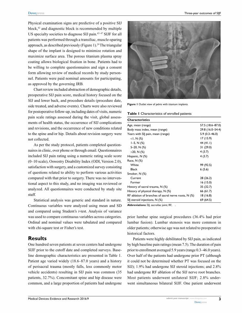
also closely related to patient responses to specific SIJ-related
questions (Figure 2). Twenty-seven (25.2%) patients had non-
SIJ lumbar spine or hip-related surgeries during follow-up
(eg, lumbar spine fusion, kyphoplasty, and hip replacement)
and 26 (24.3%) had nonsurgical procedures related to the
spine or hip (eg, rhizotomies, repeat SIJ injection, facet or
bursa injections). Patients who underwent subsequent spine
or hip surgeries had somewhat smaller improvement in SIJ
pain (–4.3 vs –5.0, P=0.2710) and somewhat higher final
ODI score (32.9 vs 26.6, P=0.1582). However, satisfaction
rates were similar across groups.
DiscussionSIJ dysfunction is an important and often overlooked chronic
health condition. It is associated with conditions commonly
treated surgically48 and was diagnosed at all sites on the
basis of typical history, physical examination findings, and a
confirmatory diagnostic anesthetic block of the SIJ produc-
ing acute pain relief. This diagnostic method is commonly
practiced in the US, is recommended by specialty societ-
ies,43–47 and was used in prospective clinical trials.49–51 Joint
fusion is a commonly performed procedure in modern spine
surgery, and evidence to support minimally invasive SIJF
sivasankaran
Sticky Note
Marked set by sivasankaran
sivasankaran
Sticky Note
Marked set by sivasankaran
sivasankaran
Sticky Note
Marked set by sivasankaran
sivasankaran
Sticky Note
Marked set by sivasankaran
sivasankaran
Sticky Note
Marked set by sivasankaran
sivasankaran
Sticky Note
Marked set by sivasankaran
sivasankaran
Sticky Note
Marked set by sivasankaran
djcher
Text Box
PROOF

Medical Devices: Evidence and Research 2016:9 submit your manuscript | www.dovepress.com
Dovepress
Dovepress
5
Three-year outcomes of SIJF
Table 3 Prospective self-reported outcomes
Measure N %
SatisfactionVery satisfied 72 67.3Somewhat satisfied 22 20.6Somewhat dissatisfied 4 3.7Very dissatisfied 8 7.5Don’t want to answer 1 0.9Have SIJ fusion again?Definitely have 74 69.2Probably have 15 14.0Probably not have 11 10.3Definitely not have 6 5.6Don’t want to answer 1 0.9Walking statusWalk without assistance 80 74.8Walk with assistive device 21 19.6Cannot walk, SIJ 2 1.9Cannot walk, something else 3 2.8Refused to answer 1 0.9Walking compared with baselineBetter 73 68.2About same 12 11.2Worse due to SIJ 10 9.3Worse due to something else 10 9.3Refused to answer 2 1.9Sitting compared with baselineBetter 69 64.5About same 21 19.6Worse due to SIJ 8 7.5Worse due to something else 7 6.5Refused to answer 2 1.9Sleeping compared with baselineBetter 63 58.9About same 23 21.5Worse due to SIJ 10 9.3Worse due to something else 10 9.3Refused to answer 1 0.9Working compared with baselineBetter 49 45.8About same 27 25.2Worse due to SIJ 9 8.4Worse due to something else 19 17.8Refused to answer 3 2.8Pain medications compared with baselineLess 70 65.4About same 25 23.4More 11 10.3Refused to answer 1 0.9Getting up from chair compared with baselineBetter 64 59.8About same 26 24.3Worse due to SIJ 5 4.7Worse due to something else 11 10.3Refused to answer 1 0.9Up and down stairs compared with baselineBetter 59 55.1About same 20 18.7
Measure N %Worse due to SIJ 10 9.3Worse due to something else 13 12.1Refused to answer 4 3.7Getting in and out of car compared with baselineBetter 61 57.0About same 24 22.4Worse due to SIJ 9 8.4Worse due to something else 12 11.2Refused to answer 1 0.9
Abbreviation: SIJ, sacroiliac joint.
using titanium implants is increasing, with a large number of
published retrospective case series,30,31,37,52–54 including some
with 4-35 and 5-year follow-up,34 comparative case series,37–39
and prospective multicenter trials.40,41 Revision rates after
SIJF are low and have decreased over time.55 Moreover, in-
trial health care utilization data from the landmark INSITE
study showed that minimally invasive SIJF is cost-effective
compared with nonsurgical treatment.56 Ignoring the SIJ
during workup of chronic low back pain may increase health
expenditures due to misdiagnosis and potentially failed lum-
bar fusion surgeries.57
Several short-term outcome studies, including a descrip-
tion of operative parameters, have been published. But for
two retrospective cohorts,34,35 little information is available
regarding long-term outcomes after SIJF. In our study, with
a minimum follow-up of 3 years, outcomes after SIJF using
titanium implants were excellent, with large improvements
in SIJ pain and only moderate residual follow-up disability
(Oswestry) scores. Mean ODI at follow-up (28.2) in the
current study was similar to 12-month values observed in
a prospective randomized trial40 (mean 28.1) and 12-month
values in a prospective multicenter single-arm study41 (mean
31.4). Although, like in most trials of spine surgery proce-
dures/devices, ODI is not restored to 0 (ie, complete absence
of disability related to back pain), our results suggest stability
of improved function over time. Self-rated improvement in
various activities of daily living associated with chronic SIJ
dysfunction was high. Satisfaction with the surgical proce-
dure was high, and most patients had the procedure again.
Improvements in SIJ pain and final ODI values were highly
correlated with responses to specific questions about activi-
ties of daily living commonly impaired in SIJ dysfunction.
Smokers appeared to have somewhat smaller improvements, a
finding common in orthopedic trials.58 This finding might be
in sync with similar findings from other fusion procedures.
Complications related to SIJF itself were uncommon and
sivasankaran
Sticky Note
Marked set by sivasankaran
djcher
Text Box
PROOF

Medical Devices: Evidence and Research 2016:9submit your manuscript | www.dovepress.com
Dovepress
Dovepress
6
Sachs et al
100 <0.0001
<0.0001 <0.0001
<0.0001
Satisfaction
Walk status
Sitting for long periods
Walking compared with prior to surgery
Have surgery again
OD
I75
50
25
Very satisfied
Walk without assistance
Better
About same
Worse due to SIJ
Worse due to something else
Sleeping
Better
About same
Worse due to SIJ
Worse due to something else
Better
About same
Worse due to SIJ
Worse due to something else
Walk with assistive device
Cannot walk, S
IJ
Cannot walk, something else
Definitely have
Probably have
Probably not have
Definitely not have
Somewhat satisfied
Somewhat dissatisfied
Very dissatisfied
0
100
OD
I
75
50
25
0
<0.0001 <0.0001100
OD
I
75
50
25
0
100
OD
I
75
50
25
0
100
OD
I
75
50
25
0
100
OD
I
75
50
25
0
Figure 2 (Continued)
sivasankaran
Sticky Note
Marked set by sivasankaran
sivasankaran
Sticky Note
Marked set by sivasankaran
djcher
Text Box
PROOF

Medical Devices: Evidence and Research 2016:9 submit your manuscript | www.dovepress.com
Dovepress
Dovepress
7
Three-year outcomes of SIJF
100 <0.0001
<0.0001
<0.0001
<0.0001
Ability to work
In and out of chair
In and out of car
Up/down stairs
Pain medications
OD
I
75
50
25
0
100
OD
I
75
50
25
0
100
OD
I
75
50
25
0
<0.0001100
OD
I
75
50
25
0
100
OD
I
75
50
25
0Better
About same
Worse due to SIJ
Worse due to something else
Better
About same
Worse due to SIJ
Worse due to something else
Better
About same
Worse due to SIJ
Worse due to something else
Better
About same
Worse due to SIJ
Worse due to something else
Less
About sameMore
Figure 2 Relationship between ODI at last follow-up and responses to specific questions related to SIJ. Notes: Dots show values for individual patients; box plots show interquartile ranges (box), whiskers show highest/lowest values within 1.5 interquartile range. Green dots are mean values. Number in upper left-hand corner shows P-value from analysis of variance.Abbreviations: ODI, Oswestry Disability Index; SIJ, sacroiliac joint.
djcher
Text Box
PROOF

Medical Devices: Evidence and Research 2016:9submit your manuscript | www.dovepress.com
Dovepress
Dovepress
8
Sachs et al
relatively minor. The reoperation rate was low and mostly
related to two factors: symptomatic implant malposition
and recurrence of symptoms due to suboptimal implant
placement and/or nonunion. Combined with the results from
prospective trials, our study indicates that minimally invasive
transiliac SIJF with triangular titanium implants is an excel-
lent treatment option for patients with SIJ dysfunction who
have failed nonsurgical treatments.
Patients in our study (and in our practices in general) were
complex, and many patients were subsequently diagnosed
with other conditions of the spine or hip and underwent either
surgical (25.2%) or nonsurgical (24.3%) procedures for such
conditions. It is likely that the same underlying pathology that
causes spine disease (eg, trauma or osteoarthritic degenera-
tion) also causes SIJ dysfunction. Although we postulated that
final ODI and satisfaction rates would be related to the need
to undergo subsequent non-SIJ surgery for these associated
conditions, we found only a modest relationship between these
factors. However, we do note that the co-occurrence of other
spine/hip conditions may result in poorer pain scores. That
is, patients with other conditions might experience pain (eg,
at Fortin’s point) that they attribute to the SIJ, which would
result in higher disability scores. Our results may therefore be
somewhat conservative with respect to overall improvement
after SIJF. Importantly, no evidence suggests that these other
hip/spine conditions are caused by SIJF. The rates of subse-
quent surgeries in prospective trials have been low. Moreover,
finite element analysis has suggested only minimal increases
in adjacent segment motion after SIJF.59 (Because the SIJ
moves only minimally during normal daily life, stabilization
and long-term fusion are not expected to increase adjacent
stresses.) Taken together, these data suggest that SIJ dysfunc-
tion can be identified in clinical practice, and SIJF can be an
effective treatment in the long term. However, many patients
are complex, with multiple pain sources, and treatment of
other lumbar spine conditions remains challenging.
Advantages of our study include its combined retro-
spective and prospective multicenter design, enrollment of
patients in different geographic areas, and various practice
types (private, teaching, hybrid, etc). These characteristics
enhance the study’s generalizability.
Our study is limited by several factors. Our study was
retrospective by design and could be subject to biases inher-
ent in this design. Some patients did not participate because
of inability to make contact or patient refusal. Although this
could have contributed a bias to our results, the directionality
of such bias is not known.
Methods to diagnose SIJ pain may have varied across sites
and time; however, the diagnostic algorithm is considered
standard, and typically included history, findings on at least
three physical examination tests that stress the SIJ, and a
confirmatory diagnostic SIJ anesthetic block.
Physical therapy is often provided to patients with chronic
low back pain, but not all patients in our cohort underwent
such treatment. However, there is no high-quality evidence
that physical therapy is effective in chronic SIJ pain.
Baseline ODI scores were not available in most patients,
limiting our ability to determine per patient improvements
in this commonly reported parameter. However, follow-up
ODI scores were similar to those reported in two prospective
multicenter US trials at 12 months.49,60 Residual ODI scores
were higher than those reported in studies of degenerative
lumbar spondylolisthesis61 or lumbar stenosis,62 but whether
this reflects patient complexity or effectiveness of SIJF is
not known.
We did not perform standardized long-term imaging of
the SIJ. In the absence of clinical signs suggestive of implant
loosening (eg, failure of SIJ pain to improve after SIJF or
pain recurrence), routine cross-sectional imaging of the SIJ
may have little, if any, clinical value. One study of the same
SIJF procedure showed a high rate of growth across the SIJ
at 5 years.34
Mean follow-up in our cohort was 3.7 years, which
represents one of the longest postoperative experiences for
this procedure reported to date. However, continued follow-
up of such patients may help to define even longer term
(8–10 years) outcomes.
Many patients in our cohort had concomitant spine dis-
ease at baseline and a substantial fraction underwent other
spine surgeries or interventional spine or hip procedures.
Such interventions may have limited improvements in ODI
or affected patients’ abilities to perform activities of daily
living. Our data do not allow us to discern whether the rate
of subsequent non-SIJ surgeries was high or low; rather these
data reflect the complexity of the patient population. Many
patients had multiple pain generators. Whether some patients
underwent SIJF when the underlying diagnosis was different
could not be determined. However, responses at 3+ years
appeared positive and consistent with improvements seen
in prospective clinical trials of SIJF.
Patients participating in this study represent the earliest
use of the iFuse device; it is possible that with increased
experience with the device, both patient selection and tech-
nical aspects related to the procedure may have improved.
sivasankaran
Sticky Note
Marked set by sivasankaran
sivasankaran
Sticky Note
Marked set by sivasankaran
djcher
Text Box
PROOF

Medical Devices: Evidence and Research 2016:9 submit your manuscript | www.dovepress.com
Dovepress
Dovepress
9
Three-year outcomes of SIJF
Finally, we did not collect data regarding opioid use in
this study. Prospective trials have shown decreases in opioid
use after minimally invasive SIJF.49,60
Our study collected outcomes of patients treated through
a transiliac lateral approach, with implants designed to resist
rotation after implantation (due to the triangular shape) and
for biological fixation in bone. Other devices are available
to perform SIJF; however, because these devices differ
from those we used, it is unknown whether our results are
applicable to such devices. Moreover, our results may not
apply to other surgical approaches to SIJF, such as direct
posterior or combined approaches, allograft-only fusions,
distraction arthrodesis using cannulated or hollow screws,
or other procedures.
ConclusionIntermediate- to long-term follow-up after lateral, transiliac
SIJF using titanium implants shows durable, clinically
important improvements in pain and disability, with high
satisfaction rates. Both complication and reoperation rates
were low. Improvements may be limited in the presence of
concomitant spine disease.
AcknowledgmentsThe authors acknowledge the Jodi Bowling, Mychelle Santos,
Natalie Kline, Katelyn Griffis, Cristy Newman, Michelle
Vogt, Denise Barnes, Lori Latham, Laurie Doredant, and
Beth Short for subject recruitment and data collection. The
authors acknowledge SI-BONE for study sponsorship and
SI-BONE staff for help with study management, statistical
analysis, and assistance with manuscript preparation. All
authors conducted clinical research as part of prospective
trials sponsored by SI-BONE.
DisclosureDoctors Harvey and Kondrashov are consultants of SI-
BONE. The other authors report no other conflicts of interest
in this work.
References 1. Sturesson B, Selvik G, Udén A. Movements of the sacroiliac joints. A
roentgen stereophotogrammetric analysis. Spine. 1989;14(2):162–165. 2. Ha K-Y, Lee J-S, Kim K-W. Degeneration of sacroiliac joint after
instrumented lumbar or lumbosacral fusion: a prospective cohort study over five-year follow-up. Spine. 2008;33(11):1192–1198.
3. Vleeming A, Schuenke MD, Masi AT, Carreiro JE, Danneels L, Willard FH. The sacroiliac joint: an overview of its anatomy, function and potential clinical implications. J Anat. 2012;221(6):537–567.
4. Erichsen JA. Lecture on the sacro-iliac disease. Lancet. 1859;73(1845): 25–27.
5. Sakamoto N, Yamashita T, Takebayashi T, Sekine M, Ishii S. An elec-trophysiologic study of mechanoreceptors in the sacroiliac joint and adjacent tissues. Spine. 2001;26(20):E468–E471.
6. Szadek KM, Hoogland PV, Zuurmond WW, de Lange JJ, Perez RS. Nociceptive nerve fibers in the sacroiliac joint in humans. Reg Anesth Pain Med. 2008;33(1):36–43.
7. Fortin J, Dwyer A, West S, Pier J. Sacroiliac joint: pain referral maps upon applying a new injection/arthrography technique. Part I: asymp-tomatic volunteers. Spine. 1994;19(13):1475–1482.
8. Dreyfuss P, Henning T, Malladi N, Goldstein B, Bogduk N. The ability of multi-site, multi-depth sacral lateral branch blocks to anesthetize the sacroiliac joint complex. Pain Med. 2009;10(4):679–688.
9. Sembrano JN, Polly DW. How often is low back pain not coming from the back? Spine. 2009;34(1):E27–E32.
10. Bernard TN, Kirkaldy-Willis WH. Recognizing specific characteristics of nonspecific low back pain. Clin Orthop. 1987;(217):266–280.
11. Liliang P-C, Lu K, Liang C-L, Tsai Y-D, Wang K-W, Chen H-J. Sac-roiliac joint pain after lumbar and lumbosacral fusion: findings using dual sacroiliac joint blocks. Pain Med. 2011;12(4):565–570.
12. Eno J-J, Boone C, Bellino M, Bishop J. The prevalence of sacroiliac joint degeneration in asymptomatic adults. J Bone Joint Surg Am. 2015;97(11):932–936.
13. Boden SD. The use of radiographic imaging studies in the evaluation of patients who have degenerative disorders of the lumbar spine. J Bone Joint Surg Am. 1996;78(1):114–124.
14. Patel N, Gross A, Brown L, Gekht G. A randomized, placebo-controlled study to assess the efficacy of lateral branch neurotomy for chronic sacroiliac joint pain. Pain Med. 2012;13(3):383–398.
15. Cohen SP, Hurley RW, Buckenmaier CC, Kurihara C, Morlando B, Dragovich A. Randomized placebo-controlled study evaluating lateral branch radiofrequency denervation for sacroiliac joint pain. Anesthesiol-ogy. 2008;109(2):279–288.
16. Patel N. Twelve-month follow-up of a randomized trial assessing cooled radiofrequency denervation as a treatment for sacroiliac region pain. Pain Pract. 2016;16(2):154–167.
17. Smith-Petersen MN. Arthrodesis of the sacroiliac joint. A new method of approach. J Bone Jt Surg. 1921;3(8):400–405.
18. Mixter W, Barr J. Rupture of the intervertebral disc with involvement of the spinal canal. N Engl J Med. 1934;211(5):210–215.
19. McGuire RA, Chen Z, Donahoe K. Dual fibular allograft dowel technique for sacroiliac joint arthrodesis. Evid Based Spine Care J. 2012;3(3):21–28.
20. Kibsgård TJ, Røise O, Stuge B. Pelvic joint fusion in patients with severe pelvic girdle pain – a prospective single-subject research design study. BMC Musculoskelet Disord. 2014;15:85.
21. Buchowski JM, Kebaish KM, Sinkov V, Cohen DB, Sieber AN, Kostuik JP. Functional and radiographic outcome of sacroiliac arthrodesis for the disorders of the sacroiliac joint. Spine J. 2005;5(5):520–528. [discus-sion 529].
22. Giannikas KA, Khan AM, Karski MT, Maxwell HA. Sacroiliac joint fusion for chronic pain: a simple technique avoiding the use of metal-work. Eur Spine J. 2004;13(3):253–256.
23. Belanger TA, Dall BE. Sacroiliac arthrodesis using a posterior midline fascial splitting approach and pedicle screw instrumentation: a new technique. J Spinal Disord. 2001;14(2):118–124.
24. Berthelot JM, Gouin F, Glemarec J, Maugars Y, Prost A. Possible use of arthrodesis for intractable sacroiliitis in spondyloarthropathy: report of two cases. Spine. 2001;26(20):2297–2299.
25. Waisbrod H, Krainick JU, Gerbershagen HU. Sacroiliac joint arthrodesis for chronic lower back pain. Arch Orthop Trauma Surg. 1987;106(4):238–240.
26. Schütz U, Grob D. Poor outcome following bilateral sacroiliac joint fusion for degenerative sacroiliac joint syndrome. Acta Orthop Belg. 2006;72(3):296–308.
27. Ashman B, Norvell D, Hermsmeyer J. Chronic sacroiliac joint pain: fusion versus denervation as treatment options. Evid Based Spine Care J. 2010;1(03):35–44.
sivasankaran
Sticky Note
Marked set by sivasankaran
sivasankaran
Sticky Note
Marked set by sivasankaran
djcher
Text Box
PROOF

Medical Devices: Evidence and Research 2016:9submit your manuscript | www.dovepress.com
Dovepress
Dovepress
10
Sachs et al
28. Lorio MP, Polly DW Jr, Ninkovic I, Ledonio CGT, Hallas K, Andersson G. Utilization of minimally invasive surgical approach for sacroiliac joint fusion in surgeon population of ISASS and SMISS membership. Open Orthop J. 2014;8:1–6.
29. Rudolf L. Sacroiliac joint arthrodesis-MIS technique with titanium implants: report of the first 50 patients and outcomes. Open Orthop J. 2012;6:495–502.
30. Sachs D, Capobianco R. Minimally invasive sacroiliac joint fusion: one-year outcomes in 40 patients. Adv Orthop. 2013;2013:536128.
31. Cummings J Jr, Capobianco RA. Minimally invasive sacroiliac joint fusion: one-year outcomes in 18 patients. Ann Surg Innov Res. 2013;7(1):12.
32. Schroeder JE, Cunningham ME, Ross T, Boachie-Adjei O. Early results of sacro–iliac joint fixation following long fusion to the sacrum in adult spine deformity. HSS J. 2013;10(1):30–35.
33. Gaetani P, Miotti D, Risso A, et al. Percutaneous arthrodesis of sacro-iliac joint: a pilot study. J Neurosurg Sci. 2013;57(4):297–301.
34. Rudolf L, Capobianco R. Five-year clinical and radiographic outcomes after minimally invasive sacroiliac joint fusion using triangular implants. Open Orthop J. 2014;8:375–383.
35. Vanaclocha VV, Verdú-López F, Sánchez-Pardo M, et al. Minimally invasive sacroiliac joint arthrodesis: experience in a prospective series with 24 patients. J Spine. 2014;3:5.
36. Sachs D, Capobianco R, Cher D, et al. One-year outcomes after minimally invasive sacroiliac joint fusion with a series of triangular implants: a multicenter, patient-level analysis. Med Devices(Auckl). 2014;7:299–304.
37. Graham Smith A, Capobianco R, Cher D, et al. Open versus minimally invasive sacroiliac joint fusion: a multi-center comparison of periop-erative measures and clinical outcomes. Ann Surg Innov Res. 2013;7 (1):14.
38. Ledonio CGT, Polly DW, Swiontkowski MF. Minimally invasive versus open sacroiliac joint fusion: are they similarly safe and effective? Clin Orthop. 2014;472(6):1831–1838.
39. Ledonio C, Polly D, Swiontkowski MF, Cummings J. Comparative effectiveness of open versus minimally invasive sacroiliac joint fusion. Med Devices (Auckl). 2014;2014(7):187–193.
40. Polly DW, Cher DJ, Wine KD, et al. Randomized controlled trial of minimally invasive sacroiliac joint fusion using triangular titanium implants vs nonsurgical management for sacroiliac joint dysfunction: 12-month outcomes. Neurosurgery. 2015;77(5):674–690.
41. Duhon BS, Cher DJ, Wine K, Kovalsky D, Lockstadt H. Triangular titanium implants for minimally invasive sacroiliac joint fusion: a prospective study. Glob Spine J. [In press].
42. Szadek KM, van der Wurff P, van Tulder MW, Zuurmond WW, Perez RS. Diagnostic validity of criteria for sacroiliac joint pain: a systematic review. J Pain. 2009;10(4):354–368.
43. Pauza KJ, Aprill C, Bogduk N, et al. Educational Guidelines for Inter-ventional Spinal Procedures. American Academy of Physical Medicine and Rehabilitation; 2008:1–48. Available from: http://www.aapmr.org/practice/guidelines/documents/edguidelines.pdf.
44. Manchikanti L, Abdi S, Atluri S, et al. An update of comprehensive evidence-based guidelines for interventional techniques in chronic spinal pain. Part II: guidance and recommendations. Pain Physician. 2013;16(2 suppl):S49–S283.
45. Bogduk N, editor. Sacroiliac joint access. Practice Guidelines for Spinal Diagnostic and Treatment Procedures. 2nd ed. San Francisco, CA: International Spine Intervention Society; 2013:523–555.
46. American Society of Anesthesiologists Task Force on Chronic Pain Man-agement, American Society of Regional Anesthesia and Pain Medicine. Practice guidelines for chronic pain management: an updated report by the American Society of Anesthesiologists Task Force on Chronic Pain Management and the American Society of Regional Anesthesia and Pain Medicine. Anesthesiology. 2010;112(4):810–833.
47. Merskey H, Bogduk N [webpage on the Internet]. Classification of Chronic Pain: Descriptions of Chronic Pain Syndromes and Definitions of Pain Terms. Reprinted 2002; 1994. Available from: http://www.iasp-pain.org/FreeBooks?navItemNumber=677. Accessed May 13, 2016.
48. Cher DJ, Reckling WC. Quality of life in preoperative patients with sacroiliac joint dysfunction is at least as depressed as in other lumbar spinal conditions. Med Devices (Auckl). 2015;8:395–403.
49. Polly DW, Cher DJ, Wine KD, et al; INSITE Study Group. Random-ized controlled trial of minimally invasive sacroiliac joint fusion using triangular titanium implants vs nonsurgical management for sacroiliac joint dysfunction: 12-month outcomes. Neurosurgery. 2015;77(5): 674–691.
50. Duhon B, Bitan F, Lockstadt H, Kovalsky D, Cher D, Hillen T; SIFI Study Group. Triangular titanium implants for minimally invasive sacroiliac joint fusion: 2-year follow-up from a prospective multicenter trial. Int J Spine Surg. 2016;10:Article13.
51. Sturesson B, Kools D, Pflugmacher R, Gasbarrini A, Prestamburgo D, Dengler J. Six-month outcomes from a randomized controlled trial of minimally invasive SI joint fusion vs. conservative management. Eur Spine J. 2016. [in press].
52. Rudolf L. Sacroiliac joint arthrodesis-MIS technique with titanium implants: report of the first 50 patients and outcomes. Open Orthop J. 2012;6(1):495–502.
53. Rudolf L. MIS fusion of the SI joint: does prior lumbar spinal fusion affect patient outcomes? Open Orthop J. 2013;7:163–168.
54. Sachs D, Capobianco R. One year successful outcomes for novel sac-roiliac joint arthrodesis system. Ann Surg Innov Res. 2012;6(1):13.
55. Cher DJ, Reckling WC, Capobianco RA. Implant survivorship analysis after minimally invasive sacroiliac joint fusion using the iFuse Implant System. Med Devices (Auckl). 2015;8:485–492.
56. Cher DJ, Frasco MA, Arnold RJ, Polly DW. Cost-effectiveness of minimally invasive sacroiliac joint fusion. Clinicoecon Outcomes Res. 2016;8:1–14.
57. Polly D, Cher D. Ignoring the sacroiliac joint in chronic low back pain is costly. Clinicoecon Outcomes Res. 2016;8:23–31.
58. Bydon M, De la Garza-Ramos R, Abt NB, et al. Impact of smoking on complication and pseudarthrosis rates after single- and 2-level postero-lateral fusion of the lumbar spine. Spine. 2014;39(21):1765–1770.
59. Lindsey D, Kiapour A, Yerby S, Goel V. Sacroiliac joint fusion minimally affects adjacent lumbar segment motion: a finite element study. Int J Spine Surg. 2015;9:64.
60. Duhon B, Cher D, Wine K, Kovalsky D, Lockstadt H; on behalf of the SIFI Study Group. Triangular titanium implants for minimally invasive sacroiliac joint fusion: a prospective study. Global Spine J. 2016;6(3):257–269.
61. Weinstein JN, Lurie JD, Tosteson TD, et al. Surgical versus nonsurgical treatment for lumbar degenerative spondylolisthesis. N Engl J Med. 2007; 356(22):2257–2270.
62. Weinstein JN, Tosteson TD, Lurie JD, et al. Surgical versus nonopera-tive treatment for lumbar spinal stenosis four-year results of the spine patient outcomes research trial. Spine (Phila Pa 1976). 2010;35(14): 1329–1338.
djcher
Text Box
PROOF

Medical Devices: Evidence and Research 2016:9 submit your manuscript | www.dovepress.com
Dovepress
Dovepress
Medical Devices: Evidence and Research
Publish your work in this journal
Submit your manuscript here: https://www.dovepress.com/medical-devices-evidence-and-research-journal
Medical Devices: Evidence and Research is an international, peer-reviewed, open access journal that focuses on the evidence, technology, research, and expert opinion supporting the use and application of medical devices in the diagnosis, monitoring, treatment and management of clinical conditions and physiological processes. The identification of novel
devices and optimal use of existing devices which will lead to improved clinical outcomes and more effective patient management and safety is a key feature. The manuscript management system is completely online and includes a quick and fair peer-review system. Visit http://www. dovepress.com/testimonials.php to read real quotes from authors.
Dovepress
11
Three-year outcomes of SIJF