Druk Functional Polymers for Targeted Delivery of Nucleic Acid Drugs 2009 Macromolecular Bioscience
13
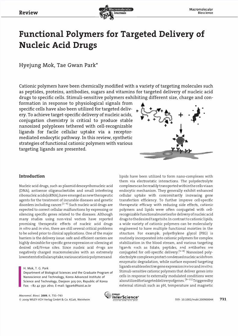
Functional Polymers for Targeted Delivery of Nucleic Acid Drugs Hyejung Mok, Tae Gwan Park* Introduction Nucleic acid drugs, such as plasmid desoxyribonucleic acid (DNA), antis ense oligo nucleotide s and small inter fering ribon uclei c acids(siRNA),have emerg ed as new therap eutic agents for the treatment of incurable diseases and genetic disorders including cancer. [1–4] Such nucleic acid drugs are expected to correct cellular malfunctions by expressing or silencing specific genes related to the diseases. Although man y stu die s using non-vi ral vec tors have report ed pr omis ing therapeuti c ef fect s of nucl eic acid dr ugs in vitro and in vivo, there are still several critical problems to be solved prior to clinical applications. One of the major barriers is the delivery issue: safe and efficient carriers are highl y desir able for speci fic gene expres sion or silen cing at desired cell/t iss ue sites. Since nucleic acid drugs are negat ively charged macromolecules with an extre mely lowextentofcellularuptake,variouscationicpolymersand lipids have been utilized to form nano-complexes with them via elect rosta tic interactions. The polye lect rolyt e comple xescan berea dil y tra nsportedwit hinthe cells viaan endocytic mechanism. They generally exhibit enhanced cel lul ar upt ake wit h con comita ntl y increasin g gene trans fecti on effici ency. To furthe r impr ove cell- speci fic thera peuti c efficac y with reduc ing side effects, cati onic pol ymers and lip ids wer e oft en con jug ate d wit h cel l- recog niza ble funct ionalmoietiesfor deliv ery of nucle ic acid drugs to thedesir ed tar getsite.In con tra st to cat ion ic lip ids , a wide variety of cationic polymers can be molecularly engineered to have multiple functional moieties in the structure. For exa mpl e, pol yet hyl ene gly col (PE G) is routinely incorporated into cationic polymers for complex stabilization in the blood stream, and various targeting li gands such as folate, pept ides, and anti bo di es ar e conjugated for cell-specific delivery. [5–8] Nanosized poly- electrolyt e comp lexes prote ct conde nsednucleic acidsfrom enzymatic degradation, while surface exposed targeting ligan dsenableselective gene expressioninvivo and invitro. Stimuli-sensitive cationic polymers that deliver genes into cells in response to externally modulated conditions were alsoutilizedfortargeteddeliveryofgenes. [9–11] Triggeredby external stimuli such as pH, temperature and magnetic Review H. Mok, T. G. Park Department of Biological Sciences and the Graduate Program of Nanoscience and Technology, Korea Advanced Institute of Science and Technology, Daejeon 305-701, Republic of Korea Fax: þ82 42 350 2610; E-mail: [email protected] Cationic polymers have been chemically modified with a variety of targeting molecules such as peptides, proteins, antibodies, sugars and vitamins for targeted delivery of nucleic acid drugs to specific cells. Stimuli-sensitive polymers exhibiting different size, charge and con- formation in response to physiological signals from specific cells have also been utilized for targeted deliv- ery. To achieve target-specific delivery of nucleic acids, con jugati on che mis try is cri tic al to pro duc e stable nanosized polyplexes tethered with cell-recognizable li gands for facile ce ll ul ar uptake vi a a re ce pt or- mediated endocytic pathway. In this review, synthetic strategies of functional cationic polymers with various targeting ligands are presented. Macromol. Biosci. 2009, 9 , 731–743 2009 WILEY-VCH Verlag GmbH & Co. KGaA, Weinheim DOI: 10.1002/mabi.200900044 731
-
Upload
alchemik1515 -
Category
Documents
-
view
218 -
download
0
Transcript of Druk Functional Polymers for Targeted Delivery of Nucleic Acid Drugs 2009 Macromolecular Bioscience
8/11/2019 Druk Functional Polymers for Targeted Delivery of Nucleic Acid Drugs 2009 Macromolecular Bioscience
http://slidepdf.com/reader/full/druk-functional-polymers-for-targeted-delivery-of-nucleic-acid-drugs-2009-macromolecular 1/13
Functional Polymers for Targeted Delivery of
Nucleic Acid Drugs
Hyejung Mok, Tae Gwan Park*
Introduction
Nucleic acid drugs, such as plasmid desoxyribonucleic acid
(DNA), antisense oligonucleotides and small interfering
ribonucleic acids(siRNA),have emerged as newtherapeutic
agents for the treatment of incurable diseases and genetic
disorders including cancer.[1–4] Such nucleic acid drugs are
expected to correct cellular malfunctions by expressing or
silencing specific genes related to the diseases. Although
many studies using non-viral vectors have reported
promising therapeutic effects of nucleic acid drugs
in vitro and in vivo, there are still several critical problems
to be solved prior to clinical applications. One of the major
barriers is the delivery issue: safe and efficient carriers are
highly desirable for specific gene expression or silencing at
desired cell/tissue sites. Since nucleic acid drugs are
negatively charged macromolecules with an extremely
lowextentofcellularuptake,variouscationicpolymersand
lipids have been utilized to form nano-complexes withthem via electrostatic interactions. The polyelectrolyte
complexescan be readily transported withinthe cells viaan
endocytic mechanism. They generally exhibit enhanced
cellular uptake with concomitantly increasing gene
transfection efficiency. To further improve cell-specific
therapeutic efficacy with reducing side effects, cationic
polymers and lipids were often conjugated with cell-
recognizable functionalmoietiesfor delivery of nucleic acid
drugs to thedesired targetsite.In contrast to cationic lipids,
a wide variety of cationic polymers can be molecularly
engineered to have multiple functional moieties in the
structure. For example, polyethylene glycol (PEG) is
routinely incorporated into cationic polymers for complex
stabilization in the blood stream, and various targeting
ligands such as folate, peptides, and antibodies are
conjugated for cell-specific delivery.[5–8] Nanosized poly-
electrolyte complexesprotect condensed nucleic acidsfrom
enzymatic degradation, while surface exposed targeting
ligands enableselective geneexpressioninvivo and invitro.
Stimuli-sensitive cationic polymers that deliver genes into
cells in response to externally modulated conditions were
alsoutilizedfortargeteddeliveryofgenes. [9–11]Triggeredby
external stimuli such as pH, temperature and magnetic
Review
H. Mok, T. G. Park
Department of Biological Sciences and the Graduate Program of
Nanoscience and Technology, Korea Advanced Institute of
Science and Technology, Daejeon 305-701, Republic of Korea
Fax: þ82 42 350 2610; E-mail: [email protected]
Cationic polymers have been chemically modified with a variety of targeting molecules such
as peptides, proteins, antibodies, sugars and vitamins for targeted delivery of nucleic acid
drugs to specific cells. Stimuli-sensitive polymers exhibiting different size, charge and con-
formation in response to physiological signals from
specific cells have also been utilized for targeted deliv-
ery. To achieve target-specific delivery of nucleic acids,
conjugation chemistry is critical to produce stable
nanosized polyplexes tethered with cell-recognizable
ligands for facile cellular uptake via a receptor-
mediated endocytic pathway. In this review, synthetic
strategies of functional cationic polymers with various
targeting ligands are presented.
Macromol. Biosci. 2009, 9 , 731–743
2009 WILEY-VCH Verlag GmbH & Co. KGaA, Weinheim DOI: 10.1002/mabi.200900044 731

8/11/2019 Druk Functional Polymers for Targeted Delivery of Nucleic Acid Drugs 2009 Macromolecular Bioscience
http://slidepdf.com/reader/full/druk-functional-polymers-for-targeted-delivery-of-nucleic-acid-drugs-2009-macromolecular 2/13
field,site-specificgenetransfectioncouldbeachievedatthe
desired target site. Among many targeted polymeric
delivery systems reported in the literature, this review
mainly focuses on a few examples of cell-specific delivery
systems for nucleic acid drugs with an emphasis on various
types of cationic polymers, available targeting ligands,
conjugation strategies, and therapeutic applications.
Several examples of targeted delivery systems triggered
by external stimuli are also briefly introduced.
Cationic Polymers for Gene Delivery
Synthetic and natural cationic polymers have been
popularly used as non-viral carriers for gene therapy due
to their far less cytotoxicities as compared to that of viral
carriers. Poly(L-lysine) (PLL) is one of the most commonly
used cationic polymers for gene delivery [Figure 1(A)]. PLL
can effectively form nano-sized polyelectrolyte complexes
with nucleic acid drugs due to primary e-amine groups of
lysine residues. However, PLL has only the primary amine
groups in the backbone, which are insufficient for facil-
itating the endosome escape of polyelectrolyte complexesinto the cytosol area. To confer an endosome escape
property to PLL, endosome breaking peptides, such as
polyhistidine and KALA peptide, were also conjugated to
enhance gene delivery efficiency.[12,13]
Poly(ethylenimine)(PEI)showsfarsuperiorgenedelivery
efficiency to PLL due to its high charge density and good
buffering capacity, which enable the formation of more
compact polyelectrolyte complexes with nucleic acids
that have enhanced endosome escape ability. In particular,
branched PEI has primary, secondary, and tertiary
amine groups, providing sufficient buffering activity at
endosomal pH for facile endosome escape of the nucleic
acids by the ‘‘proton sponge’’ effect [Figure 1(B)].[14]
However, high molecular weight PEI shows severe cyto-
toxicityproblems,makingits clinical application difficultin
spite of its excellent delivery efficiency. Linear PEI, having
only the secondary amine groups in the backbone except
for the terminal primary amine groups, is known to have
much lower cytotoxicity than branched PEI, while its
transfection efficiency is similar to that of the branched
one [Figure 1(C)].[15] In some cases, linear PEI (22k) even
showed higher transfection efficiency than branched PEI
(25k) for plasmid DNA delivery.[16] To reduce the cytotoxi-
city of PEI concomitantly maintaining the delivery
efficiency, cleavable and biodegradable PEI polymers havebeen synthesized.[17]
Poly(b-amino ester)s are biodegradable cationic poly-
mers with ester linkages in the backbone, which facilitates
the decomplexationof polyelectrolyte complexesat low pH
endosomal conditions [Figure 1(D)]. Poly(b-amino ester)s
were prepared by conjugation of primary or secondary
amine monomers to various di-acrylate monomers via a
Michael-type addition reaction. According to previous
reports, a familyof poly(b-amino ester) derivatives showed
more enhanced gene transfection efficiency with reduced
cell cytotoxicity, compared to PEI, which might be
attributed to the cleavage of poly(b-amino ester) backbone
in an acidic condition.[18,19]
Poly(amidoamine) (PAMAM) dendrimers are the most
intensively studied cationic dendrimers for gene delivery
[Figure 1(E)]. PAMAMdendrimers havebeenpopularly used
as carriers for targeted delivery of anticancer drugs, as well
as nucleic acid drugs.[20] PAMAM wasprepared by stepwise
polymerization from an initiator core, such as ammonia
and ethylenediamine.[21] As the number of generations
increased, transfection efficiency was enhanced, but cell
cytotoxicity also increased.[21,22] The cytotoxicity of
PAMAM dendrimer generation 4 [molecular weight
H. Mok, T. G. Park
Hyejung Mok is currently a postdoctoral associate
in Professor Tae Gwan Park’s laboratory, Depart-
ment of Biological Sciences at Korea Advanced
Institute of Science and Technology (KAIST). She
received Ph.D. from KAIST under thesupervisionof
Professor Tae Gwan Park in 2008. She published 17papers during her Ph.D. study. Her main research
interests lie in novel siRNA and protein delivery
systems based on polymeric biomaterials.
Tae Gwan Park received a B.S. in chemical
technology from Seoul National University in
1980, an M.S. in biological sciences from Korea
Advanced Institute of Science and Technology in
1983, and a Ph.D. in bioengineering from the
University of Washington in 1990 under the
direction of Prof. Allan S. Hoffman. Following a
postdoctoral research associate experience
(1990–1991) at the Massachusetts Institute of
Technology in Prof. Robert Langer’s laboratory,he joined Temple University, School of Pharmacy
as an assistant professor. In 1995, he returned to
Korea and became a professor at the Korea
Advanced Institute of Science and Technology.
He received the Nanotechnology Innovative
Research Award, Korea (2006), and KAIST Research
Award, Korea (2007). More recently, he received
the 2009 Clemson Award for Contributions to the
Literature from the Society for Biomaterials. His
research interests include nanobiomaterial-based
drug delivery systems, gene therapy, and tissue
engineering. He has published over 203 papers in
SCI journals, received 30 domestic and foreign
patents, and licensed out several technologies.He currently serves as an editorial board member
of Bioconjugate Chemistry, J. Controlled Release,
Pharmaceutical Research, Macromolecular Bio-
science and J. Bioactive and Compatible Polymers.
732
Macromol. Biosci. 2009 , 9 , 731–743
2009 WILEY-VCH Verlag GmbH & Co. KGaA, Weinheim DOI: 10.1002/mabi.200900044

8/11/2019 Druk Functional Polymers for Targeted Delivery of Nucleic Acid Drugs 2009 Macromolecular Bioscience
http://slidepdf.com/reader/full/druk-functional-polymers-for-targeted-delivery-of-nucleic-acid-drugs-2009-macromolecular 3/13
(MW)¼14215Da]wassimilartothatofPLL(MW ¼56 kDa).
The optimal generation number for efficient transfection
was dependent on the cell types.[21] After intravenous
injection of generation 9 PAMAM dendrimer/plasmid DNA
complexes at an N/P ratio of 5, significant gene expression
was observed only in the lung tissue, suggesting that
PAMAM dendrimer could be applied as an effective
pulmonary gene carrier.[23] PAMAM dendrimer (generation
7)/luciferase siRNA complexes exhibited up to 80% of
gene silencing effect at an N/P ratio of 10 in luciferase
expressing A549 cells.[24]
One of the naturally occurring cationic polymers useful
for gene carriers is chitosan.[25] Chitosan is a linear and
biodegradable polysaccharide composed of b-(1,4)-linked
D-glucosamin and N -acetyl-D-glucosamine. Chitosan is
known to be biocompatible and non-toxic because it can
be degraded into N -acetylglucosamine by lysozyme in the
body.[26] Water soluble chitosan with a low-molecular-
weight (22 kDa) especially exhibited better gene transfec-
tion efficiency and improved cell viabi-
lity for plasmid DNA delivery than PLL
(20kDa) in vitro,[25] suggesting that
chitosan could be a useful carrier candi-
date for gene therapy.
Cationic polymers and nucleic acidsform polyelectrolyte complex nanopar-
ticles mainly by electrostatic interac-
tions. The resultant nano-complexes,
polyplexes, can be easily aggregated or
disintegrated depending on environ-
mental conditions such as pH, salt
concentration and the presence of other
charged molecules. The stability issue of
polyplexes is particularly serious in the
bloodstream due to the non-specific
adsorption of serum proteins on the
surface. PEG has been commonly con-
jugated to cationic polymers to enhance
the stability of polyplexes in vitro and
in vivo.[27–29] PEGylated polycation/DNA
complexes showed reduced toxicity
while significantly prolonging blood
circulation time after intravenous
administration, compared to un-PEGy-
lated polycation/DNA complexes.[28]
PEGylated nanoparticles also exhibited
passive tumor targeting via the
enhanced permeability and retention
(EPR) effect.[30] Until now, various block
and graft copolymers with PEG havebeen synthesized and characterized for
gene delivery applications.[13,27–29] To
prepare an A/B-type PLL/PEG block copo-
lymer, N -carboxyanhydrideof z-protected
L-lysine was polymerized from a-methoxy-v-amino PEG as
an initiator.[27] The transfection efficiency of PLL/PEG block
copolymer/DNA complexes increased 6-fold, compared to
that of PLL/DNA complexes in 293cells.[27] Comb-type PEG-
grafted PLL copolymers were also prepared by reacting
primary amine groups(aand e aminogroups)ofPLLwithan
amine reactive succinimidyl succinate PEG derivative.[13]
While there was no significant difference in the transfec-
tion efficiency of plasmid DNA between PLL complexes
and PEG-graft-PLL complexes in serum deficient medium,
PEG-graft-PLL complexes showed a 6 times higher transfec-
tion efficiency in 10% serum containing medium than
PLL complexes.[13] This was attributed to reduced adsorp-
tion of serum proteins and enhanced stability of polyelec-
trolyte complexes via PEGylation in the serum medium.
Moreover, PEGylated PEI exhibited longer blood circulation
and more reduced acute toxicity than PEI by decreasing
non-specific accumulation in the liver after intravenous
administration.[28,29]
Functional Polymers for Targeted Delivery of Nucleic Acid Drugs
Figure 1. (A) Poly(L-lysine) (PLL), (B) branched PEI, (C) linear PEI, (D) poly(b-amino ester),and (E) poly(amidoamine) (PAMAM).
Macromol. Biosci. 2009, 9 , 731–743
2009 WILEY-VCH Verlag GmbH & Co. KGaA, Weinheim www.mbs-journal.de 733

8/11/2019 Druk Functional Polymers for Targeted Delivery of Nucleic Acid Drugs 2009 Macromolecular Bioscience
http://slidepdf.com/reader/full/druk-functional-polymers-for-targeted-delivery-of-nucleic-acid-drugs-2009-macromolecular 4/13
Conjugation Strategies for Targeting Ligandsfor Receptor/Ligand Interaction-MediatedDelivery of Nucleic Acid Drugs
Cationic polymers with multiple functional groups were
covalentlyor non-covalently conjugatedwithcell recogniz-able targeting ligands, such as peptides, proteins, anti-
bodies, sugars, vitamins and chemicals. Specific interac-
tions between ligands and receptors on the cellular
membrane can mediate receptor-mediated endocytosis of
polyelectrolyte complexes via a clathrin-dependent or a
clathrin-independent mechanism, resulting in much
enhanced cellular uptake.[31] As listed in Table 1, various
targeting ligands have been directly conjugated to cationic
functional polymers for efficient delivery of therapeutic
genes to specific cells.
Four different strategies for immobilizing targeting
ligandsontothesurfaceofDNA/polymernano-complexes
are described in this paper: ligand-polymer conjugate;
ligand-linker-polymer conjugate; ligand-nucleic acid
conjugate; ligand-linker-nucleic acid conjugate (Figure 2).
Various ligand molecules are commonly conjugated to
cationic functional polymers with and without a linker
polymer (spacer). To expose targeting ligands onto the
outer surface of polyplexes while maintaining the
complex stability, the targeting ligand was terminally
tethered to the distal end of a hydrophilic linker polymer,
such as PEG, and the ligand-PEG conjugate was covalently
linked to cationic polymers. Direct conjugation of target-ing ligands to therapeutic short chain oligodeoxynucleo-
tide and siRNA nucleic acids instead of circular plasmid
DNA often exhibited target-specific cellular uptake even
without using carrier polymers, although this could
partially impair the activity of nucleic acid drugs during
the conjugation process.[32] It should be noted that
nucleic acid-ligand conjugates cannot easily escape from
endosome to cytosol after receptor mediated endocytosis,
resulting in limited therapeutic effects.[33] Therefore, to
facilitate the endosome escape, carrier polymers should
also be required to conjugate with various fusogenic
moieties that destabilize the cell membrane at acidic
pH. PEG was also chemically conjugated to oligonucleo-
tide and siRNA via a cleavable linkage so that the
PEG-nucleic acid conjugate might form a more stable
polyelectrolyte complex than the naked one in serum
containing medium.
H. Mok, T. G. Park
Table 1. A list of representative targeted polymeric delivery systems for nucleic acids.
Targeting ligand Polymer Target receptor Target cell/tissue Cell line Ref.
peptide RGD peptide PEI integrin receptor an
b3 h uman melanoma cell Mewo cell [36]PEI-PEG [37]poly(b-amino ester) [38]
LHRH peptide siRNA-PEG LHRH receptor tumor tissue A2780 [40]PAMAM [41]
protein lactoferrin PAMAM-PEG lactoferrin receptor brain brain capillaryendothelial cell(BCEC)
[42]
PEI bronchial epithelial cell BEAS-2B cell [43]transferrin PAMAM-PEG transferrin receptor brain BCEC, in vivo [42][45]EGF biotin-PEG EGF receptor tumor tissue A431 cell [8]RAP (receptor-associatedprotein)
PLL, PDL LDL receptor human hepatoma cell HepG2 cell [48]
antibody anti-JL1 monoclonal antibody PLL JL-1 human leukemia T cell Molt 4 cell [52]chimeric anti-EGF receptorantibody (cetuximab)
PAMAM EGF receptor tumor tissue [53]
anti-HER-2 antibody
(Herceptin, Trastuzumab)
PEI human EGF receptor tumor tissue Sk-Br-3 cell [5]
SKOV-3 cells [16]anti-CD90 antibody
(Trastuzumab)
PEI CD90 fibroblast WI26SV40 [16]
anti-GAD antibody PEI-PEG GAD islet cell MIN6 cell [7]sugar galactose, lactose PEI-PVP asialoglycoprotein
receptorhepatoma cell HepG2 cell [54]
siRNA-PEG HuH-7 cell [56]mannose PLL mannose receptor macrophage cell,
dendritic cellprimarymacrophagecell, in vivo
[57][59]
chitosan RAW264.7 [58]vitamin folate PLL-PEG folate receptor tumor tissue KB cell [6]
PEI-PEG [61–63]ODN-PEG [64]
734
Macromol. Biosci. 2009 , 9 , 731–743
2009 WILEY-VCH Verlag GmbH & Co. KGaA, Weinheim DOI: 10.1002/mabi.200900044

8/11/2019 Druk Functional Polymers for Targeted Delivery of Nucleic Acid Drugs 2009 Macromolecular Bioscience
http://slidepdf.com/reader/full/druk-functional-polymers-for-targeted-delivery-of-nucleic-acid-drugs-2009-macromolecular 5/13
Targeting Ligand-MediatedDelivery of Nucleic Acid Drugs
Peptides
Arg-Gly-Asp (RGD) PeptideThe three-amino-acid sequence peptide
RGD has been extensively used as a cell-
recognizable ligand for integrin receptor
an b3/a
n b5 targeting.[34–37] In particular,
integrin receptor an b3/a
n b5 has been
known tobe specifically over-expressed in
angiogenic endothelial cells of the tumor
tissue, which enables the RGD peptide to
be used as a tumor targeting ligand.
Linear RGD(RGDC) and cyclic RGD(cRGD)
with one (CGRGDSPC) or two disulfide
bonds (ACDCRGDCFCG) have beenemployed for cell-specific gene delivery.
Among these, cRGD peptide with two
disulfide bonds showed 200-fold and 20-
fold higher potency than linear RGD and
cRGD with one disulfide bond, respec-
tively.[35] A thiol group of cysteine
modified RGD peptide (linear RGDC)
was used to conjugate to carrier poly-
mers.[36] Primary amine groups of PEI
were activated with a heterofunctional
crosslinker of N -succinimidyl-3-(2-pyri-
dyldithio)propionate (SPDP), and the
SPDP-activated PEI was reacted with
the thiol group of cysteine modified
RGD peptide via a disulfide exchange
reaction to prepare a RGD/PEI conjugate
[Figure 3(A)]. The resultant RGD/PEI
conjugate with a substitution degree of
4.6% showed 50 times higher gene
expression of luciferase plasmid than
that of the unmodified PEI for Mewo cells
(human melanoma cells). However, no
significant difference in luciferase gene
expression wasobservedin A549 cells,an
integrin receptor an b3 negative cell.Cysteine-modified RGD peptide was also
conjugated directly to poly(b-amino
ester) via a disulfide bond for integrin
receptor targeting.[38] An N-terminal
amine group of cRGD peptide without a
cysteine residue was also used for the
conjugation reaction with carrier poly-
mers or linker polymers.[37] The terminal
amine group of cRGD was activated with
a heterofunctional PEG, NHS/PEG/VS
Functional Polymers for Targeted Delivery of Nucleic Acid Drugs
Figure 2. Schematic illustration for targeted delivery of polymer/nucleic acid complexesusing four kinds of strategy, polymer-ligand conjugate, polymer-linker-ligand conju-gate, nucleic acid-ligand conjugate, and nucleic acid-linker-ligand conjugate, via recep-tor mediated endocytosis.
Figure 3. (A) RGD conjugated PEI, (B) LHRH- PEG-siRNA conjugate.
Macromol. Biosci. 2009, 9 , 731–743
2009 WILEY-VCH Verlag GmbH & Co. KGaA, Weinheim www.mbs-journal.de 735
8/11/2019 Druk Functional Polymers for Targeted Delivery of Nucleic Acid Drugs 2009 Macromolecular Bioscience
http://slidepdf.com/reader/full/druk-functional-polymers-for-targeted-delivery-of-nucleic-acid-drugs-2009-macromolecular 6/13
( N -hydroxysuccinimide/PEG/vinylsulfone), in anhydrous
dimethylformamide (DMF) for 2 h to minimize the activity
loss of vinyl sulfone groups after the reaction.The resulting
cRGD/PEG/VS conjugate was grafted to primary amine
groups of PEI. The cRGD/PEI conjugate exhibited that one
RGD substitution per PEI molecule might be sufficientenough to show good cell binding affinity to the integrin
receptor an b3/a
n b5.[37]
LHRH
Luteinizing hormone-releasing hormone (LHRH) has been
known to be a good tumor targeting ligand because
LHRH receptor is commonly over-expressed in various
cancer cells including prostate, breast, and ovarian
cancer cells.[39,40] Instead of the native LHRH peptide
(Glu-His-Trp-Ser-Tyr-Gly-Leu-Arg-Pro-Gly), LHRH analogs
(Gln-His-Trp-Ser-Tyr-DLys-Leu-Arg-Pro or Pyr-His-Trp-Ser-
Tyr-DLys-Leu-Arg-Pro-Gly), in which the glycine residue at
the position 6 was replaced by D-lysine, have been used as a
commercially available therapeutic peptide drug (Leupro-
lide acetate) to treat prostate cancer and endometriosis.
Since the N-terminal amine group and the C-terminal
carboxylic acid group of LHRH analog are blocked by
pyroglutamation and amidation, respectively, the e-amino
group of the lysine residue in the backbone was conjugated
to cationic polymers.[40] For direct conjugation of LHRH
peptide to PAMAM, the amino groups of PAMAM were
modified with carboxylic acid groups via ring-opening
acylation with glutaric anhydride, and these carboxylic
acid groups were then conjugated with the e amine groupof D-Lys6-LHRH via 1-ethyl-3-(3-dimethylaminopropyl)-
carbodiimide hydrochloride (EDC) chemistry.[41] When
using a linker polymer, PEG, for the conjugation of LHRH,
PEG with a terminal carboxylic acid group was used for the
conjugation to siRNA.[40]Afterblockinganaminegroupofa
heterobifunctional PEG derivative, NH2–PEG–COOH, using
SPDP, theremnant carboxylic acid group wasconjugated to
the e amine group of D-Lys6 of the LHRH peptide using EDC.
The resulting SPDP activated PEG/LHRH was reduced to
produce a free thiol group. An amine group at the terminal
5’-sense double strand siRNA was activated with SPDP and
reacted with thiol/PEG/LHRH to prepare a siRNA/PEG/
LHRH conjugate [Figure 3(B)]. PEI (25k) was used as a core
condensing polymer for siRNA delivery. Gene silencing
effects by siRNA/PEG/LHRH/PEI and siRNA/PEG/PEI were
analyzed in twotypes of human ovarian cancercells, A2780
(LHRH receptor over-expressing cell) and SK-OV-3 (LHRH
receptor deficient cell). While similar gene silencing
extents by siRNA/PEG/LHRH/PEI and siRNA/PEG/PEI were
shown in SK-OV-3 cells, siRNA/PEG/LHRH/PEI exhibited a
20% higher gene silencing effect than siRNA/PEG/PEI in
A2780 cells, which was attributed to LHRH receptor
mediated targeting.
Proteins
Lactoferrin and Transferrin
Lactoferrin, an iron-binding glycoprotein, is a globular
protein with a molecular weight of 80kDa, which can bind
ferricionsreversibly.Lactoferrincouldbeusedasalungandbrain targeting ligand because lactoferrin receptors are
dominantly expressed in brain capillaries and bronchial
epithelial cells.[42,43] It is known that lactoferrin-mediated
transcytosis occurredin BBB(blood brainbarrier),openinga
new possibility for brain delivery of nanoparticulate gene
carriers.[42] To prepare a lactoferrin-PEI conjugate, vicinal
hydroxyl groups of N -acetylneuraminic acid in lactoferrin
were oxidized to form amine-reactive aldehyde groups,
which were then conjugated to primary amine groups of
b-PEI (MW¼25 kDa). While the lactoferrin/PEI (lactoferrin:
PEI¼1: 17.2 molar ratio)conjugates showed a 5-fold higher
transfection efficiency than PEI for bronchial epithelial
BEAS-2B cells, no significant difference in transfection
efficiency was observed for alveolar epithelial cells, A549
cells. Transferrinis alsoa glycoprotein that transportsferric
ions, which is over-expressed about ten-fold e for tumor
cells as compared to normal cells.[44] Primary amine
groups of transferrin were used to conjugate with cationic
polymers. PAMAM-PEG-targeting ligand conjugates
were prepared using a heterobifunctional PEG derivative
[a-maleimidyl-v- N -hydroxylsuccinimidylpoly(ethylene
glycol) (NHS-PEG-MAL), MW¼3400Da).[42,45] After pri-
mary amine groups of PAMAM were conjugated to NHS-
PEG-MAL, the resulting conjugate was reacted with
thiolated transferrin that was prepared by using a Traut’sreagent, to form PAMAM-PEG-transferrin conjugate. The
extentofluciferasegeneexpressioninbrainusingPAMAM-
PEG-transferrin as a carrier was about two-fold higher than
when PAMAM or PAMAM-PEG was used after intravenous
injection. To compare the targeting activity of lactoferrin
to that of transferrin, PAMAM-PEG-lactoferrin was also
prepared based on a similar reaction procedure. The amine
groups of lactoferrin were converted to thiol groups using
N -succinimidyl-S-acetylthioacetate (SATA), which were
conjugated to PAMAM-PEG. PAMAM-PEG-lactoferrin
exhibited 2 times higher gene transfection efficiency
thanPAMAM-PEG-transferrininbraincapillaryendothelial
cells (BCEC) in vitro and in the brain after intravenous
injection in vivo. Thus, itcan bededuced thatlactoferrin isa
little more efficient than transferrin for brain targeting of
nucleic acid drugs.
Ligand Proteins
The epidermal growth factor (EGF) receptor is known to be
greatly over-expressed in tumor tissues about 100-fold
higher than normal tissues.[44] EGF could be internalized
within the cells via EGF receptor-mediated endocytosis by
H. Mok, T. G. Park
736
Macromol. Biosci. 2009 , 9 , 731–743
2009 WILEY-VCH Verlag GmbH & Co. KGaA, Weinheim DOI: 10.1002/mabi.200900044

8/11/2019 Druk Functional Polymers for Targeted Delivery of Nucleic Acid Drugs 2009 Macromolecular Bioscience
http://slidepdf.com/reader/full/druk-functional-polymers-for-targeted-delivery-of-nucleic-acid-drugs-2009-macromolecular 7/13
forming dimer complexes. Thus, EGF has been popularly
usedasagoodtargetingmoietyfortumortissues.EGFhasa
small molecular weight about 6 kDa (53 amino acids) with
three internal disulfide bonds.[46,47] Amine groups of EGF
have been commonly used for the conjugation reaction
with polymers in order to avoid interaction with intramo-lecular disulfide bonds. Because EGF has two lysine
residues, three amine groups (two e-amino groups of
Lys28 and Lys 48, and the a amino group of N-terminal)
could be used for the conjugation.[47] According to our
previous studies, only the N-terminal mono-PEGylated EGF
fully maintained its activities such as cell proliferation and
receptor binding as much as the native EGF. Thus, the
polymer conjugation site of EGF is an important issue for
targeting. To minimize the loss of targeting activity by
green fluorescent protein (GFP) through non-specific
conjugation of EGF to polymers, EGF could be physically
immobilized onto polymer/nucleic acid complexes via
biotin/streptavidin interaction.[8] N -terminal mono-PEGy-
lated and biotinylated EGF (biotin-PEG-EGF) was specifi-
cally separated by size-exclusion chromatography. After
preparing PEI/plasmid DNA complexes, streptavidins were
coatedonto thecomplexesvia ionic interaction. Then, biotin-
PEG-EGF was immobilized onto streptavidin-PEI-DNA
complexes via biotin-streptavidin interaction. PEI/plasmid
complexes coated with N-terminal specifically PEGylated
EGF exhibited significantly higher transfection efficiency
than those with non-specifically PEGylated EGF (multi-
PEGylated EGF). Luciferase gene expression by biotin-PEG-
EGF/streptavidin-PEI-DNA complexes was about ten fold
higher than that byLipofectamine andPEI forA431,a humanepidermoid carcinoma cell line, highly expressing EGF
receptor.
For the delivery of nucleic acid drugs via a low-density
lipoprotein (LDL) receptor-mediated pathway, a receptor-
associated protein (RAP) to be used as a targeting ligand
was also conjugated to cationic polymers. RAP (39–
44kDa) is known to be expressed in various tissues such as
liver and brain, and it shows binding affinity to LDL. RAP-
LDL complexes are internalized into cells via receptor-
mediated endocytosis.[48,49] The expression level of LDL
receptors on the cell membrane markedly increased in
rapidly dividing cells such as malignant cells. RAP proteins
containing a C-terminal cysteine residue instead of a
heparin binding domain were cloned and expressed in the
E. coli system. Purified cysteine-modified RAP protein was
reacted with SPDP (heterofunctional crosslinker) activated
poly(D-lysine) (PDL) or PLL to prepare a PDL-RAP or PLL-RAP
conjugate. PDL-RAP conjugate showed enhanced gene
transfection efficiency compared to that of PDL and PLL-
RAP, probably because plasmid DNA complexes with PDL-
RAP were able to be readily internalized within human
hepatoma HepG2 cells via LDL receptor mediated endocy-
tosis. In addition, PDL-RAP/DNA complexes were more
resistant to degradation both in the lysosomal compart-
ment and in the cytosolic condition than PLL-RAP/DNA
complexes.
Antibodies
Antibodies (150kDa) composed of twolarge heavy chains
and two small light chains have been used as a good
targeting ligand due to their superior specificity towards
target molecules. In particular, increasing number of
therapeutic monoclonal antibodies approved by FDA has
shown their values for targeted therapy in cancer, immune
diseases, and heart diseases.[50] Besides whole antibodies,
genetically engineered low-molecular-weight fragments,
such as single chain variant fragment (scFv) (25kDa),
dibody (55 kDa), minibody (80 kDa), and scFv-Fc (105 kDa),
have been conjugated to chemical drugs for targeted
delivery.[51] The major challenge for antibody conjugation
to polymers is to synthesize a homogeneous conjugate
population while maintaining its antigen binding activity.
Thus, site-specific conjugation of antibodies with polymers
is a critical issue especially because high molecular weight
antibodies have many reactive functional groups for
conjugation. Non-specific antibody conjugation might be
apt to damage the target binding ability partially or
completely.
For conjugating polymers to antibodies without impair-
ing an antigen binding region, the glycosylated part has
been used as an optimal site for conjugation.[52,53]
Previously, a carbohydrate-directed antibody modification
method was reported for the preparation of PLL-antibodyconjugate.[52] Anti-JL1 antibody, a monoclonal antibody
against JL1 antigen (stage II-specific human T cell lympho-
cyte differentiation antigen), was used to target human
leukemia T cells. Two hydroxyl groups in the sugar ring of
anti-JL1antibodywereconvertedtoaldehydegroupsbythe
periodate oxidation reaction via the hydrazide group of a
heterofunctional crosslinker, 3-(2-pyridyldithio)propionyl-
hydrazide (PDPH) [Figure 4(A)]. To prepare a thiolated PLL,
thee-aminogroupofPLLwasmodifiedwithsuccinimidyl-3-
(2-pyridyldithio)propionate(SPDP), and dithiothreitol(DTT)
was treated to the conjugate. The resulting 2-pyridyl
disulfide groups of antibody were reacted to thiolated PLL
via a disulfide linkage to prepare an anti-JL1 antibody-PLL
conjugate (Ab-PLL). The Ab-PLL conjugate exhibited a good
targeting effect for plasmid DNA delivery. The extent of
galactosidase gene expression by DNA/Ab-PLL complexes
(about 300 nm in diameter at a polymer/DNA weight ratio
of 5) was about 10 times higher than that by DNA/PLL
complexes, and about 2 times higher than that by the
commercial reagent Lipofectin for Molt 4 cells, human
leukemia T cells. A chimeric anti-EGF receptor monoclonal
antibody Cetuximab (IMC-C225), a brain targeting moiety,
was also oxidized in a similar method to convert the sugar
Functional Polymers for Targeted Delivery of Nucleic Acid Drugs
Macromol. Biosci. 2009, 9 , 731–743
2009 WILEY-VCH Verlag GmbH & Co. KGaA, Weinheim www.mbs-journal.de 737
8/11/2019 Druk Functional Polymers for Targeted Delivery of Nucleic Acid Drugs 2009 Macromolecular Bioscience
http://slidepdf.com/reader/full/druk-functional-polymers-for-targeted-delivery-of-nucleic-acid-drugs-2009-macromolecular 8/13
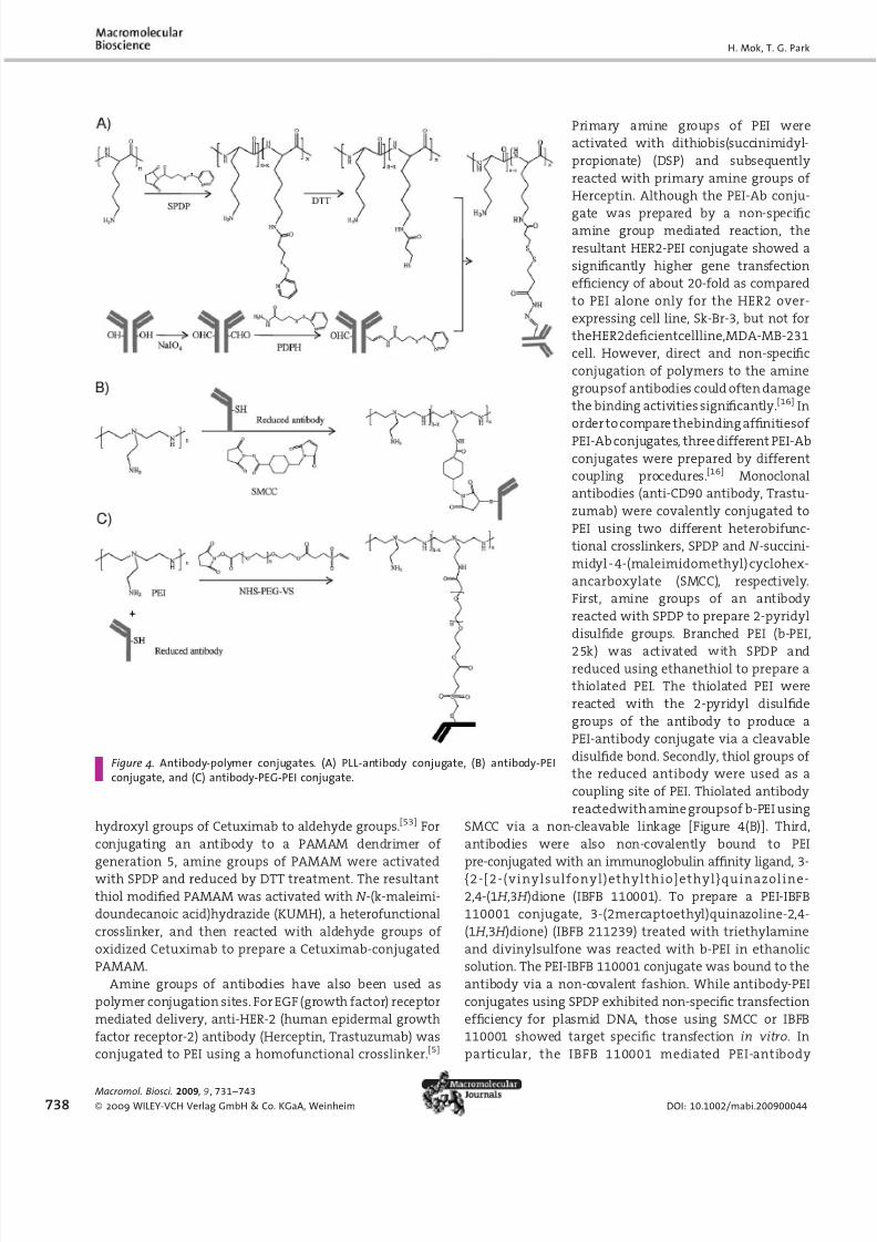
hydroxyl groups of Cetuximab to aldehyde groups.[53] For
conjugating an antibody to a PAMAM dendrimer of
generation 5, amine groups of PAMAM were activated
with SPDP and reduced by DTT treatment. The resultant
thiol modified PAMAM was activated with N -(k-maleimi-
doundecanoic acid)hydrazide (KUMH), a heterofunctional
crosslinker, and then reacted with aldehyde groups of
oxidized Cetuximab to prepare a Cetuximab-conjugated
PAMAM.
Amine groups of antibodies have also been used as
polymer conjugation sites. For EGF (growth factor) receptor
mediated delivery, anti-HER-2 (human epidermal growth
factor receptor-2) antibody (Herceptin, Trastuzumab) was
conjugated to PEI using a homofunctional crosslinker.[5]
Primary amine groups of PEI were
activated with dithiobis(succinimidyl-
propionate) (DSP) and subsequently
reacted with primary amine groups of
Herceptin. Although the PEI-Ab conju-
gate was prepared by a non-specificamine group mediated reaction, the
resultant HER2-PEI conjugate showed a
significantly higher gene transfection
efficiency of about 20-fold as compared
to PEI alone only for the HER2 over-
expressing cell line, Sk-Br-3, but not for
theHER2deficientcellline,MDA-MB-231
cell. However, direct and non-specific
conjugation of polymers to the amine
groupsof antibodies could often damage
the binding activities significantly.[16] In
order to compare thebinding affinitiesof
PEI-Ab conjugates, three different PEI-Ab
conjugates were prepared by different
coupling procedures.[16] Monoclonal
antibodies (anti-CD90 antibody, Trastu-
zumab) were covalently conjugated to
PEI using two different heterobifunc-
tional crosslinkers, SPDP and N -succini-
midyl - 4-(maleimidomethyl) cyclohex-
ancarboxylate (SMCC), respectively.
First, amine groups of an antibody
reacted with SPDP to prepare 2-pyridyl
disulfide groups. Branched PEI (b-PEI,
25k) was activated with SPDP andreduced using ethanethiol to prepare a
thiolated PEI. The thiolated PEI were
reacted with the 2-pyridyl disulfide
groups of the antibody to produce a
PEI-antibody conjugate via a cleavable
disulfide bond. Secondly, thiol groups of
the reduced antibody were used as a
coupling site of PEI. Thiolated antibody
reactedwith amine groupsof b-PEI using
SMCC via a non-cleavable linkage [Figure 4(B)]. Third,
antibodies were also non-covalently bound to PEI
pre-conjugated with an immunoglobulin affinity ligand, 3-
{2-[2-(vinylsulfonyl)ethylthio]ethyl}quinazoline-
2,4-(1 H ,3 H )dione (IBFB 110001). To prepare a PEI-IBFB
110001 conjugate, 3-(2mercaptoethyl)quinazoline-2,4-
(1 H ,3 H )dione) (IBFB 211239) treated with triethylamine
and divinylsulfone was reacted with b-PEI in ethanolic
solution. The PEI-IBFB 110001 conjugate was bound to the
antibody via a non-covalent fashion. While antibody-PEI
conjugates using SPDP exhibited non-specific transfection
efficiency for plasmid DNA, those using SMCC or IBFB
110001 showed target specific transfection in vitro. In
particular, the IBFB 110001 mediated PEI-antibody
H. Mok, T. G. Park
Figure 4. Antibody-polymer conjugates. (A) PLL-antibody conjugate, (B) antibody-PEIconjugate, and (C) antibody-PEG-PEI conjugate.
738
Macromol. Biosci. 2009 , 9 , 731–743
2009 WILEY-VCH Verlag GmbH & Co. KGaA, Weinheim DOI: 10.1002/mabi.200900044

8/11/2019 Druk Functional Polymers for Targeted Delivery of Nucleic Acid Drugs 2009 Macromolecular Bioscience
http://slidepdf.com/reader/full/druk-functional-polymers-for-targeted-delivery-of-nucleic-acid-drugs-2009-macromolecular 9/13
conjugate demonstrated the highest transfection efficiency
only forthe targetcells. This was likelyto be because amine
groupsintheFabregionoftheantibodywereblockedbyPEI
to a greater extent, losing antigen-antibody binding
capacity.
Free thiol groups of antibodies after reduction are alsogood functional sites for conjugation. Anti-GAD antibody,
specifically targeting GAD (glutamic acid decarboxylase)
that is only detected in pancreatic islet cells, was used for
target specific gene delivery. Thiol groups of reduced Fab
fragment in the GAD antibody reacted with amine groups
of PEI using a heterofunctional PEG (NHS-PEG-VS, N -
hydroxysuccinimide-PEG-vinylsulfone) to prepare a PEI-
PEG-Fab conjugate [Figure 4(C)].[7] The extent of luciferase
gene expression by the resultant PEI-PEG-Fab was over 3
times higher than that by PEI-PEG for GAD expressing
mouse insulinoma cells (MIN6 cells). The enhanced gene
expression was competitively inhibited by the addition of
freeGADantibody,provingthatGADantibodyspecificgene
expression occurred.
Sugars
Galactose
Galactose, a monosaccharide, has been used as a targeting
moiety for hepatoma cell targeting via asialoglycoprotein
receptors-mediated endocytosis.[54,55] Lactose, composed of
b-D-galactose and b-D-glucose through a b-1,4-glycosidic
linkage, could be also used as a targeting moiety for
hepatocyte targeting due to its terminal galactose. Galac-tose moieties were incorporated into amine groups of
polymers via a reductive amination reaction by the
addition of sodium cyanoborohydride.[54,55] Using lactose,
a galactose moiety was conjugated to the amine group of
PEI or amine functionalized PEG of poly(DMAEMA-NVP)-
block-PEG for specific targeting of plasmid DNA to
asialoglycoprotein receptor of hepatocytes. Galactosylated
PEI-poly(vinylpyrrolydone) showed about a 10-fold higher
transfection efficiency of luciferase plasmid DNA than that
of PEI for hepatoma HepG2 cells.[54] For targeted delivery of
siRNA, lactose was conjugated to the terminal end of PEG-
siRNA.[56] Usinglactose-PEG-acrylate, a thiolfunctionalized
siRNA (5’-modified sense strand siRNA) was conjugated to
the terminal of PEG via a Michael-type reaction. After the
conjugation reaction, unmodified antisense siRNA was
annealed to lactose-PEG-sense siRNA. The resulting lactose-
PEG-siRNA was acid-cleavable due to the presence of an
internalb-thiopropionate linkage. PLL (8.3 kDa) was used as
a core condensing polymer. Before complexation, the
e-amine group of PLL was thiolated using 2-iminothiolane
to form disulfide-crosslinked polyelectrolyte complexes
with lactose-PEG-siRNA. While naked siRNA/PLL showed
only 20% of gene silencing effect, lactose-PEG-siRNA/PLL
complexes exhibited 60% gene silencing effect for HuH-7
cells (human hepatoma cells).
Mannose
Glycoproteins with mannose, glucose, fucose and
N -acetylglucosamine units could be recognized by man-nose receptor-overexpressing cells such as macrophages
and dendritic cells and readily internalized via mannose
receptor-mediated endocytosis.[57] Because mannose has
been highlyexpressed on thesurfaces of bacteria and yeast,
mannose receptorsare over-expressed in thecellsrelated to
innate immune response.[58] Thus, mannose has been used
as a targeting moiety into macrophages and dendriticcells,
especially for the delivery of DNA vaccines.[59] Mannose
moieties were conjugated to amine groups of polymers
such as PLL and chitosan using mannopyranosylphenyl
isothiocyanate.[57,58] Vaccination of DNA encoding ovalbu-
minusingmannosylatedPLLcouldinduceboththeCD8and
CD4 T-cell immune response and antibody response, while
DNA complexed with naked PLL showed negligible effects
in vivo. The resulting induction of the immune response by
DNA/mannosylatedPLLcomplexescouldalsoinhibittumor
growth significantly in a tumor mice model.[59]
Folate
It has been known that folate receptor, a glycosylpho-
sphatidylinositol-anchored glycoprotein, is over-expressed
in several tumor tissues.[60] Folate (folic acid), a vitamin B9
(MW441Da), binds to the folate receptor with a high
binding affinity of K d109 M.[61] Thus, folate has been
considered as a useful targeting moiety for tumor or cancercell specific gene delivery. Although folate has two a and g
carboxylic acidgroupsavailablefor polymer conjugation, it
is known that only polymer conjugates viathe g -carboxylic
acid group maintain binding affinity to the folate
receptor.[61–63] For targeted delivery of plasmid DNA via
folate receptor mediated endocytosis, folate was conju-
gated at the PEG terminal of PEG-PLL.[6] After activating the
carboxylic acid group of folate using dicyclohexylcarbodii-
mide (DCC)/ N -hydroxysuccinimide (NHS), the terminal
amine group of NH2PEGCOOH (MW¼3 400) was
reacted to prepare a folate-PEG-COOH conjugate, which
was coupled to the a amino group of the e-amine-blocked
PLL [poly(e-CBZ-L-lysine), MW¼1 000]. After the de-block-
ing, the resultant folate-PEG-PLL conjugate formed stable
polyelectrolyte complexes with plasmid DNA with a size of
100nm. However, folate-PEG-PLL complexes showed
marginally enhanced transfection efficiency due to the
insufficient charge density and lack of endosome escaping
ability. To improve transfection yield, PEI (MW¼25000Da)
was additionally added to form polyelectrolyte complexes.
Folate-PEG-PLL/PEI/plasmid DNA complexes showed sig-
nificantly enhanced gene transfection efficiency, not for
A549 cells, folate-receptor deficient cells, but for KB cells,
Functional Polymers for Targeted Delivery of Nucleic Acid Drugs
Macromol. Biosci. 2009, 9 , 731–743
2009 WILEY-VCH Verlag GmbH & Co. KGaA, Weinheim www.mbs-journal.de 739

8/11/2019 Druk Functional Polymers for Targeted Delivery of Nucleic Acid Drugs 2009 Macromolecular Bioscience
http://slidepdf.com/reader/full/druk-functional-polymers-for-targeted-delivery-of-nucleic-acid-drugs-2009-macromolecular 10/13
folate receptor over-expressing cells.[6] The luciferase
expression of folate-PEG-PLL/PEI/luciferase plasmid DNA
complexes was about 10 times higher than that of
Lipofectamine or PEI for KB cells. Using a similar conjuga-
tion scheme, folate-PEG grafted PEI (folate-PEG-PEI) was
prepared for folate receptor targeted delivery of siRNA,antisense oligodeoxynucleotides (ODN), and plasmid
DNA.[64–66] DCC/NHS activated carboxylic acid group of
folate and then it was coupled with the amine group of a
heterofunctionalNH2PEGCOOHderivative.Then,amine
groups of PEI were conjugated to the activated carboxylic
acid group of folate-PEGCOOH by DCC/NHS [Figure 5(A)].
The relative transfectionlevel of plasmid DNAusing folate-
PEG-PEI was 12 times higher than commercially available
carriers such as Lipofectamine and PEI for folate receptor
positive cell line, KB cells.[64] GFP gene expression was
reduced below20% by anti-GFP siRNAplasmidDNA/folate-
PEG-PEI complexes, which was 2 times more efficient than
that by Lipofectamine and PEI.[65] Folate-PEG-PEI carriers
were utilized fortargeteddeliveryof notonly plasmid DNA,
but also oligonucleotide such as antisense ODN and
siRNA.[66] The gene silencing effect by siRNA or ODN/
folate-PEG-PEI complexeswas over 2 times higherthan that
by siRNAor ODN/PEI complexesfor thefolate receptor over-
expressing cell line, KB cells.
Folate was also conjugated to the terminal of antisense
ODN-PEG [Figure 5(B)].[67] Ag carboxylic acidgroup of folate
was transformed to a thiol group through a DCC/NHS
reaction with cystamine. An amine group of 50
- end aminemodified antisense ODN and thiol group of folate was
conjugated to the terminal of heterobifunctional NHS-PEG-
maleimide(NHS-PEG-MAL). The gene silencingefficiency of
ODN-PEG-folate/PEI,PEI,andLipofectaminecomplexeswas
20% for A549 cells (folate receptor deficient cells), while
they exhibited over 80% for KB cells (folate receptor over-
expressing cells).
Targeted Delivery of Nucleic Acid Drugs viaEnvironmentally Triggered Signals
The cell-specific delivery of nucleic aciddrugs usingcationic
carriers conjugatedwith a proper targeting ligand has often
not been accomplished as desired because of a limited
number of targeting ligands available for the cell recogni-
tion. Thus, environmental signal triggered delivery of
nucleic acids has been attempted as an
alternative option for targeted delivery
without employing cell-specific target-
ing ligands. Various stimuli-sensitive
cationic polymershave beensynthesized
and utilized for the targeted delivery of
nucleic acid drugs in vitro and in vivo
(Table 2). Physical properties of DNApolyplexes, such as size, surface charge
and surface shielding, can be changed
due to the response to environmental
signals secreted from specific cells, lead-
ing to enhanced cellular uptake into the
desired tissue (Figure 6).
Tumor tissue is well known to have
an acidic extracellular environment at
about pH5.8–7.4, which has been used
as an external signal for tumor target-
ing of various nanoparticles.[9,68]
Endosomal pH is also acidic at pH 5.5,
while cytosolic pH is neutral.[69] Mod-
ification of cationic polymers with a pH-
sensitive moiety allows the tumor cell
targeted or endosome-targeted delivery
of nucleic acid drugs. Poly(methacryloyl
sulfadimethoxine) (PSD), a pH-respon-
sive polymer, has a negative charge
at physiological conditions (pH¼7.4),
while it becomes neutral at acidic
pH6.6.[9] A terminal carboxylic acid
group of mPEG was activated with
H. Mok, T. G. Park
Figure 5. (A) folate-PEG-PEI conjugate and (B) folate-PEG-antisense ODN conjugate.
740
Macromol. Biosci. 2009 , 9 , 731–743
2009 WILEY-VCH Verlag GmbH & Co. KGaA, Weinheim DOI: 10.1002/mabi.200900044

8/11/2019 Druk Functional Polymers for Targeted Delivery of Nucleic Acid Drugs 2009 Macromolecular Bioscience
http://slidepdf.com/reader/full/druk-functional-polymers-for-targeted-delivery-of-nucleic-acid-drugs-2009-macromolecular 11/13
DCC/NHS and sequentially conjugated to the end of PSD.
Plasmid DNA/PEI complexes with a positive surface charge
were coated with negatively charged PSD-PEGconjugate at
neutral pH via ionic interactions. Physical immobilization
of PSD-PEG onto DNA/PEI complexes reduced surface
charge and cytotoxicity of the complexes. However, PSD-
PEG can be detached from the DNA/PEI complexes at
pH¼6.6 due to the charge reversion from negative to
positive charge. This reversion could mediate enhanced
transfection efficiency and cytotoxicity at acidic pH. This
gene delivery system viaan acidicpH-sensitive mode could
be applied to the tumor tissue specific gene therapy. More
recently, PLL conjugated with citraconic anhydride was
utilized as pH-sensitive surface charge reversal materials
for enhanced cellular uptake of quantum dots, adenovirus,
and nucleic acids at acidic pH.[68]
For temperature triggered gene delivery, a copolymer of
PEI and poly(NIPAM-VP) was used as a carrier polymer.[10]
Compared to PEI/DNA complexes, poly(NIPAM-VP)-block-
PEI was prepared by radical polymerization of N -isopropy-
lacrylamide (NIPAM) and 1-vinyl-2-pyrrolidinone (VP) from
PEI as a starting block. Poly(NIPAM-VP)-block-PEI/DNA
complexes at an N/P ratio of 6 exhibited reduced
cytotoxicity. While PEI/DNA complexes showed high
transfection efficiency regardless of hyperthermia, poly-
(NIPAM-VP)-block-PEI/DNAcomplexes exhibited hightrans-fection efficiency only in a hyperthermia condition by
forming large aggregates which make them more effective
for endosome escape. While poly(NIPAM-VP)-block-PEI/
DNAcomplexesare stabilizedby hydrophilic copolymers at
37 8C, the complexes were aggregated above 40 8C due to a
phase transition of poly(NIPAM) from hydrophilic to
hydrophobic property.The poly(NIPAM-VP)-block-PEIdeliv-
ered a 10-fold higher amount of plasmid DNA to tumor
tissue underthe hyperthermia conditionthan branched PEI
(25kDa) in a mouse model.[70] Moreover, hyperthermia
induced gene delivery by poly(NIPAM-VP)-block-PEI, which
significantly reduced non-specific delivery of plasmid DNA
to normal tissues such as lung, heart, liver and kidney.
A magnetic field could be also used as a targeting signal
for gene delivery.[11] To prepare magnetic field sensitive
nanoparticles, superparamagnetic nanoparticles (MNP)
were loaded into poly(D,L-lactide) (PLA) nanoparticles by
using an emulsification-solvent evaporation method.
Magnetite loaded PLA nanoparticles were coated with PEI
via ionic interactions to complex plasmid DNA. PEI coated
magnetite-PLA nanoparticles showed an enhanced serum
stabilityof plasmid DNA. A magneticfield (15min exposure
to a magnetic field, 500 G) specifically triggered gene
Functional Polymers for Targeted Delivery of Nucleic Acid Drugs
Table 2. Stimuli-sensitive targeted delivery systems for nucleic acids.
Targeting
signal
Stimuli Polymer Target
cell/tissue
Cell line Ref.
pH sensitive low pH (pH6.6) PEI/poly(methacryloylsulfa-
dimethoxine)-PEG
tumor cell A2780 (human ovarian
cancer cell)
[9]
temperature
sensitive
hyperthermia (42 8C) poly(NIPAM-VP)-block-PEI tumor cell mouse tumor model
(neuroblastoma
Neuro2A cells)
[10]
magnetic field m agnet PEI-coated magnetite-PLA
nanoparticle
A10 (rat aortic smooth
muscle cell), BAEC
[11]
Figure 6. Targeted delivery of nucleic acid drugs in response toenvironmental signals.
Macromol. Biosci. 2009, 9 , 731–743
2009 WILEY-VCH Verlag GmbH & Co. KGaA, Weinheim www.mbs-journal.de 741
8/11/2019 Druk Functional Polymers for Targeted Delivery of Nucleic Acid Drugs 2009 Macromolecular Bioscience
http://slidepdf.com/reader/full/druk-functional-polymers-for-targeted-delivery-of-nucleic-acid-drugs-2009-macromolecular 12/13
expression of plasmid DNA for A10 cells (rat aortic smooth
muscle cells) and bovine aortic endothelial (BAEC) cells.
Conclusion
A variety of carrier polymers have been proposed for
targeted delivery of nucleic acids, and some of them
exhibited exciting invivo results. For clinical applications of
nucleic acid drugs, efficient and targeted delivery issues
should be addressed by molecular engineering of carrier
polymers functionalized with targeting moieties. Many
conjugation strategiesto targetligandsfor carrier polymers
have been proposed to achieve maximum delivery
efficiency and targeting effect. Recent development of
smart multifunctional polymer carriers with enhanced
complex stability, prolonged circulation in the blood
stream, improved cellular uptake at a target site, facile
endosome escape, and minimal hindrance for intracellulargene processing could provide opportunities for clinical
applications of therapeutic nucleic acids.
Acknowledgements: This study was supported by the National
Research Laboratory project and KOSEF grant (R31-2008-000-10071-0) from the Ministry of Education, Science and Technology ,and the Nanomedicine Center grant from the Ministry of Health
and Welfare, Republic of Korea.
Received: January 29, 2009; Revised: April 11, 2009; Accepted:April 15, 2009; DOI: 10.1002/mabi.200900044
Keywords: bioengineering; conjugated polymers; drug deliverysystems; functionalization of polymers; structure-propertyrelations
[1] T.G. Park, J. H.Jeong, S. W.Kim, Adv. Drug. Deliv. Rev. 2006, 58,467.
[2] F. D. Ledley, Pharm. Res. 1996, 13 , 1595.[3] D. J. Glover, H. J. Lipps, D. A. Jans, Nat. Rev. Genet. 2005, 6, 299.[4] L. Aagaard, J. J. Rossi, Adv. Drug. Deliv. Rev. 2007, 59 , 75.
[5] S. J. Chiu, N. T. Ueno, R. J. Lee, J. Controlled Release 2004, 97 ,357.
[6] K. C. Cho, S. H. Kim, J. H. Jeong, T. G. Park, Macromol. Biosci.
2005, 5, 512.[7] J. H. Jeong, M. Lee, W. J. Kim, J. W. Yockman, T. G. Park, Y. H.
Kim, S. W. Kim, J. Controlled Release 2005, 107 , 562.[8] H. Lee, T. H. Kim, T. G. Park, J. Controlled Release 2002, 83, 109.[9] V. A.Sethuraman, K. Na, Y. H. Bae, Biomacromolecules2006, 7 ,
64.[10] A. Zintchenko, M. Ogris,E. Wagner, Bioconjug. Chem. 2006, 17 ,
766.[11] M. Chorny, B. Polyak, I. S. Alferiev, K. Walsh, G. Friedman, R. J.
Levy, FASEB J 2007, 21, 2510.
[12] M. L. Read, S. Singh, Z. Ahmed, M. Stevenson, S. S. Briggs, D.Oupicky, L. B. Barrett, R. Spice, M. Kendall, M. Berry, J. A.Preece, A. Logan, L. W. Seymour, Nucleic Acids Res. 2005, 33,e86.
[13] H. Lee,J. H.Jeong, T.G. Park, J. Controlled Release 2002, 79, 283.[14] O. Boussif, F. Lezoualc’h, M. A. Zanta, M. D. Mergny, D. Scher-
man, B. Demeneix, J. P. Behr, Proc. Natl. Acad. Sci. USA 1995,92, 7297.[15] J.H. Jeong,S. H.Song,D. W.Lim, H.Lee, T.G. Park, J. Controlled
Release 2001, 73 , 391.[16] C. Strehblow,M. Schuster,T. Moritz, H.C. Kirch, B. Opalka, J. B.
Petri, J. Controlled Release 2005, 102 , 737.[17] Y. H. Kim, J. H. Park, M. Lee, T. G. Park, S. W. Kim, J. Controlled
Release 2005, 103 , 209.[18] A. Akinc, D. M. Lynn, D. G. Anderson, R. Langer, J. Am. Chem.
Soc. 2003, 125, 5316.[19] A. Akinc, D. G. Anderson, D. M. Lynn, R. Langer, Bioconjug.
Chem. 2003, 14 , 979.[20] S. Svenson, D. A. Tomalia, Adv. Drug Deliv. Rev. 2005, 57 , 2106.[21] J. F. Kukowska-Latallo, A. U. Bielinska, J. Johnson, R. Spindler,
D. A. Tomalia,J. R. Baker, Jr., Proc. Natl. Acad. Sci. USA 1996, 93,
4897.[22] R. Duncan, L. Izzo, Adv. Drug Deliv. Rev. 2005, 57 , 2215.[23] J. F. Kukowska-Latallo, E. Raczka, A. Quintana, C. Chen, M.
Rymaszewski, J. R. Baker, Jr., Hum. Gene Ther. 2000, 11, 1385.[24] J. Zhou, J. Wu, N. Hafdi, J. P. Behr, P. Erbacher, L. Peng, Chem.
Commun. 2006, 2362.[25] M. Lee, J. W. Nah, Y. Kwon, J. J. Koh, K. S. Ko, S. W. Kim, Pharm.
Res. 2001, 18 , 427.[26] T. Chandy, C. P. Sharma, Biomater. Artif. Cells Artif. Organs
1990, 18 , 1.[27] M. A. Wolfert, E. H. Schacht, V. Toncheva, K. Ulbrich, O.
Nazarova, L. W. Seymour, Hum. Gene Ther. 1996, 7 , 2123.[28] T. Merdan, K. Kunath, H. Petersen, U. Bakowsky, K. H. Voigt, J.
Kopecek, T. Kissel, Bioconjug. Chem. 2005, 16, 785.[29] S. J. Sung, S. H. Min, K. Y. Cho, S. Lee, Y. J. Min, Y. I. Yeom, J. K.
Park, Biol. Pharm. Bull. 2003, 26, 492.[30] S. H. Kim, J. H. Jeong, S. H. Lee, S. W. Kim, T. G. Park, J. Control
Release 2008, 129 , 107.[31] L. M. Bareford, P. W. Swaan, Adv. Drug Deliv. Rev. 2007, 59 ,
748.[32] T. C. Chu, K. Y. Twu, A. D. Ellington, M. Levy, Nucleic Acids Res.
2006, 34 , e73.[33] A. M. Derfus, A. A. Chen, D. H. Min, E. Ruoslahti, S. N. Bhatia,
Bioconjug. Chem. 2007, 18 , 1391.[34] L. Beljaars, G. Molema, D. Schuppan, A. Geerts, P. J. De Bleser,
B. Weert, D. K. Meijer, K. Poelstra, J. Biol. Chem. 2000, 275,12743.
[35] E. Koivunen, B. Wang, E. Ruoslahti, Biotechnology 1995, 13 ,265.
[36] K. Kunath, T. Merdan, O. Hegener, H. Haberlein, T. Kissel,
J. Gene Med. 2003, 5, 588.[37] W. Suh, S. O. Han, L. Yu, S. W. Kim, Mol. Ther. 2002, 6 , 664.[38] G. T. Zugates, D. G. Anderson, S. R. Little, I. E. Lawhorn,
R. Langer, J. Am. Chem. Soc. 2006, 128 , 12726.[39] S. S. Dharap, Y. Wang, P. Chandna, J. J. Khandare, B. Qiu,
S. Gunaseelan, P. J. Sinko, S. Stein, A. Farmanfarmaian, T.Minko, Proc. Natl. Acad. Sci. USA 2005, 102 , 12962.
[40] S. H. Kim, J. H. Jeong, S. H. Lee, S. W. Kim, T. G. Park, Bioconjug.
Chem. 2008, 19 , 2156.[41] X. Bi, X. Shi, J. R. Baker, Jr., J. Biomater. Sci. Polym. Ed. 2008, 19,
131.[42] R. Huang, W. Ke, Y. Liu, C. Jiang, Y. Pei, Biomaterials 2008, 29,
238.
H. Mok, T. G. Park
742
Macromol. Biosci. 2009 , 9 , 731–743
2009 WILEY-VCH Verlag GmbH & Co. KGaA, Weinheim DOI: 10.1002/mabi.200900044

8/11/2019 Druk Functional Polymers for Targeted Delivery of Nucleic Acid Drugs 2009 Macromolecular Bioscience
http://slidepdf.com/reader/full/druk-functional-polymers-for-targeted-delivery-of-nucleic-acid-drugs-2009-macromolecular 13/13
[43] M. Elfinger, C. Maucksch, C. Rudolph, Biomaterials 2007, 28 ,3448.
[44] A. Agarwal, S. Saraf, A. Asthana, U. Gupta, V. Gajbhiye, N. K.Jain, Int. J. Pharm. 2008, 350 , 3.
[45] R. Q. Huang, Y. H. Qu, W. L. Ke, J. H. Zhu, Y. Y. Pei, C. Jiang, FASEB J 2007, 21, 1117.
[46] H.Lee,I. H.Jang, S. H.Ryu,T. G. Park, Pharm. Res. 2003, 20, 818.[47] H. Lee, T. G. Park, Pharm. Res. 2002, 19, 845.[48] T. G. Kim, S. Y. Kang, J. H. Kang, M. Y. Cho, J. I. Kim, S. H. Kim,
J. S. Kim, Bioconjug. Chem. 2004, 15 , 326.[49] M. Marino, L. Chiovato, S. Lisi, A. Pinchera, R. T. McCluskey,
Mol. Endocrinol. 2001, 15, 1829.[50] A. M. Wu, P. D. Senter, Nat. Biotechnol. 2005, 23 , 1137.[51] D. Schrama, R. A. Reisfeld, J. C. Becker, Nat. Rev. Drug Discov.
2006, 5, 147.[52] W. Suh, J. K. Chung, S. H. Park, S. W. Kim, J. Controlled Release
2001, 72 , 171.[53] G. Wu, R. F. Barth, W. Yang, M. Chatterjee, W. Tjarks, M. J.
Ciesielski, R. A. Fenstermaker, Bioconjug. Chem. 2004, 15, 185.[54] S. E. Cook, I. K. Park, E. M. Kim, H. J. Jeong, T. G. Park, Y. J. Choi,
T. Akaike, C. S. Cho, J. Controlled Release 2005, 105, 151.
[55] D. W. Lim, Y. I. Yeom, T. G. Park, Bioconjug. Chem. 2000, 11 ,688.
[56] M. Oishi, Y. Nagasaki, K. Itaka, N. Nishiyama, K. Kataoka, J. Am. Chem. Soc. 2005, 127 , 1624.
[57] T. Ferkol, J. C. Perales, F. Mularo, R. W. Hanson, Proc. Natl.
Acad. Sci. USA 1996, 93 , 101.
[58] H.L.Jiang,M.L.Kang,J.S.Quan,S.G.Kang,T.Akaike,H.S.Yoo,C. S. Cho, Biomaterials 2008, 29, 1931.
[59] C. K. Tang, J. Lodding, G. Minigo, D. S. Pouniotis, M. Plebanski,A. Scholzen, I. F. McKenzie, G. A. Pietersz, V. Apostolopoulos,
Immunology 2007, 120 , 325.[60] S. D. Weitman, A.G. Weinberg, L.R. Coney, V.R. Zurawski, D.S.
Jennings, B. A. Kamen, Cancer Res. 1992, 52 , 6708.[61] S. Wang, R. J. Lee, C. J. Mathias, M. A. Green, P. S. Low, Bioconjug. Chem. 1996, 7 , 56.
[62] S. Bhattacharya, A. Franz, X. Li,B. Jasti, J. Drug Target. 2008, 16,780.
[63] J. Sudimack, R. J. Lee, Adv. Drug Deliv. Rev. 2000, 41 , 147.[64] K.C. Cho,J. H.Jeong, H.J. Chung,C. O.Joe, S.W. Kim,T. G.Park,
J. Controlled Release 2005, 108 , 121.[65] S. H. Kim, J. H. Jeong, K. C. Cho, S. W. Kim, T. G. Park,
J. Controlled Release 2005, 104 , 223.[66] S. H. Kim, H. Mok, J. H. Jeong, S. W. Kim, T. G. Park, Bioconjug.
Chem. 2006, 17 , 241.[67] S. H. Kim, J. H. Jeong, H. Mok, S. H. Lee, S. W. Kim, T. G. Park,
Biotechnol. Prog. 2007, 23, 232.[68] H. Mok, J. W. Park, T. G. Park, Bioconjug. Chem. 2008, 19,
797.[69] V. Knorr, L. Allmendinger, G. F. Walker, F. F. Paintner,
E. Wagner, Bioconjug. Chem. 2007, 18 , 1218.[70] A. Schwerdt, A. Zintchenko, M. Concia, N. Roesen, K. D. Fisher,
L. H. Lindner, R. D. Issels, E. Wagner, M. Ogris, Human Gene
Ther. 2008, 19, 1283.
Functional Polymers for Targeted Delivery of Nucleic Acid Drugs
Macromol. Biosci. 2009, 9 , 731–743
2009 WILEY-VCH Verlag GmbH & Co. KGaA, Weinheim www.mbs-journal.de 743