
Drug design for ever, from hype to hope
14
PERSPECTIVE Drug design for ever, from hype to hope G. Seddon • V. Lounnas • R. McGuire • T. van den Bergh • R. P. Bywater • L. Oliveira • G. Vriend Received: 22 November 2011 / Accepted: 5 December 2011 / Published online: 18 January 2012 Ó The Author(s) 2012. This article is published with open access at Springerlink.com Abstract In its first 25 years JCAMD has been dissemi- nating a large number of techniques aimed at finding better medicines faster. These include genetic algorithms, COM- FA, QSAR, structure based techniques, homology model- ling, high throughput screening, combichem, and dozens more that were a hype in their time and that now are just a useful addition to the drug-designers toolbox. Despite mas- sive efforts throughout academic and industrial drug design research departments, the number of FDA-approved new molecular entities per year stagnates, and the pharmaceutical industry is reorganising accordingly. The recent spate of industrial consolidations and the concomitant move towards outsourcing of research activities requires better integration of all activities along the chain from bench to bedside. The next 25 years will undoubtedly show a series of translational science activities that are aimed at a better communication between all parties involved, from quantum chemistry to bedside and from academia to industry. This will above all include understanding the underlying biological problem and optimal use of all available data. Keywords Drug design Á Protein modeling Á QSAR Á G-protein coupled receptors Á Translational research Á Review Introduction Life expectancy of man, and especially man in the western world, increased by more than 2 days per week for the whole previous century [1]. Much of this dramatic increase is to the credit of hygiene, but medicines, and especially antibiotics and vaccines, have contributed significantly too. In the first world war, for example, almost as many soldiers died of disease as of bullets [2]. During the second world war this unfortunate situation got ‘remedied’ by the intro- duction of sulphonamides and penicillin [3]. At this moment medical doctors around the world can write prescriptions for tens of thousands of medicines [4], and an even larger number is available of herbal medicines, homeopathic wonder-cures, and other preparations for which the medicinal value has not been proven [5]. Most medicines function by interacting with proteins in the body. Of the more than twenty thousand protein types in our body less than five hundred are targeted by all these medicines [6]. This, of course, gives hope for the future of drug design because most proteins are still available as a target for which a blockbuster drug can be designed. Electronic supplementary material The online version of this article (doi:10.1007/s10822-011-9519-9) contains supplementary material, which is available to authorized users. G. Seddon Adelard Institute, Manchester, UK V. Lounnas Á G. Vriend (&) CMBI, Radboud University Nijmegen Medical Centre, Geert Grooteplein 26–28, 6525 GA Nijmegen, The Netherlands e-mail: [email protected] R. McGuire BioAxis Research, Bergse Heihoek 56, Berghem 5351 SL, The Netherlands T. van den Bergh Bio-Prodict, Dreijenplein 10, 6703 HB Wageningen, The Netherlands R. P. Bywater Magdalen College, Oxford, UK L. Oliveira Sao Paulo Federal University (UNIFESP), Sao Paulo, Brazil 123 J Comput Aided Mol Des (2012) 26:137–150 DOI 10.1007/s10822-011-9519-9
Transcript of Drug design for ever, from hype to hope

PERSPECTIVE
Drug design for ever, from hype to hope
G. Seddon • V. Lounnas • R. McGuire •
T. van den Bergh • R. P. Bywater • L. Oliveira •
G. Vriend
Received: 22 November 2011 / Accepted: 5 December 2011 / Published online: 18 January 2012
� The Author(s) 2012. This article is published with open access at Springerlink.com
Abstract In its first 25 years JCAMD has been dissemi-
nating a large number of techniques aimed at finding better
medicines faster. These include genetic algorithms, COM-
FA, QSAR, structure based techniques, homology model-
ling, high throughput screening, combichem, and dozens
more that were a hype in their time and that now are just a
useful addition to the drug-designers toolbox. Despite mas-
sive efforts throughout academic and industrial drug design
research departments, the number of FDA-approved new
molecular entities per year stagnates, and the pharmaceutical
industry is reorganising accordingly. The recent spate of
industrial consolidations and the concomitant move towards
outsourcing of research activities requires better integration
of all activities along the chain from bench to bedside. The
next 25 years will undoubtedly show a series of translational
science activities that are aimed at a better communication
between all parties involved, from quantum chemistry to
bedside and from academia to industry. This will above all
include understanding the underlying biological problem
and optimal use of all available data.
Keywords Drug design � Protein modeling � QSAR �G-protein coupled receptors � Translational research �Review
Introduction
Life expectancy of man, and especially man in the western
world, increased by more than 2 days per week for the
whole previous century [1]. Much of this dramatic increase
is to the credit of hygiene, but medicines, and especially
antibiotics and vaccines, have contributed significantly too.
In the first world war, for example, almost as many soldiers
died of disease as of bullets [2]. During the second world
war this unfortunate situation got ‘remedied’ by the intro-
duction of sulphonamides and penicillin [3].
At this moment medical doctors around the world can
write prescriptions for tens of thousands of medicines [4],
and an even larger number is available of herbal medicines,
homeopathic wonder-cures, and other preparations for
which the medicinal value has not been proven [5]. Most
medicines function by interacting with proteins in the body.
Of the more than twenty thousand protein types in our body
less than five hundred are targeted by all these medicines
[6]. This, of course, gives hope for the future of drug design
because most proteins are still available as a target for
which a blockbuster drug can be designed.
Electronic supplementary material The online version of thisarticle (doi:10.1007/s10822-011-9519-9) contains supplementarymaterial, which is available to authorized users.
G. Seddon
Adelard Institute, Manchester, UK
V. Lounnas � G. Vriend (&)
CMBI, Radboud University Nijmegen Medical Centre, Geert
Grooteplein 26–28, 6525 GA Nijmegen, The Netherlands
e-mail: [email protected]
R. McGuire
BioAxis Research, Bergse Heihoek 56, Berghem 5351 SL,
The Netherlands
T. van den Bergh
Bio-Prodict, Dreijenplein 10, 6703 HB Wageningen,
The Netherlands
R. P. Bywater
Magdalen College, Oxford, UK
L. Oliveira
Sao Paulo Federal University (UNIFESP), Sao Paulo, Brazil
123
J Comput Aided Mol Des (2012) 26:137–150
DOI 10.1007/s10822-011-9519-9

Despite massive, world-wide efforts the number of new
molecular entities (NMEs) that the FDA approves per year
for use as medicines certainly isn’t growing [7], while the
amount of money involved goes up much faster than
inflation [7] even when we include Obama’s Troubled
Asset Relief Program [8].
This journal (JCAMD) has published many, many arti-
cles on techniques that according to the authors of these
articles were the holy grail for drug design, and that in
today’s reality are just good tools used in this process.
Following a path familiar in science, someone has a good
idea, gives it a name and publishes it. Others follow suit
and publish improvement after improvement, after which
yet others start testing all similar methods. An example is
the use of support vector machines for ligand selection.
This was introduced in 2000 [10] and only 3 years of
improvements were needed before the first comparison
methods were published [11]. Figure 1 illustrates the des-
peration of pharmaceutical industries. The ever increasing
costs mainly result from development and marketing [12]
and, unfortunately for us, not from research. This might
explain why each time a new drug design research tool gets
published pharmaceutical industries immediately jump on
it and give it a hype status.
A brief history of tools
The first hype in drug design was born out of the famous
article by Hol [13] in which he coined the name ‘rational
drug design’ for all protein-structure based techniques,
thereby implicitly calling all methods that actually worked,
such as screening or luck, irrational; see Fig. 2.
It is not by eye that we can determine either the fitness
of a ligand for a pocket, or the safety or efficacy of a drug.
It does not seem illogical to assume that the founders of
JCAMD were at least subconsciously dealing with the
oversimplification implied by Fig. 2 when they started this
journal. And we believe that most articles published in
JCAMD have dealt with aspects of drug design ‘left out’ of
Fig. 2. The advent of faster computers like first the VAX/
VMS, then supercomputers such as the CRAY, and finally
the PC, have allowed scientist to numerically solve
chemical problems of ever increasing size and complexity.
Semi-empirical quantum calculation methods have been
devised to calculate the chemically relevant aspects of the
electronic wave-functions associated with small organic
molecules and thus compute their 3D dimensional struc-
tures as well as the energy of their conformers [15–19]. All
the techniques derived in this domain are referred to as
ligand-based drug design. In parallel, the development of
molecular mechanics force fields combined with the fact
that Newton’s equations of motion could be solved for
entire proteins in their aqueous environments were true
innovations in the investigation of the structure function
relationships [20–28]. Thus, not only the geometry and the
potential energy surface of macromolecular assemblies
could be calculated but also their dynamic and thermody-
namic properties [29, 30]. For the early computational
chemists this opened the perspective of testing at will the
energy of interactions between protein targets and large
collections of small molecule ligands [29, 31, 32]. The
original thoughts that this would replace experimental
validation processes, though, have long been shown to be a
nice dream at best. The perception that the underlying
Fig. 1 Amount of money spent in billion US$/NME (after Munos
[7]). Munos summarises these numbers eloquently in his 2009 review,
but you are also encouraged to read the commentary by Firestone [9]
Fig. 2 Methotrexate in the active site pocket of dihydrofolate
reductase (PDBid:4DFR [14]). Next to a monochrome picture
showing this same fit, Hol wrote: ‘‘As to whether a drug can actually
reach its target, e.g. the active center of an enzyme, is primarily a
spatial problem. Assuming that the structures of both components are
known, computer graphics can help in checking the suitability of a
potentially active substance. As example, the structure of the complex
formed between a bacterial dihydrofolate-reductase, NADP and the
anticancer drug methotrexate (gray dots) is shown on the right. As
one can see, it fits’’ [13]
138 J Comput Aided Mol Des (2012) 26:137–150
123

mechanism of protein–ligand recognition would be
unravelled and would thus allow what ever since has been
called structure-based drug design has never looked so
clear and promising as at that particular moment in 1986.
With the exception of a very small fraction of ligands
that are purely rigid, most bioactive ligands have a number
of rotatable bonds that make them flexible. The values of
the torsion angles in ligands are determined by the valence
electrons of the atoms. The development of empirical
molecular mechanics force field in the late 1970s [33, 34]
have allowed for the in silico determination of the geom-
etries (low energy conformers) of ligands in vacuo.
Application of these methods relies on two underlying
assumptions: (1) that the conformation of the dissolved
ligand corresponds closely to its gas-phase conformation
[35]; and (2) that the biologically active conformation of
the ligand is likely to be found among the set of low energy
conformers of the isolated ligand [36, 37]. The combined
knowledge of the ligand structure (determined by NMR or
X-ray), the measured binding affinities, and the spatial
overlay of the low energy conformations should then be
sufficient to establish a structure activity relationship [38]
and pinpoint the spatial organization of the recurrent
chemical features correlated with activity (pharmaco-
phore). This paved the way for a series of successes for
ligand-based drug design [e.g. 39, 40, 16]. However,
although it seems fairly reasonable at first sight, both
assumptions in practice proved to be incomplete and/or
insufficient [41–50].
The computational process by which the complementary
aspects between a ligand and a receptor binding site can be
ascertained has been explored with the design of specifi-
cally dedicated docking programmes. Early docking
methods were based uniquely on assessing the shape
complementarity [51] between a pocket in the 3D structure
of a protein and low energy conformers of a ligand. The
approach was computationally cumbersome due to the
need to systematically search all possible ligand orienta-
tions within the pocket and scoring each of these poses by
its steric hindrance. Subsequent developments have taken
place in several directions: improved scoring functions
[52–63] different ways to deal with ligand flexibility [60,
64–72], and most recently also ways to deal with receptor
flexibility [73–78]. Fundamental research has been per-
formed into directions such as desolvation energies
[79–83], or other aspects of the force fields used for scoring
docking poses [66, 78, 84–96].
The idea to calculate from first principles all atomic
motions occurring in an active enzyme in its aqueous
environment has attracted many scientists to computer
aided molecular design. Starting with the atomic loci
obtained from the X-ray structure of en enzyme it can be
envisaged to integrate Newton’s equations [29, 31]. A
series of snapshots describing the trajectory of the enzyme
over time could thus be produced and ensemble average
properties calculated based on Boltzmann’s ergodic
hypothesis. The near infinite computer time needed for
such experiments muted this field till concepts from
alchemy could be embraced. In silico, one is not bound by
the sequential order of events that govern paths between
states, and hence so-called thermodynamic cycle methods
could be developed that replaced chemical steps with
alchemical steps that in principle should lead to the same
outcome [29, 30, 97, 98].
Comparative Molecular Field Analysis (CoMFA) is
based on the overlay of active ligands [99, 100–102]. Ini-
tially, the technique was more a concept than an effective
tool as computer power was very limited and molecular
descriptors as well as dedicated algorithms needed to be
developed [103]. The underlying idea that the 3D dimen-
sional steric/non-bonded (Van der Waals) and electrostatic
potential fields generated by the spatial organization of the
chemical features around the scaffold of a ligand (Fig. 3)
play a fundamental role in the biomolecular receptor rec-
ognition was so intuitively right and the technique made a
break-through in 1988 [99]. Examples of the application of
the method are plentiful [100]. About 15% of all articles in
JCAMD refer to the use of this technique, refined and
applied in all sorts of ways to produce the overly famous
quantitative structure activity relationship (QSAR) equa-
tions. However, CoMFA suffers from three drawbacks: (1)
the alignment of the ligands in the pocket must be either
known or gambled correctly; (2) the method has been
established for rigid or quasi-rigid classes of molecules
(e.g. steroids); and (3) the detailed influence of the protein
pocket is not known which means that any feature that is
not implicitly present in the training set will be missed
[104–109]. These nearly fatal drawbacks prevented the
generalization of the method as a standalone solution to
rational drug design. Certainly, the best way to apply
CoMFA is to combine it with a pharmacophore model and
a carefully conducted conformational study of the ligands
[110].
Many drug design projects include at some stage
knowledge of the 3D structure of the target protein, and
homology modelling is normally used when neither X-ray
nor NMR derived coordinates are available [111–118].
Many computer programs were written for this purpose
[119–123] and the CASP competition [124] illustrates
every 2 years where the field stands. Presently, YASARA
seems to be performing very well [122], but many labs are
working hard so this situation might change again in the
future. For example, methods are under development that
use PLIM [125] to provide a first fix on the ligand docking
site where-after steered Molecular Dynamics is used to
continue the trajectory to convergence.
J Comput Aided Mol Des (2012) 26:137–150 139
123

Similar to the CASP competitions, the GPCR-DOCK
[126, 127] competitions have evaluated the quality of
docking software, but with the additional complexity that
the target structures needed to be modelled before docking
could be attempted. In recent years a whole series of
studies have been published in which homology modelling,
combined with other tools, proved a viable replacement for
the cumbersome experimental determination of target
structures [111, 114, 128, 129]. The good performance of
two Dutch teams [130] in the recent GPCR Dock compe-
tition [127] beautifully illustrates the often mentioned fact
that even the best tools only perform well in the hands of
good scientists [131]. In this latter article we find the
interesting quote ‘‘Interestingly enough, it is the model
built with most human intervention which proves to be the
best’’.
In the early 1990s the radical new idea emerged that
instead of the virtual and/or real screening of large libraries
of already existing molecules to identify new bioactive
hits, one could rather attempt to construct entirely new
synthesizable molecular entities solely based on the
knowledge of the active site of the pharmaceutical target
enzyme. [132–134]. To do so, small organic fragments
composed of few atoms only must be assembled in silico
inside the binding sites of enzymes in such a way that
optimal protein–ligand, steric, and electronic complemen-
tarity is achieved [84, 125, 135–141]. The major problem
of this approach arises from the complexity of the active
site landscape and the combinatorial vastness of all
possible arrangements of fragments in the volume delin-
eated by an enzyme active site [66, 142–144]. How to
choose the first fragment and where should it be positioned
and oriented with respect to the inner surface of the binding
pocket or cleft [143, 145, 146]? Which next fragment
should be attached to it? [147]? The genetic algorithms [66,
142, 148–150] have been invented which allow this con-
cept to be realized within a tractable amount of computer
time by performing random transformations on a ligand
collection. These transformations are selection, mutation,
and crossover, and are reminiscent of the corresponding
evolutionary processes in biology underlying the optimi-
sation of genes, hence their name ‘genetic algorithms’.
Experience shows that these algorithms provide solutions
that nicely fit the objective function, although it often is
difficult to understand exactly why [66].
Randomly screening very large libraries containing up
to 105 or even 106 chemical entities in in vitro enzymatic
assays to produce leads has been the central paradigm of
the pharmaceutical industry across the 1990s. However,
after years of operating very expensive screening facilities,
it has been realized that the hits produced were not of the
expected quality. For example, often a bias is observed
toward too lipophilic compounds that are impossible to
optimize. Compared to the actual number of chemically
entities (*infinite) the any amount of compounds that can
be screened via this process is essentially zero [151, 152].
In parallel, computational chemists had inferred that
screening could be successfully operated virtually
throughout computers at all stages in the drug design
process from hit identification via hit optimization to lead
optimization [153–162]. In each of these three stages vir-
tual libraries can be created and filtered either using
chemometrics to exclude molecules that obviously aren’t
drug-like because of their predicted solubility or ADME/
Tox properties, or using 3D chemical molecular descriptors
(pharmacophores), or using docking results. Thus, libraries
of compound that do not actually exist can be screened and
a much smaller, manageable number of compounds
selected. This is of particular advantage at the stage of lead
optimization, when only few compounds are left. Scaffolds
of lead compounds usually carry a number of branching
points were chemical variation is allowed. The in silico
creation of combinatorial libraries of all the variant com-
pounds is a dramatically faster process than its in vitro
counterpart [163–165].
One of the main difficulties in establishing reliable and/
or transferrable QSAR equations is that, even within a class
of chemical analogues, ligand affinities may not respond
linearly to the variation of one or several of the molecular
descriptors that have been identified as related to activity.
For instance, across a series of chemical substituents sorted
by increasing polarity the measured affinity may respond
Fig. 3 Contour representation of key features from a CoMFA
analysis of a series of coumarin substrates and inhibitors of
cytochrome P4502A5 [Poso et al, adapted from the publicly available
UCLA Chemistry 125 course]. The red and blue regions indicate
positions where it would be favourable, respectively unfavourable to
place a negative charge and the green/yellow regions where it would
be favourable/unfavourable to locate bulky groups
140 J Comput Aided Mol Des (2012) 26:137–150
123

linearly only for a restricted number of them because steric
hindrance or global effect such as desolvation may penalize
the binding of slightly larger groups. The modification of a
branched group at another point around the scaffold may
however allow some of the previously excluded ligands to
become highly active. Indeed the mere addition of one
methyl group may result in a sudden tenfold leap in
potency, dramatically increasing ligand efficiency [166,
167]. It was demonstrated that these problems could be
circumvented using artificial intelligence methods (neural
network, support vector machine, etc.) that are insensitive
to the spatial alignment of the ligand scaffolds and that are
able to recognize particular combinations of properties
distributed around the scaffold of a set of active ligands
[168–170]. Artificial intelligence can be ubiquitously
implemented at various stages in the rational drug design
process to improve results that can be otherwise be more
uncertainly obtained with classical methods, especially
when assessing general properties that are the result of the
subtle combination of many different factors in relation to
others such as drug-likeness [171]. Various examples of
artificial intelligence applications and their limitations have
been published in JCAMD [172–175]. Notwithstanding the
utility of artificial intelligence, normal intelligence remains
useful in avoiding some of the all too common pitfalls in
the derivation and application of QSAR models [176].
We apologise to the many authors of methods that didn’t
make it into the above list (see ESM Table 1). Much good
work has been done that the editors certainly wouldn’t allow
us to include because citing all 1,200 articles published in
JCAMD in the first 25 years would perhaps be a bit exces-
sive. We could have mentioned the work by Che on privilege
structures [177], or by Lotta et al. [178] on multivariate PLS
modelling of data sets. The recent work by Zhou et al. [179]
on the use of DFT calculation to accurately assess the
existence of intermolecular H-bonds in docking instances.
Sarmah et al’s [180] work on solvent effects also added
significantly to the drug-designers toolbox, but the methods
described in these articles didn’t achieve hype status.
Where do we stand today?
The rapid increase in costs of developing and marketing
new medicines is not leaving the pharmaceutical industry
untouched. Recent years have seen a strong concentration
of activities in terms of mergers, buy-outs, and closures [7].
It may simply be, that a research-intensive industry like the
pharmaceutical industry does not lend itself to the type of
management that is common in consumer goods, fashion
and footwear. It seems a paradox, though, that the high
costs associated with drug design are caused by develop-
ment, marketing, and legal fees, but when it comes to cost-
reduction research departments are, euphemistically called,
consolidated. The past years have also seen a consolidation
of methods. JCAMD has published a large series of articles
in which multiple methods have been combined. [22, 128,
129, 181–185]. All these pipelines and otherwise combined
methods speed up the use of the existing tools, and allow
them to be applied to ever larger numbers of small mole-
cules in ever shorter times.
Actually, there is a new hype raging at the moment, and
it is called ‘translational science’. In the Wikipedia we find
under translational research: ‘‘In the field of medicine, for
example, it is used to translate the findings in basic
research more quickly and efficiently into medical practice
and, thus, meaningful health outcomes, whether those are
physical, mental, or social outcomes’’. In a sense, the
recent spate of articles on combining existing techniques
into more easily applicable super-tools fit nicely to this
translational paradigm. It must be stated, though, that the
translation science hype is feeling stiff competition from
systems biology [186] and modelling pharmacokinetics and
pharmacodynamics [187]. Between the lines we read in
translational science that the pharmaceutical industry has
finally realized that our deep lack of understanding of all
aspects of the interaction of a medicine with a human being
is the main cause for luck still being the most determining
factor in the drug design process. Consequently, we see the
out-sourcing budgets of the large pharmaceutical industries
go up [188], and more and more fundamental research
performed in academia is finding its way to small and
medium size enterprises (SMEs) where it can be incorpo-
rated in their lean and mean research machines [9]. Big
pharma will at some time buy either their products or the
whole SMEs and convert validated targets and leads or
even Phase I products into new medicines.
This new paradigm will probably also be proven a hype
soon; only time can tell if translational research will rescue
the pharmaceutical industry, or that it will only better
illustrate what it all is that we don’t know yet. It remains a
fact that better understanding the underlying biology, better
treatment of all available data, and more intelligent com-
binations of data, information, and knowledge must be
beneficial for the drug design process and thus, on the long
run, for all of us.
If the pharmaceutical industry wants academia more
involved in the drug design process they could themselves
make a giant first step by making available all (or at least
very many) X-ray structures of protein–ligand complexes.
We estimate that the number of PDB [189, 190] files col-
lecting computer dust in the pharmaceutical industry is
considerably bigger than the 75,000 structures now in the
PDB. We have discussed this possibility with industrial
crystallographers who realized that they were sometimes
sitting on thousands of structure files for which secrecy was
J Comput Aided Mol Des (2012) 26:137–150 141
123
no longer an issue. They remained nevertheless hesitant to
even consider discussing with their management the
release of these data in fear of paranoia based rejection.
Another often heard rejection criterion is that they are a bit
ashamed for these data because often these files have not
been refined any further than was needed to answer the
biological or pharmaceutical question at hand. We offer to
set up a database for these files, and we offer to re-refine all
industrial structures of protein–ligand complexes. We will
then only release those coordinates to the wider public that
pass certain minimal validation criteria [191]. Obviously,
the files in this system will remain the property of the
depositors. If one day deposition of coordinate files into the
PDB becomes significantly easier, we can consider
depositing all files in the PDB on behalf of the original
depositors. It might seem a bold promise to re-refine per-
haps even 100,000 structures, but the PDB_REDO exper-
iment [192–196] shows that today this can be done. In
PDB_REDO we significantly improved 85% of all pres-
ently available PDB files that were solved by X-ray. It
seems likely that structures that often have been minimally
refined can be improved even easier. One can even
envisage that industries would like to look back at their
own coordinates after we went through the elaborate and
time consuming refinement process for them; in manage-
ment speak that would be the ultimate win–win situation.
Dealing with data, information, and knowledge:
from hype to hope?
Despite massive efforts in the design of tools, databases,
robotic techniques, and management innovations, luck
seems to be at the basis of the discovery of most new
medicines [197]. The blockbuster Viagra is probably the
best illustration of the opportunism that we tend to call
serendipity [198].
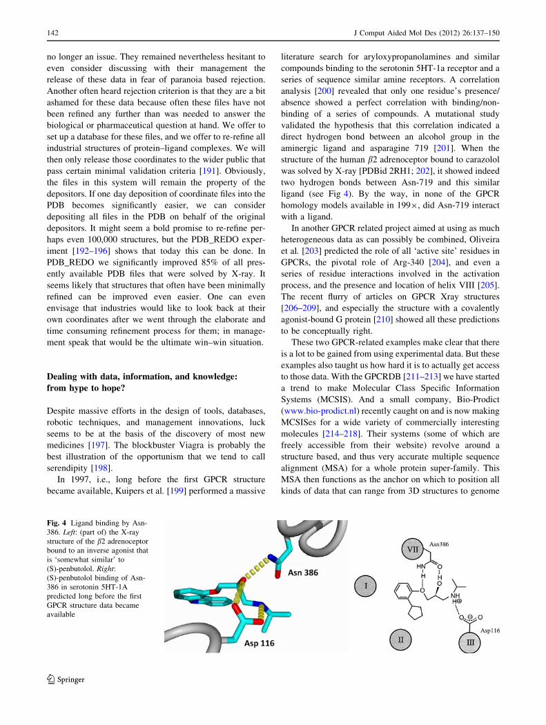
In 1997, i.e., long before the first GPCR structure
became available, Kuipers et al. [199] performed a massive
literature search for aryloxypropanolamines and similar
compounds binding to the serotonin 5HT-1a receptor and a
series of sequence similar amine receptors. A correlation
analysis [200] revealed that only one residue’s presence/
absence showed a perfect correlation with binding/non-
binding of a series of compounds. A mutational study
validated the hypothesis that this correlation indicated a
direct hydrogen bond between an alcohol group in the
aminergic ligand and asparagine 719 [201]. When the
structure of the human b2 adrenoceptor bound to carazolol
was solved by X-ray [PDBid 2RH1; 202], it showed indeed
two hydrogen bonds between Asn-719 and this similar
ligand (see Fig 4). By the way, in none of the GPCR
homology models available in 1999, did Asn-719 interact
with a ligand.
In another GPCR related project aimed at using as much
heterogeneous data as can possibly be combined, Oliveira
et al. [203] predicted the role of all ‘active site’ residues in
GPCRs, the pivotal role of Arg-340 [204], and even a
series of residue interactions involved in the activation
process, and the presence and location of helix VIII [205].
The recent flurry of articles on GPCR Xray structures
[206–209], and especially the structure with a covalently
agonist-bound G protein [210] showed all these predictions
to be conceptually right.
These two GPCR-related examples make clear that there
is a lot to be gained from using experimental data. But these
examples also taught us how hard it is to actually get access
to those data. With the GPCRDB [211–213] we have started
a trend to make Molecular Class Specific Information
Systems (MCSIS). And a small company, Bio-Prodict
(www.bio-prodict.nl) recently caught on and is now making
MCSISes for a wide variety of commercially interesting
molecules [214–218]. Their systems (some of which are
freely accessible from their website) revolve around a
structure based, and thus very accurate multiple sequence
alignment (MSA) for a whole protein super-family. This
MSA then functions as the anchor on which to position all
kinds of data that can range from 3D structures to genome
Fig. 4 Ligand binding by Asn-
386. Left: (part of) the X-ray
structure of the b2 adrenoceptor
bound to an inverse agonist that
is ‘somewhat similar’ to
(S)-penbutolol. Right:(S)-penbutolol binding of Asn-
386 in serotonin 5HT-1A
predicted long before the first
GPCR structure data became
available
142 J Comput Aided Mol Des (2012) 26:137–150
123

related data, from mutation studies to ligand binding con-
stants, or from sequence correlation patterns to the predic-
tion of mutations that enhance the protein’s stability. As the
most powerful information tends to be carefully hidden in
the literature, an extensive set of literature-mining scripts
aids with the extraction of, for example, mutation infor-
mation. In fact, it was shown that the suite of mutation data
extracting scripts reaches a much better coverage than can
be obtained by human experts [214–218].
A recent development that will aid the drug hunters of
the future is the Utopia PDF reader [213, 219]. Vroling et al.
[213] showed how this programmable PDF reader could be
used to directly couple data in articles on GPCRs to the
GPCRDB. This intelligent hyperlinking has a series of
benefits. First, the residue numbering problem gets solved
because the reader can ask the GPCRDB for the position in
the GPCR MSA of any residue mentioned in the article, and
it can even modify or correct the sequence numbers in the
article if needed. Much good GPCR mutation data was
published in the pre-GPCR-structure era that ended with the
opsin structure article [220], and often these data were
misinterpreted because of the poor quality of the available
homology models [221]. The Utopia-GPCR PDF reader can
correct those interpretations thereby salvaging old, high
quality experimental data for future use. Figure 5 shows an
image from an old mutation study [222] in which the
authors describe several ground-breaking mutations in the
guinea pig histamine H1 receptor, building and validating a
homology model using these data, and arguing, for exam-
ple, that residue Trp161 plays an important role in receptor-
ligand binding. This assumption was based on the effect of
the mutation on receptor function, leading to a model in
which Trp161 was modelled in the ligand-binding site. By
contrast, the GPCRDB generated annotation listed in the
sidebar of the reader indicates that this residue, located in
TM IV, points towards the membrane and possibly interacts
with cholesterol. This is a completely different situation
from that proposed by the authors. Looking at the model
provided by the GPCRDB, based on the latest crystal
structures, it can be seen that a direct role of Trp161 in
receptor-ligand binding is highly unlikely.
Folkerstma et al. (2005) analyzed nearly 100 nuclear
receptor (NR) ligand binding domains. Combined with
manually curated multiple sequence alignments, key posi-
tions in the ligand binding pocket were identified that had
specific interactions with functionally diverse compounds.
For example, residues at position 26 in Fig. 6 were shown
to only have interactions with antagonists. This analysis
required a substantial amount of work: categorizing struc-
tures and compounds, creating multiple sequence align-
ments, analyzing ligand contacts, and transferring the
results into a homogeneous residue numbering scheme (the
so-called 3D numbers). With the 3DM information system
[223; see the help movie], these analyses can today be
performed in a matter of minutes [215, 217, 224].
More than 100 articles were found that discuss the
effects of mutating this residue on the ligand binding of the
receptor. In all these articles this same residue has 14
different residue numbers ranging from 52 to 709. The use
of a common 3D numbering scheme enables transfer of
heterogeneous information between protein family mem-
bers. Figure 7 shows 40 antagonists in red and 70 agonists
in blue. In this example, a hundred articles had to be ‘read’
to extract all available mutation information for this single
position mutated in 22 different receptor—species combi-
nations. That these 100 articles had to be found among
Fig. 5 Left, one page from the Histamine H1 article by Wieland et al.
[222] in which Trp161 is suggested to interact with the ligand while
the PDF reader sidebar shows today’s interpretation that this
tryptophan is facing outwards towards the lipid or a dimer partner.
The original picture of the modelled active site is shown enlarged in
the middle panel while the right hand side figure is a plot of the
GPCRDB-derived model of this receptor. The GPCRDB does not
(yet) dock ligands, so the ligand is represented by a hand-added grayball
J Comput Aided Mol Des (2012) 26:137–150 143
123

100,000 PubMed entries that contain NR information is a
whole different story in itself.
If, one day, all structures of NR-ligand complexes that
now are scattered over inaccessible industrial hard disks
could be concentrated in one system, then we could con-
sider asking much more elaborate questions. We could
consider correlating aspects of ligands with protein atom
characteristics, or we could analyse if residues not
contacting the ligand have an influence on binding or
activation, etcetera.
It is not only important to get as much information as
possible stored in systems amenable to scrutiny, but it is
also important to realize that for every one bioinformati-
cian or drug hunter there are one hundred scientists who do
not use molecular software regularly. Project Hope aims to
predict the molecular phenotype of point mutations that
were shown causally related to human disease states [225].
This system attempts in all stages of user interaction to
cater for human geneticists who typically do not use
molecular software at all. Hope only asks the user to cut-n-
paste the sequence, and click the residue mutated and the
mutation residue type. It then builds a homology model if
needed, calls dozens of servers and services in seven
countries, combines all possible information and writes a
final report that can be directly used in publications, but,
more importantly, that is written without using any bioin-
formatics jargon and even has a build-in dictionary that
explains terms such as ‘active site’, ‘salt-bridge’, or ‘tor-
sion angle’ in human genetics understandable terms. Hope
thus is the ultimate translation machine because in doing
translational research it even translates between the
researchers.
We believe that the recent spate of consolidations in the
pharmaceutical industry is not a problem but an opportu-
nity. Mankind needs medicines, and now that pushing ones
luck is slowly becoming a less successful technique, only
research can find them. This research can progress rapidly
if the thousands and thousands of X-ray structures of pro-
tein–ligand complexes would find their way from hard-
disks behind pharmaceutical industry firewalls to the public
domain. Drug design research in the next 25 years will
revolve around ever broader collaborations, ever more
holistic understanding of the drug—human interactions,
Fig. 6 Bargraph showing the
number of ligand contacts per
residue extracted from 776
nuclear receptor ligand binding
domain structures plotted as
function of their 3D numbers.
The blue bars represent the
number of contacts with
agonistic compounds. The redbars indicate the number of
contacts with antagonistic
compounds. The residue with
3D number 26 is only bound to
antagonistic compounds
Fig. 7 Cartoon representation of two superposed representative NR
structures (one bound to an agonist; one bound to an antagonist).
These two structures, obviously, differ most in the location of Helix
12. The blue ligands are agonists; the red ones are antagonists. The
ligands were placed in the same orientation as found in their native
PDB file. All PDB files were superposed on the representative NR
structures. Residue 26, for which the antagonist interaction had been
mentioned in the literature, is shown in yellow, as is residue for which
Fig. 6 also indicates antagonist interactions, albeit with less antag-
onists than residue 26. This role of residue 29 might represent a novel
finding. Figure made with the 3DM-plugin for the YASARA—
WHAT IF suite
144 J Comput Aided Mol Des (2012) 26:137–150
123

and ever better use of the available data, information, and
knowledge.
Acknowledgments VL and GV acknowledge financial support
from NBIC, and TIPharma, TvdB appreciate the support from Bio-
Prodict (www.bio-prodict.com). The authors thank Jan Kelder for
critically reviewing the manuscript. Elmar Krieger helped with YA-
SARA, Maarten Hekkelman, Coos Baakman, Bas Vroling, Wilmar
Teunissen, Barbara van Kampen, provided technical support. The
authors mention with pleasure the many stimulating discussions with
Sander Nabuurs, Daniel Girones, Gijs Schaftenaar, Friedrich Ripp-
mann, Ad IJzerman, Margot Beukers, Isabel Duarte, Christof Francke,
Henk-Jan Joosten, Jacob de Vlieg.
Open Access This article is distributed under the terms of the
Creative Commons Attribution Noncommercial License which per-
mits any noncommercial use, distribution, and reproduction in any
medium, provided the original author(s) and source are credited.
References
1. Life expectancy (2011) http://en.wikipedia.org/wiki/Life_expect
ancy. Accessed 6 Dec 2011
2. World War I casualties (2011) http://en.wikipedia.org/wiki/
World_War_I_casualties. Accessed 6 Dec 2011
3. Medecine and World War II (2011) http://www.historylearning
site.co.uk/medicine_and_world_war_two.htm. Accessed 16 Feb
2011
4. Snell ES, Griffin JP (1985) How many medicines are there? Br
Med J 290:773–774
5. Lewington A (1993) Medicinal plants and plant extracts: a
review of the importation into Europe. Traffic network report.
Traffic International, Cambridge, UK
6. Overington JP, Al-Lazikani B, Hopkins AL (2006) How many
drug targets are there? Nat Rev Drug Discov. doi:10.1038/
nrd2199
7. Munos B (2009) Lessons from 60 years of pharmaceutical
innovation. Nat Rev Drug Discov. doi:10.1038/nrd2961
8. Troubled Asset Relief Program (2011) http://en.wikipedia.org/
wiki/Troubled_Asset_Relief_Program. Accessed 6 Dec 2011
9. Firestone RA (2011) Lessons from 54 years of pharmaceutical
research Nat Rev Drug Discov. doi:10.1038/nrd2961-c1
10. Robert B, Matthew T, Sean H, Bernard B (2000) Drug design by
machine learning: support vector machine for pharmaceutical
data analysis. Proceedings of the AISB’00 symposium on arti-
ficial intelligence in bioinformatics. pp 1–4
11. Byvatov E, Fechner U, Sadowski J, Schneider G (2003) Com-
parison of support vector machine and artificial neural network
systems for drug/nondrug classification. J Chem Inf Comput Sci
43:1882–1889
12. Gagnon MA, Lexchin J (2008) The cost of pushing pills: a new
estimate of pharmaceutical promotion expenditures in the Uni-
ted States. PLoS Med. doi:10.1371/journal.pmed.0050001
13. Hol WGJ (1986) Protein crystallography and computer-graphics
toward rational drug design. Angew Chem Int Ed Engl 25:
767–778
14. Bolin JT, Filman DJ, Matthews DA, Hamlin RC, Kraut J (1982)
Crystal structures of Escherichia coli and Lactobacillus casei
dihydrofolate reductase refined at 1.7 A resolution. I. General
features and binding of methotrexate. J Biol Chem 257:
13663–13672
15. Thompson PE, Manallack DT, Blaney FE, Gallagher T (1992)
Conformational studies on (?)-anatoxin-a and derivatives.
J Comput Aided Mol Des 6:287–298
16. Ruiz J, Lopez M, Mila J, Lozoya E, Lozano JJ, Pouplana R
(1993) QSAR and conformational analysis of the antiinflam-
matory agent amfenac and analogues. J Comput Aided Mol Des
7:183–198
17. Aleman C, Perez JJ (1993) SCF-MO study of the polyglycine II
structure. J Comput Aided Mol Des 7(2):241–250
18. Oyasu H, Nakanishi I, Tanaka A, Murano K, Matsuo M (1995)
Conformational studies on the four stereoisomers of the novel
anticholinergic 4-(dimethylamino)-2-phenyl-2-(2-pyridyl)pen-
tanamide. J Comput Aided Mol Des 9:171–180
19. Schaftenaar G, Noordik JH (2000) Molden: a pre- and post-
processing program for molecular and electronic structures.
J Comput Aided Mol Des 14:123–134
20. Manzetti S, McCulloch DR, Herington AC, van der Spoel D
(2003) Modeling of enzyme-substrate complexes for the me-
talloproteases MMP-3, ADAM-9 and ADAM-10. J Comput
Aided Mol Des 17:551–565
21. Duran D, Aviyente V, Baysa C (2004) Solvent effect on the
synthesis of clarithromycin: a molecular dynamics study.
J Comput Aided Mol Des 18:145–154
22. Curioni A, Mordasini T, Andreoni W (2004) Enhancing the
accuracy of virtual screening: molecular dynamics with quan-
tum-refined force fields. J Comput Aided Mol Des 18:773–784
23. Hammond PS, Wu Y, Harris R, Minehardt TJ, Car R, Schmitt JD
(2005) Protonation-induced stereoisomerism in nicotine: confor-
mational studies using classical (AMBER) and ab initio (Car-Par-
rinello) molecular dynamics. J Comput Aided Mol Des 19:1–15
24. Roccatano D, Sbardella G, Aschi M, Amicosante G, Bossa C,
Nola AD, Mazza F (2005) Dynamical aspects of TEM-1 beta-
lactamase probed by molecular dynamics. J Comput Aided Mol
Des 19:329–340
25. Chipot C, Rozanska X, Dixit SB (2005) Can free energy cal-
culations be fast and accurate at the same time? Binding of low-
affinity, non-peptide inhibitors to the SH2 domain of the src
protein. J Comput Aided Mol Des 19:765–770
26. Fanelli F, De Benedetti PG (2006) Inactive and active states and
supramolecular organization of GPCRs: insights from compu-
tational modeling. J Comput Aided Mol Des 20:449–461
27. Bharatham K, Bharatham N, Kwon YJ, Lee KW (2008)
Molecular dynamics simulation study of PTP1B with allosteric
inhibitor and its application in receptor based pharmacophore
modeling. J Comput Aided Mol Des 22:925–933
28. Eyrisch S, Helms V (2009) What induces pocket openings on
protein surface patches involved in protein-protein interactions?
J Comput Aided Mol Des 23:73–86
29. van Gunsteren WF, Berendsen HJ (1987) Thermodynamic cycle
integration by computer simulation as a tool for obtaining free
energy differences in molecular chemistry. J Comput Aided Mol
Des 1:171–176
30. Hansson T, Marelius J, Aqvist J (1998) Ligand binding affinity
prediction by linear interaction energy methods. J Comput
Aided Mol Des 12:27–35
31. Wilcox GL, Quiocho FA, Levinthal C, Harvey SC, Maggiora
GM, McCammon JA (1988) Symposium overview. Minnesota
conference on supercomputing in biology: proteins, nucleic
acids, and water. J Comput Aided Mol Des 1:271–281
32. Wimmer E (1988) Future in biomolecular computation. J Com-
put Aided Mol Des 1:283–290
33. Allinger NL (1977) Conformational-analysis. 130. Mm2—
hydrocarbon force-field utilizing V1 and V2 torsional terms.
J Am Chem Soc 99:8127–8134
34. Stewart JJP (1990) Special issue—Mopac—a semiempirical
molecular-orbital program. J Comput Aided Mol Des 4:1–45
J Comput Aided Mol Des (2012) 26:137–150 145
123

35. Allen FH, Harris SE, Taylor R (1996) Comparison of conformer
distributions in the crystalline state with conformational ener-
gies calculated by ab initio techniques. J Comput Aided Mol Des
10:247–254
36. Klebe G, Mietzner T (1994) A fast and efficient method to
generate biologically relevant conformations. J Comput Aided
Mol Des 8:583–606
37. Bostrom J, Norrby PO, Liljefors T (1998) Conformational
energy penalties of protein-bound ligands. J Comput Aided Mol
Des 12:383–396
38. Mayer D, Naylor CB, Motoc I, Marshall GR (1987) A unique
geometry of the active site of angiotensin-converting enzyme
consistent with structure-activity studies. J Comput Aided Mol
Des 1:3–16
39. Martin J, Andrews P (1987) Conformation-activity relationships
of opiate analgesics. J Comput Aided Mol Des 1:53–72
40. Martin YC, Bures MG, Danaher EA, DeLazzer J, Lico I, Pavlik
PA (1993) A fast new approach to pharmacophore mapping and
its application to dopaminergic and benzodiazepine agonists.
J Comput Aided Mol Des 7:83–102
41. Lakdawala A, Wang M, Nevins N, Liotta D, Rusinska-Roszak
D, Lozynski M, Snyder JP (2001) Calculated conformer ener-
gies for organic molecules with multiple polar functionalities are
method dependent: taxol (case study). BMC Chem Biol. doi:
10.1186/1472-6769-1-2
42. Vieth M, Hirst JD, Brooks CL (1998) Do active site confor-
mations of small ligands correspond to low free-energy solution
structures? J Comput Aided Mol Des 12:563–572
43. Klebe G (1995) Toward a more efficient handling of confor-
mational flexibility in computer-assisted modelling of drug
molecules. Perspect Drug Discov Des 3:85–105
44. Ota N, Agard DA (2001) Binding mode prediction for a flexible
ligand in a flexible pocket using multi-conformation simulated
annealing pseudo crystallographic refinement. J Mol Biol
314:607–617
45. Diller DJ, Merz KM Jr (2002) Can we separate active from
inactive conformations? J Comput Aided Mol Des 16:105–112
46. Huse M, Kuriyan J (2003) The conformational plasticity of
protein kinases. Cell 109:275–282
47. Teague SJ (2003) Implications of protein flexibility for drug
discovery. Nat Rev Drug Discov 2:527–541
48. Chouard T (2005) Structural biology: breaking the protein rules.
Nature 471:151–153
49. Dyson HJ, Wright PE (2005) Intrinsically unstructured proteins
and their functions. Natl Rev Mol Cell Biol 6:197–208
50. Uversky VN, Dunker AK (2010) Understanding protein non-
folding. Biochim Biophys Acta 1804:1231–1264
51. Kuntz ID, Blaney JM, Oatley SJ, Langridge R, Ferrin TE (1982)
A geometric approach to macromolecule-ligand interactions.
J Mol Biol 161:269–288
52. Bohm HJ (1994) The development of a simple empirical scoring
function to estimate the binding constant for a protein-ligand
complex of known three-dimensional structure. J Comput Aided
Mol Des 8:243–256
53. Meng EC, Kuntz ID, Abraham DJ, Kellogg GE (1994) Evalu-
ating docked complexes with the HINT exponential function
and empirical atomic hydrophobicities. J Comput Aided Mol
Des 8:299–306
54. Jain AN (1996) Scoring noncovalent protein-ligand interactions:
a continuous differentiable function tuned to compute binding
affinities. J Comput Aided Mol Des 10:427–440
55. Wang R, Lai L, Wang S (2002) Further development and
validation of empirical scoring functions for structure-based
binding affinity prediction. J Comput Aided Mol Des
16:11–26
56. Kelly MD, Mancera RL (2003) A new method for estimating the
importance of hydrogen-bonding groups in the binding site of a
protein. J Comput Aided Mol Des 17:401–414
57. Muryshev AE, Tarasov DN, Butygin AV, Butygina OY, Alek-
sandrov AB, Nikitin SM (2003) A novel scoring function for
molecular docking. J Comput Aided Mol Des 17:597–605
58. Morley SD, Afshar M (2004) Validation of an empirical RNA-
ligand scoring function for fast flexible docking using Ribodock.
J Comput Aided Mol Des 18:189–208
59. Tame JR (2005) Scoring functions—the first 100 years. J Com-
put Aided Mol Des 19:445–451
60. Jain AN (2009) Surflex-Dock 2.1: robust performance from
ligand energetic modeling, ring flexibility, and knowledge-based
search. J Comput Aided Mol Des 21:281–306
61. Cincilla G, Vidal D, Pons M (2009) An improved scoring
function for suboptimal polar ligand complexes. J Comput
Aided Mol Des 23:143–152
62. Dobes P, Fanfrlık J, Rezac J, Otyepka M, Hobza P (2011)
Transferable scoring function based on semiempirical quantum
mechanical PM6-DH2 method: CDK2 with 15 structurally
diverse inhibitors. J Comput Aided Mol Des 25(3):223–235
63. Tondel K, Anderssen E, Drablos F (2006) Protein alpha shape
(PAS) dock: a new gaussian-based score function suitable for
docking in homology modelled protein structures. J Comput
Aided Mol Des 20:131–144
64. Miller MD, Kearsley SK, Underwood DJ, Sheridan RP (1994)
FLOG: a system to select ‘quasi-flexible’ ligands complemen-
tary to a receptor of known three-dimensional structure.
J Comput Aided Mol Des 8:153–174
65. Kearsley SK, Underwood DJ, Sheridan RP, Miller MD Flexi-
bases: a way to enhance the use of molecular docking methods.
J Comput Aided Mol Des 8:565–582
66. Oshiro CM, Kuntz ID, Dixon JS (1995) Flexible ligand docking
using a genetic algorithm. J Comput Aided Mol Des 9:113–130
67. Knegtel RM, Bayada DM, Engh RA, von der Saal W, van Ge-
erestein VJ, Grootenhuis PD Comparison of two implementa-
tions of the incremental construction algorithm in flexible
docking of thrombin inhibitors. J Comput Aided Mol Des
13:167–183
68. Makino S, Ewing TJ, Kuntz ID (1999) DREAM??: flexible
docking program for virtual combinatorial libraries. J Comput
Aided Mol Des 13:513–532
69. Ewing TJ, Makino S, Skillman AG, Kuntz ID (2001) DOCK
4.0: search strategies for automated molecular docking of flex-
ible molecule databases. J Comput Aided Mol Des 15:411–428
70. Hindle SA, Rarey M, Buning C, Lengaue T (2002) Flexible
docking under pharmacophore type constraints. J Comput Aided
Mol Des 16:129–149
71. Grasselli M, Cascone O, Birger Anspach F, Delfino JM (2002)
On the molecular interaction between lactoferrin and the dye
Red HE-3b. A novel approach for docking a charged and highly
flexible molecule to protein surfaces. J Comput Aided Mol Des
16:917–934
72. Bursulaya BD, Totrov M, Abagyan R, Brooks CL 3rd (2003)
Comparative study of several algorithms for flexible ligand
docking. J Comput Aided Mol Des 17:755–763
73. Bottegoni G, Kufareva I, Totrov M, Abagyan R (2008) A new
method for ligand docking to flexible receptors by dual alanine
scanning and refinement (SCARE). J Comput Aided Mol Des
22:311–325
74. Zhao Y, Sanner MF (2008) Protein-ligand docking with multiple
flexible side chains. J Comput Aided Mol Des 22:673–679
75. Kang L, Li H, Jiang H, Wang X (2009) An improved adaptive
genetic algorithm for protein-ligand docking. J Comput Aided
Mol Des 23:1–12
146 J Comput Aided Mol Des (2012) 26:137–150
123

76. Jain AN (2009) Effects of protein conformation in docking:
improved pose prediction through protein pocket adaptation.
J Comput Aided Mol Des 23:355–374
77. Garden DP, Zhorov BS (2010) Docking flexible ligands in
proteins with a solvent exposure- and distance-dependent
dielectric function. J Comput Aided Mol Des 24:91–105
78. Totrov M, Abagyan R (2008) Flexible ligand docking to mul-
tiple receptor conformations: a practical alternative. Curr Opin
Struct Biol 18:178–184
79. Bohm HJ, Klebe G (1996) What can we learn from molecular
recognition in protein-ligand complexes for the design of new
drugs? Angew Chem Int Ed Engl 35:2589–2614
80. Shoichet BK, Leach AR, Kuntz ID (1999) Ligand solvation in
molecular docking. Protein Sruct Funct Genet 34:4–16
81. Gohlke H, Klebe G (2002) Approaches to the description and
prediction of the binding affinity of small-molecule ligands to
macromolecular receptors. Angew Chem Int Ed 41:2644–2676
82. Demchuk E, Wade RC (1996) Improving the continuum
dielectric approach to calculating pK(a)s of ionizable groups in
proteins. J Phys Chem 100:17373–17387
83. Nielsen JE, Vriend G (2001) Optimizing the hydrogen-bond
network in Poisson-Boltzmann equation-based pK(a) calcula-
tions. Protein Stuct Funct Genet 43:403–412
84. Rarey M, Kramer B, Lengauer T, Klebe G (1996) A fast flexible
docking method using an incremental construction algorithm.
J Mol Biol 261:470–489
85. Goodsell DS, Morris GM, Olson AJ (1996) Automated docking
of flexible ligands: applications of AutoDock. J Mol Recognit
9:1–5
86. Friesner RA, Banks JL, Murphy RB, Halgren TA, Klicic JJ,
Mainz DT, Repasky MP, Knoll EH, Shelley M, Perry JK, Shaw
DE, Francis P, Shenkin PS (2004) A new approach for rapid,
accurate docking and scoring. 1. Method and assessment of
docking accuracy. J Med Chem 47:1739–1749
87. Jones G, Willett P, Glen RC, Leach AR, Taylor R (1997)
Development and validation of a genetic algorithm for flexible
docking. J Mol Biol 267:727–748
88. Hartmann C, Antes I, Lengauer T (2009) Docking and scoring
with alternative side-chain conformations. Proteins 74:712–726
89. Taylor RD, Jewsbury PJ, Essex JW (2003) FDS: flexible ligand
and receptor docking with a continuum solvent model and soft-
core energy function. J Comput Chem 24:1637–1656
90. Muegge I (2006) PMF scoring revisited. J Med Chem
49:5895–5902
91. Englebienne P, Moitessier N (2009) Docking ligands into flex-
ible and solvated macromolecules. 4. Are popular scoring
functions accurate for this class of proteins? J Chem Inf Model
49:1568–1580
92. Oda A, Tsuchida K, Takakura T, Yamaotsu N, Hirono S (2006)
Comparison of consensus scoring strategies for evaluating
computational models of protein-ligand complexes. J Chem Inf
Model 46:380–391
93. Foloppe N, Hubbard R (2006) Towards predictive ligand design
with free-energy based computational methods? Curr Med
Chem 13:3583–3608
94. Jain AN (2006) Scoring functions for protein-ligand docking.
Curr Protein Pept Sci 7:407–420
95. Robertson TA, Varani G (2007) An all-atom, distance-depen-
dent scoring function for the prediction of protein-DNA inter-
actions from structure. Proteins 66:359–374
96. Rajamani R, Good AC (2007) Ranking poses in structure-based
lead discovery and optimization: current trends in scoring
function development. Curr Opin Drug Discov Devel
10:308–315
97. Tembre BL, McCammon JA (1984) Ligand-receptor interac-
tions. Comput Chem 8:281–283
98. Ferguson DM, Radmer RJ, Kollman PA (1991) Determination
of the relative binding free-energies of peptide inhibitors to the
Hiv-1 protease. J Med Chem 34:2654–2659
99. Cramer RD, Patterson DE, Bunce JD (1988) Comparative
molecular-field analysis (Comfa).1. Effect of shape on binding
of steroids to carrier proteins. J Am Chem Soc 110:5959–5967
100. Norinder U (1990) Experimental design based 3-D QSAR
analysis of steroid-protein interactions: application to human
CBG complexes. J Comput Aided Mol Des 4:381–389
101. Bursi R, Grootenhuis PD (1999) Comparative molecular field
analysis and energy interaction studies of thrombin-inhibitor
complexes. J Comput Aided Mol Des 13:221–232
102. Zhang Z, An L, Hu W, Xiang Y (2007) 3D-QSAR study of
hallucinogenic phenylalkylamines by using CoMFA approach.
J Comput Aided Mol Des 21:145–153
103. Cramer RD, Milne M (1979) Lattice model–general paradigm
for shape-related structure-activity correlation. Abstracts of
papers of the American chemical society, 19th ACS Meeting
COMP 44
104. Nicklaus MC, Milne GW, Burke TR Jr (1992) QSAR of con-
formationally flexible molecules: comparative molecular field
analysis of protein-tyrosine kinase inhibitors. J Comput Aided
Mol Des 6:487–504
105. Rault S, Bureau R, Pilo JC, Robba M (1992) Comparative
molecular field analysis of CCK-A antagonists using field-fit as
an alignment technique. A convenient guide to design new
CCK-A ligands. J Comput Aided Mol Des 6:553–568
106. Calder JA, Wyatt JA, Frenkel DA, Casida JE (1993) CoMFA
validation of the superposition of six classes of compounds
which block GABA receptors non-competitively. J Comput
Aided Mol Des 7:45–60
107. Kroemer RT, Hecht P (1995) A new procedure for improving
the predictiveness of CoMFA models and its application to a set
of dihydrofolate reductase inhibitors. J Comput Aided Mol Des
9:396–406
108. Gohda K, Mori I, Ohta D, Kikuchi T (2000) A CoMFA analysis
with conformational propensity: an attempt to analyze the SAR of
a set of molecules with different conformational flexibility using a
3D-QSAR method. J Comput Aided Mol Des 14:265–275
109. Manchester J, Czerminski R (2009) CAUTION: popular
‘‘Benchmark’’ data sets do not distinguish the merits of 3D
QSAR methods. J Chem Inf Model 49:1449–1454
110. Kharkar PS, Reith ME, Dutta AK (2008) Three-dimensional
quantitative structure-activity relationship (3D QSAR) and
pharmacophore elucidation of tetrahydropyran derivatives as
serotonin and norepinephrine transporter inhibitors. J Comput
Aided Mol Des 22:1–17
111. Park H, Lee S (2004) Homology modeling, force field design,
and free energy simulation studies to optimize the activities of
histone deacetylase inhibitors. J Comput Aided Mol Des
18:375–388
112. Tomich CH, da Silva P, Carvalho I, Taft CA (2005) Homology
modeling and molecular interaction field studies of alpha-glu-
cosidases as a guide to structure-based design of novel proposed
anti-HIV inhibitors. J Comput Aided Mol Des 19:83–92
113. Rossi KA, Markwalder JA, Seitz SP, Chang CH, Cox S, Bois-
clair MD, Brizuela L, Brenner SL, Stouten PF (2005) Under-
standing and modulating cyclin-dependent kinase inhibitor
specificity: molecular modeling and biochemical evaluation of
pyrazolopyrimidinones as CDK2/cyclin A and CDK4/cyclin D1
inhibitors. J Comput Aided Mol Des 19:111–122
114. Schlegel B, Laggner C, Meier R, Langer T, Schnell D, Seifert R,
Stark H, Holtje HD, Sippl W (2007) Generation of a homology
model of the human histamine H(3) receptor for ligand docking
and pharmacophore-based screening. J Comput Aided Mol Des
21:437–453
J Comput Aided Mol Des (2012) 26:137–150 147
123

115. Katritch V, Byrd CM, Tseitin V, Dai D, Raush E, Totrov M,
Abagyan R, Jordan R, Hruby DE (2007) Discovery of small
molecule inhibitors of ubiquitin-like poxvirus proteinase I7L
using homology modeling and covalent docking approaches.
J Comput Aided Mol Des 21:549–558
116. Neves MA, Simoes S, Sa e Melo ML (2010) Ligand-guided
optimization of CXCR4 homology models for virtual screening
using a multiple chemotype approach. J Comput Aided Mol Des
24:1023–1033
117. Knehans T, Schuller A, Doan DN, Nacro K, Hill J, Guntert P,
Madhusudhan MS, Weil T, Vasudevan SG (2011) Structure-
guided fragment-based in silico drug design of dengue protease
inhibitors. J Comput Aided Mol Des 25:263–274
118. Eberini I, Daniele S, Parravicini C, Sensi C, Trincavelli ML,
Martini C, Abbracchio MP (2011) In silico identification of new
ligands for GPR17: a promising therapeutic target for neuro-
degenerative diseases. J Comput Aided Mol Des 25:743–752
119. Sali A, Blundell TL (1993) Comparative protein modelling by
statisfaction of spatial restraints. J Mol Biol 234:779–815
120. Vriend G (1990) WHAT IF: a molecular modeling and drug
design program. J Mol Graph 8:52–56
121. Reichelt J, Dieterich G, Kvesic M, Schomburg D, Heinz DW
(2005) BRAGI: linking and visualization of database informa-
tion in a 3D viewer and modeling tool. Bioinformatics
21:1291–1293
122. Krieger E, Joo K, Lee J, Lee J, Raman S, Thompson J, Tyka M,
Baker D, Karplus K (2009) Improving physical realism, ste-
reochemistry, and side-chain accuracy in homology modeling:
four approaches that performed well in CASP8. Proteins
9:114–122
123. Thompson J, Baker D (2011) Incorporation of evolutionary
information into Rosetta comparative modeling. Protein Struct
Func Bioinfo 79:2380–2388
124. CASP1 proceedings (1995) Protein Struct Funct Genet 23:
295–460
125. Harris MR, Kihlen M, Bywater RP (1993) PLIM: a protein-
ligand interaction modeller. J Mol Recognit 6:111–115
126. Michino M, Abola E; GPCR Dock 2008 participants, Brooks CL
3rd, Dixon JS, Moult J, Stevens RC (2009) Community-wide
assessment of GPCR structure modelling and ligand docking:
GPCR Dock 2008 Nat Rev Drug Discov 8:455–463
127. Kufareva I, Rueda M, Vsevolod K, Stevens RC, Abagyan R,
GPCR Dock 2010 participants Status of GPCR modeling and
docking as reflected by community wide GPCR Dock 2010
assessment. Structure 19:1108–1126
128. Broer BM, Gurrath M, Holtje HD (2003) Molecular modelling
studies on the ORL1-receptor and ORL1-agonists. J Comput
Aided Mol Des 17:739–754
129. Miguet L, Zhang Z, Barbier M, Grigorov MG (2006) Compar-
ison of a homology model and the crystallographic structure of
human 11beta-hydroxysteroid dehydrogenase type 1 (11be-
taHSD1) in a structure-based identification of inhibitors.
J Comput Aided Mol Des 20:67–81
130. Roumen L, Sanders MP, Vroling B, De Esch IJ, De Vlieg J,
Leurs R, Klomp JP, Nabuurs SB, De Graaf C (2011) The pitfalls
and challenges of predicting GPCR-ligand interactions. Phar-
maceuticals 4:1196–1215
131. Henriques ES, Floriano WB, Reuter N, Melo A, Brown D,
Gomes JA, Maigret B, Nascimento MA, Ramos MJ (2001) The
search for a new model structure of beta-factor XIIa. J Comput
Aided Mol Des 15:309–322
132. Brown N, McKay B, Gasteiger J (2004) The de novo design of
median molecules within a property range of interest. J Comput
Aided Mol Des 18:761–771
133. Belda I, Madurga S, Llora X, Martinell M, Tarrago T, Piqueras
MG, Nicolas E, Giralt E (2005) ENPDA: an evolutionary
structure-based de novo peptide design algorithm. J Comput
Aided Mol Des 19:585–601
134. Zaliani A et al (2009) Second-generation de novo design: a view
from a medicinal chemist perspective. J Comput Aided Mol Des
135. Goodford PJ (1985) A computational-procedure for determining
energetically favorable binding-sites on biologically important
macromolecules. J Med Chem 28:849–857
136. Wade RC, Clark KJ, Goodford PJ (1993) Further development
of hydrogen-bond functions for use in determining energetically
favorable binding-sites on molecules of known structure.1.
Ligand probe groups with the ability to form 2 hydrogen-bonds.
J Med Chem 36:140–147
137. Rotstein SH, Murcko MA (1993) GroupBuild: a fragment-based
method for de novo drug design. J Med Chem 36:1700–1710
138. Willett P (1995) Genetic algorithms in molecular recognition
and design. Trends Biotechnol 13:516–521
139. Bohm HJ (1992) The computer program LUDI: a new method
for the de novo design of enzyme inhibitors. J Comput Aided
Mol Des 6:61–78
140. Bohm HJ (1992) LUDI: rule-based automatic design of new
substituents for enzyme inhibitor leads. J Comput Aided Mol
Des 6:593–606
141. Clark DE, Frenkel D, Levy SA, Li J, Murray CW, Robson B,
Waszkowycz B, Westhead DR (1995) PRO-LIGAND: an
approach to de novo molecular design. 1. Application to the
design of organic molecules. J Comput Aided Mol Des 9:13–32
142. Westhead DR, Clark DE, Frenkel D, Li J, Murray CW, Robson
B, Waszkowycz B (1995) PRO-LIGAND: an approach to de
novo molecular design. 3. A genetic algorithm for structure
refinement. J Comput Aided Mol Des 9:139–148
143. Rarey M, Wefing S, Lengauer T (1996) Placement of medium-
sized molecular fragments into active sites of proteins. J Comput
Aided Mol Des 10:41–54
144. Barakat MT, Dean PM (1995) The atom assignment problem in
automated de novo drug design. 1. Transferability of molecular
fragment properties. J Comput Aided Mol Des 9:341–350
145. Rotstein SH, Murcko MA (1993) GenStar: a method for de novo
drug design. J Comput Aided Mol Des 7:23–43
146. Roe DC, Kuntz ID (1995) BUILDER v.2: improving the
chemistry of a de novo design strategy. J Comput Aided Mol
Des 9:269–282
147. Leach AR, Kilvington SR (1994) Automated molecular design:
a new fragment-joining algorithm. J Comput Aided Mol Des
8:283–298
148. Golberg DE (1989) Genetic algorithms in search, optimization
and machine learning. Addison-Wesley, New York
149. Todorov NP, Dean PM (1997) Evaluation of a method for
controlling molecular scaffold diversity in de novo ligand
design. J Comput Aided Mol Des 11:175–192
150. Schneider G, Lee ML, Stahl M, Schneider P (2000) De novo
design of molecular architectures by evolutionary assembly of
drug-derived building blocks. J Comput Aided Mol Des
14:487–494
151. Blum LC, Reymond JL (2009) 970 million druglike small
molecules for virtual screening in the chemical universe data-
base GDB-13. J Am Chem Soc 131:8732–8733
152. Kubinyi H (1992) HTS technologies—IBC informa conference.
IDrugs 4:168–173
153. Sudarsanam S, Virca GD, March CJ, Srinivasan S (1992) An
approach to computer-aided inhibitor design: application to
cathepsin L. J Comput Aided Mol Des 6:223–233
154. Bohm HJ, Banner DW, Weber L (1999) Combinatorial docking
and combinatorial chemistry: design of potent non-peptide
thrombin inhibitors. J Comput Aided Mol Des 13:51–56
155. Filikov AV, Mohan V, Vickers TA, Griffey RH, Cook PD,
Abagyan RA, James TL (2000) Identification of ligands for
148 J Comput Aided Mol Des (2012) 26:137–150
123

RNA targets via structure-based virtual screening: HIV-1 TAR.
J Comput Aided Mol Des 14:593–610
156. Furet P, Meyer T, Mittl P, Fretz H (2001) Identification of cylin-
dependent kinase 1 inhibitors of a new chemical type by
structure-based design and database searching. J Comput Aided
Mol Des 15:489–495
157. Afantitis A, Melagraki G, Sarimveis H, Koutentis PA, Marko-
poulos J, Igglessi-Markopoulou O (2006) Investigation of
substituent effect of 1-(3, 3-diphenylpropyl)-piperidinyl pheny-
lacetamides on CCR5 binding affinity using QSAR and virtual
screening techniques. J Comput Aided Mol Des 20:83–95
158. Kortagere S, Welsh WJ (2006) Development and application of
hybrid structure based method for efficient screening of ligands
binding to G-protein coupled receptors. J Comput Aided Mol
Des 20:789–802
159. Talevi A, Bellera CL, Castro EA, Bruno-Blanch LE (2007) A
successful virtual screening application: prediction of anticon-
vulsant activity in MES test of widely used pharmaceutical and
food preservatives methylparaben and propylparaben. J Comput
Aided Mol Des 21:527–538
160. Irwin JJ (2008) Community benchmarks for virtual screening.
J Comput Aided Mol Des 22:193–199
161. Krueger BA, Weil T, Schneider G (2009) Comparative virtual
screening and novelty detection for NMDA-GlycineB antago-
nists. J Comput Aided Mol Des 23:869–881
162. Didziapetris R, Dapkunas J, Sazonovas A, Japertas P (2010)
Trainable structure-activity relationship model for virtual
screening of CYP3A4 inhibition. J Comput Aided Mol Des
24:891–906
163. Joseph-McCarthy D (2002) An overview of in silico design and
screening: toward efficient drug discovery. Curr Drug Discov
20–23
164. Bajorath J (2002) Virtual screening: methods, expectations, and
reality. Curr Opin Drug Discov Dev 2:24–28
165. Rarey M, Stahl M (2001) Similarity searching in large combi-
natorial chemistry spaces. J Comput Aided Mol Des 15:
497–520
166. Hopkins AL, Groom CR, Alex A (2004) Ligand efficiency: a
useful metric for lead selection. Drug Discov Today 9:430–431
167. Abad-Zapatero C, Metz JT (2005) Ligand efficiency indices as
guideposts for drug discovery. Drug Discov Today 10:464–469
168. Jain AN, Koile K, Chapman D (1994) Compass: predicting
biological activities from molecular surface properties. Perfor-
mance comparisons on a steroid benchmark. J Med Chem
37:2315–2327
169. Handschuh S, Goldfuss B, Chen J, Gasteiger J, Houk KN (2000)
Steroid binding by antibodies and artificial receptors: explora-
tion of theoretical methods to determine the origins of binding
affinities and specificities. J Comput Aided Mol Des 14:611–629
170. Jain AN, Dietterich TG, Lathrop RH, Chapman D, Critchlow RE
Jr, Bauer BE, Webster TA, Lozano-Perez T (1994) A shape-
based machine learning tool for drug design. J Comput Aided
Mol Des 8:635–652
171. Walters WP, Murcko MA (2002) Prediction of ‘drug-likeness’.
Adv Drug Deliv Rev 54:255–271
172. Jung E, Choi SH, Lee NK, Kang SK, Choi YJ, Shin JM, Choi K,
Jung DH (2011) Machine learning study for the prediction of
transdermal peptide. J Comput Aided Mol Des 25:339–347
173. Sapre NS, Gupta S, Pancholi N, Sapre N (2008) Molecular
docking studies on tetrahydroimidazo-[4, 5, 1-jk][1, 4]-benzo-
diazepinone (TIBO) derivatives as HIV-1 NNRT inhibitors.
J Comput Aided Mol Des 22:69–80
174. Hirst JD, King RD, Sternberg MJ (1994) Quantitative structure-
activity relationships by neural networks and inductive logic
programming. II. The inhibition of dihydrofolate reductase by
triazines. J Comput Aided Mol Des 8:421–432
175. Livingstone DJ, Manallack DT, Tetko IV (1997) Data modelling
with neural networks: advantages and limitations. J Comput
Aided Mol Des 11:135–142
176. Doweyko AM (2008) QSAR: dead or alive? J Comput Aided
Mol Des 22:81–89
177. Che Y, Brooks BR, Marshall GR (2006) Development of small
molecules designed to modulate protein-protein interactions.
J Comput Aided Mol Des 20:109–130
178. Lotta T, Taskinen J, Backstrom R, Nissinen E (1992) PLS mod-
elling of structure-activity relationships of catechol O-methyl-
transferase inhibitors. J Comput Aided Mol Des 6:253–272
179. Zhou H, Lai WP, Zhang Z, Li WK, Cheung HY (2009) Com-
putational study on the molecular inclusion of andrographolide
by cyclodextrin. J Comput Aided Mol Des 23:153–162
180. Sarmah P, Deka RC (2009) DFT-based QSAR and QSPR
models of several cis-platinum complexes: solvent effect.
J Comput Aided Mol Des 23:343–354
181. Singh N, Cheve G, Ferguson DM, McCurdy CR (2006) A
combined ligand-based and target-based drug design approach
for G-protein coupled receptors: application to salvinorin A, a
selective kappa opioid receptor agonist. J Comput Aided Mol
Des 20:471–493
182. Zheng M, Yu K, Liu H, Luo X, Chen K, Zhu W, Jiang H (2006)
QSAR analyses on avian influenza virus neuraminidase inhibi-
tors using CoMFA, CoMSIA, and HQSAR. J Comput Aided
Mol Des 20:549–566
183. Clark RD (2008) A ligand’s-eye view of protein binding.
J Comput Aided Mol Des 22:507–521
184. Caballero J, Quiliano M, Alzate-Morales JH, Zimic M, Deharo E
(2011) Docking and quantitative structure-activity relationship
studies for 3-fluoro-4-(pyrrolo[2, 1-f][1, 2, 4]triazin-4-yloxy)
aniline, 3-fluoro-4-(1H-pyrrolo[2, 3-b]pyridin-4-yloxy)aniline,
and 4-(4-amino-2-fluorophenoxy)-2-pyridinylamine derivatives
as c-Met kinase inhibitors. J Comput Aided Mol Des 25:349–369
185. Yoo J, Medina-Franco JL (2011) Homology modeling, docking
and structure-based pharmacophore of inhibitors of DNA
methyltransferase. J Comput Aided Mol Des 25:555–567
186. Papp B, Notebaart RA, Pal C (2011) Systems-biology approaches
for predicting genomic evolution. Nat Rev Genet 12:591–602
187. Breimer DD (2008) PK/PD modelling and beyond: impact on
drug development. Pharm Res 25:2720–2722
188. Swati Chaturvedi Outsourcing in Pharmaceutical Industry
(2011) http://www.bionity.com/en/whitepapers/49803/outsour
cing-in-pharmaceutical-industry.html. Accessed 6 Dec 2011
189. Protein Data Bank (2011) http://www.pdb.org. Accessed 6 Dec
2011
190. Berman HM, Westbrook J, Feng Z, Gilliland G, Bhat TN,
Weissig H, Shindyalov IN, Bourne PE (2000) The protein data
bank. Nucleic Acids Res 28:235–242
191. Read RJ, Adams PD, Arendall WB 3rd, Brunger AT, Emsley P,
Joosten RP, Kleywegt GJ, Krissinel EB, Lutteke T, Otwinowski
Z, Perrakis A, Richardson JS, Sheffler WH, Smith JL, Tickle IJ,
Vriend G, Zwart PH (2011) A new generation of crystallo-
graphic validation tools for the protein data bank. Structure 19:
1395–1412
192. PDB redo (2011) http://www.cmbi.ru.nl/pdb_redo/. Accessed 29
Nov 2011
193. Joosten RP, Vriend G (2007) PDB improvement starts with data
deposition. Science 317:195–196
194. Joosten RP, Joosten K, Cohen SX, Vriend G, Perrakis A (2011)
Automatic rebuilding and optimization of crystallographic
structures in the PDB. Bioinformatics Epub ahead of print
195. Joosten RP, Salzemann J, Bloch V, Stockinger H, Berglund A,
Blanchet C, Bongcam-Rudloff E, Combet C, Da Costa AL,
Deleage G, Diarena M, Fabbretti R, Fettahi G, Flegel V, Gisel
A, Kasam V, Kervinen T, Korpelainen E, Mattila K, Pagni M,
J Comput Aided Mol Des (2012) 26:137–150 149
123

Reichstadt M, Breton V, Tickle IJ, Vriend G (2009)
PDB_REDO: automated re-refinement of X-ray structure mod-
els in the PDB. J Appl Cryst 42:376–384
196. Joosten RP, Womack T, Vriend G, Bricogne G (2009) Re-
refinement from deposited X-ray data can deliver improved
models for most PDB entries. Acta Crystallogr D Biol Crys-
tallogr 65:176–185
197. Ban TA (2006) The role of serendipity in drug discovery. Dia-
logues Clin Neurosci 8:244–335
198. Nurnberg HG, Hensley PL, Gelenberg AJ, Fava M, Lauriello J,
Paine S (2003) Treatment of antidepressant-associated sexual
dysfunction with sildenafil: a randomized controlled trial.
JAMA 289:56–64
199. Kuipers W, Link R, Standaar PJ, Stoit AR, Van Wijngaarden I,
Leurs R, Ijzerman AP (1997) Study of the interaction between
aryloxypropanolamines and Asn386 in helix VII of the human
5-hydroxytryptamine1A receptor. Mol Pharmacol 51:889–896
200. Kuipers W, Oliveira L, Paiva ACM, Rippman F, Sander C,
IJzerman AP (1996) In: Findlay J (ed) Membrane protein
models. Bios Scientific Publishers, Oxford
201. GPCRDB information system for G protein-coupled receptors
(2011) http://www.gpcr.org/7tm/. Accessed 6 Dec 2011
202. Cherezov V, Rosenbaum DM, Hanson MA, Rasmussen SG,
Thian FS, Kobilka TS, Choi HJ, Kuhn P, Weis WI, Kobilka BK,
Stevens RC (2007) High-resolution crystal structure of an
engineered human beta2-adrenergic G protein-coupled receptor.
Science 318:1258–1265
203. Oliveira L, Paiva ACM, Vriend G (1993) A common motif in
G-protein-coupled seven transmembrane helix receptors.
J Comp aided Mol Des 7:649–658
204. Oliveira L, Paiva ACM, Sander C, Vriend G (1994) A common
step for signal transduction in G protein-coupled receptors.
Trends Pharmacol Sci 15:170–172
205. Oliveira L, Paiva ACM, Vriend G (1999) A low resolution
model for the interaction of G proteins with G protein-coupled
receptors. Protein Eng 12:1087–1095
206. Wacker D, Brown MA, Fenalti G, Katritch V, Abagyan R,
Cherezov V, Stevens RC (2010) Conserved binding mode of
human beta(2) adrenergic receptor inverse agonists and antag-
onist revealed by X-ray crystallography. J Am Chem Soc
132:11443–11445
207. Standfuss J, Xie G, Edwards PC, Burghammer M, Oprian DD,
Schertler GFX (2007) Crystal structure of a thermally stable
rhodopsin mutant. J Mol Biol 372:1179–1188
208. Serrano-Vega MJ, Magnani F, Shibata Y, Tate CG (2008)
Conformational thermostabilization of the b1-adrenergic recep-
tor in a detergent-resistant form of the b1-adrenergic receptor in
a detergent-resistant form. Proc Natl Acad Sci USA 105:
877–882
209. Rasmussen SG, Choi HJ, Rosenbaum DM, Kobilka TS, Thian
FS, Edwards PC, Burghammer M, Ratnala VR, Sanishvili R,
Fischetti RF, Schertler GF, Weis WI, Kobilka BK (2007)
Crystal structure of the human beta2 adrenergic G-protein-
coupled receptor. Nature 450:383–877
210. Rosenbaum DM, Zhang C, Lyons JA, Holl R, Aragao D, Arlow
DH, Rasmussen SGF, Choi HJ, DeVree BT, Sunahara RK, Chae
PS, Gellman SH, Dror RO, Shaw DE, Weis WI, Caffrey M,
Gmeiner P, Kobilka BK (2011) Structure and function of an
irreversible agonist-beta(2) adrenoceptor complex. Nature
469:236–240
211. Horn F, Bettler E, Oliveira L, Campagne F, Cohen FE, Vriend G
(2003) GPCRDB information system for G protein-coupled
receptors. Nucleic Acids Res 31:294–297
212. Horn F, Weare J, Beukers MW, Horsch S, Bairoch A, Chen W,
Edvardsen O, Campagne F, Vriend G (1998) GPCRDB: an
information system for G protein-coupled receptors. Nucleic
Acids Res 26:275–279
213. Vroling B, Thorne D, McDermott P, Attwood TK, Vriend G,
Pettifer S (2011) Integrating GPCR-specific information with
full text articles. BMC Bioinfo 12:362
214. Joosten HJ, Han Y, Niu W, Vervoort J, Dunaway-Mariano D,
Schaap PJ (2008) Identification of fungal oxaloacetate hydro-
lyase within the isocitrate lyase/PEP mutase enzyme super-
family using a sequence marker-based method. Proteins 70:
157–166
215. Kuipers RK, Joosten HJ, van Berkel WJ, Leferink NG, Rooijen
E, Ittmann E, van Zimmeren F, Jochens H, Bornscheuer U,
Vriend G, dos Santos VA, Schaap PJ (2010) 3DM: systematic
analysis of heterogeneous superfamily data to discover protein
functionalities. Proteins 78:2013–2101
216. Kourist R, Jochens H, Bartsch S, Kuipers R, Padhi SK, Gall M,
Bottcher D, Joosten HJ, Bornscheuer UT (2010) The alpha/beta-
hydrolase fold 3DM database (ABHDB) as a tool for protein
engineering. Chembiochem 11:1635–1643
217. Kuipers R, van den Bergh T, Joosten HJ, Lekanne dit Deprez
RH, Mannens MM, Schaap PJ (2010) Novel tools for extraction
and validation of disease-related mutations applied to Fabry
disease. Hum Mutat 31:1026–1032
218. Cerdobbel A, De Winter K, Aerts D, Kuipers R, Joosten HJ,
Soetaert W, Desmet T (2011) Increasing the thermostability of
sucrose phosphorylase by a combination of sequence- and
structure-based mutagenesis. Protein Eng Des Sel 24:829–834
219. Attwood TK, Kell DB, McDermott P, Marsh J, Pettifer SR,
Thorne D (2009) Calling international rescue: knowledge lost in
literature and data landslide! Biochem J 424:317–333
220. Palczewski K, Kumasaka T, Hori T, Behnke CA, Motoshima H,
Fox BA, Letrong I, Teller T, Okada T, Stenkamp RE, Ya-
mamoto M, Miyano M (2000) Crystal structure of rhodopsin: a
G protein-coupled receptor. Science 289:739–745
221. Oliveira L, Hulsen T, Lutje Hulsik D, Paiva AC, Vriend G
(2004) Heavier-than-air flying machines are impossible. FEBS
Lett 564:269–273
222. Wieland K, ter Laak AM, Smit MJ, Kuhne R, Timmerman H,
Leurs R (1999) Mutational analysis of the antagonist-binding
site of the histamine H(1) receptor. J Biol Chem 274:
29994–30000
223. Nuclear Receptors 3DM (2011) https://fungen.wur.nl/?sfamid=
NR&mode=alignment. Accessed 9 Dec 2011
224. Stenson PD, Cooper DN (2010) Prospects for the automated
extraction of mutation data from the scientific literature. Hum
Genomics 5:1–4
225. Venselaar H, Te Beek TA, Kuipers RK, Hekkelman ML, Vriend
G (2010) Protein structure analysis of mutations causing inher-
itable diseases. An e-Science approach with life scientist
friendly interfaces. BMC Bioinfo 11:548
150 J Comput Aided Mol Des (2012) 26:137–150
123