Dr Stuart Conway Organic Option II ... - University of...
15
Dr Stuart Conway Organic Option II: Chemical Biology University of Oxford 1 Organic Chemistry Option II: Chemical Biology Dr Stuart Conway Department of Chemistry, Chemistry Research Laboratory, University of Oxford email: [email protected] Teaching webpage (to download handouts): http://conway.chem.ox.ac.uk/Teaching.html Recommended books: Biochemistry 4 th Edition by Voet and Voet, published by Wiley, ISBN: 9780470570951. Foundations of Chemical Biology by Dobson, Gerrard and Pratt, published by OUP (primer) ISBN: 0199248990
Transcript of Dr Stuart Conway Organic Option II ... - University of...

Dr Stuart Conway Organic Option II: Chemical Biology University of Oxford
1
Organic Chemistry Option II: Chemical Biology
Dr Stuart Conway Department of Chemistry, Chemistry Research Laboratory, University of Oxford email: [email protected] Teaching webpage (to download hand-‐outs): http://conway.chem.ox.ac.uk/Teaching.html
Recommended books: Biochemistry 4th Edition by Voet and Voet, published by Wiley, ISBN: 978-‐0-‐470-‐57095-‐1. Foundations of Chemical Biology by Dobson, Gerrard and Pratt, published by OUP (primer) ISBN: 0-‐19-‐924899-‐0
Dr Stuart Conway Organic Option II: Chemical Biology University of Oxford
2
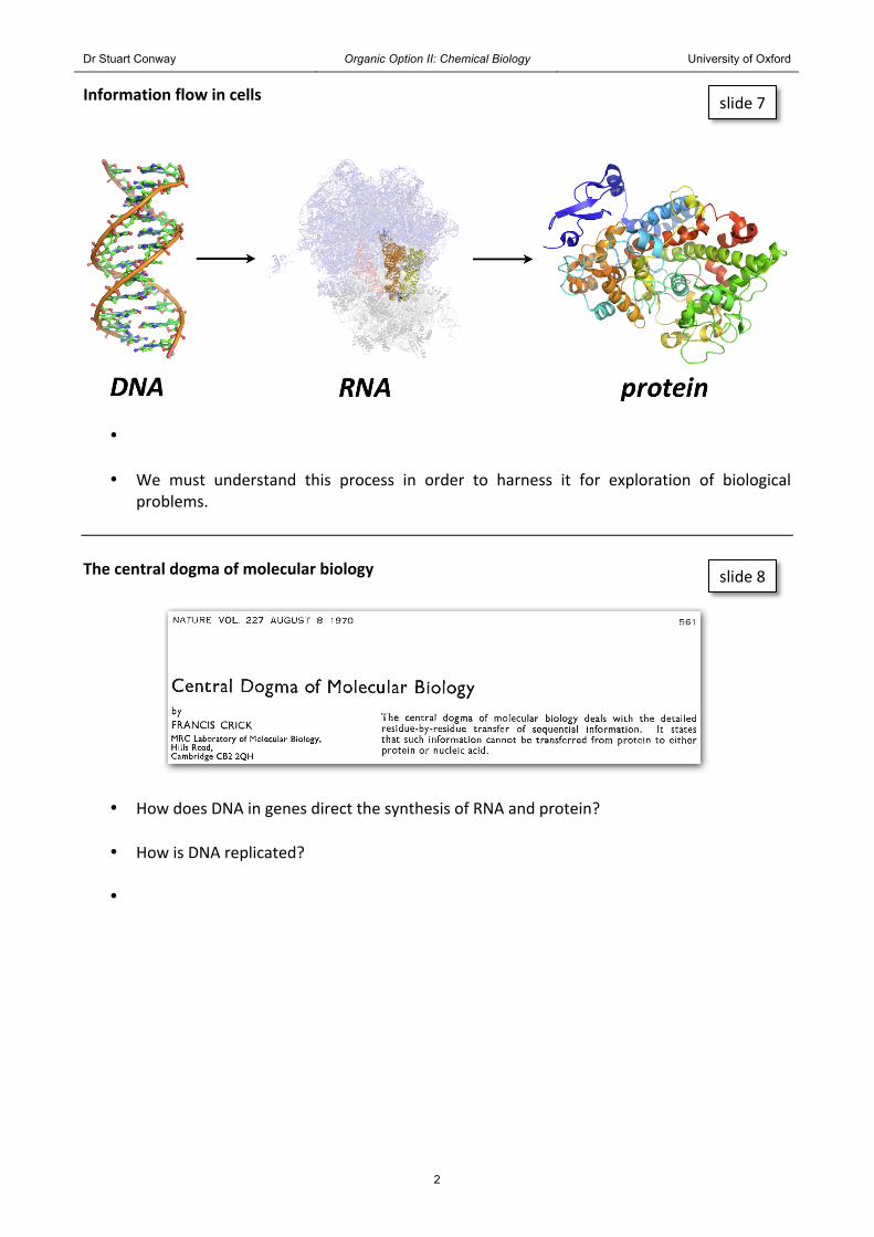
Information flow in cells
•
• We must understand this process in order to harness it for exploration of biological problems.
The central dogma of molecular biology
• How does DNA in genes direct the synthesis of RNA and protein?
• How is DNA replicated?
•
slide 7
slide 8
Dr Stuart Conway Organic Option II: Chemical Biology University of Oxford
3
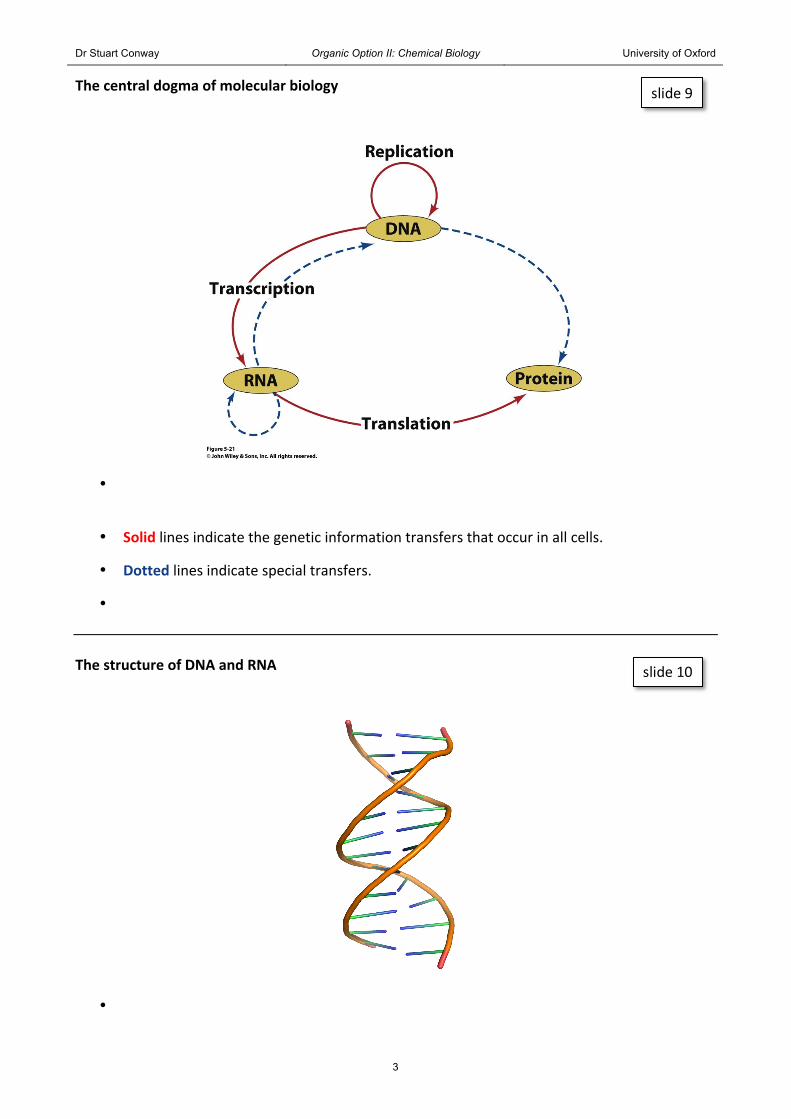
The central dogma of molecular biology
•
• Solid lines indicate the genetic information transfers that occur in all cells.
• Dotted lines indicate special transfers.
• The structure of DNA and RNA
•
slide 9
slide 10
Dr Stuart Conway Organic Option II: Chemical Biology University of Oxford
4
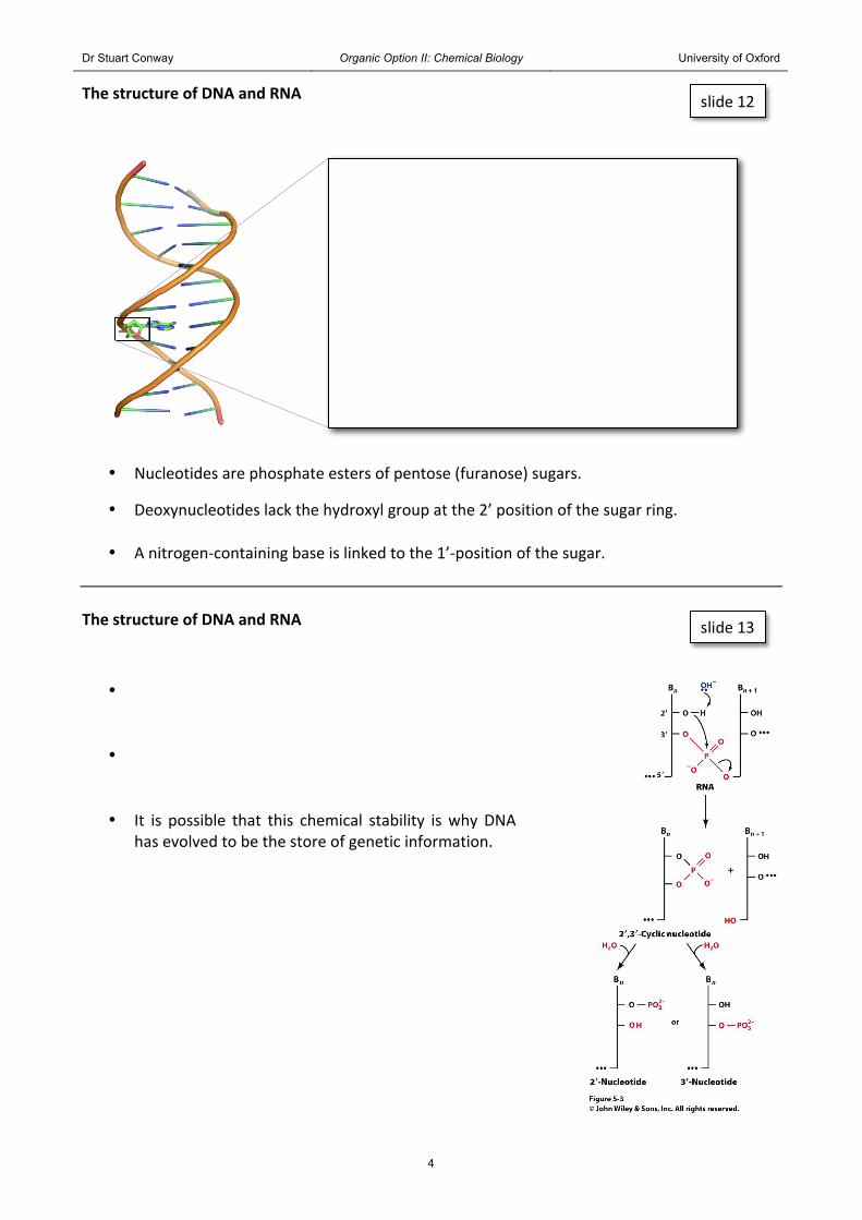
The structure of DNA and RNA
• Nucleotides are phosphate esters of pentose (furanose) sugars.
• Deoxynucleotides lack the hydroxyl group at the 2’ position of the sugar ring.
• A nitrogen-‐containing base is linked to the 1’-‐position of the sugar. The structure of DNA and RNA
•
•
• It is possible that this chemical stability is why DNA has evolved to be the store of genetic information.
slide 12
slide 13
Dr Stuart Conway Organic Option II: Chemical Biology University of Oxford
5
The structure of DNA and RNA
• The nitrogen bases are planar, aromatic and heterocyclic.
• They are usually either purine or pyrimidine derivatives.
The structure of DNA and RNA
• The major purine components of nucleic acids are adenine and guanine.
• The purines form glycosidic bonds to ribose via their N9 atoms.
The structure of DNA and RNA
• The major pyrimidine components of nucleic acids are cytosine, uracil and thymine (5-‐methyluracil).
• Uracil occurs mainly in RNA whereas thymine occurs mainly in DNA.
• The pyrimidines form glycosidic bonds to ribose via their N1 atoms.
slide 14
slide 15
slide 16
Dr Stuart Conway Organic Option II: Chemical Biology University of Oxford
6
The structure of DNA and RNA
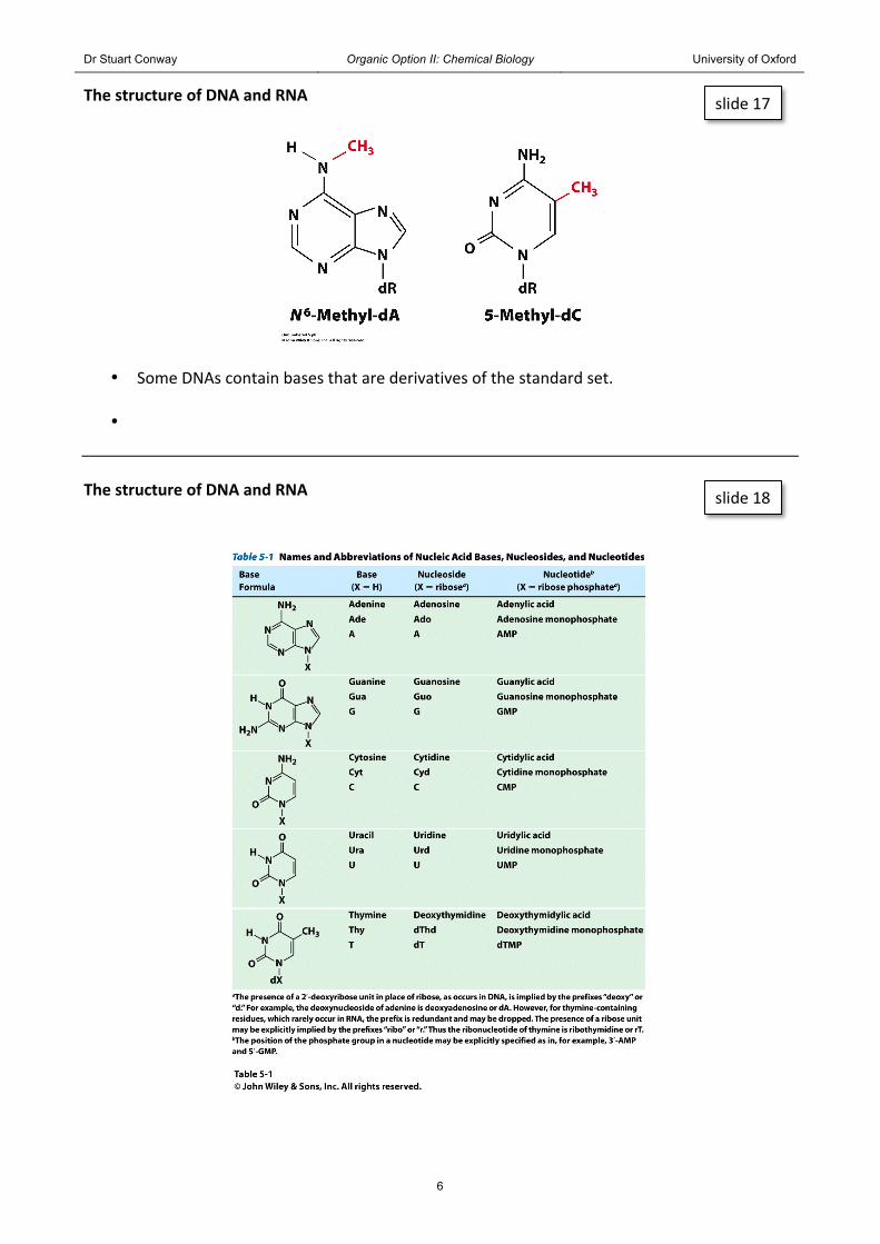
• Some DNAs contain bases that are derivatives of the standard set.
• The structure of DNA and RNA
slide 17
slide 18

Dr Stuart Conway Organic Option II: Chemical Biology University of Oxford
7
The structure of DNA and RNA
Nucleotide: adenosine monophosphate (R = OH in RNA and H in DNA)
Nucleoside: adenosine (R = OH in RNA and H in DNA)
Base: adenine
The structure of DNA and RNA
•
• The phosphate groups bridge the 3’-‐ and 5’-‐positions of successive sugar residues.
• The phosphate groups are deprotonated
at physiological pH, hence nucleic acids are polyanions in the cell.
•
slide 19
slide 20

Dr Stuart Conway Organic Option II: Chemical Biology University of Oxford
8
The structure of DNA and RNA
• Nucleic acids were first isolated in 1869 and the presence of these molecules in cells was demonstrated a few years later.
• In the 1930s and 1940s it was widely believed that nucleic acids had a monotonously
repeating sequence of all four bases = the so called “tetranucleotide hypothesis”.
• It was generally assumed that genes, known to be carriers of genetic information, were proteins.
• See Biochemistry pages 85-‐89 to see the experiments that proved DNA is the carrier of
genetic information. The structure of DNA and RNA
• Erwin Chargaff was the first to show that DNA contains equal numbers of adenine and thymine residues (A = T) and equal numbers of cytosine and guanine residues (C = G).
• These relationships are known as
“Chargaff’s rules”.
• Although not specifically stated by Chargaff, this observation suggests some form of base pairing in the (then unknown) structure of DNA.
slide 21
slide 22

Dr Stuart Conway Organic Option II: Chemical Biology University of Oxford
9
The structure of DNA and RNA
•
• The planes of the bases are nearly perpendicular to the helix axis.
• Each base is hydrogen bonded to a base on the opposite strand to form a planar base pair. Complementary base pairing
• The most remarkable feature of the Watson and Crick structure is that it can accommodate
only two types of base pairs.
• Each adenine residue must pair with a thymine residue and vice versa.
slide 25
slide 26

Dr Stuart Conway Organic Option II: Chemical Biology University of Oxford
10
Complementary base pairing
• Each guanine residue must pair with a cytosine residue and vice versa.
• The geometries of these A:T and G:C pairs , the so-‐called Watson-‐Crick base pairs, mean
that these base pairs are interchangeable in the double helix.
slide 27
Dr Stuart Conway Organic Option II: Chemical Biology University of Oxford
11
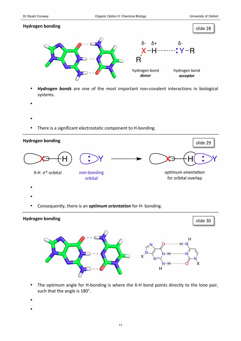
Hydrogen bonding
• Hydrogen bonds are one of the most important non-‐covalent interactions in biological systems.
•
•
• There is a significant electrostatic component to H-‐bonding.
Hydrogen bonding
•
•
• Consequently, there is an optimum orientation for H-‐ bonding.
Hydrogen bonding
• The optimum angle for H-‐bonding is where the X-‐H bond points directly to the lone pair, such that the angle is 180°.
•
•
slide 28
slide 29
slide 30

Dr Stuart Conway Organic Option II: Chemical Biology University of Oxford
12
Complementary base pairing
• The H-‐bond donor and acceptor patterns are such that A can only bind to T and G can only
bind to C.
• As A can only bind to T and G can only bind to C, we can immediately understand Chargaff’s rules.
• In addition, the Watson-‐Crick structure allows for any sequences of bases on one
polynucleotide strand if the opposite strand has the complementary sequence.
• This structure also suggests that hereditary information is encoded in the sequence of bases on either strand.
NN
NH
HN
NX N
NH
O CH3
XOHN
N
ON
NX N
N
N
XON
H
HH
HHdonor
acceptor
acceptor
donor
acceptor
donor
donor acceptor
donor
acceptor
adenine thymine guanine cytosine
slide 31

Dr Stuart Conway Organic Option II: Chemical Biology University of Oxford
13
DNA structure
• DNA has three major helical forms, B-‐DNA, A-‐DNA and Z-‐DNA.
• B-‐DNA is the biologically predominant form of DNA it forms a right-‐handed helix with major and minor grooves.
• When relative humidity is reduced to 75%, B-‐DNA undergoes a reversible conformational
change to A-‐DNA.
• A-‐DNA forms a wide, flatter helix than B-‐DNA.
• The base pairs of A-‐DNA are tilted 20 ° with respect to the helix axis.
• Certain DNA sequences can form a left-‐handed helix that has been called Z-‐DNA.
• It is not clear whether Z-‐DNA has any biological significance -‐ it may play a role in regulating DNA transcription.
advanced slide 32 & 33

Dr Stuart Conway Organic Option II: Chemical Biology University of Oxford
14
RNA structure
•
• Transfer RNA (see later) resembles an “L” shape, being made up of two short helical regions connected by a hinge.
•
RNA structure
• Hydrogen bonding in helical RNA occurs between cytosine and guanine as in DNA.
• Cytosine is replaced by uracil, which forms complementary hydrogen bonds with adenine.
slide 34
slide 35
Dr Stuart Conway Organic Option II: Chemical Biology University of Oxford
15
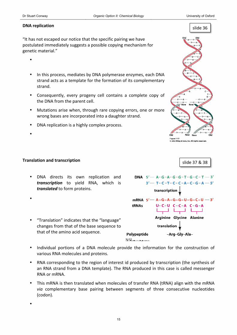
DNA replication
“It has not escaped our notice that the specific pairing we have postulated immediately suggests a possible copying mechanism for genetic material.”
•
• In this process, mediates by DNA polymerase enzymes, each DNA strand acts as a template for the formation of its complementary strand.
• Consequently, every progeny cell contains a complete copy of the DNA from the parent cell.
• Mutations arise when, through rare copying errors, one or more wrong bases are incorporated into a daughter strand.
• DNA replication is a highly complex process.
•
Translation and transcription
• DNA directs its own replication and transcription to yield RNA, which is translated to form proteins.
•
• “Translation” indicates that the “language” changes from that of the base sequence to that of the amino acid sequence.
• Individual portions of a DNA molecule provide the information for the construction of various RNA molecules and proteins.
• RNA corresponding to the region of interest id produced by transcription (the synthesis of an RNA strand from a DNA template). The RNA produced in this case is called messenger RNA or mRNA.
• This mRNA is then translated when molecules of transfer RNA (tRNA) align with the mRNA via complementary base pairing between segments of three consecutive nucleotides (codon).
•
slide 36
slide 37 & 38