DR. SAIDUNNISA Associated Professor Department of Biochemistry Nucleic acid Structure and DNA...
49
DR. SAIDUNNISA Associated Professor Department of Biochemistry Nucleic acid Structure and DNA packing
-
Upload
pauline-cross -
Category
Documents
-
view
219 -
download
3
Transcript of DR. SAIDUNNISA Associated Professor Department of Biochemistry Nucleic acid Structure and DNA...

DR. SAIDUNNISA
Associated Professor
Department of Biochemistry
Nucleic acid Structure and
DNA packing

Learning Objectives
At the end of the session student shall be able to:
Define nucleic acid and list the different types of purines and pyrimidine bases
Define nucleoside and nucleotide. Learn the different types of nucleosides and
nucleotides and list the biologically important forms present in humans
List the synthetic analogs and their use in clinical medicine

Friedrich Miescher in 1869
Isolated what he called nuclein from the nuclei of pus cells.
Nuclein was shown to have acidic properties, hence it became called nucleic acid

The distribution of nucleic acids in the eukaryotic cell
DNA is found in the nucleus.
Small amounts in mitochondria.
RNA is found throughout the cell.

Nucleic acids
There are two types of nucleic acids:
1. DNA (Deoxy ribonucleic acid)
2. RNA (Ribonucleic acid) Serve as transmitters of genetic information.
Nucleic acids are the polymers of nucleotides.

Major Bases
These are planar aromatic heterocyclic compounds.

SugarsSpot the difference

Structure of nucleosides
H
In purines the 9th nitrogen is linked with C1’ of sugar by beta N -glycosidic bond.
In pyrimidines the N1 binds with C1’ of ribose with beta N-glycosidic linkage.

Structure of nucleotides
A phosphate group
Nucleotides have three characteristic components:A nitrogenous base(pyrimidines or purine)
A pentose sugar
The C5’ is phosphorelated with phospho anhydride bonds.

Nucleotide nomenclature

Biologically important nucleotides
Adenosine derivatives: AMP, ADP, ATP. cAMP,
Guonosine derivatives: GMP, GDP GTP, cGMP
Uridine derivatives: UDP UTP, UMP, UDP-Glucose
Cytidine derivatives: CTP, CDP, CMP Miscellaneous: PAPS (active phosphate),
SAM (active sulphate)

Synthetic Analogs
These are used as therapeutic agents by the oncologist to treat various malignancies.
Purine analougs: e.g 6 mercapto –purine, 6 thio –guanine are used
in treating leukemia. Allopurinol is used in treating gout. Pyramidine analougs: 5 fluorouracil, thio-uracil are used in treating
hyperthyroidism. Azathymidine: used in the treatment of HIV
infection.

Purine/Pyrimidine analogs

The nucleotides in nucleic acids are joined by phosphodiester bonds.
The C3’ of ribose binds with C5’ of next nucleotide with phosphodiester bonds.

Example of nucleotides in nucleic acids

Reading sequence
A nucleic acid polymer has a
free 5’-phosphate group at
one end and a free 3’-OH
group at the other end The sequence is read from
the free 5’-end using the
letters of the bases This example reads 5’—A—C—G—T—3’

P
P
P
P
P
P
Adding in the basesTHE SUGAR-PHOSPHATE BACKBONE
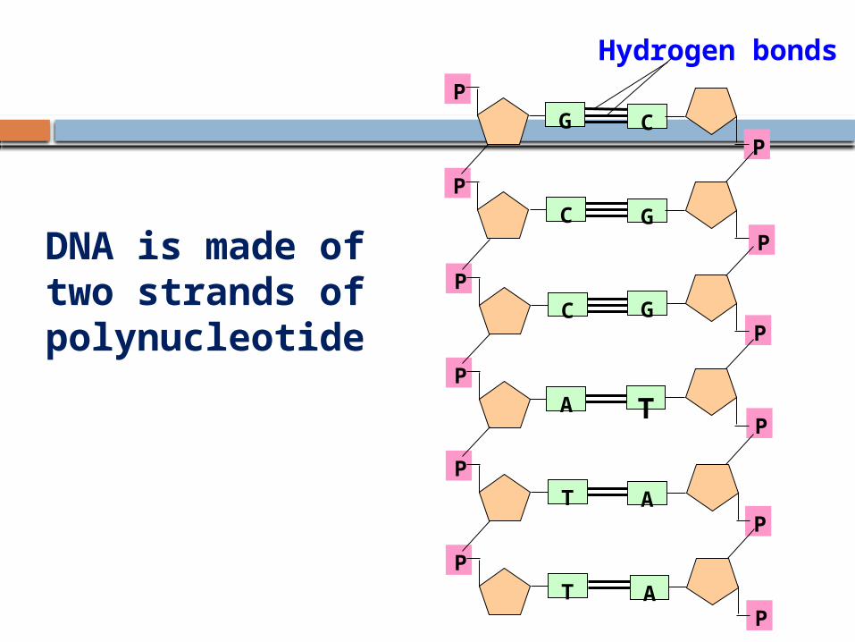
DNA is made of two strands of polynucleotide
P
P
P
P
P
P
C
G
G
T
A
A
P
P
P
P
P
P
G
C
C
A
T
T
Hydrogen bonds

DNA Stabilization– Complementary Base Pairing
Hydrogen bonds hold the bases and thus the two DNA strands together.
These are weaker than covalent bonds and allow the DNA strands to separate during replication and transcription.

Concept of Base-pairing
The ratio of purine to pyrimidine bases in the DNA molecule is always equal.
This is known as Chargaff's base pairing rule. Adenine base of one strand of DNA is hydrogen
bonded to a thymine in the opposite strand; while the guanine is hydrogen bonded to cytosine.
There are 2 hydrogen bonds present between adenine and thymine while 3 hydrogen bonds present between guanine and cytosine.

Nucleic Acid Structure“Base Pairing”
T A AG C C
3’
T C GG TA
3’ 5’
5’
DNA base-pairing is antiparallel
Concept of base pairing is essential for determining the mechanism of:1. Replication2. Transcription3. Translation4. Base-paring allows
one strand of DNA to serve as a template for the synthesis of the other strand.

Antiparallel
Watson and Crick concluded that the two strands of DNA run in opposite directions.
On one strand the 5’-carbon of the sugar is above the 3’-carbon. This strand is said to run in a 5’ to 3’ direction.
On the other strand the 3’-carbon is above the 5’-carbon.
This strand is said to run in a 3’to 5’ direction.
• The strands are antiparallel this concept of directionality of nucleic acid strands is essential for understanding the mechanisms of replication and transcription.
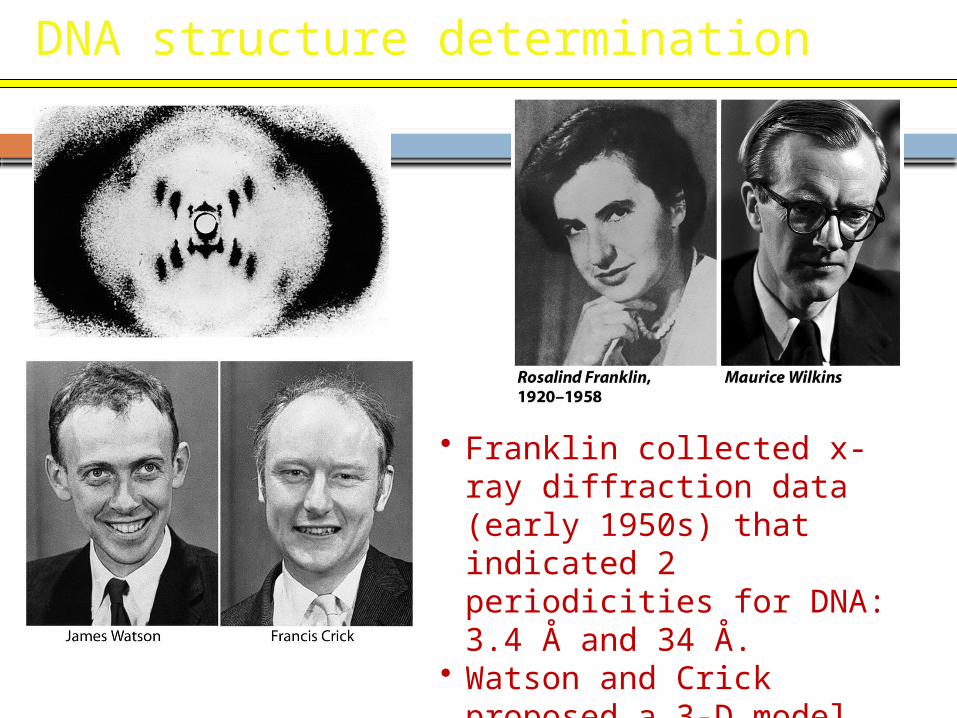
DNA structure determination
• Franklin collected x-ray diffraction data (early 1950s) that indicated 2 periodicities for DNA: 3.4 Å and 34 Å.
• Watson and Crick proposed a 3-D model accounting for the data.

Types of DNA
It exists in 6 different forms A-E and Z.
The B- form a right handed helix with 10 base pairs per turn is the most predominant form under physiological conditions.
Z-form is a left handed helix with 12 base pairs per turn.

Primary Structure of DNA
The primary structure of a nucleic acid is the nucleotide sequence
The nucleotides in nucleic acids are joined by phosphodiester bonds.
The C3’ of ribose binds with C5’ of next nucleotide with phosphodiester bonds.

Secondary Structure of DNA
Two polydeoxyribonucleotide strands coiled around a central axis forming a double stranded helix.
This type of model was first proposed by Watson and Crick for which they were awarded the noble prize in 1962 .

Secondary Structure: DNA Double Helix
• DNA consists of two helical chains wound around the same axis in a right-handed fashion aligned in an antiparallel fashion.
• There are 10.5 base pairs, or 36 Å, per turn of the helix.
• Alternating deoxyribose and phosphate groups on the backbone form the outside of the helix.
• The planar purine and pyrimidine bases of both strands are stacked inside the helix.

Secondary structure
The two strands that are equidistant from each other are twisted at the top and bottom, they form a double helix.
In the double helix of DNA, the base pairs that join the two stands are stacked like a spiral staircase along the central axis of the molecule.
The aromatic rings of bases are hydrophobic electrons of the adjacent base pairs interact, generating stacking forces that in addition to the hydrogen bonding of the base pairs, help to stabilize the helix.

DNA-Physical characters
Separation of DNA strands occurs
due to rupture in hydrogen bonds when DNA is placed in an acid – alkali or to increase temperatures. This is known as denaturation where the helical structure is lost , the phosphodiester bonds are not broken.
Melting temperature(Tm) is defined as the temperature at which half of the helical structure of DNA is lost.

Renaturation
Renaturation or reannealing or hybridization is a process in which the separated complementary DNA strands can reform the double helix when an acid alkali is removed and with decrease in temperature.
The process where a single stranded DNA anneals with complementary strands of RNA is called Hybridization which is extensively used in research and clinical testing.

Melting temperatures (Tm)
Melting temperatures (Tm) of DNA molecules with different nucleotide compositions. At a wavelength of 260 nm, single-stranded DNA has a higher relative absorbance than does double-stranded DNA. When 2 strands are separated, absorbance may increase by 30-40%.
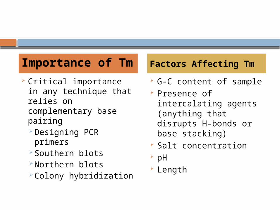
Critical importance in any technique that relies on complementary base pairing Designing PCR
primers Southern blots Northern blots Colony hybridization
G-C content of sample Presence of intercalating
agents (anything that disrupts H-bonds or base stacking)
Salt concentration pH Length
Importance of Tm Factors Affecting Tm

The two strands of the double helix separate reversibly at high temperatures
The temperature at which this “denaturation” or “melting” occurs (tm) depends on the pH and salt concentration, and increases approximately linearly with the GC content of the DNA.
The temperature at which this “denaturation” or “melting” occurs (tm) depends on the pH and salt concentration, and increases approximately linearly with the GC content of the DNA.
If the temperature is lowered, the strands recombine. The rate of recombination is inversely proportional to the complexity of the DNA.
If the temperature is lowered, the strands recombine. The rate of recombination is inversely proportional to the complexity of the DNA.
30 40 50 60% GC

dsDNA
ssDNAnucleotides
dA
dC
dG
dU
The conjugated p-electron systems of the purine & pyrimidine bases absorb strongly in the UV.
The conjugated p-electron systems of the purine & pyrimidine bases absorb strongly in the UV.
The absorbance of double-stranded DNA (dsDNA) at 260 nm is less than that of either single-stranded DNA (ssDNA) or the free bases. This is called “hypochromism.”
The absorbance of double-stranded DNA (dsDNA) at 260 nm is less than that of either single-stranded DNA (ssDNA) or the free bases. This is called “hypochromism.”
Double-stranded and single-stranded DNA differ in their optical absorption at 260 nm

Higher Organization of DNA
Supercoils In higher organisms the linear DNA is twisted
around its own axis when a supercoil is formed it can be either right handed or positive direction same as B form DNA or left handed or negative twisted in the opposite direction.
Topoisomerases and gyrases are enzymes that can release or insert the supercoils
DNA Packing
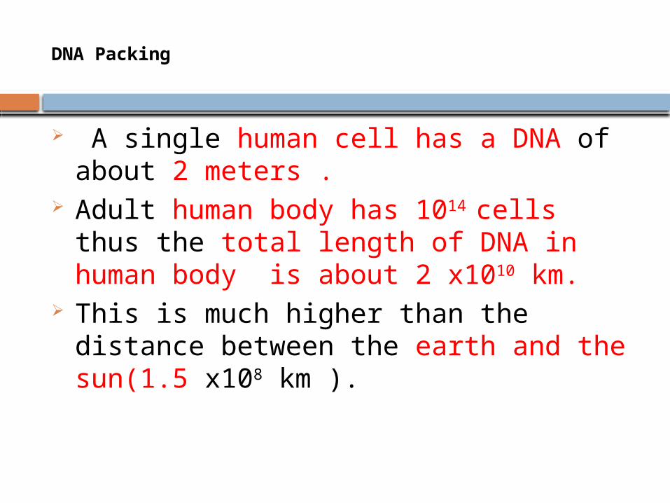
A single human cell has a DNA of about 2 meters .
Adult human body has 1014 cells thus the total length of DNA in human body is about 2 x10¹⁰ km.
This is much higher than the distance between the earth and the sun(1.5 x10⁸ km ).

DNA packing conti….
The length of DNA in the nucleus is far greater than the size of the compartment in which it is contained.
To fit into this compartment the DNA has to be condensed in some manner.
The degree to which DNA is condensed is expressed as its packing ratio.
Packing ratio - the length of DNA divided by the length into which it is packaged

Histones
So the DNA molecule is highly packed with the help of histones such that it can be accommodated within nuclear limit of 5 microns
These are basic proteins and are of 5 types used for packing DNA.
They are H1, H2A, H2B, H3 and H4.

Nucleosomes
Nucleosomes: - Whole DNA is not packed as a single coil. Instead it is present as small coils known as Nucleosomes "bead-like" structure.
Usually DNA is coiled around the octamer, which is made up of 2 units of H2A, H2B, H3, H4 histones and DNA and approximately it takes 2 turns around the histone octamer.

Nucleosome conti….
Each nucleosome is joined by a linker DNA and H1 types of histones.
The nucleosomes along with linker DNA and histones appear as beads on a string under electron microscope.
Further coiling of nucleosomes forms chromatin fiber. Thus the long threadlike DNA molecule is folded into chromosomes.

The second level of packing is the coiling of beads in a helical structure called chromatin fiber
that is found in both interphase chromatin and mitotic chromosomes.
This structure increases the packing ratio to about 40.

The final packaging occurs when the fiber is organized in loops, scaffolds and domains that give a final packing ratio of about 1000 in interphase chromosomes and about 10,000 in mitotic chromosomes
The subunit designation of the chromosome is chromatin. The fundamental unit of chromatin is the nucleosome


DNA Packing

Motivate your Answer
What is the relationship between Nucleosomes, chromatin fibers and the scaffold structure with respect to the organization of DNA in the nucleus?

46
Learning Check NA1
Write the complementary base sequence for the matching strand in the following DNA section:
-A-G-T-C-C-A-A-T-G-C-
• • • • • • • • • • • • • • • • • • • •

47
Solution NA1
Write the complementary base sequence for the matching strand in the following DNA section:
-A-G-T-C-C-A-A-T-G-C- • • • • • • • • • •
• • • • • • • • • •
-T-C-A-G-G-T-T-A-C-G-

Why does DNA contain T rather than U?
N
CHC
O
HN
CHCO
C
NH2
N
N
CHCO
CH
cytosine uracil
H2O
Cytosine deaminates non-enzymatically to form uracil. If this happens in DNA, it constitutes a mutation. Cells have a proof-reading system that recognizes the error and replaces the U by C.
Cytosine deaminates non-enzymatically to form uracil. If this happens in DNA, it constitutes a mutation. Cells have a proof-reading system that recognizes the error and replaces the U by C.
Deamination of cytosine is of less consequence in RNA, because RNA is not the permanent repository of genetic information.
Deamination of cytosine is of less consequence in RNA, because RNA is not the permanent repository of genetic information.

The phosphate groups of DNA and RNA are negatively charged
A phosphodiester group has a pKa of about 1, and so will always be ionized and negatively charged under physiological conditions (pH ~7).
Nucleic acids require counterions such as Mg2+, polyamines, histones or other proteins to balance this charge.
A phosphodiester group has a pKa of about 1, and so will always be ionized and negatively charged under physiological conditions (pH ~7).
Nucleic acids require counterions such as Mg2+, polyamines, histones or other proteins to balance this charge.
5’
3’
HO-CH2
ON
O
O P O CH2
OO
N
O
O P O CH2
OO
N
O
O P O CH2
OO
O-PO32
N
+M
+M
+M
+M