DR KARANU JK. Most common primary malignancy of bone It is a plasma cell disorder- monoclonal...
53
DR KARANU JK
-
Upload
andrew-wade -
Category
Documents
-
view
221 -
download
0
Transcript of DR KARANU JK. Most common primary malignancy of bone It is a plasma cell disorder- monoclonal...

DR KARANU JK

Most common primary malignancy of bone
It is a plasma cell disorder- monoclonal neoplasms related to
each other by virtue of their development from common progenitors in the B lymphocyte lineage
Thoughts that plamacytoma is simply an early, isolated form of multiple myeloma
There are two important variants of myeloma, solitary bone plasmacytomaextramedullary plasmacytoma

Solitary bone plasmacytoma is a single lytic bone lesion without marrow plasmacytosis
Extramedullary plasmacytomas usually involve the submuscosal lymphoid tissue of the nasopharynx or paranasal sinuses without marrow plasmacytosis.

The cause of myeloma is not known
Incresed frequency in those exposed to radiation
Seen more frequently among farmers, wood workers, leather workers and those exposed to petroleum products

Chromosomal alterations identified:13q14 deletions17p13 deletions11q abnormalities
Common translocationst(11;14)(q13;q32) and t(4;14)(p16;q32)

Overexpression of myc or ras genes has been noted in some cases
Mutations in p53 and Rb1 have also been described

No common molecular pathogenesis has yet emerged
Represents more than 40% of primary bone cancers
Peak incidence is in 5th to 7th decades
2:1 male predominance
Blacks have nearly twice the incidence of whites.
Yearly incidence is around 4 per 100000

Knh multiple myeloma 337 cases in 5 years, average of 67 per year
Plasmacytoma 16 cases in 5 years, average of 3 per year

Multiple myeloma cells bind via cell surface adhesion molecules to bone marrow stromal cells and extracellular matrix.
This triggers multiple myeloma cell growth, survival, drug resistance and migration in the bone marrow milieu

The cell effect is due to direct multiple myeloma and bone marrow stromal cell interaction , as well as induction of cytokines
Cytokines involved include IL6, insulin like growth factor 1, vascular endothelial growth factorstromal cell derived growth factor

Growth, drug resistance and migration are mediated via Ras/Raf/mitogen- activated protein kinase, PI3-K/Akt and protein kinase c signaling cascades

The clinical manifestation of all the plasma cell disorders relate to expansion of the neoplastic cells
secretion of cell products- immunoglobulins, lymphokines
Host’s response to the tumour

Bone pain most common complaint- precipitated my movement
WeaknessWeight lossAnaemia and thrombocytopeniaPeripheral neuropathyHypercalcaemiaRenal failure

Pathological fractures
Symptoms are usually of short duration because of the aggressive nature of the disease
The spine is the most common location followed by the ribs and pelvis.

Hypercalcaemia, osteoporosis, pathological fracture, lytic bone lesions, bone painTumor expansion, osteoclast activating factor, osteoblast inhibitory factors
Renal failure-Hypercalcaemia, light chain deposition, urate nephropathy, drugs

Easy fatigue- anaemiaBone marrow infiltration, haemolysis, decreased erythropoietin levels
Recurrent infectionsHypogammaglobulinaemia, low cd4 count, decreased neutrophil migration

Neurologic symptomsHyper viscosity, croglobulinemia, Hypercalcaemia, nerve compression, POEMS syndrome
Nausea and vomitingRenal failure, Hypercalcaemia

The classic triad of myeloma isi. Marrow plasmacytosis
(>10%)- CD138+, monoclonal
ii. Lytic bone lesionsiii. Serum and/ or urine M
component

Monoclonal gammopathies of uncertain significance1% go on to develop myelomaM protein in serum<30g/lBone marrow clonal plasma cells<10%
No evidence of other B cell proliferative disorder

Clinical evaluation of patients with myeloma includes a careful physical examination searching for tender bones and masses.
Chest and bone radiographs may reveal lytic lesions or diffuse osteopenia

Multiple ‘punched out’ sharply demarcated, purely lytic lesions without any surrounding reactive sclerosis


The lack of reactive bone formation is shown by the fact that most lesions are negative on bone scan

MRI offers a sensitive means to document extent of bone marrow infiltration and cord or root compression in patients with pain syndromes

Histologically multiple myeloma appears as sheets of plasma cells
These are small round blue cells clock face nuclei abundant cytoplasm perinuclear clearing or halo
Amyloid production can be abundant and may be pathognomonic for the disease


Serum immunoelectrophoresis shows monoclonal gammopathy
A 24 hr urine specimen quantitate protein excretionconcentrated aliquot is used for electrophoresis and immunologic typing of any M component

The serum M component will be IgG in 53%, IgA in 25%, and IgD in 1%.
20% of patients will have only light chains in serum and urine.

Fewer than 1% of patients will have no identifiable M component
The heat test for detecting Bence Jones proteins is falsely negative in 50% of patients with light chain myeloma

Complete blood count with differential may reveal anaemia
ESR is elavated
Serum chemistries calcium, urea, nitrogen, creatinine and uric acid levels may be elevated.

Solitary plasmacytoma pathological differentials may include chronic osteomyelitis with abundant plasma cellsPlasmcytoma has monoclonal light
chains whereas in COM they are polyclonal
Myeloma cells stain positive for natural killer antigen CD56, whereas reactive cells do not

In poorly differentiated cases lymphoma could be a differential
Lymphoma cells usually stain positive for CD45 (leukocyte common antigen)
and CD20 ( a B cell marker),

Stage 1 Hb>10g/dl,serum calcium <3 mmol/l, normal bone xray or solitary lesion,
low M component production

Stage 2- neither fitting stage 1 or 2
Stage 3- one or more of the following:Hb <8.5g/dlSerum calcium >3 mmol/lAdvanced lytic bone lesionHigh M component production

10% of patients will have an indolent course- slow progression
These patients only require antitumor therapy when the disease becomes symptomatic anaemia, hypercalcaemia,
progressive lytic bone lesions, progressive rise in serum myeloma protein levels or recurrent infections.

Primary treatment of multiple myeloma is chemotherapy
Symptomatic bone lesions usually respond rapidly to radiation treatment 40 Gy
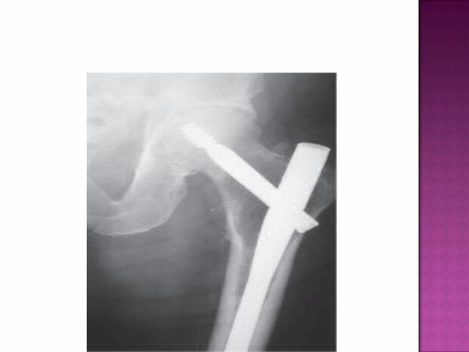
Treatment of impending or actual pathological fractures of the spine, acetabulum, proximal femur or proximal humerus

Patients with symptomatic and/ or progressive myeloma require therapeutic intervention
2 sorts of such therapySystemic therapy to control the
progression of myelomaSymptomatic supportive care to
prevent serious morbidity from the complications of the disease

Standard treatment for newly diagnosed cases depends on whether the patient is a candidate for high dose chemotherapy with autologous stem cell transplant
Transplant candidates avoid alkylating agents such as melphalan
Glucorticoids, vincristine, doxorubicin, thalidomide

Supportive care directed at the anticipated complicationsHypercalcaemia-bisphosphonates,
glucocorticoids,hydration, natriuresis
Prophylactic IV gamma globulin in recurrent serious infections
Anaemia- erythropoietin, along with haematinics

Short life expectancy in these patients operations aimed to earliest resumption of full activityTumor debulking
Internal fixation augmented with methacrylate
Cemented total joint arthroplasty or hemiarthroplasty


In most patients local radiation treatment should be instituted approximately 3 weeks after surgery or when the wound appears to be healed.

No conclusive evidence on the corelation
Aggressive course and worse prognosis in HIV
Multiple myeloma can accelerate progression of HIV infection
HIV plays a major role in the evolution of malignant plasma cell tumors

Patients with solitary plasmacytoma without evidence of systemic involvement have a better prognosis
More than half of patients who present with a solitary plasmacytoma eventually go on to develop multiple myeloma.

Patients in stage 1A have a median survival of more than 5 years and those in 3B about 15 months
The median overall survival is 5-6 years with subsets of patients surviving over 10 years.

The major causes of death are progressive myeloma,renal failure, sepsis or therapy related acute leukaemia or myelodysplasia.
Nearly a quarter die of myocardial infarction, chronic lung disease, diabetes or stroke.

ALIVE DEAD TOTAL CASES
2008 43 15 582009 41 20 612010 50 22 722011 43 31 742012 48 24 72

ALIVE DEAD TOTAL CASES
2008 2 1 32009 2 1 32010 1 2 32011 4 1 52012 1 1 2