Pancreas Neoplasms IPMN HCC - Avera Health Plans · - mural nodule greater than 10 mm in height was...
22
3/21/2016 1 Pancreas Neoplasms Ahmad Bashar Abdulkarim, MD, PhD, FACS Avera Health System March/2016 Pancreas Neoplasms • Pancreas – Pancreas duct adenocarcinoma (PDAC) – IPMN – Pancreas cysts • Liver – Cholangiocarcinoma – HCC – Adenoma Pancreas Duct Adenocarcinoma (PDAC) • The overall 5-year survival is 6%. • However, survival of patients with early stage pancreatic cancer is significantly better. • Importantly, there is a significant window of opportunity for early detection and treatment between the first genetic alteration in a cell in the pancreas and development of full-blown pancreatic cancer. Pancreas Duct Adenocarcinoma (PDAC) • The most common precursor to invasive PDAC, pancreatic intraepithelial neoplasia (PanIN), is microscopic. • In addition to this microscopic lesion, there are two macroscopically discernible cystic precursor lesions in the pancreas. These cystic precursor lesions are intraductal papillary mucinous neoplasm (IPMN) and mucinous cystic neoplasm (MCN). PDAC- Genetics • Mathematical modeling of genetic data suggests that the genetic evolution of PDAC takes almost 12 years from the earliest genetic alteration in a precursor lesion to the development of a full-blown invasive cancer. • Invasive PDAC is one of the best understood tumors at the genetic level. They are genetically very complex, with wide- spread chromosome abnormalities, numerous losses and gains of large segments of DNA, and on average more than 60 exomic alterations in each cancer. • PanINs are small microscopic lesions that are <5 mm. They are composed of a flat or papillary neoplastic epithelium. Yachida S. et al. Oncogene, 2013;32:5253 Iacobuzio-Donahue CA. Gut 2012;61:1085 PDAC • Three grades of dysplasia can distinguished in PanIN lesions. – PanIN-1A and PanIN-1B have low-grade dysplasia. – PanIN-2 is considered intermediate-grade dysplasia and shows mostly papillary epithelium with mild to moderate cytological atypia. – PanIN-3 is considered high-grade dysplasia (carcinoma in situ). Of note, PanINs are often surrounded by lobular parenchymal atrophy which, when multifocal, can be detected by endoscopic ultrasound and may serve as a biomarker in patients at high-risk for PDAC. Brune K. et al. Am J Surg Pathol 2006;30:1067
Transcript of Pancreas Neoplasms IPMN HCC - Avera Health Plans · - mural nodule greater than 10 mm in height was...
3/21/2016
1
Pancreas Neoplasms
Ahmad Bashar Abdulkarim, MD, PhD, FACS
Avera Health System
March/2016
Pancreas Neoplasms
• Pancreas – Pancreas duct adenocarcinoma (PDAC)
– IPMN
– Pancreas cysts
• Liver – Cholangiocarcinoma
– HCC
– Adenoma
Pancreas Duct Adenocarcinoma (PDAC)
• The overall 5-year survival is 6%. • However, survival of patients with early stage
pancreatic cancer is significantly better. • Importantly, there is a significant window of
opportunity for early detection and treatment between the first genetic alteration in a cell in the pancreas and development of full-blown pancreatic cancer.
Pancreas Duct Adenocarcinoma (PDAC)
• The most common precursor to invasive PDAC, pancreatic intraepithelial neoplasia (PanIN), is microscopic.
• In addition to this microscopic lesion, there are two macroscopically discernible cystic precursor lesions in the pancreas. These cystic precursor lesions are intraductal papillary mucinous neoplasm (IPMN) and mucinous cystic neoplasm (MCN).
PDAC- Genetics
• Mathematical modeling of genetic data suggests that the genetic evolution of PDAC takes almost 12 years from the earliest genetic alteration in a precursor lesion to the development of a full-blown invasive cancer.
• Invasive PDAC is one of the best understood tumors at the genetic level. They are genetically very complex, with wide-spread chromosome abnormalities, numerous losses and gains of large segments of DNA, and on average more than 60 exomic alterations in each cancer.
• PanINs are small microscopic lesions that are <5 mm. They are
composed of a flat or papillary neoplastic epithelium. Yachida S. et al. Oncogene, 2013;32:5253
Iacobuzio-Donahue CA. Gut 2012;61:1085
PDAC
• Three grades of dysplasia can distinguished in PanIN lesions. – PanIN-1A and PanIN-1B have low-grade dysplasia. – PanIN-2 is considered intermediate-grade dysplasia and shows mostly papillary
epithelium with mild to moderate cytological atypia. – PanIN-3 is considered high-grade dysplasia (carcinoma in situ). Of note, PanINs are often surrounded by lobular parenchymal atrophy which, when
multifocal, can be detected by endoscopic ultrasound and may serve as a biomarker in patients at high-risk for PDAC.
Brune K. et al. Am J Surg Pathol 2006;30:1067
3/21/2016
2
Pancreas Cystic Lesions (PCLs)
• Two macroscopically discernible cystic precursor
lesions in the pancreas:
– intraductal papillary mucinous neoplasm (IPMN)
– mucinous cystic neoplasm (MCN)
Pancreas Cystic Lesions (PCLs)
• CT scan: 3% of asymptomatic individuals have pancreatic cysts, approximately 25% of which is consistent with an IPMN.
• The prevalence of IPMN is equal in men and women; the majority of patients are diagnosed around 60 years of age.
• IPMNs can macroscopically be categorized in three groups: – 10-35% arises in the main pancreatic duct (MD), – 40-65% in a branch duct (BD), and – 15-40% involves both the main and BDs (mixed type).
de Jong K, et al. Clin Gastroenterol Hepatol 2010;8:806
Pancreatic Cystic Lesions (PCLs)
• Pancreatic cystic lesion prevalence based on age group:
– 0.5% in those less than 40 years of age
– 25% in those 70-79 years of age
– 37% in those aged 80 or over
Matsubara S, et al. Pancreas 2012;41:1241
IPMN
The clinico-pathologic behavior of combined(mixed)-type IPMN is similar to that of MD-IPMN.
PDAC
• 62% of MD and 58% of mixed type IPMNs have high-grade dysplasia, and that 44% of MD and 45% of mixed type IPMNs have an associated invasive carcinoma.
• In contrast, only 24% of BD-IPMNs have high-grade dysplasia, and 17%
an associated invasive carcinoma. • consensus guidelines for the management of IPMN and MCN were
established in 2012. These guidelines advise that most IPMNs that involve the MD should be surgically resected because of their high rate of malignancy, whereas surgical indications for BD-IPMNs include the presence of “high-risk stigmata” such as mural nodules and symptomatology.
Tanaka M, et al. Pancreatology 2012;12:183
International consensus guidelines for surgical resection of pancreatic cysts
• Perform endoscopic ultrasound, and if any of these features are present:
• • Definite mural nodule
• • Main duct features suspicious for involvement
• • Cytology: suspicious or positive for malignancy
• Consider resection if clinically appropriate
• MCN Surgical resection is recommended for all surgically fit patients. Observation may be considered in elderly frail patients. In patients with MCNs of <4 cm without mural nodules, parenchyma-sparing resections (i.e., middle pancreatectomy) and distal pancreatectomy with spleen preservation as well as laparoscopic procedures should be considered

3/21/2016
3
IPMNs • Histologically, IPMNs can be categorized as gastric foveolar,
intestinal, pancreato-biliary, or oncocytic type based on the direction of differentiation of the neoplastic epithelium as defined by histology and immuno-labeling.
• Gastric-foveolar IPMNs are lined by epithelium resembling foveolar epithelium of the gastric mucosa . Associated invasive carcinomas are rare.
• Intestinal IPMNs resemble villous adenomas of the gastrointestinal tract.
• Pancreato-biliary IPMNs are usually high-grade lesions with complex architecture with cribriforming papillae and bridging.
IPMNs
• Oncocytic IPMNs, also known as intraductal oncocytic.
• papillary neoplasms (IOPNs), are morphologically the most complex lesions.
ITPN
• Intraductal tubulo-papillary neoplasms (ITPN) are
recognized as an intraductal neoplasm distinct from IPMNs.
• These lesions are rare, and their precise relationship to the other IPMN subtypes remains to be defined.
• ITPN are the most recently recognized pancreatic intraductal neoplasm.
• These lesions are always high-grade.
Pancreatic Cystic Lesions (PCLs)
• \\\
Neoplastic Cysts Non-neoplastic Cysts
Epithelial Non-epithelial
Cystic Neoplasms of the Pancreas
• Epithelial neoplasms – Serous cystadenoma* – Mucinous cystic neoplasm (MCN) and MCN-associated
carcinoma* – Intraductal papillary mucinous neoplasm (IPMN) and – IPMN-associated carcinoma* – Solid pseudopapillary neoplasm* – Pancreatic ductal adenocarcinoma with cystic degeneration* – Cystic pancreatic endocrine neoplasm (CPEN)* – Acinar cystadenoma and cystadenocarcinoma – Dermoid cyst (cystic teratoma) – Intraductal papillary variant of acinar cell carcinoma – Intraductal tubulopapillary neoplasm
Classification of pancreatic cystic lesions (PCLs)

3/21/2016
4
Cystic Neoplasms of the Pancreas
• Non-epithelial
– Lymphangioma
– Epidermoid cyst
– Cystic pancreatic hamartoma
– Mesothelial cyst
Cystic Neoplasms of the Pancreas
• Lesions resembling pancreatic cystic neoplasms – Pseudocyst* – Lymphoepithelial cyst (epidermoid cyst) – Mucinous nonneoplastic cyst – Enteric duplication cysts – Endometrial cyst – Hydatid cyst – Retention cyst – Accessory splenic cyst – Cystic pheochromocytoma – Cystic gastrointestinal stromal tumor – Squamoid cyst
Morphological details of the common cysts of the pancreas
Pseudocyst Serous MCN IPMN
Cystic Neoplasms of the Pancreas
• Calcifications in a cyst wall are highly suggestive of a mucinous cystadenoma, rather than a pseudocyst.
Unilocular pancreatic pseudocyst with debris
Differentiation from Pseudocysts EUS with FNA
• Amylase
• CEA
• Cytology
– Debris
– Inflammatory cells
– Histiocytes
– Epithelial cells

3/21/2016
5
IPMN-EUS
CEA
-Reflects the presence of a mucinous epithelium. and
-It is elevated in both IPMNs and MCNs.
-It is mainly beneficial to distinguish mucinous cysts from non-mucinous.
-It does not differentiate IPMNs from MCNs -It does not differentiate benign IPMNs from malignant IPMNs.
Pancreatic Cystic Lesions (PCLs)
The World Health Organization (WHO) classified IPMNs into three subgroups according to degree of dysplasia:
(I) IPMN with low- or intermediate-grade dysplasia;
(II) IPMN with high-grade dysplasia (carcinoma in situ); and
(III) IPMN with an associated invasive carcinoma.
Fish-eye ampulla: Pathognomonic for IPMN
IPMN
They are:
Usually found in the head of the pancreas
Solitary cystic lesion, but in
20% to 30% multifocal
5% to 10% involve the pancreas diffusely
IPMN
• Signs of malignancy on EUS in IPMN: - main pancreatic duct (>10 mm) in MD-IPMN and - large tumors (>40 mm), BD-IPMN - irregular septae, BD-IPMN; - mural nodule greater than 10 mm in height was associated
with malignancy in both MD-IPMN and BD-IPMN. - Large unilocular cystic component, focal hypoechoic mass,
thick septations and thickening of cyst wall are also features of malignant or potentially malignant lesions.
International consensus guidelines for surgical resection of
pancreatic cysts-2012
• High risk stigmata: – Obstructive jaundice with cystic pancreas head lesion – Enhancing solid component within cyst – Main pancreatic duct >10 mm In size
• Worrisome features:
– Pancreatitis – Cyst >3 cm – Thickened enhancing cyst walls – Non-enhancing mural nodule – Main pancreatic duct 5-9 mm – Abrupt change in caliber of pancreatic duct with distal
pancreatic atrophy Tanaka M, et al. Pancreatology 2012;12:183
3/21/2016
6
International consensus guidelines for surgical resection of pancreatic cysts
To date,
there have been no consistent predictive factors for malignancy in MD-IPMN, including the degree of MPD dilation, presence of symptoms, or mural nodules.
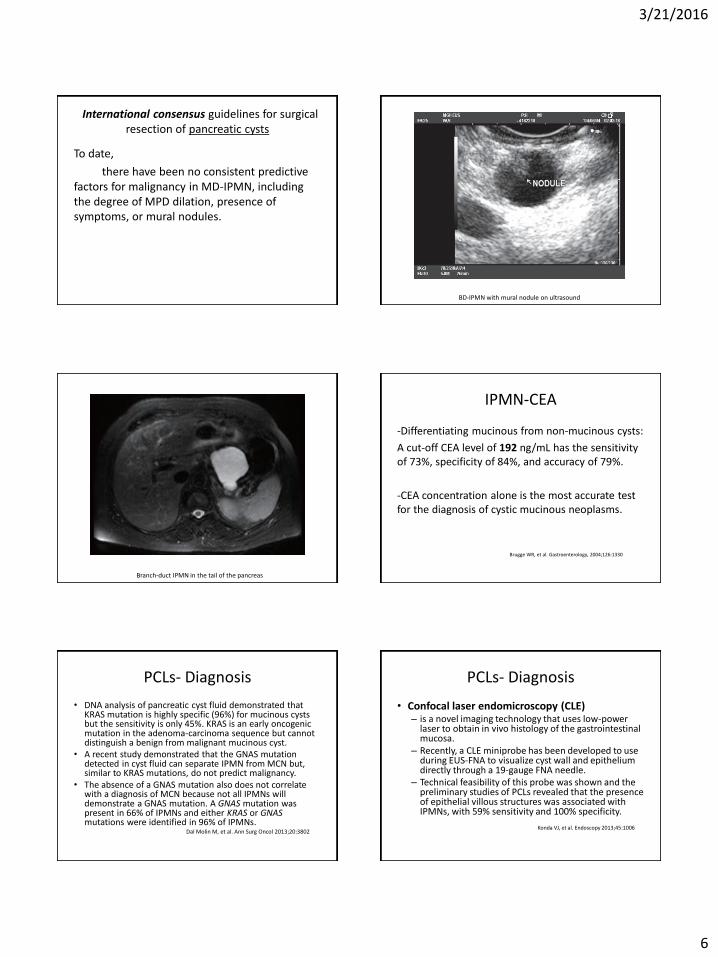
BD-IPMN with mural nodule on ultrasound
Branch-duct IPMN in the tail of the pancreas
IPMN-CEA
-Differentiating mucinous from non-mucinous cysts:
A cut-off CEA level of 192 ng/mL has the sensitivity of 73%, specificity of 84%, and accuracy of 79%.
-CEA concentration alone is the most accurate test for the diagnosis of cystic mucinous neoplasms.
Brugge WR, et al. Gastroenterology, 2004;126:1330
PCLs- Diagnosis
• DNA analysis of pancreatic cyst fluid demonstrated that KRAS mutation is highly specific (96%) for mucinous cysts but the sensitivity is only 45%. KRAS is an early oncogenic mutation in the adenoma-carcinoma sequence but cannot distinguish a benign from malignant mucinous cyst.
• A recent study demonstrated that the GNAS mutation detected in cyst fluid can separate IPMN from MCN but, similar to KRAS mutations, do not predict malignancy.
• The absence of a GNAS mutation also does not correlate with a diagnosis of MCN because not all IPMNs will demonstrate a GNAS mutation. A GNAS mutation was present in 66% of IPMNs and either KRAS or GNAS mutations were identified in 96% of IPMNs.
Dal Molin M, et al. Ann Surg Oncol 2013;20:3802
PCLs- Diagnosis
• Confocal laser endomicroscopy (CLE) – is a novel imaging technology that uses low-power
laser to obtain in vivo histology of the gastrointestinal mucosa.
– Recently, a CLE miniprobe has been developed to use during EUS-FNA to visualize cyst wall and epithelium directly through a 19-gauge FNA needle.
– Technical feasibility of this probe was shown and the preliminary studies of PCLs revealed that the presence of epithelial villous structures was associated with IPMNs, with 59% sensitivity and 100% specificity.
Konda VJ, et al. Endoscopy 2013;45:1006
3/21/2016
7
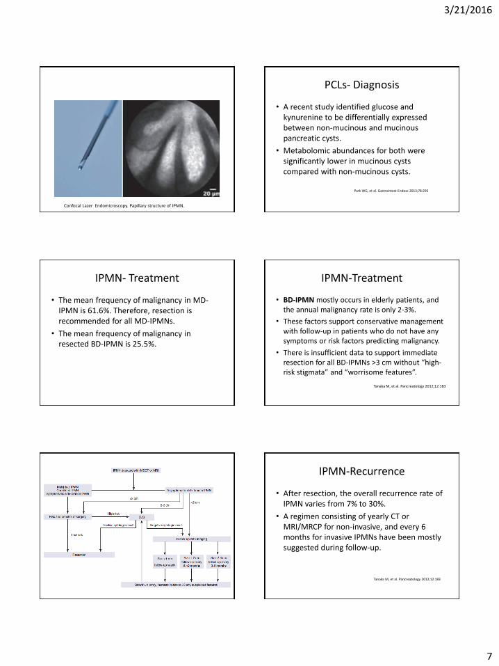
Confocal Lazer Endomicroscopy. Papillary structure of IPMN.
PCLs- Diagnosis
• A recent study identified glucose and kynurenine to be differentially expressed between non-mucinous and mucinous pancreatic cysts.
• Metabolomic abundances for both were significantly lower in mucinous cysts compared with non-mucinous cysts.
Park WG, et al. Gastrointest Endosc 2013;78:295
IPMN- Treatment
• The mean frequency of malignancy in MD-IPMN is 61.6%. Therefore, resection is recommended for all MD-IPMNs.
• The mean frequency of malignancy in resected BD-IPMN is 25.5%.
IPMN-Treatment
• BD-IPMN mostly occurs in elderly patients, and the annual malignancy rate is only 2-3%.
• These factors support conservative management with follow-up in patients who do not have any symptoms or risk factors predicting malignancy.
• There is insufficient data to support immediate resection for all BD-IPMNs >3 cm without “high-risk stigmata” and “worrisome features”.
Tanaka M, et al. Pancreatology 2012;12:183
IPMN-Recurrence
• After resection, the overall recurrence rate of IPMN varies from 7% to 30%.
• A regimen consisting of yearly CT or MRI/MRCP for non-invasive, and every 6 months for invasive IPMNs have been mostly suggested during follow-up.
Tanaka M, et al. Pancreatology 2012;12:183

3/21/2016
8
IPMN-Non-Surgical Treatment
• The combination of ethanol and paclitaxel injection resulted in elimination of the cysts, as determined by CT scanning, in 29/47 (62%) of patients, in a median follow-up period of 21.7 months.
• RFA is an emerging treatment.
Mucinous Cystic Neooplasms (MCN)
• MCNs are defined as:
– Cyst-forming epithelial neoplasms
– Usually without communication with the pancreatic duct, and,
– Composed of columnar, mucin-producing ductal epithelium with an underlying ovarian-type stroma.
Mucinous Cystic Neooplasms (MCN)
• Similar to IPMNs, MCNs are classified according to the grade of dysplasia:
– (I) MCN with low or intermediate-grade dysplasia; (II) MCN with high-grade dysplasia; and,
– (III) MCN with an associated invasive carcinoma.
• The lesions may be unilocular or multilocular.
MCNs
• MCNs almost exclusively occur in women, with a peak incidence in the fifth decade.
• The body and the tail of the pancreas are predominantly affected.
• Up to one-third of MCNs are reported to harbor an invasive carcinoma.
• Risk factors for the presence of malignancy include: – large tumor size, – associated mass or mural nodules, – and advanced age.
MCNs- Diagnosis
• Routine labs are not diagnostic.
• CT/MRI/EUS help in making a diagnosis and differentiate benign for malignant lesions.
MCNs-Management
• Current consensus guideline advocates that all MCNs should be resected.
• Non-invasive MCNs require no surveillance after resection.
3/21/2016
9
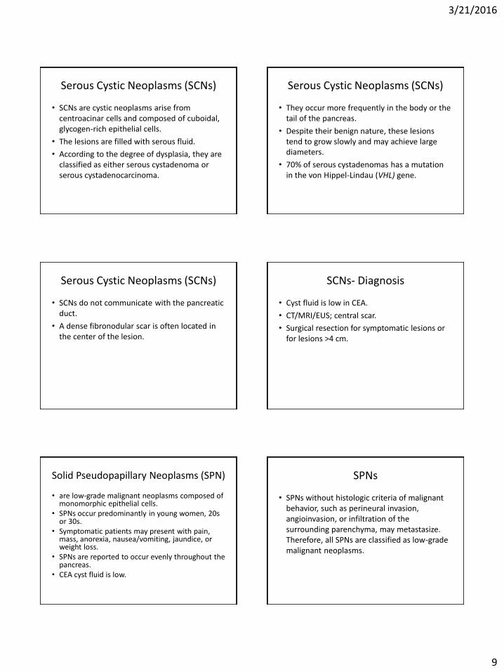
Serous Cystic Neoplasms (SCNs)
• SCNs are cystic neoplasms arise from centroacinar cells and composed of cuboidal, glycogen-rich epithelial cells.
• The lesions are filled with serous fluid.
• According to the degree of dysplasia, they are classified as either serous cystadenoma or serous cystadenocarcinoma.
Serous Cystic Neoplasms (SCNs)
• They occur more frequently in the body or the tail of the pancreas.
• Despite their benign nature, these lesions tend to grow slowly and may achieve large diameters.
• 70% of serous cystadenomas has a mutation in the von Hippel-Lindau (VHL) gene.
Serous Cystic Neoplasms (SCNs)
• SCNs do not communicate with the pancreatic duct.
• A dense fibronodular scar is often located in the center of the lesion.
SCNs- Diagnosis
• Cyst fluid is low in CEA.
• CT/MRI/EUS; central scar.
• Surgical resection for symptomatic lesions or for lesions >4 cm.
Solid Pseudopapillary Neoplasms (SPN)
• are low-grade malignant neoplasms composed of monomorphic epithelial cells.
• SPNs occur predominantly in young women, 20s or 30s.
• Symptomatic patients may present with pain, mass, anorexia, nausea/vomiting, jaundice, or weight loss.
• SPNs are reported to occur evenly throughout the pancreas.
• CEA cyst fluid is low.
SPNs
• SPNs without histologic criteria of malignant behavior, such as perineural invasion, angioinvasion, or infiltration of the surrounding parenchyma, may metastasize. Therefore, all SPNs are classified as low-grade malignant neoplasms.

3/21/2016
10
SPNs- Diagnosis and Treatment
• CT/MRI/EUS
• Surgical resection.
Liver Neoplasms
Benign Liver Neoplasms
• Hemangioma
• Focal Nodular Hyperplasia
• Hepatic Adenoma
Hepatocellular Adenoma (HA)
• Rare tumors. Incidence: 1/100,000. F>M. • The incidence of HCA is 30 times greater in oral
contraceptive users compared to nonusers. • A dose-dependent association and spontaneous
regression following the withdrawal of estrogens have also been described.
• There is variable expression of estrogen receptor on HA (26-73%); therefore, some HA do not show regression on hormone therapy withdrawal.
Buhler H, Gastroenterology 1982; 82:775 Torbenson M, 2002;15(3): 189
Hepatocellular Adenoma
• Risk Factors:
– Oral contraceptives
– Female gender
– Anabolic steroids
– Hepatic Adenomatosis (>10 adenomas)
– Glycogen storage disease type I (von Gierke) and type III
Hepatocellular Adenoma
• Bordeaux classification: HCAs are divided into four subgroups based on clear genetic differences: The presence of hepatocyte nuclear factor 1α (HNF1A) mutations,
accounts for 30-40%. The presence of activating mutations of β-catenin, accounts for 10-
20% of cases. This triggers a signaling pathway that stimulates HCC. Inflammatory response group: 40-50%, have increased risk of
malignant degeneration. Unclassified: No genetic alterations, 10-25%.
Bioulac-Sage P, Hepatology 2007; 46:740

3/21/2016
11
Need to bring pictures from paper: Liver Adenoma 4
Hepatocellular Adenoma
• Malignancy: – Overall malignant transformation frequency of
4.2%.
• Rupture and Bleeding – Prevalence of 20-30% of lesions larger lesions >5
cm.
van Aalten SM, Br J Surg 2012; 99:911
Hepatocellular Adenoma
• Hepatic Adenomatosis:
– Presence of >10 adenomas in the liver.
Flejou JF, Gastroenterology 1985;89(5): 1132
Differential: FNH
A central scar with divergences to the periphery is visible on T2-weighting and is reminiscent of a “spoke wheel”
Venous phase T1-weighted MRI. FNH
Hepatocellular Adenoma Management
• Surgical resection is recommended for high-risk adenomas: – lesions ≥5 cm in size or increasing in size over time,
– adenomas with evidence of internal hemorrhage,
– adenomas occurring in males, which have increased malignant risk,
– those with positive nuclear β-catenin immunohistochemical staining,
– older females with no history of use of oral contraceptives.

3/21/2016
12
Hepatocellular Adenoma Management
• Low risk adenomans:
<<Less than 5 cm in size occurring in young females on oral contraceptive pills>>
Discontinue use of oral contraceptive pills,
perform intermittent surveillance imaging.
Malignant Liver Neoplasms
• Cholangiocarcinoma
• Hepatocellular Carcinoma (HCC)
Cholangiocarcinoma
• Epithelial cell malignancy arising from varying locations within the biliary tree showing markers of cholangiocyte differentiation.
Cholangiocarcinoma
• Classification
Based on anatomical location:
Intrahepatic (10%) Perihilar (50%) Distal (40%)
Cholangiocarcinoma
• Intrahepatic Cholangiocarcinoma:
Is defined as a cholangiocarcinoma located proximal to the second degree bile ducts.
Cholangiocarcinoma
• Perihilar Cholangiocarcinoma:
Is localized to the area between the second degree bile ducts and the insertion of the cystic duct into the common bile duct.

3/21/2016
13
Cholangiocarcinoma
• Distal Cholangiocarcinoma:
Is confined to the area between the origin of the cystic duct and ampulla of Vater.
Cholangiocarcinoma
• Most cholangiocarcinomas are well, moderately, and poorly differentiated adenocarcinomas.
Liver Cancers
• Cholangiocarcinoma
• Hepatocellular cancer
• Mixed hepatocellular-cholangiocellular carcinomas <1%
Cholangiocarcinoma
• Risk Factors: – Cirrhosis – Hepatitis C – Hepatitis B – Primary sclerosing cholangitis (PSC); [5-10% life time incidence] – Bile duct cystic disorders [life time incidence: 6-30%] – hepatobiliary flukes, Opisthorchis viverrini and Clonorchis sinensis – Hepatolithiasis [7% increased risk] – Biliary-enteric drainage – Genetic – Metabolic Syndrome – Diabetes – Obesity
Cholangiocarcinoma- Diagnosis
• Typical radiological features of
cholangiocarcinoma include progressive contrast
uptake throughout both arterial and venous phases
of a cross-sectional imaging study.
• By contrast, hepatocellular carcinoma lesions are
associated with hyperenhancement in the arterial
phase and contrast washout in the venous phase of
a contrast-enhanced imaging study.
Potential cells of origin in intrahepatic cholangiocarcinoma

3/21/2016
14
• Notch signalling is vital in cell fate determination and regulates biliary duct formation.49 Its involvement in cholangiocarcinoma biology was reported in several studies.
• Notch pathway activation was implicated in conversion of mature adult hepatocytes into precursors of intrahepatic cholangiocarcinoma in two preclinical models involving cell fate tracing techniques.20,50 These studies challenge the theory that cholangiocarcinoma cells are derived from cholangiocytes, peribiliary glandular cells, or hepatic progenitor cells. They also emphasise the plasticity of liver cells regarding their differentiated state, and draw attention to transcriptome studies identifying overlap in hepatocellular carcinoma and cholangiocarcinoma signatures.
Intrahepatic cholangiocarcinoma Clinical classification and diagnosis
• Intrahepatic cholangiocarcinoma can be classified
morphologically by growth patterns as mass-
forming, periductal-infiltrating, intraductal,
superficial spreading, and undefined subtypes.
• The superficial spreading and intraductal subtypes
are associated with the best prognosis and
• Periductal and mass-forming subtypes with the
worst.
Intrahepatic cholangiocarcinoma Clinical classification and diagnosis
• Typical radiological features of cholangiocarcinoma include:
– Progressive contrast uptake throughout both arterial and venous phases of a cross-sectional imaging study.
– By contrast, hepatocellular carcinoma lesions are associated with hyperenhancement in the arterial phase and contrast washout in the venous phase of a contrast-enhanced imaging study.
Intrahepatic cholangiocarcinoma Clinical classification and diagnosis
• A strong enhancing rim and irregular shape on gadoxetic acid-enhanced MRI favors mixed hepatocellular-cholangiocellular carcinoma, and
• lobulated shape, weak rim, and a target appearance favors a mass-forming intrahepatic cholangiocarcinoma.
Intrahepatic cholangiocarcinoma Clinical classification and diagnosis
• Carbohydrate antigen 19-9 (CA 19-9) is a traditional serum biomarker used for cholangiocarcinoma diagnosis.
– In patients with primary sclerosing cholangitis the most reliable cutoff for intrahepatic cholangiocarcinoma is 129 U/mL, which provides sensitivity, specificity, and adjusted positive predictive values of 79%, 98%, and 57%, respectively.
Intrahepatic cholangiocarcinoma Clinical classification and diagnosis
• Studies suggest that mixed hepatocellular-cholangiocellular carcinomas have a distinct appearance on cross-sectional imaging studies.
• A strong enhancing rim and irregular shape on gadoxetic acid-enhanced MRI favors mixed hepatocellular-cholangiocellular carcinoma, and
• lobulated shape, weak rim, and a target appearance favors a mass-forming intrahepatic cholangiocarcinoma.
3/21/2016
15
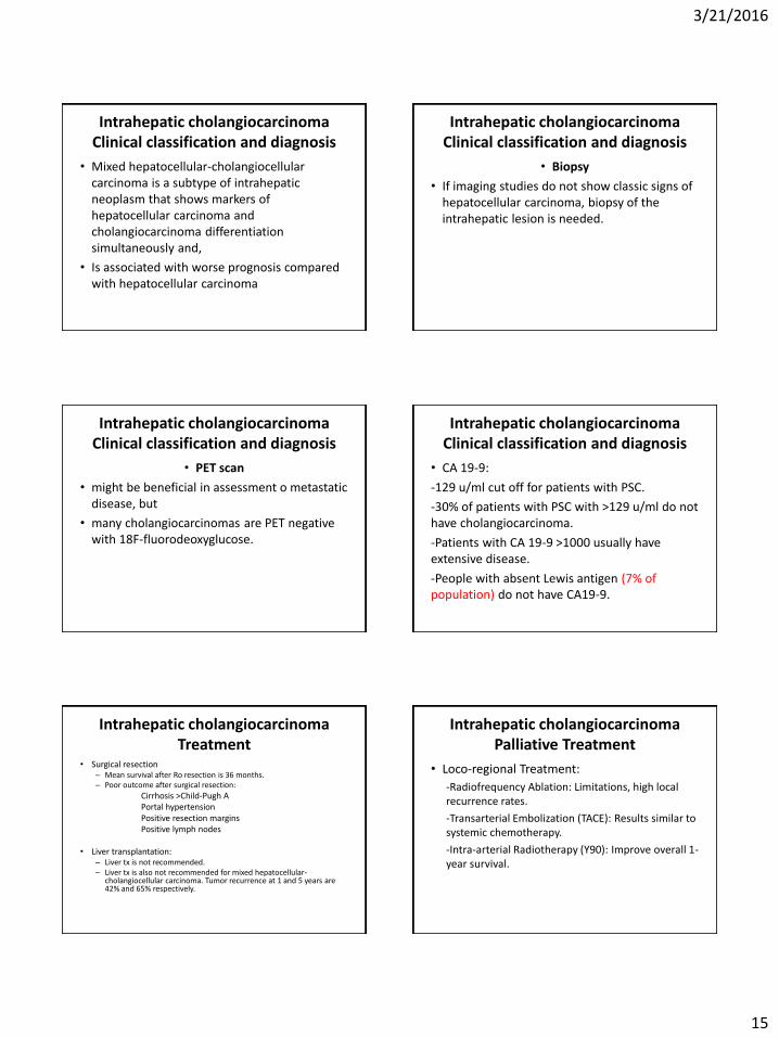
Intrahepatic cholangiocarcinoma Clinical classification and diagnosis
• Mixed hepatocellular-cholangiocellular carcinoma is a subtype of intrahepatic neoplasm that shows markers of hepatocellular carcinoma and cholangiocarcinoma differentiation simultaneously and,
• Is associated with worse prognosis compared with hepatocellular carcinoma
Intrahepatic cholangiocarcinoma Clinical classification and diagnosis
• Biopsy
• If imaging studies do not show classic signs of hepatocellular carcinoma, biopsy of the intrahepatic lesion is needed.
Intrahepatic cholangiocarcinoma Clinical classification and diagnosis
• PET scan
• might be beneficial in assessment o metastatic disease, but
• many cholangiocarcinomas are PET negative with 18F-fluorodeoxyglucose.
Intrahepatic cholangiocarcinoma Clinical classification and diagnosis
• CA 19-9:
-129 u/ml cut off for patients with PSC.
-30% of patients with PSC with >129 u/ml do not have cholangiocarcinoma.
-Patients with CA 19-9 >1000 usually have extensive disease.
-People with absent Lewis antigen (7% of population) do not have CA19-9.
Intrahepatic cholangiocarcinoma Treatment
• Surgical resection – Mean survival after Ro resection is 36 months. – Poor outcome after surgical resection:
Cirrhosis >Child-Pugh A Portal hypertension Positive resection margins Positive lymph nodes
• Liver transplantation:
– Liver tx is not recommended. – Liver tx is also not recommended for mixed hepatocellular-
cholangiocellular carcinoma. Tumor recurrence at 1 and 5 years are 42% and 65% respectively.
Intrahepatic cholangiocarcinoma Palliative Treatment
• Loco-regional Treatment:
-Radiofrequency Ablation: Limitations, high local recurrence rates.
-Transarterial Embolization (TACE): Results similar to systemic chemotherapy.
-Intra-arterial Radiotherapy (Y90): Improve overall 1-year survival.

3/21/2016
16
Perihilar Cholangiocarcinoma
A. Exophytic
B. Intraductal
1- Infiltrating
2- Papillary: Favorable prognosis
3- Tubulo-papillary: Favorable prognosis
Perihilar Cholangiocarcinoma Evaluation
• Jaundice: Most common presentation (90%)
• CA 19-9: (IgG4-related cholangiopathy)
• CT scan
• MRI/MRCP
• EUS
Perihilar Cholangiocarcinoma Evaluation
Perihilar Cholangiocarcinoma Evaluation
Perihilar Cholangiocarcinoma
• The inclusion criteria include:
– unresectable perihilar cholangiocarcinoma
– 3 cm or less in radial diameter
– without intrahepatic or extrahepatic metastases.
Hepatocellular Carcinoma (HCC)
• Worldwide, – HCC represents the sixth most common cancer.
– It is the third most common cause of cancer- related deaths.
Bruix J, Hepatology. 2011; 53(3):1020
3/21/2016
17
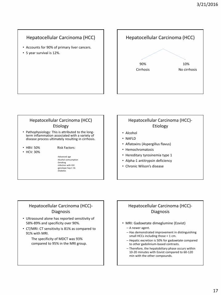
Hepatocellular Carcinoma (HCC)
• Accounts for 90% of primary liver cancers.
• 5 year survival is 12%.
Hepatocellular Carcinoma (HCC)
90% 10%
Cirrhosis No cirrhosis
Hepatocellular Carcinoma (HCC) Etiology
• Pathophysiology: This is attributed to the long-term inflammation associated with a variety of disease process ultimately resulting in cirrhosis.
• HBV: 50% Risk Factors: • HCV: 30% -Advanced age
-Alcohol comsumption -Smoking -infection with HIV -genotype hep C 1b -Diabetes
Hepatocellular Carcinoma (HCC)- Etiology
• Alcohol
• NAFLD
• Aflatoxins (Aspergillus flavus)
• Hemochromatosis
• Hereditary tyrosinemia type 1
• Alpha-1 antitrypsin deficiency
• Chronic Wilson’s disease
Hepatocellular Carcinoma (HCC)- Diagnosis
• Ultrasound alone has reported sensitivity of 58%-89% and specificity over 90%.
• CT/MRI: CT sensitivity is 81% as compared to 91% with MRI.
The specificity of MDCT was 93% compared to 95% in the MRI group.
Hepatocellular Carcinoma (HCC)- Diagnosis
• MRI: Gadoxetate dimeglumine (Eovist) – A newer agent.
– Has demonstrated improvement in distinguishing small HCCs including those < 1 cm.
– Hepatic excretion is 50% for gadoxetate compared to other gadolinium-based contrasts.
– Therefore, the hepatobiliary phase occurs within 10-20 minutes with Eovist compared to 60-120 min with the other compounds.

3/21/2016
18
Hepatocellular Carcinoma (HCC)- Diagnosis
• AFP sensitivity has been reported to range from 25% to 65% for detecting HCC as a screening tool.
Hepatocellular Carcinoma (HCC)- Diagnosis
• Prognostigating factors:
– Micro RNA.
– Gene expression: e.g. HN1, RAN, RAMP3, KRT19, and TAF9.
– Protein glycosylation.
Hepatocellular Carcinoma (HCC)- Treatment
Resection Liver Transplant
5 year disease free survival _ +
5 year overall survival + +
10 year survival _ +
Hepatocellular Carcinoma (HCC)- Treatment
• In patients with underlying liver disease, the best outcome after liver resection is in: – a normal bilirubin,
– hepatic venous pressure gradient ≤ 10 mmHg
– a small isolated tumor (≤ 3 cm)
Jemal et al, CA Cancer J Clin 2008, 58; 71-96.
Lovet et al, Seminar Liver Des, 2005; 25: 181-200
Hepatocellular Carcinoma (HCC)- Treatment
• In 1996, the Milan group defined a group of patients who could achieve excellent survival of 75% at four years.

3/21/2016
19
Hepatocellular Carcinoma (HCC)- Treatment Milan Criteria
– Single tumor < 5 cm, – Three lesions or less with none greater than 3 cm, – No distant metastasis – No lymph node involvement – No lympho-vascular invasion
Mazzaferro V, N Engl J Med 1996; 334: 693
Hepatocellular Carcinoma (HCC)- Treatment
• The UCSF criteria/2001 which showed a 75% survival at 5 years:
– Single tumor ≤ 6.5 cm,
– Three or fewer tumors, all ≤ 4.5 cm,
– Total tumor diameter of ≤ 8 cm.
Hepatocellular Carcinoma (HCC)- Treatment
• Barcelona Clinic Liver Cancer Group criteria:
– 1 tumor <7 cm,
– 3 tumors <5 cm,
– 5 tumors <3 cm,
or down-staging to Milan criteria with pre-transplant adjuvant therapies.
(over 50% 5-year survival))
Llovet JM et al. Lancet 2003; 362: 1907.
Hepatocellular Carcinoma (HCC)- Treatment
• European Metro Group created:
– Metroticket criteria, which consists of nodule size plus tumor number ≤7.
(71% 5-year survival)
Mazzaferro V et al. Lancet Oncol 2009; 10: 35-43
Hepatocellular Carcinoma (HCC)- Treatment
• RFA/microwave is recommended for
– tumors ≤ 3 cm with up to three lesions treated simultaneously
– a single lesion ≤ 5 cm in pts who are not surgical candidates
– can be used in both Child-Pugh Class A and B liver disease relatively safely.
Hepatocellular Carcinoma (HCC)- Treatment
• TACE is the treatment of choice in larger and later staged tumors.
• The rational behind its use is the hepatic artery becomes the main feeder to the tumor. This procedure delivers treatment directly to the tumor through the segmental branch, limiting damage to surrounding normal hepatic parenchyma.
• Directed therapy – Chemotherapeutics : doxorubicin, mitomycin-C, cisplatin..
– Then uses vascular embolization material to cease blood flow and induce cellular injury to the tumor.
3/21/2016
20
Hepatocellular Carcinoma (HCC)- Treatment- TACE
• The general recommendations: – lesions < 8 cm, > 3 lesions,
– with no evidence of extrahepatic extension or lymph node involvement, and
– with patients with relatively preserved liver function including Child-Pugh Class A and B liver disease.
– Mortality <2%.
European Association for Study of Liver; Eur J Cancer 2012; 48: 599-641
Hepatocellular Carcinoma (HCC)- Treatment- TACE
• TACE can reduce the percentage of post-transplant recurrence (17% with treatment vs 36% without treatment).
Porrett PM, et al. Liver Transpl 2006; 12: 665
Hepatocellular Carcinoma (HCC)- Treatment- TACE
• TACE with drug eluting beads (DEB) have recently shown to improve side effects and improve outcome.
Salem R et al. Gastroenterology 2011; 140: 497-507.
3/21/2016
21
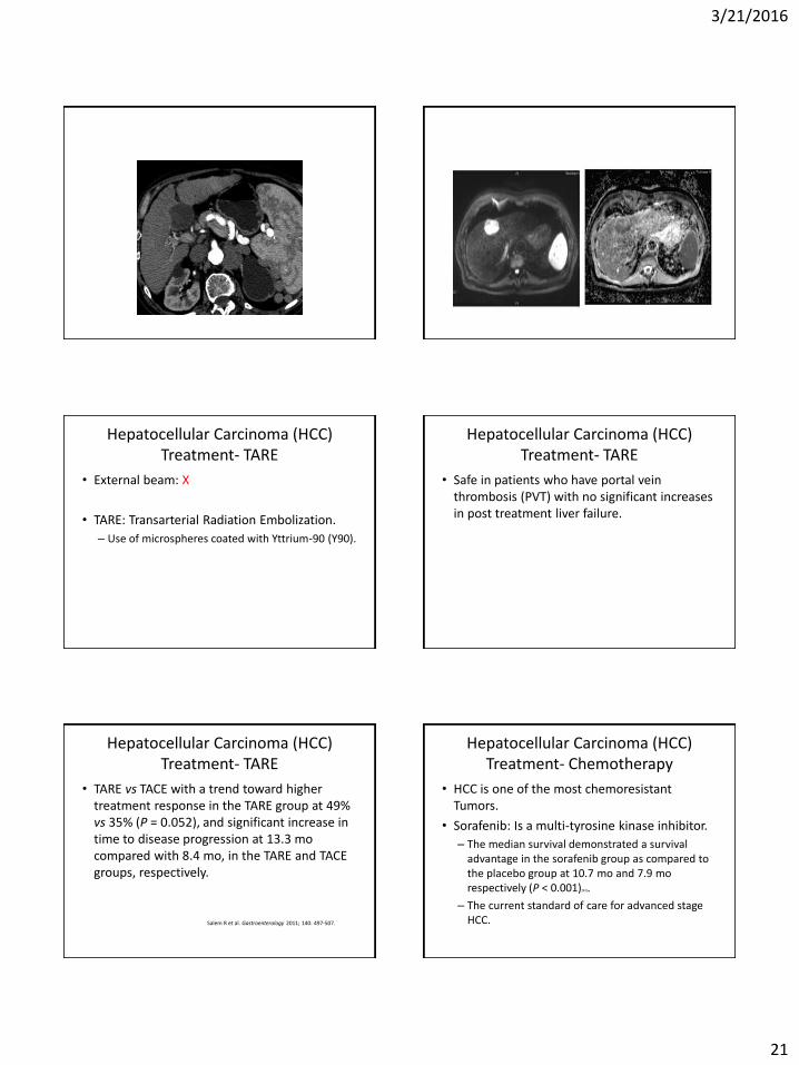
Hepatocellular Carcinoma (HCC) Treatment- TARE
• External beam: X
• TARE: Transarterial Radiation Embolization.
– Use of microspheres coated with Yttrium-90 (Y90).
Hepatocellular Carcinoma (HCC) Treatment- TARE
• Safe in patients who have portal vein thrombosis (PVT) with no significant increases in post treatment liver failure.
Hepatocellular Carcinoma (HCC) Treatment- TARE
• TARE vs TACE with a trend toward higher treatment response in the TARE group at 49% vs 35% (P = 0.052), and significant increase in time to disease progression at 13.3 mo compared with 8.4 mo, in the TARE and TACE groups, respectively.
Salem R et al. Gastroenterology 2011; 140: 497-507.
Hepatocellular Carcinoma (HCC) Treatment- Chemotherapy
• HCC is one of the most chemoresistant Tumors.
• Sorafenib: Is a multi-tyrosine kinase inhibitor.
– The median survival demonstrated a survival advantage in the sorafenib group as compared to the placebo group at 10.7 mo and 7.9 mo respectively (P < 0.001)[93].
– The current standard of care for advanced stage HCC.

3/21/2016
22
Questions
Thank you