Does a backwardly read protein sequence have a unique native...
10
Protein Engineering vol.9 no.l pp.5-14, 19% Does a backwardly read protein sequence have a unique native state? Krzysztof A.Olszewski 1 " 2 , Andrzej Kolinski 1 - 2 and Jeffrey Skolnick 1 ' 3 'Department of Molecular Biology, Scripps Research Institute, 10666 North Torrey Pines Road, La Jolla, CA 92037, USA and 2 Department of Chemistry, University of Warsaw, uJ. Pasteura 1, 02-093 Warsaw, Poland ^To whom correspondence should be addressed Amino acid sequences of native proteins are generally not palindromic Nevertheless, the protein molecule obtained as a result of reading the sequence backwards, i.e. a retro- protein, obviously has the same amino acid composition and the same hydrophobicity profile as the native sequence. The important questions which arise in the context of retro-proteins are: does a retro-protein fold to a well defined native-like structure as natural proteins do and, if the answer is positive, does a retro-protein fold to a structure similar to the native conformation of the original protein? In this work, the fold of retro-protein A, originated from the retro-sequence of the B domain of Staphylococcal protein A, was studied. As a result of lattice model simula- tions, it is conjectured that the retro-protein A also forms a three-helix bundle structure, in solution. It is also predicted that the topology of the retro-protein A three-helix bundle is that of the native protein A, rather than that correspond- ing to the mirror image of native protein A. Secondary structure elements in the retro-protein do not exactly match their counterparts in the original protein structure; however, the amino acid side chain contact pattern of the hydrophobic core is partly conserved. Keywords: lattice representation of proteins/Monte Carlo method/retro-protein AlStaphylococcal protein A/three-helix bundle Introduction The biological functions of proteins are based on their unique three-dimensional structure. Since the Anfinsen refolding experiments (Anfinsen, 1973), it is believed that the native structure of proteins is uniquely determined by their amino acid sequence. However, the problem of determining a protein's three-dimensional structure from the sequence itself remains unsolved, despite years of intensive research (see Vasquez et al., 1994). Among various attempts to solve the protein folding problem, those which utilize the reduced representation of the protein molecule seem to emerge as methods that allow study of the stability and folding of small proteins at acceptable computational cost (Skolnick and Kolinski, 1989; Chan and Dill, 1993; Godzik etal., 1993a; Liwo etal., 1993;Shakhnovich and Gutin, 1993; Hao and Scheraga, 1994; Kolinski and Skolnick, 1994a,b; Sali et al., 1994; Socci and Onuchic, 1994; Park and Levitt, 1995). In this work, using a high coordination lattice model of proteins, we studied the behavior of a novel retro-protein. We attempted to establish possible links that exist between the retro-protein structure and the structure of the original protein. Naturally occurring proteins are built from L-amino acids, consecutively connected by amide bonds in order to produce a long backbone of amide bonds with amino acid side groups, which are attached to the chiral C° carbon atoms. The amide (peptide) bond that connects consecutive C° carbons is almost planar (Corey and Pauling, 1953), since the rotation around the bond between NH and CO groups is hindered. Therefore, the conformational variety of protein structures originates from changes in the relative orientation of the consecutive peptide bond plates customarily described by dihedral angles O and *¥ (Scheraga, 1968). Thus, the conformation of the protein backbone can be roughly described by specifying only the location of the C" carbons (Oldfield and Hubbard, 1994). Let us define the retro-sequence as the backwardly read sequence of the original protein. One can attempt to rebuild the retro- sequence on the existing C™ backbone. This leads to a structure similar to the original protein but with completely translocated side-chains with respect to their original positions (Figure 1). The alternative approach is to rebuild the putative retro-protein structure using a retro-sequence but starting from the C- terminus of the original protein backbone instead of the N- terminus. The resulting structure is compatible with the original protein, but the direction of the protein backbone is opposite. Moreover, the CMT^ bonds now point in different directions, since each C" carbon (except those of glycines) is chiral, e.g. the side chains in helices will lie in the opposite direction (Figure 2). This leads to a potentially different pattern of side chain contacts. If D-amino acids were used in the rebuilding procedure instead of L-amino acids, the overlap of side chains would be greater; however, the backbone direction still remains opposite the native protein. Note, therefore, that the homology between the native protein sequence and its retro-sequence is generally very low. The result of backward reading of the protein sequence (retro-transition) will be further referred to as a retro-protein. The result of changing the absolute chirality of amino acids (chiral transition) performed on a native protein containing L-amino acids (L-protein) will be referred to as a D-protein. The backward reading of the sequence does not change the chirality of amino acids constituting a protein; therefore, it cannot produce a protein composed of D-amino acids, i.e. the retro-transition and the chiral transition are independent of each other. Since the chiral transition produces a perfect mirror image of the L-protein, it is safe to assume that the D-protein acquires the perfect mirror image structure of the L-protein upon folding. Indeed, D-HIV protease has been synthesized and shown to acquire a perfect mirror image fold of the naturally occurring HIV protease (Milton et al., 1992). Recently, another example of a D-protein, the Leu5 variant of trypsin inhibitor, has also been shown by Nielsen et al. (1994) to acquire the mirror- image form. Also, various cyclic and linear oligopeptide hormones have been synthesized that are related to each other © Oxford University Press
Transcript of Does a backwardly read protein sequence have a unique native...

Protein Engineering vol.9 no.l pp.5-14, 19%
Does a backwardly read protein sequence have a unique nativestate?
Krzysztof A.Olszewski1"2, Andrzej Kolinski1-2 andJeffrey Skolnick1'3
'Department of Molecular Biology, Scripps Research Institute, 10666 NorthTorrey Pines Road, La Jolla, CA 92037, USA and 2Department ofChemistry, University of Warsaw, uJ. Pasteura 1, 02-093 Warsaw, Poland
^To whom correspondence should be addressed
Amino acid sequences of native proteins are generally notpalindromic Nevertheless, the protein molecule obtainedas a result of reading the sequence backwards, i.e. a retro-protein, obviously has the same amino acid compositionand the same hydrophobicity profile as the native sequence.The important questions which arise in the context ofretro-proteins are: does a retro-protein fold to a welldefined native-like structure as natural proteins do and, ifthe answer is positive, does a retro-protein fold to astructure similar to the native conformation of the originalprotein? In this work, the fold of retro-protein A, originatedfrom the retro-sequence of the B domain of Staphylococcalprotein A, was studied. As a result of lattice model simula-tions, it is conjectured that the retro-protein A also formsa three-helix bundle structure, in solution. It is also predictedthat the topology of the retro-protein A three-helix bundleis that of the native protein A, rather than that correspond-ing to the mirror image of native protein A. Secondarystructure elements in the retro-protein do not exactlymatch their counterparts in the original protein structure;however, the amino acid side chain contact pattern of thehydrophobic core is partly conserved.Keywords: lattice representation of proteins/Monte Carlomethod/retro-protein Al Staphylococcal protein A/three-helixbundle
IntroductionThe biological functions of proteins are based on their uniquethree-dimensional structure. Since the Anfinsen refoldingexperiments (Anfinsen, 1973), it is believed that the nativestructure of proteins is uniquely determined by their aminoacid sequence. However, the problem of determining a protein'sthree-dimensional structure from the sequence itself remainsunsolved, despite years of intensive research (see Vasquezet al., 1994). Among various attempts to solve the proteinfolding problem, those which utilize the reduced representationof the protein molecule seem to emerge as methods that allowstudy of the stability and folding of small proteins at acceptablecomputational cost (Skolnick and Kolinski, 1989; Chan andDill, 1993; Godzik etal., 1993a; Liwo etal., 1993;Shakhnovichand Gutin, 1993; Hao and Scheraga, 1994; Kolinski andSkolnick, 1994a,b; Sali et al., 1994; Socci and Onuchic, 1994;Park and Levitt, 1995). In this work, using a high coordinationlattice model of proteins, we studied the behavior of a novelretro-protein. We attempted to establish possible links that
exist between the retro-protein structure and the structure ofthe original protein.
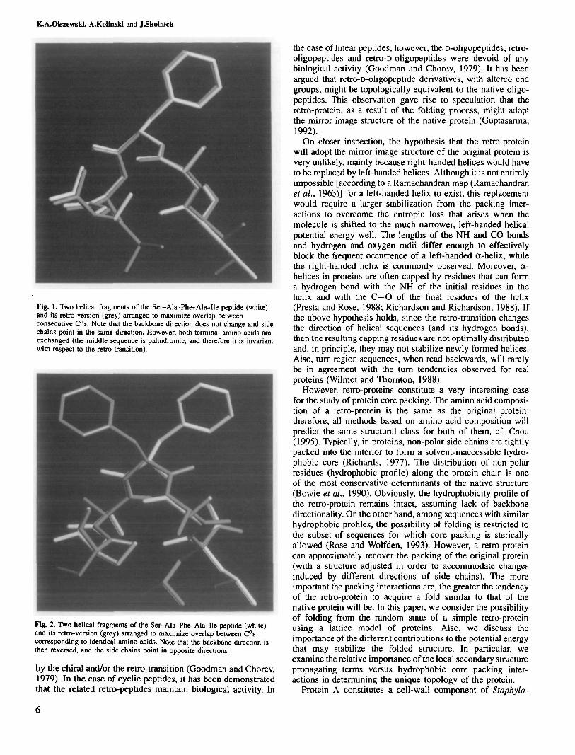
Naturally occurring proteins are built from L-amino acids,consecutively connected by amide bonds in order to producea long backbone of amide bonds with amino acid side groups,which are attached to the chiral C° carbon atoms. The amide(peptide) bond that connects consecutive C° carbons is almostplanar (Corey and Pauling, 1953), since the rotation aroundthe bond between NH and CO groups is hindered. Therefore,the conformational variety of protein structures originates fromchanges in the relative orientation of the consecutive peptidebond plates customarily described by dihedral angles O and*¥ (Scheraga, 1968). Thus, the conformation of the proteinbackbone can be roughly described by specifying only thelocation of the C" carbons (Oldfield and Hubbard, 1994). Letus define the retro-sequence as the backwardly read sequenceof the original protein. One can attempt to rebuild the retro-sequence on the existing C™ backbone. This leads to a structuresimilar to the original protein but with completely translocatedside-chains with respect to their original positions (Figure 1).The alternative approach is to rebuild the putative retro-proteinstructure using a retro-sequence but starting from the C-terminus of the original protein backbone instead of the N-terminus. The resulting structure is compatible with the originalprotein, but the direction of the protein backbone is opposite.Moreover, the CMT^ bonds now point in different directions,since each C" carbon (except those of glycines) is chiral, e.g.the side chains in helices will lie in the opposite direction(Figure 2). This leads to a potentially different pattern of sidechain contacts. If D-amino acids were used in the rebuildingprocedure instead of L-amino acids, the overlap of side chainswould be greater; however, the backbone direction still remainsopposite the native protein. Note, therefore, that the homologybetween the native protein sequence and its retro-sequence isgenerally very low. The result of backward reading of theprotein sequence (retro-transition) will be further referred toas a retro-protein. The result of changing the absolute chiralityof amino acids (chiral transition) performed on a native proteincontaining L-amino acids (L-protein) will be referred to as aD-protein. The backward reading of the sequence does notchange the chirality of amino acids constituting a protein;therefore, it cannot produce a protein composed of D-aminoacids, i.e. the retro-transition and the chiral transition areindependent of each other.
Since the chiral transition produces a perfect mirror imageof the L-protein, it is safe to assume that the D-protein acquiresthe perfect mirror image structure of the L-protein upon folding.Indeed, D-HIV protease has been synthesized and shown toacquire a perfect mirror image fold of the naturally occurringHIV protease (Milton et al., 1992). Recently, another exampleof a D-protein, the Leu5 variant of trypsin inhibitor, has alsobeen shown by Nielsen et al. (1994) to acquire the mirror-image form. Also, various cyclic and linear oligopeptidehormones have been synthesized that are related to each other
© Oxford University Press
K.A.CHswwski, A.Kolinski and J^kolnkk
Fig. 1. Two helical fragments of the Ser-AJa-Phe-Ala-Ile peptide (white)and its retro-version (grey) arranged to maximize overlap betweenconsecutive C"s. Note that the backbone direction does not change and sidechains point in the same direction. However, both terminal aniino acids areexchanged (the middle sequence is palindromic, and therefore it is invariantwith respect to the retro-transition).
Fig. 2. Two helical fragments of the Scr-Ala-Phe-Ala-Ile peptide (white)and its retro-version (grey) arranged to maximize overlap between C°scorresponding to identical amino acids. Note that the backbone direction isthen reversed, and the side chains point in opposite directions.
by the chiral and/or the retro-transition (Goodman and Chorev,1979). In the case of cyclic peptides, it has been demonstratedthat the related retro-peptides maintain biological activity. In
the case of linear peptides, however, the D-oligopeptides, retro-oligopeptides and retro-D-oligopeptides were devoid of anybiological activity (Goodman and Chorev, 1979). It has beenargued that retro-D-oligopeptide derivatives, with altered endgroups, might be topologically equivalent to the native oligo-peptides. This observation gave rise to speculation that theretro-protein, as a result of the folding process, might adoptthe mirror image structure of the native protein (Guptasarma,1992).
On closer inspection, the hypothesis that the retro-proteinwill adopt the mirror image structure of the original protein isvery unlikely, mainly because right-handed helices would haveto be replaced by left-handed helices. Although it is not entirelyimpossible [according to a Ramachandran map (Ramachandranet al., 1963)] for a left-handed helix to exist, this replacementwould require a larger stabilization from the packing inter-actions to overcome the entropic loss that arises when themolecule is shifted to the much narrower, left-handed helicalpotential energy well. The lengths of the NH and CO bondsand hydrogen and oxygen radii differ enough to effectivelyblock the frequent occurrence of a left-handed a-helix, whilethe right-handed helix is commonly observed. Moreover, cc-helices in proteins are often capped by residues that can forma hydrogen bond with the NH of the initial residues in thehelix and with the C=O of the final residues of the helix(Presta and Rose, 1988; Richardson and Richardson, 1988). Ifthe above hypothesis holds, since the retro-transition changesthe direction of helical sequences (and its hydrogen bonds),then the resulting capping residues are not optimally distributedand, in principle, they may not stabilize newly formed helices.Also, turn region sequences, when read backwards, will rarelybe in agreement with the turn tendencies observed for realproteins (Wilmot and Thornton, 1988).
However, retro-proteins constitute a very interesting casefor the study of protein core packing. The amino acid composi-tion of a retro-protein is the same as the original protein;therefore, all methods based on amino acid composition willpredict the same structural class for both of them, cf. Chou(1995). Typically, in proteins, non-polar side chains are tightlypacked into the interior to form a solvent-inaccessible hydro-phobic core (Richards, 1977). The distribution of non-polarresidues (hydrophobic profile) along the protein chain is oneof the most conservative determinants of the native structure(Bowie et al., 1990). Obviously, the hydrophobicity profile ofthe retro-protein remains intact, assuming lack of backbonedirectionality. On the other hand, among sequences with similarhydrophobic profiles, the possibility of folding is restricted tothe subset of sequences for which core packing is stericallyallowed (Rose and Wolfden, 1993). However, a retro-proteincan approximately recover the packing of the original protein(with a structure adjusted in order to accommodate changesinduced by different directions of side chains). The moreimportant the packing interactions are, the greater the tendencyof the retro-protein to acquire a fold similar to that of thenative protein will be. In this paper, we consider the possibilityof folding from the random state of a simple retro-proteinusing a lattice model of proteins. Also, we discuss theimportance of the different contributions to the potential energythat may stabilize the folded structure. In particular, weexamine the relative importance of the local secondary structurepropagating terms versus hydrophobic core packing inter-actions in determining the unique topology of the protein.
Protein A constitutes a cell-wall component of Staphylo-

Native structure of retro-protein A
coccus aureus that binds to an Fc domain of immunoglobulins.Its extracellular part consists of five highly homologousdomains designated E, D, A, B and C, respectively (Table I).The B domain of Staphylococcal protein A, complexed withthe Fc portion of human polyclonal immunoglobulin G, hasbeen crystallized and the structure of the complex has beensolved. The B domain part of the complex consists of twohelices, from Gin 10 to Leu 18 (helix I) and from Glu26 toAsp37 (helix IT), which are packed together to form anantiparallel helical hairpin (Deisenhofer, 1981). The three-dimensional solution structure of the B domain has also beendetermined by NMR spectroscopy (Gouda et al, 1992) in theabsence of the complexing immunoglobulin. In water, it formsa stable three-helix bundle motif with helix I (Gin 10 to His 19)tilted with respect to the antiparallel hairpin formed by helicesII (Glu26 to Asp37) and m (Ser42-Ala55). The N-terminalresidues up to Glu9 and the C-terminal residues from Gln56to the terminal lysine do not exhibit ordered structure. Theabsence of the third helix in the crystal structure of the Bdomain complex with immunoglobulin is probably induced bycrystal contacts (Gouda et al, 1992).
The distribution of secondary structural elements in thesolution structure of the B domain of protein A agrees withthe capping properties of helical termini (Presta and Rose,1988; Richardson and Richardson, 1988). The first helix isfairly well capped at the N-terminus by Asnl2, and the C-terminus by His 19. The second helix N-cap Asn26-Glu25-Glu26 is perfect, while the C-cap is marked only by the Lys36residue. In the third helix, Asn44, together with Ser42, agreeswith the capping properties at the N-terminus, and the C-capis formed by the Lys50, Lys51 and Asn53. Also, Pro21 andAsn22 that constitute the first turn are highly expected in theirpositions (Wilmot and Thornton, 1988). The second turn(between helices FI and IH) is even more exemplary, beingbuilt from Asp38-Pro39-Ser40-Gln41 (Wilmot andThornton, 1988).
Structures of the other domains of protein A have not beenreported previously. However, since all domains of protein Aare at least 80% homologous to each other and also bind toimmunoglobulin, we assume that the overall structure isconserved within the family of domains of protein A. Moreover,in order to remove an Asn-Gly pair from the native sequenceof the B domain of protein A, the so-called protein Z, hasbeen proposed, and subsequently expressed as a single pointmutation of the B domain involving Gly30 and Ala30 (G30A)(Nilsson et al, 1987). Its NMR structure (Lyons et al., 1993)reveals a three-helix bundle topology for protein Z in solution.
Recently, a lattice model of proteins has been used toredesign the B domain of protein A so that its mutant preservesthe three-helix bundle topology, but has a different overallchirality of the global fold (Olszewski et al., 1995). We haveshown that, although the native topology of the three-helixbundle is strongly conserved, it is possible to find, by anextensive search of possible mutations, a putative mutant thatmay exhibit the topological mirror image structure. Moreover,additional studies of protein A mutations have proved that thelattice model used here can differentiate between the twotopological alternatives of the three-helix bundle, thereforeencouraging its application to the study of the retro-proteinfolding simulations and packing interaction studies.
The outline of the remainder of this paper is as follows. Inthe Methods section, we briefly present the lattice model ofproteins and the interaction scheme used. The model is
essentially the same as that used in our previous studies(Kolinski and Skolnick, 1994a; Olszewski et al, 1995); how-ever, for the reader's convenience, we describe concisely thelattice representation of the protein chain and the variouscontributions to the force field. Also, the algorithm for all-atom model building is discussed. In the Results section, wediscuss in detail the lattice simulations of retro-protein A; thisis then followed by the all-atom model building. Additionalanalysis of secondary structure predictions corroborates ourpredicted structure of retro-protein A.
Methods
A 90-component, high-coordination lattice model used forthe protein backbone representation (Kolinski and Skolnick,1994a) was constructed by making all possible permutationsof the components of the generic vectors (3,1,1), (3,1,0),(3,0,0), (2,2,1) and (2,2,0), with the lattice unit length equalto 1.22 A. These vectors connect consecutive 0*3 along theprotein backbone, thus serving as virtual bonds. No backboneatoms other than the C°s are explicitly used, and only consecut-ive pairs of vectors that form protein-like angles betweenvirtual bonds (i.e. from 72.5 to 154°) are permitted. The latticerepresentations of a high-resolution library protein C" carbonare within 0.7 A r.m.s. (root mean square deviation of C"carbons) of their continuous space representations (Godziket al, 1993b). A library of single ball rotamers is used torepresent amino acid side chains. Rotamers are located at theside chain center of mass positions and depend on the localgeometry of the protein backbone. The number of allowedrotamers is different for different amino acids and varies fromone rotamer (e.g. for alanine) to a maximum of 58 rotamersfor arginine, in certain backbone configurations. The localaccuracy of this side chain center of mass representationis approximately 1 A r.m.s. On combining the side chainrepresentation into the model, the overall intrinsic geometricalaccuracy of the model decreases to about 2 A r.m.s. MonteCarlo simulations of protein folding on the above lattice areperformed by accepting or rejecting small movements of theprotein backbone on the basis of the asymmetric Metropoliscriterion (Metropolis et al, 1953). These movements includepredefined local two- and three-virtual-bond moves and alsolong-distance moves designed to enhance the search of con-formational space. The latter moves are generated by concertedsequences of overlapping three-bond motions. In addition,random changes of rotamer positions are allowed to facilitatethe packing of side chains (Kolinski and Skolnick, 1994a).
The potential energy used consists of five terms, viz.
+ WNNENN
(1)
generated by the analysis of the library of high-resolution PDBstructures of globular proteins (Kolinski and Skolnick, 1994a)with the weighting factors w for the various energy contribu-tions equal to w,^, = 1, wbb = 0.5, w^ = 1.75, w^T = 2and wNN = 0.25.
In particular, the local, sequence-dependent termN- 3
2, ( U 24 3) (2)
involves two consecutive C? to Cf+ 3 and C?+ , to Cf+ 4 (theindex / represents consecutive C"s) chiral distances,i.e. fj, i + ij + 2 and ?i + ij + 24 + 3> respectively. The chiral

K.A.OIszewskl, A.Kolinski and J-Skolnick
distance is defined as follows: f,jt = sign ((b,- Xby)-bt) II b, + b, + bk II , where b, is a virtual bond thatconnects Cf to C? + ). The subscripts in E ^ (Equation 2)signify the amino acid sequence dependence of the term. Sincetwo overlapping Cf—Cf+ 3 chiral distances are involved, Epmp
propagates protein-like elements of secondary structure alongthe protein backbone. In addition to this local term, aneffective interaction between Cf and Cf that simulates theformation of a hydrogen bond between backbone atoms of the(th and y'th residues was introduced:
, ± Uj± i )5 , v (3)
where bjj = 1 when amino acids 1 and j form a hydrogen-bonded pair and otherwise 6V = 0. £" and £"" are equal to-0 .5 kT. Amino acids 1 and j form a hydrogen-bonded pairwhen, and only when, the following geometrical criteriaare satisfied:
(Kolinski and Skolnick, 1995). Attractive interactions aremodified by a factor
/ = 1 - {cos2[Z(u,, u,)] - cos2(20°))2 (8)
which is dependent upon the angle between the vector u,- =r,+2 - r,_2 and the corresponding vector Uy = r,-+2 - r;_2 inorder to induce proper supersecondary structure packing.The angle ZCii/.u,) represents the relative orientation of thesecondary structure in the vicinity of the /th residue withrespect to the secondary structure surrounding the yth residue,and 20° is the most probable packing angle of helices.
The ENN supplemental term was designed to reproduce theoccurrence of protein-like side-chain contact maps of globularproteins. An artificial neural network with error back-propaga-tion has been trained to recognize frequently occurring 7 X 7fragments of side chain contact maps (Milik et al., 1995). Foreach pair ij, if the 7 X 7 fragment of the side chain contactmap centered at ij is recognized by the neural network as
Kb, - 1- bd-Tij \^ 0,,^ (4)Kb, - 1 - b,)-rlV l« OmiX
where Tjj is a vector connecting Cf to Cf. Rmm = 4.6 A,/?„,„ = 7.3 A and a^^ = 13.4 A2. This interaction componentis sequence independent and also non-directional, since themodels do not specify the location of backbone atoms otherthan C*. Proton donors and acceptors are not differentiated.Every amino acid but proline can form up to two hydrogenbonds, whereas proline can participate in only one hydrogenbond. The hydrogen-bonding scheme accounts for theco-operativity of hydrogen bond formation by introducing aneffective interaction between adjacent pairs of hydrogen bonds.
A one-body, centrosymmetric burial potential,
(5)
reflects the radial distribution of distances /f from the ithamino acid side chain to the center of mass of the protein[where 5 is the expected radius of gyration, calculated for aclosely packed protein (Kolinski and Skolnick, 1994a)]. Thepurpose of the burial energy is to enforce the compaction ofthe protein; therefore, the E^ component is small for compactstates and dominates denatured states. It serves as a drivingforce in the initial stages of the folding process (Kolinski andSkolnick, 1994a) by narrowing the conformational space searchto compact or near-to-compact states.
The final packing of the protein core depends on moredetailed packing interactions. They are modeled by the com-bination of a pairwise, soft-core repulsion augmented by asquare-well potential, derived as a potential of mean forcefrom the frequency of close contact occurrences betweenamino acids:
(6)
where / andy indicate interacting amino acids ; andy, and equals
E^, for r,j <Ey, for Rff =fa, for R™0, for ri4 ^
Ru and e,v s* 0R,j and e v < 0
(7)
The radius of repulsion, Ry, depth e^ and limits Ry of thesquare-well width are publicly available by anonymous ftp
8
Table I.domains
No.
1011121314151617181920212223242526272829303132333435363738394041424344454647484950515253
Sequencesand of the
E
GinGinAsnAlaPheTyrGinValLeuAsnMetProAsnLeuAsnAlaAspGinArgAsnGlyPhelieGinSerLeuLysAspAspProSerGinSerAlaAsnValLeuGlyGluAlaGinLysLeuAso
of all Staphylococcal protein A three-helixretro-protein A (backwardly read B domain
D
GinGinSerAlaPheTyrGluHeLeuAsnMetProAsnLeuAsnGluAlaGinArgAsnGlyPheHeGinSerLeuLysAspAspProSerGinSerThrAsnVaJLeuGlyGluAlaLysLysLeuAsn
A
GinGinAsnAlaPheTyrGluHeLeuAsnMetProAsnLeuAsnGluGluGinArgAsnGlyPheHeGinSerLeuLysAspAspProSerGinSerAlaAsnLeuLeuSerGluAlaLysLysLeuAsn
B
GinGinAsnAlaPheTyrGluHeLeuHisLeuProAsnLeuAsnGluGluGinArgAsnGlyPhelieGinSerLeuLysAspAspProSerGinSerAlaAsnLeuLeuAlaGluAlaLysLysLeuAsn
C
GinGinAspAlaPheTyrGluHeLeuHisLeuProAsnLeuThrGluGluGinArgAsnGlyPheHeGinSerLeuLysAspAspProSerVaJSerLysGluHeLeuAlaGluAlaLysLysLeuAsn
bundlesequence).
retro
AsnLeuLysLysAlaGluAlaLeuLeuAsnAlaSerGinSerProAspAspLysLeuSerGinHePheGlyAsnArgGinGluGluAsnLeuAsnProLeuHisLeuHeGluTyrPheAlaAsnGinGin
Only amino acids that correspond to well defined secondary structure forthe experimental B domain structure are shown.

being protein-like, then the pair interaction well depth ismodified in the following way:
-I- f) If.. (Q\
where
2 , x7£^'(10)
and the summation in Equation 10 is performed over the 7 X 7fragment of the appropriate contact map; cu = 1 if side chainsk and / are in contact and 0 otherwise. Therefore, the neuralnetwork term simulates the average effective many-bodycomponent of the potential energy responsible for the mutualpacking of super secondary structure elements.
Previously, the same lattice model with a similar interactionscheme was successfully applied by Kolinski, Skolnick andco-workers to the simulation of the folding process of smallhelical proteins (Kolinski and Skolnick, 1994b), coiled coils
Table IL Average energies for retro-protein A incorresponding to the native topology E0*1 and tobasin Em, respectively (AE™ - m v = P * - E"1*)
T
1.00.90.8
-187.4-219.9-252.2
-182.2-207.5-239.1
isothermalthe mirror
simulationsimage topology
\rvat - inv
-5.2-12.4-13.1
Native structure of retro-protein A
(Vieth et al., 1994) and crambin (Kolinski and Skolnick,1994b). However, here the rotamer energy (Kolinski andSkolnick, 1994a) has not been used, and the many-bodycomponent of Kolinski and Skolnick (1994a) has been replacedby the neural network, packing regularizing term (Milik et al.,1995). Exactly the same model has been used in the recentanalysis of protein A mutations (Olszewski et al., 1995).
The lattice models obtained in the course of the simulationsare subsequently transformed into full atom models in orderto test the consistency of our results at the atomic resolutionlevel. All atom models are built from lattice C™ backbonestructures of retro-protein A using the following procedure.First, the complete backbone and C^ carbons positions arereconstructed using the method of Milik.M., Kolinski.A. andSkolnick, J. (unpublished), which is based on a statisticalanalysis of the peptide plate orientation with respect to threeconsecutive C" virtual bonds. All-atom models were thencompleted by rebuilding the side chains using the CHARMMpackage (Brooks et al., 1983). The resulting structures wereinitially relaxed in vacuo using the CHARMM all-atom poten-tial [PARAM19 parameter set with polar hydrogens (Brookset al., 1983)] and then relaxed in an 8 A water shell. We usedthe TIP3P water model (Jorgensen et al., 1983) and periodicboundary conditions to prevent the waters from evaporating.The protein has been almost completely immersed in a boxcontaining about 800 water molecules. For each structure, weperformed a few iterations of a relaxation procedure thatconsisted of ten heating and ten cooling MD simulations. Eachheating cycle starts at 50 K and ends at 700 K during the 4 pscycle; then, the system is cooled to 50 K during the 5 ps
aa
^g.a
LH
LI
ZJ
5NR
QE
E,
\^/a.a
VS
QS
PD
DK
.̂j ~ "j
tu
z
? 19
k .
•
• • •
•
• . "
29
• r•
m
•
J1
•
39
m '
•
• •
49
• •
•• •
9 19 29 39 49N L K K A E A L L N A S Q S P D D K L S Q I F G N R Q E E N L N P L H L I EYFANQQ
Fig. 3. Average contact map for the retro-protein A in the native topology (above diagonal) and in the mirror image topology (below diagonal).
KA.CHszewski, A.Kolinski and J-Skolnlck
9 19 29 39
9 19 29 39 49N L K K A E A L L N A S Q S P D D K L S Q I F G N R Q E E N L N P L H L I EYFANQQ
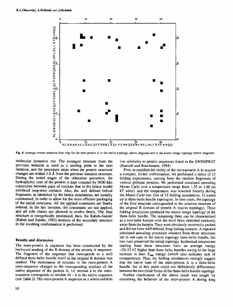
Fig. 4. Average contact retention time map for the retro-protein A in the native topology (above diagonal) and in the mirror image topology (below diagonal).
molecular dynamics run. The averaged structure from theprevious iteration is used as a starting point to the nextiteration, and the procedure stops when the protein structuralchanges are within 1.0 A from the previous iteration structure.During the initial stages of the relaxation procedure, thehydrophobic core of the protein is kept compact by NOE-likeconstraints between pairs of residues that in the lattice modelexhibited long-time contacts. Also, the well defined helicalfragments, as identified by the lattice simulations, are initiallyconstrained, in order to allow for the more efficient packagingof the initial structures. All the applied constraints are finallyrelaxed. In the last iteration, the constraints are not applied,and all side chains are allowed to evolve freely. The finalstructure is energetically minimized; then, the Kabsh-Sander(Kabsh and Sander, 1983) analysis of the secondary structurein the resulting conformation is performed.
Results and discussionThe retro-protein A sequence has been constructed by thebackward reading of the B domain of the protein A sequence.The fragment of the sequence that corresponds to a welldefined three-helix bundle motif in the original B domain wasstudied. The numeration of residues in the retro-protein Aretro-sequence changes to the corresponding residues of thenative sequence of the protein A, i.e. residue it in the retro-sequence corresponds to residue 54 - k in the native sequence(see Table I). The retro-protein A sequence as a whole exhibits
low similarity to protein sequences listed in the SWISSPROT(Bairoch and Boeckmann, 1994).
First, to establish the ability of the retro-protein A to acquirea compact, folded conformation, we performed a series of 15folding experiments, starting from the random fragments ofvarious globular proteins. We performed simulated annealingMonte Carlo over a temperature range from 1.55 to 1.00 (inkT units), and the temperature was lowered linearly duringthe Monte Carlo run. Out of 15 folding simulations, 12 endedup in three-helix bundle topologies. In nine cases, the topologyof the final structure corresponded to the solution structure ofthe original B domain of protein A (native topology). Threefolding simulations produced the mirror image topology of thethree-helix bundle. The remaining three can be characterizedas a two-helix hairpin with the third helix stretched randomlyaway from the hairpin. They were obviously incorrectly packedand did not have well defined, long-lasting contacts. A repeatedsimulated annealing procedure initiated from those structuresled in one case to the native topology three-helix bundle, buttwo runs preserved the initial topology. Isothermal simulationsstarting from those structures have an average energy-20-25 kT higher than three helix bundles owing to the largeincrease in their E^ energy (which also indicates lack ofcompactness). Thus, the folding simulations strongly suggestthat the native state of the retro-protein A is a three-helixbundle, but at this point could not definitively differentiatebetween the two chiral forms of the three-helix bundle topology.
Further clarification of the above result was sought byexamining the behavior of the retro-protein A during long
10
Native structure of retro-proteJii A
19 29 39
QQNAFYEI LHLPRL ME EQRNGF I QSLKDDPSQSANLLAEAKKLN
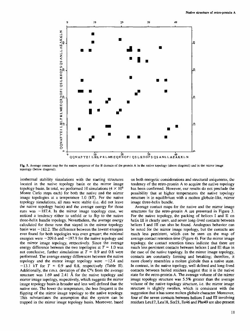
Fig. 5. Average contact map for the native sequence of the B domain of the protein A in the native topology (above diagonal) and in the mirror imagetopology (below diagonal).
isothermal stability simulations with the starting structureslocated in the native topology basin or the mirror imagetopology basin. In total, we performed 10 simulations (4 X 10⁶
Monte Carlo steps each) for both the native and the mirrorimage topologies at a temperature 1.0 (kT). For the nativetopology simulations, all runs were stable (i.e. did not leavethe native topology basin) and the average energy for thoseruns was -187.4. In the mirror image topology case, wenoticed a tendency either to unfold or to flip to the nativethree-helix bundle topology. Nevertheless, the average energycalculated for those runs that stayed in the mirror topologybasin was —182.2. The difference between the lowest energiesever found for both topologies was even greater; the minimalenergies were —209.6 and —197.9 for the native topology andthe mirror image topology, respectively. Since the averageenergy difference between the two topologies at T = 1.0 wasnot conclusive, further simulations at T = 0.9 and 0.8 wereperformed. The average energy differences between the nativetopology and the mirror image topology were -12.4 and-13.1 kT for T = 0.9 and 0.8, respectively (Table II).Additionally, the r.m.s. deviation of the C"s from the averagestructure was 1.69 and 2.41 A for the native topology andmirror image topology, respectively, which suggests the mirrorimage topology basin is broader and less well defined than thenative one. The lower the temperature, the less frequent is theflipping of the mirror image topology to the native topology.This substantiates the assumption that the system can betrapped in the mirror image topology basin. Moreover, based
on both energetic considerations and structural uniqueness, thetendency of the retro-protein A to acquire the native topologyhas been confirmed. However, our results do not preclude thepossibility that at higher temperatures the native topologystructure is in equilibrium with a molten globule-like, mirrorimage three-helix bundle.
Average contact maps for the native and the mirror imagestructures for the retro-protein A are presented in Figure 3.For the native topology, the packing of helices I and II onhelix HI is clearly seen, and seven long-lived contacts betweenhelices I and in can also be found. Analogous behavior canbe noted for the mirror image topology, but the contacts aremuch less persistent, which can be seen on the map ofaverage contact retention time (Figure 4). For the mirror imagetopology, the contact retention times indicate that there aremuch less persistent contacts between helices I and IH than inthe case of the native topology. In the mirror image topology,contacts are constantly forming and breaking; therefore, itmore closely resembles a molten globule than a native state.In contrast, in the native topology, well defined and long-livedcontacts between buried residues suggest that it is the nativestate for the retro-protein A. The average volume of the mirrorimage topology structure was 5.5% greater than the averagevolume of the native topology structure, i.e. the mirror imagestructure is slightly swollen, which is consistent with thesuggestion that it has some molten globule character. Moreover,four of the seven contacts between helices I and HI involvingresidues Leul7, Leul8, Ser21, De46 and Phe49 are also present
11

ICA.OUzewski, A.Kollnski and J^kolnick
0.0 r-i
I -5.08.
Ig
12c';= -10.0S.
-15.0
native topologymirror image
10 20 30 40amino acid number in the retro-protein A sequence
50
Fig. 6. Decomposition of the average pair interaction energy into one-body terms corresponding to the consecutive amino acids in the retro-sequence ofprotein A.
in the native structure of the native B domain of protein A(Figure 5). In addition, contacts of Leu45 (helix HI) with Ile31and Phe32 (helix II), and also Phe32 and Leu28 with Leu 18(helix I), are invariant with respect to the retro-transition. Intotal, eight long-lived contacts (i.e. over half of the totalpersistent contacts) that contribute to the stabilization of thenative three-helix bundle topology are conserved with respectto the retro-transition. However, the decomposition of the pairinteraction energy terms into single residue components doesnot reveal significant differences between the native andthe mirror image topologies (Figure 6). The average £pair
contribution per side chain at T = 0.8 is -7.23 and -7.21 forthe native topology and the mirror image topology, respectively.Hence, although the proper hydrophobic core packing of theretro-protein A is a necessary condition to assemble the foldedstructure, we conclude that it is not sufficient to direct theprotein toward the native fold during the folding process.
Although the packing of the native topology is similar tothe packing of the original protein A, the changes in thesecondary structure of the retro-protein A are significant. Thefirst helix of the retro-protein A is capped by the Asn 10 at theN-terminus and by the Gln22 and Ser23 at the C-terminus.The second turn in the protein A, which becomes the first inthe retro-protein A, is preserved, since the sequence Ser23-Pro24—Asp25 mimics the turn tendencies from real proteins.Asp26 residue caps the N-terminus of the second helix, whichends with Arg35 and Gln36 as C-terminal residues. On theother hand, the first turn region from the native protein A isno longer a rum in the retro-protein; instead, Asn31 andPro42 initiate the third helix, which is in agreement with thepreferences for N-cap residues. The third helix seems to bewell capped at the C-terminus by the asparagine and twoglutamines.
A number of the secondary structure prediction methodshave been applied to the sequence of the retro-protein A (Levin
12
Res. number
B domain sequence
Glbrat methodLevin methodDPM methodSOPMA methodPhD methodlattice modelNMR structure
Res. nuiiber
re t ro -pro te in A
Glbrat methodLevin methodDPM methodSOPMA methodPhD methodl a t t i c e nodal
NMR s t ruc tu reB domain sequence
Res. nunber
10 20 30 40 50I I I I IQQNAFYEILHLPNLNIEQRNGFIQSLKDDPSQSAMLLAEAKKLN
HHHHHHHHHHCCCCCHHHHHHHEEECCCCCCHHHHHHHHHHHHHCTCEECCEEECTTCCHHCHTHCEHEECCCSCHHHHHHHHHHHCCTCTHHHHEEHHCCCCCCHCCCEEHCHCCTCTCTHHHHHHHHHCCHHHHEEEEEECCCCCHHHCCHHHHICCCCCCCHHHHHHHHHHHCCHHHHHHHHHCCCHHHHHHHHHHHHHHCCCHHHHHHHHHHHCCHHHHHHHKHHTTTTTHHHHHHHHHHHHHTTTTHHHHHHHHHHHHHHHHHHHHHHTTTTTHHHHHHHHHHHHHTTTTHHHHHHHHHHHH
10 20 30 40 50I I I I INLKKAEALLNASQSPDDItLSQIFGHRQEENLNPLHLIEYrANQQ
HHHHHHHHHHHCCCCCHHHEEEECCCHHHCCCHHHHHHHHJ1HHHTCCHHHHHHHHCCCCCHHHHHHHHCCCCTCCCHHHHHHHHCTTCCCHHHHHHHHHTCTTTCCHCHIETCCHCCCCTCHHEEHHHHCCCHHHHHHHEEHHCCCCCCHHHHHHHCCCCCCCCCCEHHHHHHCCCCCCHHHHHHHHHCCCCCHHHHHHHHBHHCCCCCCHHHHHHHHCCCHHHHHHHHHHHCTTTTHHHHHHHHHHHTTTTHHHHHHHHHCCC
HHHHHHHHHHHHTTTTHHHHHHHHHHHHHTTTTTHHHHHHHHHHHLKKAEALLNASQSPDDKLSQirGNRQEENLNPLHLIEYFANQQ
I I I I I50 40 30 20 10
Fig. 7. Summary of the secondary structure predictions for the B domain ofprotein A and for the retro-sequence based on the B domain of the proteinA. The methods reported include the Gibrat method (Gibrat et al., 1987),Levin method (Levin et al., 1986), DPM method (Deleage and Roux, 1987),SOPMA method (Geourjon and Deleage, 1994, 1995) and PhD method(Rost and Sander, 1994). The results of lattice Monte Carlo simulation arealso reported, and in the case of the B domain those based on the NMRstructure are also presented. To facilitate the secondary structurecomparison, the B domain sequence together with the NMR structure isrepeated backwards at the bottom of the figure, so that the pattern of aminoacid side chains exactly matches the retro-B domain.
et al., 1986; Deleage and Roux, 1987; Gibrat et al., 1987;Rost and Sander, 1994; Geourjon and Deleage, 1995). All ofthem, in general, predict the existence of three helices, althoughthe helical termini locations vary (Figure 7). Nevertheless,fragments Argl2-Ala20, Arg27-Gly33 and Leu34-Ala50 arepredicted as helical by nearly all methods. The PhD methodpredictions are consistent with lattice simulations for thesecond helix termini and the C-terminus of the first helix. The

Native structure of retro-protein A
B
Fig. 8. (a) All-atom model of the retro-B domain of protein A in a space-filling representation. The hydrophobic core (dark grey) is almost completelycovered, (b) All-atom model of the retro-B domain of protein A in a ball-and-stick representation. The hydrophobic core is presented in dark grey and theother amino acids are in light grey, (c) All-atom model of the retro-B domain of protein A with a ribbon tube showing the three-helix bundle topology.
lattice prediction for the N-terminus of the third helix is alsopredicted by Gibrat et al. (1987) and Levin et al. (1986). Also,the DPM method prediction of the localization of the first turncorresponds to that of the lattice model. The middle helix inthe retro-protein A is shorter than the corresponding helix inthe native B domain (Figure 7). The first turn in the retro-protein becomes broader than the second turn in the B domainand the opposite tendency can be noticed for the other pair ofcorresponding turns.
For each folding simulation run that rendered the retro-protein A in the native topology of the protein A, an all-atommodel building procedure was performed in order to obtain amore detailed view of the retro-protein packing and to precludethe possibility of incorrect packing (e.g. due to the stericoverlap that cannot be seen in the lattice model). The protocolfor rebuilding all-atom models described in the Methodssection was used. During the relaxation procedure, we noticedthat the secondary structure became more regular as the
13
KAOlszewskJ, A.KoUnski and J-Skolnlck
retro-molecule adjusted its hydrophobic core packing. Thehydrophobic core of the final structures was well packed andsurrounded by solvent-exposed amino acids (Figure 8a and b).A few hydrophobic amino acids are exposed, but this alsotakes place in the original protein A, since the molecule is toosmall to accommodate all of its hydrophobic amino acids inthe protein core. According to the Kabsh—Sander analysis ofthe resulting structures (Figure 8c), the first helix usually startsat Leu 11 or Lysl2 and ends at Ala20 or Ser21, which agreeswell with the lattice model and with secondary structurepredictions. Asp26 initiates the second helix, but its C-terminusis not well defined. Depending on the starting point for theall-atom model rebuilding, the second helix may propagate upto Glu38 or end at Phe32. The longer the second helix is, thestronger is its tendency to slim and acquire a 310-helix shapein the last turn. The third helix is always initiated by Pro42and is usually terminated at Ala50. Thus, overall, the all-atommodels are consistent with the lattice model of the retro-protein A.
ConclusionsA three-dimensional structure of the new protein generated bythe backward reading of the B domain of Staphylococcalprotein A has been determined using the protein lattice modelapproach. The retro-protein A is predicted to acquire a welldefined native-like tertiary structure having the three-helixbundle topology. The three-helix bundle topology has two'chiral isomers', one corresponding to the native structure ofthe native sequence of protein A and the other to its topologicalmirror image. The model predicts that the topology adoptedin the native sequence of protein A is also preferred bythe retro-protein A. This finding is in contrast to previoussuggestions that the retro-protein might acquire the mirrorimage structure of the original protein. The hydrophobic corecontacts in the retro-protein A are, to a large extent, conserved.This observation suggests that hydrophobic interactions playan important role in the determination of the topology of theprotein A and the retro-protein A. However, the pair interactioncontribution to the total energy is not able by itself todistinguish between chiral alternatives of three-helix bundletopology. The secondary structure elements also shift theirpositions with respect to the structure of the original proteinto accommodate the local secondary structure preferences. Asa result, the retro-protein A in the native topology of the Bdomain of protein A has a lower energy than in the mirrorimage topology. Although our results constitute a fairly strongindication of the conservation of the global fold with respectto the backward reading of the protein sequence, the demonstra-tion of their validity awaits experimental verification.
AcknowledgementsWe thank Professor Lucjan Piela for seminal discussions. We gratefullyacknowledge NIH grant GM-37408 and the Joseph Drown Foundation fortheir partial support of this research. A.K. is an International Research Scholarof the Howard Hughes Medical Institute.
ReferencesAnfinsen,C.B. (1973) Science, 181, 223-230.BairochA and Boeckmann.B. (1984) Nucleic Acids Res., 22, 3578-3580.Bowie J.U., Reidhaar,OJ.F, Lim.W.A. and Sauer,R.T. (1990) Science, 247,
1306-1310.BrooksJ3.R., Bruccoleri,R., Olafson.B., StatesJ)., Swaminathan,S. and
Karplusjvl. (1983) J. Comput. Chem., 4, 187-217.Chan.H.S. and Dill.KA. (1993) / . Chem. Phys., 99, 2116-2127.Chou,K.C. (1995) Pwteins, 21, 319-344.
14
Corey.R.B. and Pauling,L. (1953) Proc. R. Soc. Land., B141, 10-20.DeisenhoferJ. (1981) Biochemistry, 20, 2361-2370.Deleage.G. and Roux3- (1987) Protein Engng, 1, 239-294.Geourjon.C. and Deleage.G. (1994) Protein Engng, 7, 157-164.Geourjon.C. and Deleage.G. (1995) Comput. Appl. Biosci., 9, 197-199.GibraUF., GamierJ. and Robson,B. (1987) J. Mol. Biol., 198, 425-444.GodzikA-, KolinskiA and SkolnickJ. (1993a) J. Comput.-Aided Mol Des.,
7, 397-438.GodzikA., KolinskiA and SkolnickJ. (1993b) J. Comput. Chem., 14,
1194-1202.Goodman^, and Chorev,M. (1979) Ace. Chem. Res., 12, 1-14.Gouda,H., Torigoejrl., SaitoA, Satojvl., Arata,Y. and Schimada.I. (1992)
Biochemistry, 40, 9665-9672.GuptasarmaJ3. (1992) FEBS Lett., 310, 205-210.Hao,M.H. and Scheraga,H.A. (1994) J. Phys. Chem., 98, 4940.Jorgensen.W.L., ChandrasekharJ., MaduraJ.D., lmpey,R.W. and Klein,M.L.
(1983) J. Chem. Phys., 79, 926-935.Kabsh.W. and Sander.C. (1983) Biopolymers, 22, 2577-2637.KolinskiA and SkolnickJ. (1994a) Pwteins, 18, 338-352.KolinskiA and SkolnickJ. (1994b) Proteins, 18, 353-366.KolinskiA and SkolnickJ. (1995) available at scripps.edu via anonymous
ftp in the /pub/skolnick/mutant directory.LevinJ.M., Robson.B. and GamierJ. (1986) FEBS Lett., 205, 303-308.Liwo.A., Pincus,M.R., Wawak,RJ., Rackovsky.S. and Scheraga,H.A. (1993)
Protein Sa., 2, 1715-1731.Lyons.B., Tashiro.M., CedergrenX- and Montelione.G. (1993) Biochemistry,
32, 7839-7845.Metropolis^., RosenbluthA, Rosenbluth.M., TellerA and TellerJE. (1953)
J. Chem. Phys., 21, 1087-1092.Milikjrf., KolinskiA and SkolnickJ. (1995) Protein Engng, 8, 225-236.Milton,R.C.D., Milton.S.C.F. and Kent,S.B.H. (1992) Science, 256, 1445-
1448.Nielsen.KJ., AlewoodJJ., AndrewsJ., Kent,S.B.H. and CraikJDJ. (1994)
Protein Sci., 3, 291-302.Nilsson.B., Moks.T., JanssonJ3., Abrahamsen.L.A., Elmblad,E.H.,
Henrichson.C, Jones.T. and Uhlen,M. (1987) Protein Engng, 1, 107-113.Oldfield.TJ. and Hubbard,R.E. (1994) Proteins, 18, 324-337.Olszewski.K.A., KolinskiA and SkolnickJ. (1995) Proteins, in press.ParkJi.H. and Levittjd. (1985) J. Mol. Biol., 249, 493-507.Presta,L.G. and Rose.G.D. (1988) Science, 240, 1632-1641.Ramachandran,G.N., Ramaknshnan,C. and Sasisekharan.V. (1963) J. Mol.
Biol., 7, 95.RichardsJ?. (1977) Annu. Rev. Biophys. Bioengng, 6, 151-176.RichardsonJ.S. and Richardson.D.C. (1988) Science, 240, 1648-1652.Rose,G.D. and Wolfden.R. (1993) Annu. Rev. Biophys. Biomol. Struct., 22,
381^115.Rost,B. and Sander.C. (1994) Proteins, 19, 55-72.SaliA, Shakhnovich,E.I. and Karplusjvl. (1994)/ Mol Biol., 235,1614-1636.Scheraga,H.A. (1968) Adv. Phys. Org. Chem., 6, 103-184.Shakhnovich.E.I. and GutinAM. (1993) Proc. Natl Acad. Sci. USA, 90,
7195-7199.SkolnickJ. and KolinskiA (1989) Annu. Rev. Phys. Chem., 40, 207-235.Socci.N.D. and OnuchicJ.N. (1994) /. Chem. Phys., 101, 1519-1528.Vasquez,M., Nemethy.G. and Scheraga,H.A. (1994) Chem. Rev., 94, 2183—
2239.Viethjvl., KolinskiA, Brooks.ni.C.L. and SkolnickJ. (1994) /. Mol. Biol,
237, 361-367.Wilmot,CM. and ThomtonJ.M. (1988) J. Mol. Biol., 203, 221-232.
Received September 12, 1995; revised October 30, 1995; accepted October31, 1995