DOCTORAL THESIS - Oddělení radiofrekvenční...
75
Charles University in Prague Stockholm University Faculty of Mathematics and Faculty of Science Physics DOCTORAL THESIS Mária Šoltésová Fast Dynamic Processes in Solution Studied by NMR Spectroscopy Department of Low Department of Materials and Temperature Physics Environmental Chemistry Supervisors of the doctoral thesis: doc. RNDr. Jan Lang, Ph.D. prof. Jozef Kowalewski Study programme: Physics Specialization: Biophysics, Chemical and Macromolecular Physics Prague 2013
Transcript of DOCTORAL THESIS - Oddělení radiofrekvenční...

Charles University in Prague Stockholm University
Faculty of Mathematics and Faculty of SciencePhysics
DOCTORAL THESIS
Mária Šoltésová
Fast Dynamic Processes in Solution Studiedby NMR Spectroscopy
Department of Low Department of Materials andTemperature Physics Environmental Chemistry
Supervisors of the doctoral thesis: doc. RNDr. Jan Lang, Ph.D.prof. Jozef Kowalewski
Study programme: Physics
Specialization: Biophysics, Chemical andMacromolecular Physics
Prague 2013

Doctoral Thesis 2013
This dissertation work was realized within the framework of the “Agree-ment of Joint Responsibility for PhD Education and Examination” betweenStockholm University and Charles University in Prague.
Faculty Opponent:prof. Gyula BattaDepartment of Organic ChemistryUniversity of Debrecen, Hungary
Evaluation committee:prof. Lena MälerDepartment of Biochemistry and BiophysicsStockholm University, Sweden
prof. István FuróSchool of Chemical Science and EngineeringRoyal Institute of Technology, Sweden
prof. Jaromír PlášekInstitute of PhysicsCharles University in Prague, Czech Republic
prof. Elke SchwedaDepartment of Physics, Chemistry and BiologyLinköping University, Sweden
prof. Helena ŠtepánkováDepartment of Low Temperature PhysicsCharles University in Prague, Czech Republic
Cover: Molecules of polysaccharide, ethanol, cryptophane-C, and trisaccharide andtheir spectra.
c©Mária Šoltésová, Stockholm 2013
ISBN 978-91-7447-741-2
Printed in Sweden by US-AB, Stockholm 2013
Distributor: Department of Materials and Environmental Chemistry, Stockholm University

Venované mojej rodine
Nothing in life is to be feared. It is only to be understood.
Marie Curie-Skłodowska


I declare that I carried out this doctoral thesis independently, and only with thecited sources, literature and other professional sources.
I understand that my work relates to the rights and obligations under the ActNo. 121/2000 Coll., the Copyright Act, as amended, in particular the fact thatthe Charles University in Prague has the right to conclude a license agreementon the use of this work as a school work pursuant to Section 60 paragraph 1 ofthe Copyright Act.
In Stockholm, 21st August 2013 Mária Šoltésová

Abstract
Title: Fast Dynamic Processes in Solution Studied by NMR Spectroscopy
Author: Mária Šoltésová
Department: Department of low temperature physics, Charles University in Prague,and Department of Materials and Environmental Chemistry, Stockholm University
Supervisor: doc. RNDr. Jan Lang, Ph.D., Department of low temperature physics,Charles University in Prague, and Prof. Jozef Kowalewski, Department of Materialsand Environmental Chemistry, Stockholm University
Abstract: Nuclear magnetic resonance (NMR) spectroscopy is capable to deliver adetailed information about the dynamics on molecular level in a wide range of timescales, especially if accompanied by suitably chosen theoretical tools. In this work, weemployed a set of high-resolution NMR techniques to investigate dynamics processesin several weakly interacting molecular systems in solution.
Van der Waals interactions play an important role in inclusion complexes of crypto-phane-C with chloroform or dichloromethane. The complex formation was thoroughlyinvestigated by means of 1H and 13C NMR experiments along with the quantum-chemical density functional theory (DFT) calculations. We characterized kinetics,thermodynamics, as well as fine details of structural rearrangements of the complexformation.
Internal dynamics of oligo- and polysaccharides presents a considerable chal-lenge due to possible coupling of internal and global molecular motions. Two smalloligosaccharides were investigated as test cases for a newly developed integrated ap-proach for interpreting the dynamics of the molecules with non-trivial internal flexi-bility. The approach comprised advanced theoretical tools, including stochastic mod-eling, molecular dynamics (MD) simulations, and hydrodynamic simulations.
A biologically important bacterial O-antigenic polysaccharide from E. Coli O91was addressed employing selective isotope labeling and multiple-field 13C relaxationexperiments. The complex dynamics of the polysaccharide is characterized by theconformational motion of the exocyclic groups of the sugars, superimposed to thebreathing motion of the polymeric chain.
Hydrogen bonding is another major non-covalent interaction. Dilute solutions ofethanol were chosen as a model of liquid systems containing extensive hydrogen-bonded networks. We developed a new methodology consisting of NMR diffusionmeasurements, DFT calculations, and hydrodynamic modeling and utilized it to de-termine average size of the molecular clusters of ethanol at given conditions.
Keywords: Nuclear magnetic resonance, Dynamics, Ethanol, Cryptophanes, Saccha-rides

Abstrakt
Název práce: Rychlé dynamické procesy v roztoku studované pomocí NMR
Autor: Mária Šoltésová
Katedra: Katedra fyziky nízkých teplot, Univerzita Karlova v Praze a Department ofMaterials and Environmental Chemistry, Stockholm University
Vedoucí disertacní práce: doc. RNDr. Jan Lang, Ph.D., Katedra fyziky nízkých teplot,Univerzita Karlova v Praze a prof. Jozef Kowalewski, Department of Materials andEnvironmental Chemistry, Stockholm University
Abstrakt: Nukleární magnetická rezonance (NMR) dokáže poskytnout detailní infor-mace o dynamice na molekulární úrovni v širokém oboru casových škál, zejménapokud je doplnena vhodnými teoretickými nástroji. V této práci byla použita sadatechnik NMR spektroskopie vysokého rozlišení pro výzkum dynamických procesuslabe interagujících molekulárních struktur v roztoku.
Van der Waalsovy interakce hrají duležitou roli v inkluzních komplexech kryptofanu-C s chloroformem nebo dichlormethanem. Tvorba komplexu byla podrobne zkoumánaza použití 1H a 13C NMR experimentu spolu s kvantove-chemickými výpocty. Bylacharakterizována kinetika, termodynamika, jakož i detaily strukturních zmen pri tvorbekomplexu.
Vnitrní dynamika oligo- a polysacharidu predstavuje velkou výzvu kvuli možnémuprovázání lokálního a globálního molekulárního pohybu. Dva modelové oligosacharidybyly použity pro testování nove vyvinuté integrované metody pro popis dynamikymolekul s netriviální vnitrní flexibilitou. Tato metoda spojuje pokrocilé teoretickévýpocty vcetne stochastického modelování, simulací molekulové dynamiky a hydro-dynamiky.
Antigenní bakteriální polysacharid z E. Coli O91, duležitý z biologického hlediska,byl studován za pomoci selektivního izotopového znacení a NMR relaxacních experi-mentu ve více magnetických polích. Komplexní dynamika polysacharidu je charakter-izována konformacními zmenami exocyklických skupin cukerných reziduí a omezenouinterní flexibilitou polymerního retezce.
Vodíkové vazby jsou další z duležitých nekovalentních interakcí. Zredené roz-toky ethanolu byly vybrány jako model kapalného systému obsahujícího rozsáhlousít’ vodíkových vazeb. Vyvinuli jsme novou metodologii, složenou z NMR difúzníchmerení, kvantove-chemických výpoctu a hydrodynamického modelování a aplikovaliji pro zjištení prumerné velikosti molekulových klastru ethanolu za specifických pod-mínek.
Klícová slova: Nukleární magnetická rezonance, dynamika, ethanol, kryptofan, sacharidy

List of Papers
The following papers, referred to in the text by their Roman numerals, areincluded in this thesis.
PAPER I: NMR Investigation of Guest-Host Complex between Chlo-roform and Cryptophane CZ. Takacs, M. Šoltésová, D. Kotsyubynskyy, J. Kowalewski, J.Lang, T. Brotin, and J. P. DutastaMagn. Reson. Chem., 48, 623–629 (2010).
My contribution: Taking a major part at acquiring of the NMRexperimental data including their analysis, major contribution tointerpretation of the data and discussions of the results.
PAPER II: Host-Guest Complexes between Cryptophane-C and Chloro-methanes RevisitedZ. Takacs, M. Šoltésová, J. Kowalewski, J. Lang, T. Brotin, andJ.-P. DutastaMagn. Reson. Chem., 51, 19–31 (2013).
My contribution: Taking part at acquiring of the NMR exper-imental data, contributing to interpretation of the data and dis-cussions of the results.
PAPER III: Stochastic Modeling of Flexible Biomolecules Applied to NMRRelaxation. 2. Interpretation of Complex Dynamics in Lin-ear OligosaccharidesD. Kotsyubynskyy, M. Zerbetto, M. Šoltésová, O. Engström, R.Pendrill, J. Kowalewski, G. Widmalm, and A. Polimeno,J. Phys. Chem. B, 116, 14541–14555 (2012).
My contribution: Acquiring of the NMR experimental data forthe trisaccharide and their analysis, performing the interpreta-tion of the NMR data using the DCM model.

PAPER IV: Dynamics of Exocylic Groups in the Escherichia coli O91 O-Antigen Polysaccharide in Solution Studied by Carbon-13NMR RelaxationM. Šoltésová, J. Kowalewski, and G. WidmalmJ. Biomol. NMR, Accepted for publication
My contribution: Acquiring of the NMR experimental data in-cluding their analysis, performing the interpretation of the datausing various motional models. Major contribution to discus-sions of the results and preparation of the manuscript and fig-ures.
PAPER V: Determination of Size of Molecular Clusters of Ethanol byMeans of Diffusion NMR and Hydrodynamic CalculationsM. Šoltésová, M. Peksa, L. Benda, J. Czernek, and J. Lang,in manuscript
My contribution: Sample preparation, acquiring of the NMRexperimental data including their analysis, performing hydro-dynamic simulations, interpretation of the data. Major con-tribution to discussions of the results and preparation of themanuscript and figures.
Reprints were made with permission from the publishers.

Contents
Abstract vi
Abstrakt vii
List of Papers viii
List of Figures xii
List of Tables xiv
1 Introduction 1
2 Studied systems 42.1 Cryptophanes and host-guest complexes . . . . . . . . . . . . 42.2 A trisaccharide as an example of complex internal dynamics . 62.3 Escherichia coli O-antigenic polysaccharides . . . . . . . . . 7
2.3.1 E. Coli O91 O-antigenic polysaccharide . . . . . . . . 82.4 Hydrogen-bonded clusters of ethanol . . . . . . . . . . . . . . 9
2.4.1 Hydrogen bonds . . . . . . . . . . . . . . . . . . . . 92.4.2 Hydrogen bonding and NMR . . . . . . . . . . . . . . 11
3 Theory and methods 123.1 Basic principles of NMR . . . . . . . . . . . . . . . . . . . . 12
3.1.1 Radio-frequency field . . . . . . . . . . . . . . . . . . 133.1.2 Spin density operator . . . . . . . . . . . . . . . . . 143.1.3 Magnetization vector . . . . . . . . . . . . . . . . . . 163.1.4 Bloch equations . . . . . . . . . . . . . . . . . . . . . 16
3.2 NMR interactions . . . . . . . . . . . . . . . . . . . . . . . . 173.2.1 Chemical shift . . . . . . . . . . . . . . . . . . . . . 183.2.2 Direct dipole-dipole interaction . . . . . . . . . . . . 193.2.3 J-coupling . . . . . . . . . . . . . . . . . . . . . . . 19
3.3 Translational diffusion . . . . . . . . . . . . . . . . . . . . . 20

3.4 Two-site chemical exchange . . . . . . . . . . . . . . . . . . 213.4.1 Host-guest complex formation . . . . . . . . . . . . . 233.4.2 1D NOESY experiment . . . . . . . . . . . . . . . . 24
3.5 NMR relaxation . . . . . . . . . . . . . . . . . . . . . . . . . 243.5.1 Relaxation through dipole-dipole interactions . . . . . 263.5.2 Carbon-13 relaxation . . . . . . . . . . . . . . . . . . 28
3.6 Measuring relaxation parameters . . . . . . . . . . . . . . . . 303.6.1 Inversion – recovery: T1 measurement . . . . . . . . . 303.6.2 Spin-echo: T2 measurement . . . . . . . . . . . . . . 303.6.3 Steady-State NOE . . . . . . . . . . . . . . . . . . . 31
3.7 Cross-correlated relaxation . . . . . . . . . . . . . . . . . . . 323.7.1 Redfield relaxation theory . . . . . . . . . . . . . . . 323.7.2 Relaxation interference . . . . . . . . . . . . . . . . . 333.7.3 Cross-correlated relaxation in AMX systems . . . . . 34
3.8 Motional models . . . . . . . . . . . . . . . . . . . . . . . . 353.8.1 Lipari-Szabo model . . . . . . . . . . . . . . . . . . . 363.8.2 Skrynnikov modification to the Lipari-Szabo model . . 373.8.3 Extended Lipari-Szabo model by Clore . . . . . . . . 373.8.4 Other models . . . . . . . . . . . . . . . . . . . . . . 37
3.9 Relaxation and chemical exchange . . . . . . . . . . . . . . . 38
4 Discussion of the papers 394.1 Host-guest complexes of Cryptophane-C . . . . . . . . . . . . 39
4.1.1 Cryptophane-C revisited . . . . . . . . . . . . . . . . 404.2 An integrated approach to interpretation of a complex dynam-
ics in linear oligosaccharides . . . . . . . . . . . . . . . . . . 414.2.1 The diffusive chain model . . . . . . . . . . . . . . . 424.2.2 Performance of DCM model for the interpretation of
NMR relaxations . . . . . . . . . . . . . . . . . . . . 444.3 Complex dynamics of the E. coli O91 O-antigenic
polysaccharide . . . . . . . . . . . . . . . . . . . . . . . . . 454.3.1 NMR experiments . . . . . . . . . . . . . . . . . . . 454.3.2 Interpretation of the dynamics . . . . . . . . . . . . . 47
4.4 Hydrogen-bonded molecular clusters of ethanol . . . . . . . . 494.4.1 TMS/hexane calibration experiments and simulations . 504.4.2 Viscosity determination . . . . . . . . . . . . . . . . 514.4.3 Average size of molecular clusters of ethanol . . . . . 51
Acknowledgements liii
References lv

List of Figures
1.1 Schematic representation of the approximate timescales of themolecular motions (lower part) and their possibility to detectthem by various NMR experimental techniques (upper part). . 2
2.1 Anti and syn isomer a cryptophane: two possible ways of con-nection of the aliphatic -O-(CH2)n-O- linkers to the CTB caps. 4
2.2 Examples of cryptophanes . . . . . . . . . . . . . . . . . . . 52.3 Trisaccharide, a schematic representation (left) and a stick model
(right). The relevant torsion angles ω and ψ2 are highlighted. . 62.4 Schematic representation of the E. coli lipopolysaccharide. The
O-antigenic chain consists of n ≈10–25 repeating units of 2–7 sugar residues per unit. The composition of the individualparts may vary. . . . . . . . . . . . . . . . . . . . . . . . . . 7
2.5 The structure of the biological repeating unit of E. coli O91O-antigenic polysaccharide. The specifically labeled sites re-sulting from growth on D-[1-13C]glucose (turquoise squares)and D-[6-13C]glucose (magenta circles) are shown. . . . . . . 9
2.6 Schematic representation of a hydrogen bond between two ethanolmolecules. . . . . . . . . . . . . . . . . . . . . . . . . . . . . 10
2.7 Examples of hydrogen-bonded clusters of ethanol: a cyclicpentamer (left) and a linear pentamer (right). . . . . . . . . . . 11
3.1 A double stimulated spin-echo pulse sequence for measuringthe translational diffusion with the bipolar gradient pulses andthe spin-lock before the acquisition. Appropriate phase cyclingand cleaning gradients have to be used (not shown). . . . . . . 22
3.2 The 1D NOESY sequence with double pulsed-field gradientspin-echo (DPFGSE) for determining the chemical exchangerates. Appropriate phase cycling has to be used (not shown). . 25
3.3 The energy-level diagram associated with a two-spin system . 273.4 The inversion – recovery pulse sequence for measuring the re-
laxation time T1. . . . . . . . . . . . . . . . . . . . . . . . . . 30
3.5 The CPMG spin-echo pulse sequence for measuring the relax-ation time T2. . . . . . . . . . . . . . . . . . . . . . . . . . . 31
3.6 Steady-state NOE pulse sequence. . . . . . . . . . . . . . . . 32
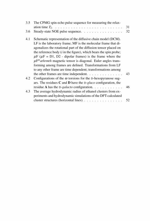
4.1 Schematic representation of the diffusive chain model (DCM).LF is the laboratory frame; MF is the molecular frame that di-agonalizes the rotational part of the diffusion tensor placed onthe reference body (i in the figure), which bears the spin probe;µF (µF = D1, D2 - dipolar frames) is the frame where theµFmathrmth magnetic tensor is diagonal. Euler angles trans-forming among frames are defined. Transformations from LFto any other frame are time dependent; transformations amongthe other frames are time independent. . . . . . . . . . . . . . 43
4.2 Configurations of the ω torsions for the D-hexopyranose sug-ars. The residues C and D have the D-gluco configuration, theresidue A has the D-galacto configuration. . . . . . . . . . . . 46
4.3 The average hydrodynamic radius of ethanol clusters from ex-periments and hydrodynamic simulations of the DFT-calculatedcluster structures (horizontal lines). . . . . . . . . . . . . . . . 52
List of Tables
2.1 Structures of the biological repeating unit of the E. coli O91O-antigen. . . . . . . . . . . . . . . . . . . . . . . . . . . . . 8
4.1 Comparison of the model-free motional parameters of residuesA–E as obtained for the [6-13C] and [1-13C] labeled O-antigen.LS stands for Lipari-Szabo model, ext. LS denotes the ex-tended Lipari-Szabo model, LS + ex. means Lipari-Szabo withthe exchange term Rex. . . . . . . . . . . . . . . . . . . . . . 48

1. Introduction
Investigation of biomolecules such as proteins, carbohydrates, nucleic acids,and possibly their complexes, is undoubtedly important, and therefore a topicof extensive interest in the chemistry- and biology-related research fields. Thesystems of biological interest are often large and complicated and their inves-tigation represents a challenge at both experimental and theoretical levels.
Not only the structure of these systems but also their interactions and dy-namics are of great importance in description of their biological function. Themotions of the molecules are strongly interrelated with their interactions andthus their biological functions. To fully understand the phenomena ongoingin the complex systems, it is sometimes essential to abandon the complicatedcases and turn to simpler, yet realistic, model systems.
An example of a phenomenon that is very complex and plays significantrole in the biological systems is hydrogen bonding. A better understandingcan be achieved by means of studying systems composed of small organicmolecules. The relative simplicity of such systems can provide an invaluableinsight into the important features of the hydrogen-bonded complexes.
Utilization of hydrogen bonding and other non-covalent interactions gaverise to the rich field of supramolecular chemistry with many promising ap-plications. Cryptophane molecules, with their ability to form supramolecularcomplexes, can serve as model systems for molecular recognition or biologicalreceptors.
Other instances are small or medium-sized molecules that can serve as testcases for theoretical models that are designed to describe complex moleculardynamics.
The studies included in this work incorporate at the first sight rather diversesystems, from small ethanol molecules, through cryptophanes and oligosac-charides, to a somewhat larger polysaccharide, all described in detail in Chap-ter 2. The common attribute, which makes these systems and associated phe-nomena interesting to explore, is their dynamics, to a large extend affected byweak molecular interactions.
Nuclear magnetic resonance (NMR) spectroscopy is one of the most im-portant spectroscopic techniques which can provide an informative insight intothe motional properties of various systems. Nowadays, the researchers canprofit from the fast developing methodology and improving sensitivity of NMR
1
that opens the possibility to employ a wide range of experiments. The basicprinciples and methods of NMR spectroscopy are explained in Chapter 3.
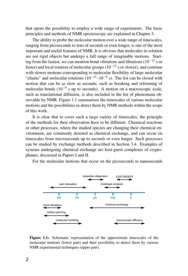
The ability to probe the molecular motion over a wide range of timescales,ranging from picoseconds to tens of seconds or even longer, is one of the mostimportant and useful features of NMR. It is obvious that molecules in solutionare not rigid objects but undergo a full range of imaginable motions. Start-ing from the fastest, we can mention bond vibrations and librations (10−12 s orfaster) and local rotation of molecular groups (10−12 s or slower), and continuewith slower motions corresponding to molecular flexibility of large molecular“chains” and molecular rotations (10−12–10−9 s). The list can be closed withmotion that can be as slow as seconds, such as breaking and reforming ofmolecular bonds (10−9 s up to seconds). A motion on a macroscopic scale,such as translational diffusion, is also included in the list of phenomena ob-servable by NMR. Figure 1.1 summarizes the timescales of various molecularmotions and the possibilities to detect them by NMR methods within the scopeof this work.
It is clear that to cover such a large variety of timescales, the principleof the methods for their observation have to be different. Chemical reactionsor other processes, where the studied species are changing their chemical en-vironment, are commonly denoted as chemical exchange, and can occur ontimescales from microseconds up to seconds or even longer. Such processescan be studied by exchange methods described in Section 3.4. Examples ofsystems undergoing chemical exchange are host-guest complexes of crypto-phanes, discussed in Papers I and II.
For the molecular motions that occur on the picoseconds to nanoseconds
ps ns μs ms s
bond vibrations
methyl spinning
molecular tumbling
chemical exchange
macroscopic diffusion
spin relaxation lineshape analysis
EXSY/NOESYrelaxation dispersion
Figure 1.1: Schematic representation of the approximate timescales of themolecular motions (lower part) and their possibility to detect them by variousNMR experimental techniques (upper part).
2

timescales, NMR spin relaxation has proven to be especially sensitive tech-nique. Theoretical and experimental aspects of the spin relaxation are de-scribed in Section 3.5. An example of dynamics which can be studied usingNMR relaxation is the internal motion of oligosaccharides and polysaccha-rides, studied in Papers III and IV.
Finally, the translational diffusion, described in Section 3.3, can be utilizedto address the hydrogen bonding phenomenon, as shown in Paper V. Here,NMR diffusion measurements were used to investigate the hydrogen-bondedclusters of ethanol.
3

2. Studied systems
2.1 Cryptophanes and host-guest complexes
Cryptophanes are medium-sized molecules known for their ability of bind-ing small molecular guests inside their cavity. Since their first synthesis andcharacterization by Collet and coworkers in 1980s [1–5], they have receivedextensive attention in both theoretical and experimental studies. Several re-views discussing the chemistry of cryptophanes have been published, the mostrecent by Brotin and Dutasta [6].
Cryptophanes consist of two cyclotribenzylene (CTB) units (the caps),connected by three aliphatic linkers of the -O-(CH2)n-O- type. There is a largevariability of cryptophanes in terms of different substituents on the caps ormodifications of the linkers. The connection of the linkers to the caps definesthe syn or anti isomer, as schematically depicted in Figure 2.1. Among thecryptophanes studied recently in our laboratory we can mention cryptophane-A, cryptophane-C, and cryptophane-D with n = 2, and cryptophane-E withn = 3, depicted in Figure 2.2.
synanti
Figure 2.1: Anti and syn isomer a cryptophane: two possible ways of connectionof the aliphatic -O-(CH2)n-O- linkers to the CTB caps.
Cryptophane-A is an anti isomer, having two identical caps, both car-rying methoxy substituents on each phenyl group. Cryptophanes C and Dhave methoxy groups only on one of the caps. They are diastereoisomers –cryptophane-C is the anti isomer and cryptophane-D is the syn isomer.
An interesting issue is the conformation of the linkers, since it affects the
4

symmetry properties of the cryptophane molecule as well as the size of thecavity. The linkers of cryptophanes C and D can be found in either gaucheor trans conformation. Moreover, there are two types of trans conformersdenoted T1 and T2, each having the -O-CH2-CH2-O- dihedral angle of 180◦, butdiffering in the relative position of the CH2 groups with respect to the oxygens.In the case of gauche conformation, the -O-CH2-CH2-O- dihedral angle is 60◦
(G−) or −60◦ (G+). In analogy with the trans case, both conformations canadditionally differ in the relative position of the CH2 groups with respect tothe oxygens. This gives four possibilities for gauche conformers, denoted G±1and G±2.
The conformation of the cryptophane molecule is defined by the conforma-tion of each of its three linkers (e.g. T1T1T1). Even if degeneracies due to thesymmetry of the molecule are considered, the number of possible conformersis large .
Cryptophanes possess a hydrophobic cavity, in which they can encapsu-late a small organic guest molecule. Among the most favorable guests arehalomethanes, for example chloroform (CHCl3), dichloromethane (CH2Cl2),chloromethane, bromomethane, iodomethane and others. The complex is heldtogether by van der Waals interactions.
The focus of this work was on cryptophane-C and its complexation withchloroform and dichloromethane, investigated in Papers I and II.
OOO
OO
O
H3CO OCH3H3COO
OO
OO
OH3CO
OCH3H3CO
OCH3
H3CO
OCH3
Cryptophane-A Cryptophane-C
Cryptophane-D Cryptophane-E
OOO
OO
H3CO OCH3H3CO
O
OOO
OO O
H3CO
OCH3H3CO
OCH3
H3CO
OCH3
Figure 2.2: Examples of cryptophanes
5

2.2 A trisaccharide as an example of complex internal dy-namics
A linear trisaccharide, composed of three mannose residues α-D-Manp-(1→2)-α-D-Manp-(1→6)-α-D-[6-13C]-Manp-OMe, was subject of the study reportedin Paper III. This relatively simple molecule is an interesting model system forboth experimental and theoretical investigation due to its complex dynamics.The internal flexibility is described by the rotation around the glycosidic link-ages, the two important torsions are defined by angles denoted ω and ψ2 inFigure 2.3.
As it was anticipated [7], the internal motion is coupled to the overall tum-bling of the molecule due to the similar timescales, thus interpretation of thedynamics using common methods becomes particularly difficult. For this rea-son, the trisaccharide became an interesting study case for testing an integratedtheoretical approach designed to deal with dynamics of molecules described asa flexible chain, i.e. consisting of rigid units linked by joints around which tor-sional motion is possible.
Paper III describes the performance of the developed approach for inter-pretation of NMR relaxation data of the trisaccharide and a pentasaccharide.
O
O
13CH2
HOHO
HOHO
O
OMe
OH
123
4 5
1'2'3'
4' 5'
6'
OHOHO 1''2''3''
4'' 5''
6''
OH
HO
OH
O
φ
φ
ψ
ψ
ω
ω
ω
pp
p
6
6
2
2
ω
ψ2
Figure 2.3: Trisaccharide, a schematic representation (left) and a stick model(right). The relevant torsion angles ω and ψ2 are highlighted.
6
2.3 Escherichia coli O-antigenic polysaccharides
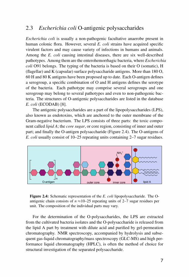
Escherichia coli is usually a non-pathogenic facultative anaerobe present inhuman colonic flora. However, several E. coli strains have acquired specificvirulent factors and may cause variety of infections in humans and animals.Among the E. coli causing intestinal diseases, there are six well-describedpathotypes. Among them are the enterohemorrhagic bacteria, where Escherichiacoli O91 belongs. The typing of the bacteria is based on their O (somatic), H(flagellar) and K (capsular) surface polysaccharide antigens. More than 180 O,60 H and 80 K antigens have been proposed up to date. Each O-antigen definesa serogroup, a specific combination of O and H antigens defines the serotypeof the bacteria. Each pathotype may comprise several serogroups and oneserogroup may belong to several pathotypes and even to non-pathogenic bac-teria. The structures of O-antigenic polysaccharides are listed in the databaseE. coli (ECODAB) [8].
The antigenic polysaccharides are a part of the lipopolysaccharides (LPS),also known as endotoxins, which are anchored to the outer membrane of theGram-negative bacterium. The LPS consists of three parts: the toxic compo-nent called lipid A; the core sugar, or core region, consisting of inner and outerpart; and finally the O-antigen polysaccharide (Figure 2.4). The O-antigens ofE. coli usually consist of 10–25 repeating units containing 2–7 sugar residues.
P
P
P
P P
NH3+
lipid Ainner coreouter coreO-antigenn
Figure 2.4: Schematic representation of the E. coli lipopolysaccharide. The O-antigenic chain consists of n ≈10–25 repeating units of 2–7 sugar residues perunit. The composition of the individual parts may vary.
For the determination of the O-polysaccharides, the LPS are extractedfrom the cultivated bacteria isolates and the O-polysaccharide is released fromthe lipid A part by treatment with dilute acid and purified by gel-permeationchromatography. NMR spectroscopy, accompanied by hydrolysis and subse-quent gas-liquid chromatography/mass spectroscopy (GLC-MS) and high per-formance liquid chromatography (HPLC), is often the method of choice forstructural investigation of the separated polysaccharide.
7

NMR spectroscopy can be also helpful in determination of the biosyn-thetic pathway of the LPS O-antigen. In Escherichia coli, two distinct mecha-nisms of the synthesis of the O-antigenic polysaccharide have been described.In some bacteria, the polymerization occurs via the transfer of the growingchain to a newly synthesized repeating unit on undecaprenolphosphate, a mem-brane bound carrier, termed Wzy-polymerase-dependent pathway. In anotherdescribed pathway, the polysaccharide is formed at the cytoplasmatic faceof the inner cell membrane by the transfer of glycosyl residues to the non-reducing end and subsequently transported to the periplasmatic face, calledABC-transporter-dependent pathway [9–11].
2.3.1 E. Coli O91 O-antigenic polysaccharide
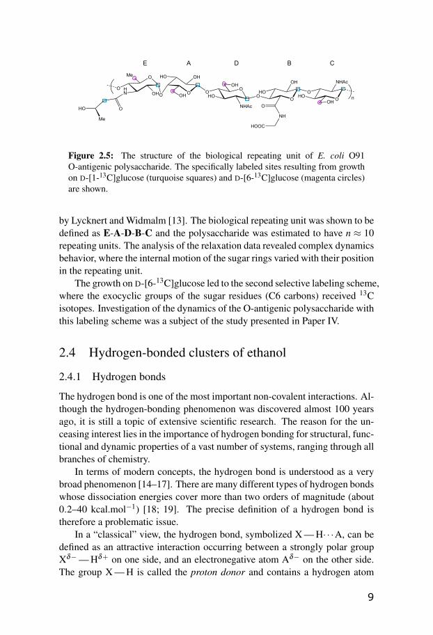
The species of our interest was the O-antigenic polysaccharide from the en-terohemorrhagic E. coli O91. The structural and biosynthetic study was per-formed by Kjellberg et al. [12] on the 13C-enriched sample isolated from bac-teria grown on uniformly labeled D-glucose. It was shown that the E. Coli O91O-antigen is composed of a pentasaccharide repeating unit, consisting of 5sugar residues denoted A–E, each of which is a hexopyranosyl entity contain-ing six carbons. The amino sugars C and D are N-acetylated, residue B carriesa glycinyl group and residue E has an (R)-3-hydroxybutyryl group as a sub-stituent. The chemical structure of the repeating unit is listed in Table 2.1 andrepresented in Figure 2.5.
Table 2.1: Structures of the biological repeating unit of the E. coli O91O-antigen.
residue sugarE →4)-α-D-Quip-3-N-[(R)-3-hydroxybutyramido]-(1→A →4)-β -D-Galp-(1→D →4)-β -D-GlcpNAc-(1→B →4)-β -D-GlcpA-6-N-Gly-(1→C →3)β -D-GlcpNAc-(1→
Additional cultures with specifically labeled D-[1-13C]glucose and D-[6-13C]glucose were grown, resulting in the labeling at, inter alia, anomeric car-bons (C1 position) and exocyclic groups (C6 position), depicted in Figure 2.5.The NMR experiments performed on these samples revealed the direct incor-poration of the enriched glucose in the polysaccharide chain.
The carbon-13 relaxation study on the site-specifically labeled O-antigenicpolysaccharide resulting from the D-[1-13C]glucose growth was later performed
8
OO
O
OO
HO
O OH
OH
HO
NHAc
OHOH
HO
A D B
O
O HN OH
E
OOH
HO
NHAc
C
O
OHO
Me
n
NH
O
HOOC
Me
Figure 2.5: The structure of the biological repeating unit of E. coli O91O-antigenic polysaccharide. The specifically labeled sites resulting from growthon D-[1-13C]glucose (turquoise squares) and D-[6-13C]glucose (magenta circles)are shown.
by Lycknert and Widmalm [13]. The biological repeating unit was shown to bedefined as E-A-D-B-C and the polysaccharide was estimated to have n ≈ 10repeating units. The analysis of the relaxation data revealed complex dynamicsbehavior, where the internal motion of the sugar rings varied with their positionin the repeating unit.
The growth on D-[6-13C]glucose led to the second selective labeling scheme,where the exocyclic groups of the sugar residues (C6 carbons) received 13Cisotopes. Investigation of the dynamics of the O-antigenic polysaccharide withthis labeling scheme was a subject of the study presented in Paper IV.
2.4 Hydrogen-bonded clusters of ethanol
2.4.1 Hydrogen bonds
The hydrogen bond is one of the most important non-covalent interactions. Al-though the hydrogen-bonding phenomenon was discovered almost 100 yearsago, it is still a topic of extensive scientific research. The reason for the un-ceasing interest lies in the importance of hydrogen bonding for structural, func-tional and dynamic properties of a vast number of systems, ranging through allbranches of chemistry.
In terms of modern concepts, the hydrogen bond is understood as a verybroad phenomenon [14–17]. There are many different types of hydrogen bondswhose dissociation energies cover more than two orders of magnitude (about0.2–40 kcal.mol−1) [18; 19]. The precise definition of a hydrogen bond istherefore a problematic issue.
In a “classical” view, the hydrogen bond, symbolized X — H· · ·A, can bedefined as an attractive interaction occurring between a strongly polar groupXδ−— Hδ+ on one side, and an electronegative atom Aδ− on the other side.The group X — H is called the proton donor and contains a hydrogen atom
9

that plays a fundamental role in the interaction. The moiety A is called theproton acceptor. In order to be able to form hydrogen bonds, the X — H grouphas to be at least slightly polar and A should supply a sterically accessibleconcentration of negative charge (usually a lone pair of electrons). An exampleof a hydrogen bond between two molecules of ethanol is depicted in Figure 2.6.Among the important characteristics distinguishing hydrogen bonds from vander Waals interactions is their directionality – a preference of linearity.
Hydrogen bonding is the key to understanding the structure and propertiesof variety of neat liquids as well as their mixtures. Particularly interesting arethose consisting of molecules containing hydroxyl groups, which can createhydrogen bonds where the oxygen acts both as the proton acceptor and donor(O — H· · ·O). This fact allows for complicated hydrogen-bonded networks inwater, low-molecular-weight alcohols and other liquids. Moreover, significantcooperative effects are observed in arrays of hydrogen bonds [20–22]. Oncea molecule is engaged as a hydrogen donor, it automatically becomes a betteracceptor, and vice versa.
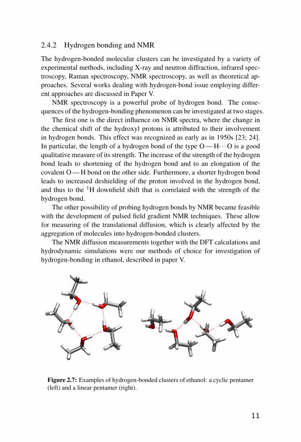
In Paper V, we have studied the hydrogen bonding in a solution of liquidethanol in a non-polar solvent. The molecules of ethanol are known to formhydrogen-bonded aggregates, called molecular clusters. Ethanol is expectedto form less complicated structures than for instance water, where the clustersof hydrogen-bonded molecules can be large and of extensive variety. Nev-ertheless, ethanol clusters can still consist of a varying number of moleculesand have quite diverse possible topologies, details of which are not completelyunderstood. Examples of pentameric cyclic and linear ethanol clusters are de-picted in Figure 2.7.
δ- δ+ δ-
Figure 2.6: Schematic representation of a hydrogen bond between two ethanolmolecules.
10
2.4.2 Hydrogen bonding and NMR
The hydrogen-bonded molecular clusters can be investigated by a variety ofexperimental methods, including X-ray and neutron diffraction, infrared spec-troscopy, Raman spectroscopy, NMR spectroscopy, as well as theoretical ap-proaches. Several works dealing with hydrogen-bond issue employing differ-ent approaches are discussed in Paper V.
NMR spectroscopy is a powerful probe of hydrogen bond. The conse-quences of the hydrogen-bonding phenomenon can be investigated at two stages.
The first one is the direct influence on NMR spectra, where the change inthe chemical shift of the hydroxyl protons is attributed to their involvementin hydrogen bonds. This effect was recognized as early as in 1950s [23; 24].In particular, the length of a hydrogen bond of the type O — H· · ·O is a goodqualitative measure of its strength. The increase of the strength of the hydrogenbond leads to shortening of the hydrogen bond and to an elongation of thecovalent O — H bond on the other side. Furthermore, a shorter hydrogen bondleads to increased deshielding of the proton involved in the hydrogen bond,and thus to the 1H downfield shift that is correlated with the strength of thehydrogen bond.
The other possibility of probing hydrogen bonds by NMR became feasiblewith the development of pulsed field gradient NMR techniques. These allowfor measuring of the translational diffusion, which is clearly affected by theaggregation of molecules into hydrogen-bonded clusters.
The NMR diffusion measurements together with the DFT calculations andhydrodynamic simulations were our methods of choice for investigation ofhydrogen-bonding in ethanol, described in paper V.
Figure 2.7: Examples of hydrogen-bonded clusters of ethanol: a cyclic pentamer(left) and a linear pentamer (right).
11

3. Theory and methods
This chapter aims at a brief description of common principles of high-resolutionnuclear magnetic resonance with the focus on dynamic processes in solutionand their investigation by NMR. Detailed description can be found in severaltextbooks, for example [25–29].
3.1 Basic principles of NMR
Nuclear magnetic resonance is a phenomenon at which resonating behavior ofnuclei with non-zero spin angular momentum III (with its components Ix, Iy, Iz
and its spin quantum number I) in external magnetic field is observed. Nucleiwith non-zero spin also possess the nuclear magnetic moment µµµ , related to thespin angular momentum of the nucleus as
µµµ = γ hIII, (3.1)
where h is the reduced Planck constant and the quantity γ is called the magne-togyric ratio or γ-factor.
When a nuclear spin is placed into an external magnetic field BBB, the fieldinteracts with the magnetic moment via Zeeman interaction, described by theZeeman Hamiltonian
H0 =−µµµ ·BBB =−γ hIII ·BBB. (3.2)
A large static magnetic field (tens of Tesla), produced usually by a super-conducting coil, is essential in high-resolution NMR. The static field is as-sumed to be oriented along the laboratory-frame z-axis. In this conventionBBB = (0,0,B0), which simplifies the Zeeman Hamiltonian in Eq. 3.2 to
H0 =−γ hIzB0. (3.3)
Solution of the time-independent Schrödinger equation with the Hamil-tonian 3.3 gives 2I + 1 eigenvectors |I,M〉, called also Zeeman eigenstates.Zeeman eigenstates are characterized by the spin quantum number I, possess-ing either integer or half-integer values, and the magnetic quantum number Min the range of (−I,−I +1, . . . , I).
12

The corresponding eigenvalues are the energies of the 2I + 1 equidistantenergy levels, characterized by the quantum number M
EM =−γ hB0M. (3.4)
The distance ∆E between the levels is ∆E = |γ| hB0.Nuclei with the nuclear spin quantum number I = 1/2 in the ground state,
referred further as spin-1/2 nuclei, are of major importance in NMR. Amongthe most commonly studied are 1H, 13C, 15N, 19F, etc. In this work, the at-tention is mostly drawn to experiments on 1H and 13C nuclei. The 1H nuclei,sometimes called also “protons”, have a γ-factor of γH = 267.522× 10⁻⁶ rads−1 T−1 and their natural abundance is practically 100 %, thus making thisisotope easily observable by NMR. The 13C isotopes, also called “carbon-13”nuclei, have the γ-factor about 4-times lower than protons (γC = 67.283×10⁻⁶
rad s−1 T−1) and their natural abundance is only 1.1 %, resulting in lowersensitivity than 1H nuclei. One way of dealing with the low sensitivity ofcarbon-13 experiments is using isotopicaly enriched samples (carbon-13 la-beling).
Spin-1/2 nuclei have two Zeeman eigenstates, usually denoted |α〉 and |β 〉,defined as
|α〉=∣∣1
2 ,+12
⟩|β 〉=
∣∣12 ,−
12
⟩. (3.5)
The spin is not restricted to the |α〉 or |β 〉 state, but may be in a superpositionof the two energy eigenstates
|ψ〉= cα |α〉+ cβ |β 〉 , (3.6)
where cα and cβ are the (normalized) superposition coefficients. In general,the spin state |ψ〉 is time-depended and it can be shown, by solving the time-dependent Schrödinger equation with the Hamiltonian 3.3, that the spin pre-cesses around the z-axis with the frequency given by
ω0 =−γB0. (3.7)
The quantity ω0 is called the Larmor frequency and it is proportional to theexternal magnetic field strength B0 and the γ-factor of the nucleus.
3.1.1 Radio-frequency field
The important part of the NMR experiment is a time-dependent external mag-netic field, oscillating at the frequency ωre f , called the radio-frequency field,or r.f. field. It is generated by the excitation coil, usually placed perpendicu-larly to the direction of the static field. The spin thus experiences two fields
13

– the static field of the magnet B0 and the many orders of magnitude smallerr.f. field BRF .
The effect of the r.f. field can be described as a perturbation to the staticmagnetic field by applying a time-dependent perturbation theory. For sim-plicity, the motion under the influence of the r.f. field is described in a framerotating around the direction of BBB0 (laboratory frame z-axis) with the angularfrequency ωre f . The detailed derivation of the motion of the nuclear spin inthe presence of the r.f. field is to be found for example in books of Levitt [25]or Keeler [26], here we just summarize the results.
It can be shown that, if the radio-frequency field is applied close to the res-onance (i.e. ωre f ≈ω0), the motion of the single spin-1/2 can be described as arotation of the angular momentum around the direction of the BBBRF (which ap-pears static in the rotating frame). The closer is the frequency of the field ωre f
to the Larmor frequency ω0 of the observed nucleus, the larger the influenceon the spin. This resonant behavior is the key principle of nuclear magneticresonance.
In a typical NMR experiment, the r.f. field is applied in short pulses and theresponse of the system, known as the free induction decay, or FID, is recordedand submitted to the Fourier transform to give an NMR spectrum.
In a special case, when the r.f. field is applied in a pulse of frequencyωre f equal to the Larmor frequency ω0 of the observed nucleus and the radio-frequency coil is placed along the x-axis (so-called x-phase pulse), the r.f. fieldcauses the rotation of the spin around x-axis by the flip angle given as
βp = τpωnut , (3.8)
where τp is the duration of the pulse. The quantity ωnut is called the nutationfrequency and it represents the magnitude of the r.f. field as
ωnut = |12
γBRF |. (3.9)
If, for example, the r.f. field is applied for a certain τp, for which the flip angleis 90◦, we denote a pulse like that a π/2-pulse. In the case of βp = 180◦, wespeak of a π-pulse.
3.1.2 Spin density operator
So far we have discussed the properties of a single spin-1/2 in the magneticfield. Here we consider a more realistic situation, in which a large amount of1/2-spins is present in a sample. A useful description of such case is the spindensity operator ρ , which describes the quantum state of the entire collection
14

of spins. The spin density operator is given as
ρ =1N ∑
j
∣∣ψ j⟩⟨
ψ j∣∣, (3.10)
where the index j samples throughout all the N spins present. In case of anensemble of identical non-interacting spin-1/2 species, where |ψ〉 is in the formof Eq. 3.6, we can write the matrix elements of the spin density operator, alsoreferred to as the density matrix
ρ =
(ραα ραβ
ρβα ρββ
)=
(cαcα∗ cαcβ∗cβ cα∗ cβ cβ∗
). (3.11)
The diagonal elements ραα and ρββ are referred to as the populations of thestates |α〉 and |β 〉. The off-diagonal elements ραβ and ρβα are called the co-herences between the states |α〉 and |β 〉. One of the advantages of the densityoperator construct is that the expectation value for an operator Q of an arbitraryobservable can be expressed as⟨
Q⟩= Tr
{ρQ}, (3.12)
where “Tr” stands for the trace of the operator.In thermal equilibrium, the coherences between the states are all zero,
while the populations of the energy levels obey the Boltzmann distribution.The thermal equilibrium density matrix for isolated spins-1/2 is given by
ρeq =exp(−H0/kBT )
Tr{
exp(−H0/kBT )} , (3.13)
where H0 is the Hamiltonian in the form of Eq. 3.3 and kB is the Boltzmannconstant.
The density matrix can be rewritten in an approximate form of
ρeq ≈( 1
2 +14B 0
0 12 −
14B
)=
12
1+12BIz, (3.14)
where 1 is the unity operator and B is the Boltzmann factor defined as
B=γ hB0
kBT. (3.15)
This means that, for positive γ , the low-energy state |α〉 is populated slightlymore than the high-energy state |β 〉. Since B is a very small number, the pop-ulation difference is exceedingly small at ordinary temperatures and fields.
15

Therefore, the polarization of the spin angular momentum vectors along thedirection of the external field in thermal equilibrium is very slight.
A consequence of this fact is the very low sensitivity of NMR comparedto other spectroscopic techniques. Nevertheless, several approaches to dealwith this fact were developed, including usage of very high static fields, signalaveraging, isotopic enrichment of samples, polarization transfer, etc.
3.1.3 Magnetization vector
Consistent with the population difference between the spin energy levels, onecan introduce the ensemble averaged nuclear magnetic moment, or magnetiza-tion vector MMM, indicating the magnitude and the direction of the net magneticmoment. The three Cartesian components of the vector are
Mz =2B(ραα −ρββ
)(3.16)
Mx =4B
Re{
ρβα
}(3.17)
My =4B
Im{
ρβα
}, (3.18)
where “Re” and “Im” denote the real and imaginary part of the density ma-trix elements, respectively. Clearly, the net magnetization is parallel to thedirection of the external magnetic field (z-axis) in thermal equilibrium.
It is possible to investigate the response of the magnetization vector tothe r.f. field BBBRF in analogy to the case of the single spin-1/2, described inSection 3.1.1. It can be shown that, consistently with the single-spin case, ther.f. field causes the rotation of the magnetization vector around the direction ofBBBRF by the angle defined by the strength and length of the pulse.
3.1.4 Bloch equations
It is experimentally observed that, after the perturbation of the spin system,the populations gradually drift towards their thermal equilibrium values andthe coherences gradually decay to zero. These observations are due to thenuclear spin relaxation. The phenomenological approach to the spin relaxationdescribes the dynamics of the magnetization in terms of Bloch equations [30]
dMz
dt= γ (MMM×BBB)z−
Mz−M0
T1(3.19)
dMx
dt= γ (MMM×BBB)x−
Mx
T2(3.20)
dMy
dt= γ (MMM×BBB)y−
My
T2. (3.21)
16
Eq. 3.19 predicts the magnetization component along the BBB0 to decay toits equilibrium value, M0. The time constant for this process is called thelongitudinal, or spin-lattice, relaxation time T1. The solution to Eq. 3.19 afterthe initial inversion of magnetization by a π-pulse can be written in the formof an exponentially recovering function
Mz(t) = M0 (1−2exp(−t/T1)) . (3.22)
Equations 3.20 and 3.21 describe the exponential decay of the transversecomponents of the magnetization, Mx and My, to zero. This process is char-acterized by the transverse, or spin-spin, relaxation time constant T2. Theevolution of the transverse components of the magnetization vector after theπ/2-pulse is expressed as
Mx,y(t) = M0 exp(−t/T2). (3.23)
Sometimes, the reciprocal values of T1 and T2, denoted relaxation rates R1and R2, are used.
The reason for introducing two different time constant is the different phys-ical origin of the transverse and longitudinal relaxation. The longitudinal re-laxation involves the energy exchange between the spin system and the sur-rounding matter (“lattice”), while the transverse relaxation involves the lossof phase coherence in the motion of individual spins. We shall discuss therelaxation phenomenon in detail further in Section 3.5.
3.2 NMR interactions
A real sample contains a large number of electrons as well as nuclei. Thenuclear spins not only experience the external magnetic field, but also interactwith each other and with the electrons present.
The complete description of the dynamics of nuclei and electrons is nearlyimpossible and, fortunately, not necessary. In order to describe the dynamics ofthe spins, one can use the nuclear spin Hamiltonian, which only describes thespin states of the nuclei, and enters the time-dependent Schrödinger equation.Further simplifications are possible, since the internal interactions of the spinsare usually much weaker compared to the interactions with the external field.Therefore, in most cases, they can be treated in terms of perturbation theory asa perturbation to the Zeeman Hamiltonian.
The effect of the spin interactions can be observed at two stages. Thefirst one is their effect on NMR spectra, second is their influence on the spinrelaxation.
17

Here we describe the most relevant interactions and their Hamiltoniansinvolved in diamagnetic systems of spins-1/2.
3.2.1 Chemical shift
The electrons surrounding nuclei cause the local magnetic field to vary on asub-molecular distance scale. The external magnetic field induces electroncurrents that generate a local magnetic field proportional to the static magneticfield, but not necessarily in the same direction. The magnetic moments of thespins react to the field induced by these electronic currents. The Hamiltonianfor the response of a single spin I to the induced magnetic field is
HCS = γ III ·σσσ ·BBB0. (3.24)
The quantity σσσ is called the chemical shift tensor. It is a property of a particularnucleus and it is in general anisotropic.
As any rank-2 Cartesian tensor, the shielding tensor can be decomposedinto its symmetric and antisymmetric part. The coordinate system, in whichthe symmetric part has only diagonal elements σXX , σYY , and σZZ , is calledthe principal axis system. The isotropic component of the symmetric chemicalshift tensor σiso is called the isotropic chemical shift and it is defined as
σiso =13(σXX +σYY +σZZ) . (3.25)
The choice of the principal axis system follows usually the convention
|σZZ−σiso| ≥ |σXX −σiso| ≥ |σYY −σiso| . (3.26)
We can also define a quantity ∆σ denoted the chemical shift anisotropy(CSA) as
∆σ = σZZ−12(σXX +σYY ) . (3.27)
The case of non-zero CSA reflects the fact that the shielding experienced bya particular nucleus depends on the orientation of the molecule bearing thenucleus with respect to the static magnetic field.
In the case of isotropic liquids, only the isotropic component σiso is impor-tant due to the motional averaging. As a consequence of the chemical shift, theLarmor frequency of the nuclei in the isotropic liquids is shifted to the value
ωCS0 =−γB0 (1+σiso) , (3.28)
causing different positions of the resonances of the nuclei with the differentelectronic environment in NMR spectra.
18

The absolute magnitude of the chemical shift is dependent on the magni-tude of the external magnetic field B0, therefore it is convenient to introduce arelative scale independent of the measuring apparatus as
δiso[ppm] =ω0−ωst
ωst×106 =
σst −σiso
1−σst×106 ≈ (σst −σiso)×106, (3.29)
where ω0 is the Larmor frequency of the particular isotope and ωst is the Lar-mor frequency of a reference compound. For experiments on 1H and 13C nu-clei, a common reference compound is the tetramethylsilane (TMS), Si(CH3)4.
The chemical shift range differs for different isotopes in correlation withtheir number of electrons. For the 1H nuclei, the chemical shift can vary intens of ppm depending on the electronic environment. For heavier nuclei, like13C, the chemical shift can range in hundreds of ppm.
3.2.2 Direct dipole-dipole interaction
The strongest interaction for 1/2-spin nuclei is the direct dipole-dipole (DD)interaction. This interaction arises between two nuclear magnetic momentsclose to each other in space. Each of the magnetic moments generates a mag-netic field to which the other spin responds. The direct dipole-dipole interac-tion between spins I and S is represented by the Hamiltonian of the followingform
HDD = bIS
[3(III ·eee)(SSS ·eee)− III · SSS
], (3.30)
where eee is the unit vector in the direction of the line connecting the two nu-clei. The magnitude of the dipole-dipole coupling is given by the dipole-dipolecoupling constant, usually denoted bIS and given by:
bIS =−µ0γIγSh4πr3
IS, (3.31)
where the distance between the two spins is rIS, and γI and γS are their magne-togyric ratios.
In the case of isotropic liquids, the dipole-dipole interaction vanishes dueto the molecular tumbling, which changes the orientation of the IS-spin axis ona time scale much faster compared to the dipole-dipole couplings. The dipole-dipole interaction therefore does not influence the appearance of liquid spectra.However, it plays an important role in nuclear spin relaxation.
3.2.3 J-coupling
The indirect dipole-dipole interaction, or J-coupling, is an interaction betweentwo nuclear spins mediated by chemical bond electrons. Two spins have a mea-surable J-coupling if they are connected through a small number of chemical
19

bonds, including hydrogen bonds. Therefore, the J-coupling provides a directspectral manifestation of the chemical bond and is an important connectionbetween NMR and chemistry. The full form of the intramolecular J-couplinginteraction between the two spins I and S is
HJ = 2π III ·JJJIS · SSS, (3.32)
where JJJIS is the J-coupling tensor, which depends on the molecular orientation.In isotropic liquids, the J-coupling tensor is averaged by the rapid moleculartumbling and attains the isotropic form given as the trace of JJJIS
JIS =13(Jxx + Jyy + Jzz) . (3.33)
The scalar constant JIS is sometimes referred to as the scalar coupling con-stant. Unlike the chemical shift, J-coupling is independent of the externalmagnetic field.
3.3 Translational diffusion
Molecules, or generally any particles, in solution are randomly changing theirpositions due to the Brownian motion. This phenomenon is known as the trans-lational diffusion. It is characterized by the translational diffusion coefficientD, defined according to the Stokes-Einstein theory [31] as
D =kBTfT
, (3.34)
where fT is the friction factor, given by
fT = 6πηrH (3.35)
for the special case of a spherical particle of hydrodynamic radius rH in asolvent of viscosity η . Combining Equations 3.34 and 3.35, it is possible toexpress the hydrodynamic radius as
rH =kBT
6πηD. (3.36)
Pulse-field gradient (PFG) NMR spectroscopy can be used to measure thetranslational diffusion of molecules. By the use of a magnetic field gradient,molecules can be spatially labeled, i.e. marked depending on their position inthe sample tube. One of the simplest pulse sequences for measuring the trans-lational diffusion is the stimulated spin echo sequence, proposed by Stejskaland Tanner [32].
20

The principle of the stimulated echo sequence lies in the application of thegradient of the external magnetic field of magnitude G in the direction of thestatic magnetic field BBB0. Owing to this gradient, the magnetic field varies alongthe z-axis according to
B(z) = B0 +G(z), (3.37)
and so does the Larmor frequency ω0 of the nuclei
ω0(z) =−γ (B0 +G(z)) . (3.38)
The position of the nuclei along the z-axis is encoded according to their phaseangle
φ(z) =−ω0(z)δ =−γB(z)δ , (3.39)
where δ is the duration of the gradient pulse.The application of the second gradient causes that only spins which did not
change their position during the delay ∆ between the gradient pulses are refo-cused and contribute to the signal. The detected signal intensity is attenuateddepending on the diffusion coefficient of the species in the sample
I(2τ,G) = I(2τ,0)e−Dγ2G2δ 2(∆−δ/3), (3.40)
where I2τ,G is the intensity of the signal at the time 2τ with the gradient applied,I2τ,0 is the signal intensity at 2τ without applied gradient, and ∆− δ/3 is thediffusion time. The translational diffusion coefficient D can be obtained by fit-ting the Gaussian decaying function of Eq. 3.40 to the experimental intensitiesvs. the gradient strength.
Recently, more elaborate experimental protocols have been developed inorder to deal with the drawbacks of the simple stimulated echo. In order tocompensate the eddy-current effects, bipolar gradients [33] or the longitudi-nal eddy-current delay (LED) [34] are used. Convection within the sample issuppressed by the use of the double-stimulated echo [35]. The pulse sequencedepicted in Figure 3.1, with the spin-lock before the acquisition to removeminor phase distortions in the spectra [36], was used in this work.
3.4 Two-site chemical exchange
Chemical exchange is in general any process at which the spins in the moleculechange their magnetic environment. This may be due to chemical reaction,isomerization, conformational changes, complexation of molecules, etc. Thesephenomena reflect in NMR spectra and can affect the spin relaxation.
21

τ/2 spin locktrd τ/2 τ/2 τ/2
tacq
13C
G
π/2 π π/2 π/2 ππ/2 π π/2
δ/2
δ/2
δ δ/2
δ/2
Δ/2
δ
Δ/2
Figure 3.1: A double stimulated spin-echo pulse sequence for measuring thetranslational diffusion with the bipolar gradient pulses and the spin-lock beforethe acquisition. Appropriate phase cycling and cleaning gradients have to be used(not shown).
The two-site chemical exchange process, at which the nuclear spin is trans-ported between two sites A and B with unequal chemical shifts ωA and ωB, canbe characterized by equation
Ak1
k−1
B, (3.41)
where k1 and k−1 are the first-order exchange rate constants. The exchangerates k1 and k−1 may in general differ if the energy barriers for forward andbackward reactions are not equal. The ratio of the rate constants is called theequilibrium constant and it is equal to the ratio of the equilibrium concentra-tions of the two species
K =k1
k−1=
[B]eq
[A]eq. (3.42)
In the case when k1 = k−1, we speak of a symmetrical two-site exchange.The effect of the chemical exchange depends on the difference in the chem-
ical shifts frequency of the two sites ∆ω = ωA−ωB compared to the exchangerate kex = k1+k−1. The spectral lineshapes are most profoundly affected by theexchange process if the system is in the intermediate exchange regime where∆ω is similar to kex.
In the case whenkex < |∆ω/2| (3.43)
we speak of the slow intermediate exchange regime, in which we observe sep-arate lines for A and B sites at their frequencies ωA and ωB. The exchangerates in this regime can be obtained by the 1D NOESY experiment (see Sec-tion 3.4.2).
22

Broadening of the lines occurs with the increase of the exchange rate kex.The point at which
kex = |∆ω/2| (3.44)
is called the cross-over point and approximately corresponds to the point wherethe spectral lines for the two sites merge to one line, called the coalescence.The single peak appears at a position given by the average of the two chem-ical shifts ωA and ωB, weighted by the equilibrium concentrations of the twospecies.
The fast intermediate exchange is characterized by
kex > |∆ω/2| (3.45)
and in this regime the further increase of the exchange rate causes narrowingof the single line. The exchange rates in the fast intermediate exchange can beevaluated for example by lineshape analysis [37; 38].
3.4.1 Host-guest complex formation
In this work, the chemical exchange considered is the encapsulation of themolecular guest (G) by the molecular host (H) leading to the host-guest (HG)complex formation. Thus, the guest is in exchange between the bulk solvent(free state) and the cavity of the host (bound state). The process can be ex-pressed by a chemical reaction
H +Gk1
k−1
HG (3.46)
with the rate constants k1 and k−1. For the interpretation of NMR experi-ments, the effective pseudo-first-order exchange rates for free-to-bound (kFB)and bound-to-free (kBF ) reaction are introduced
kFB = k1[H] (3.47)
kBF = k−1 (3.48)
kex = kFB + kBF . (3.49)
The association equilibrium constant is given according to
K =kFB
kBF [H]=
[HG]
[H][G]. (3.50)
23

The activation energies of the exchange process Ea can be determined byfitting the Arrhenius equation
k(T ) = A exp(− Ea
NAkBT
)(3.51)
to the temperature-dependent exchange rate constants, where k stands eitherfor k1 or k−1 and NA is the Avogadro constant.
3.4.2 1D NOESY experiment
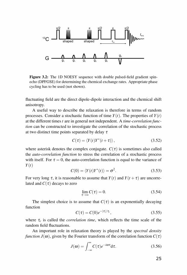
One of the most powerful aspects of NMR is the ability to detect the chemicalexchange phenomena, even when the system is in equilibrium, over a widerange of rates. The exchange rates in the slow intermediate exchange regimecan be determined for instance by the one-dimensional NOESY experiment,using the fact that chemical exchange can lead to a magnetization transfer be-tween the two sites in exchange.
The selective 1D NOESY pulse sequence with the pulsed-field gradients[39] is depicted in Figure 3.2. The sequence starts with a selective excitationof one of the sites by a technique termed excitation sculpting. This consistsof the double pulsed-field gradient spin-echo (DPFGSE), which combines theselective shaped π-pulses with the pulsed-field gradients (g1–g3) to dephasethe non-desired magnetization. Second part of the sequence contains a variablemixing time delay τm, during which the magnetization transfer, caused by theexchange, occurs. The signal is then detected at the second site by the lastπ/2-pulse.
The experiment is usually repeated for several suitably chosen values ofthe mixing time τm. As a result, the exchange-mediated intensity buildup as afunction of τm is obtained. Assuming that the chosen τm values are sufficientlyshort for observing the process in the initial rate regime, the reaction rates aregiven by the slope of the buildup curve [40; 41].
3.5 NMR relaxation
Relaxation is a process of returning of the spin system to the equilibrium aftera perturbation. As described in Section 3.1.4, the populations obey the Boltz-mann distribution and the coherences are zero in thermal equilibrium.
For spins-1/2, nuclear spin relaxation is caused by a fluctuating magneticfield at the sites of the nuclear spins. The fluctuating field originates from theweak magnetic interactions in combination with the random thermal motionof the molecules. For diamagnetic systems, the most important sources of the
24
tacq13C
G
π/2 π/2 π π/2
g1
τm
π π
shaped shaped
g1 g2 g2 g3
g3
Figure 3.2: The 1D NOESY sequence with double pulsed-field gradient spin-echo (DPFGSE) for determining the chemical exchange rates. Appropriate phasecycling has to be used (not shown).
fluctuating field are the direct dipole-dipole interaction and the chemical shiftanisotropy.
A useful way to describe the relaxation is therefore in terms of randomprocesses. Consider a stochastic function of time Y (t). The properties of Y (t)at the different times t are in general not independent. A time-correlation func-tion can be constructed to investigate the correlation of the stochastic processat two distinct time points separated by delay τ
C(τ) = 〈Y (t)Y ∗(t + τ)〉 , (3.52)
where asterisk denotes the complex conjugate. C(τ) is sometimes also calledthe auto-correlation function to stress the correlation of a stochastic processwith itself. For τ = 0, the auto-correlation function is equal to the variance ofY (t)
C(0) = 〈Y (t)Y ∗(t)〉= σ2. (3.53)
For very long τ , it is reasonable to assume that Y (t) and Y (t + τ) are uncorre-lated and C(τ) decays to zero
limτ→∞
C(τ) = 0. (3.54)
The simplest choice is to assume that C(τ) is an exponentially decayingfunction
C(τ) =C(0)e−|τ|/τc , (3.55)
where τc is called the correlation time, which reflects the time scale of therandom field fluctuations.
An important role in relaxation theory is played by the spectral densityfunction J(ω), given by the Fourier transform of the correlation function C(τ)
J(ω) =∫
∞
−∞
C(τ)e−iωτdτ. (3.56)
25

The spectral density function represents the amount of the radio-frequencypower generated by the stochastic process Y (t) at a particular frequency ω .For the time-correlation function in the form of 3.55, the spectral density is aLorentzian function:
J(ω) =C(0)2τc
1+ω2τc2 . (3.57)
For the relaxation through rank-2 interactions (such as dipole-dipole inter-action or chemical shift anisotropy), a time correlation function for the nor-malized rank-2 spherical harmonics (Yl,m with l = 2) is required. For Y (t) inthe form of
Y2,0(t) =14
√5π
[3cos2
θ(t)−1]
(3.58)
it can be shown that the relevant time correlation function for the small-steprotational diffusion of isotropically reorienting rigid body is indeed in the formof a simple exponential
C2(τ) =1
4πe−τ/τc (3.59)
with τc =1
6DR, where DR is the rotational diffusion coefficient.
In most of other cases dealing with more complex dynamics, includingflexible molecules, more complicated expressions for J(ω) have to be em-ployed (see Section 3.8).
3.5.1 Relaxation through dipole-dipole interactions
For spins-1/2, the most important relaxation mechanism is the dipole-dipoleinteraction between the nuclear magnetic moments of the spins in the spatialvicinity of each other. The dipolar relaxation parameters are also an importantsource of information about molecular dynamics.
Consider a situation with a solution containing molecules with two types ofnuclear spins-1/2, denoted I and S. We assume that no J-couplings are present,only the dipole-dipole interaction between the spins. The system is describedby four Zeeman energy levels and a set of transition probabilities between thelevels (see Figure 3.3). One can set up a set of equations describing the kineticsof the populations in the four-level system. It was shown by Solomon [42] thatthe relaxation of the longitudinal magnetization components, proportional tothe expectation values of the Iz and Sz operators, is related to the populationsof the four-level system and can be described by a set of following equations,denoted Solomon equations
ddt
[ ⟨Iz⟩⟨
Sz⟩ ] = −
[ρI σIS
σIS ρS
][ ⟨Iz⟩− Ieq
z⟨Sz⟩−Seq
z
], (3.60)
26

αIβS
αIαS
βIαS
βIβS
W1SW1I
W0
W2
W1SW1I
Figure 3.3: The energy-level diagram associated with a two-spin system
where Ieqz and Seq
z are the equilibrium longitudinal magnetizations of the twospins, and ρI and ρS are corresponding longitudinal relaxation rates. The sym-bol σIS denotes the cross-relaxation rate. The matrix at the right-hand side ofEq. 3.60 is called the relaxation matrix. The relaxation rates are associatedwith the transition probabilities in the following way
ρI = W0 +2W1I +W2 (3.61)
ρS = W0 +2W1S +W2 (3.62)
σIS = W2−W0 (3.63)
The Solomon approach uses the time-dependent perturbation theory to derivethe transition probabilities between the pairs of energy levels in Figure 3.3.The transition probabilities, induced by a random process, are proportional tospectral densities of the random perturbation evaluated at the relevant frequen-cies:
W1I =3π
10b2
ISJ(ωI) (3.64)
W1S =3π
10b2
ISJ(ωS) (3.65)
W0 =π
5b2
ISJ(ωI−ωS) (3.66)
W2 =π
5b2
ISJ(ωI +ωS). (3.67)
27

The relaxation equations for the spins I and S are coupled by the cross-relaxation term σIS and thus the general solution to the Eq. 3.60 for
⟨Iz⟩
and⟨Sz⟩
is a sum of two exponentials. Although the cross-relaxation is in prin-ciple always present in dipolar-relaxed systems, the single exponential behav-ior, postulated by Bloch (Eq. 3.22), can be obtained under certain conditions.Such conditions can be achieved, for instance, by irradiation of the I-spin witha radio-frequency field at its resonant frequency. The relaxation rate of theS-spin in such case is given by
T−11S ≡ ρS =W0 +2W1S +W2. (3.68)
This situation applies, for example, for carbon-13 (S-spin) relaxation under thebroad-band decoupling of protons (I-spins).
The irradiation of the spin I, leading to its saturation (⟨Iz⟩= 0), results also
in the modification of the steady-state (d⟨Sz⟩/dt = 0) solution to the Solomon
equations for the spin S ⟨Sz⟩
steady−state = Seqz (1+η) . (3.69)
The steady-state intensity of the S-spin signal is enhanced by a factor of (1+η),where
η =γI
γS
σIS
ρS. (3.70)
This phenomenon is referred to as the Nuclear Overhauser Effect (NOE) andthe quantity (1+η) is called the NOE enhancement factor. If the spins I and Sare of different isotopic species (as for example 13C and 1H), we talk about theheteronuclear NOE. Measurement of the enhancement factor allows for de-termination of the cross-relaxation rate σIS. This relaxation parameter carriesinformation on the relaxation mechanisms and on the molecular dynamics.
3.5.2 Carbon-13 relaxation
The main focus in this work has been put on carbon-13 NMR relaxation param-eters, since they contain valuable information about the dynamics or structureof the molecules.
For 13C nuclei directly bonded to one or more hydrogens, the dipole-dipoleinteraction with these hydrogens is normally the dominant relaxation mecha-nism. Application of a proton broadband decoupling removes not only theJ-coupling with the attached protons, but also the coupling between the tworelaxation equations in Eq. 3.60. Thus, the longitudinal carbon-13 relaxationis mono-exponential (well-defined T1) and the NOE factor is always positive.
28

Under the assumptions of the Solomon approach, i.e. only dipole-dipoleinteraction between 1H and 13C present, one can obtain expression for T1 andNOE from Eqs. 3.68 and 3.70 combined with Eqs. 3.64–3.67.
Under certain conditions, the transverse relaxation is also mono-exponentialand similar expression can be derived for the relaxation time T2
T−11,DD =
π
5b2
CH [J(ωH −ωC)+3J(ωC)+6J(ωH +ωC)] (3.71)
η =γH
γC
6J(ωH +ωC)− J(ωH −ωC)
J(ωH −ωC)+3J(ωC)+6J(ωH +ωC)(3.72)
T−12,DD =
π
10b2
CH [4J(0)+ J(ωH −ωC) +
+ 3J(ωC)+6J(ωH)+6J(ωH +ωC)] . (3.73)
The dipole-dipole coupling constant bCH for I =1H and S =13C is defined inEq. 3.31. For a rigid molecule, bCH is proportional to the inverse cube of theC-H distance rCH . Sometimes, the vibrationally averaged rCH distance has tobe used to account for the effect of molecular vibrations [43].
The simple form of the relaxation parameters in Eqs. 3.71–3.73 in the pres-ence of the proton decoupling may be retained also for more complicated spinsystems. For a carbon-13 interacting with two non-equivalent protons, it issufficient to multiply the Eqs. 3.71 and 3.73 by a factor of two.
The relaxation parameters T1, T2 and NOE are commonly referred to asthe “conventional” relaxation parameters, in contrast with the cross-correlatedrelaxation parameters that will be discussed in Section 3.7.
From the experimental point of view, it is important to notice the depen-dence of the relaxation parameters in Eqs. 3.71–3.73 on the magnetic fieldstrength (through ωH and ωC). For a given correlation time τc, in a regionwhere ω2τ2
c � 1, the relaxation parameters are practically independent of theexternal field. This region is called the extreme narrowing regime. On theother hand, a field-dependence of relaxation parameters for ω2τ2
c ≥ 1 is ob-served. Note that ω represents here all the relevant frequencies that enter thespectral densities J(ω) in Eqs. 3.71–3.73, the largest being ωH +ωC.
Carbon-13 relaxation parameters outside of the extreme narrowing regimecan provide an excellent source of information on molecular dynamics in so-lution.
We can also investigate the dependence of 1/T1 and 1/T2 on the correlationtime τc and through this also on molecular size, solvent viscosity and temper-ature. Under the extreme narrowing conditions, T1 and T2 are equal. Outsideof the extreme narrowing regime, 1/T1 reaches its maximum and further de-creases with rising τc, while 1/T2 is rising instead.
29

3.6 Measuring relaxation parameters
3.6.1 Inversion – recovery: T1 measurement
The spin-lattice relaxation time T1 can be determined by using the inversion– recovery pulse sequence (see, for example, [27]), which is depicted in Fig-ure 3.4. The pulse sequence begins with a πx-pulse which generates an in-verted population distribution, i.e. inverts the magnetization. The populationsrelax back to the thermal equilibrium during the recovery delay τ . The final(π/2)x-pulse converts the population differences into observable coherences(transverse magnetization). A recycle delay trd should be long enough (ap-proximately 5-times T1) to ensure the equilibrium conditions between the rep-etitions of the experiment.
T1 can be determined by repeating the experiment for different recoverydelays τ and fitting the exponentially recovering function of type as in Eq. 3.22to the experimental peak intensities vs. relaxation delays.
For carbon-13 T1 experiments, decoupling of protons throughout the mea-surement is usually applied to ensure that the relaxation is mono-exponential.
trd
1H decoupling
13Cτ
(π/2)x(π)x
tacq
Figure 3.4: The inversion – recovery pulse sequence for measuring the relaxationtime T1.
3.6.2 Spin-echo: T2 measurement
The basic spin-echo sequence, proposed by Hahn [44], consists of a (π/2)x-pulsefollowed by a πx-pulse, separated by a delay τ/2, where τ is the echo time.The first pulse creates the transverse magnetization, which is let to evolve fora period of τ/2. The contribution of different spins evolve with different ratesdue to the chemical shift differences, or possible inhomogeneities of the mag-netic field. After this period, the magnetization in the xy-plane is inverted bythe π-pulse. Another τ/2 after the second pulse, transverse magnetization isrefocused and a spin-echo is observed.
30
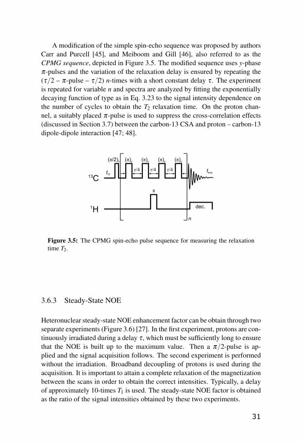
A modification of the simple spin-echo sequence was proposed by authorsCarr and Purcell [45], and Meiboom and Gill [46], also referred to as theCPMG sequence, depicted in Figure 3.5. The modified sequence uses y-phaseπ-pulses and the variation of the relaxation delay is ensured by repeating the(τ/2 – π-pulse – τ/2) n-times with a short constant delay τ . The experimentis repeated for variable n and spectra are analyzed by fitting the exponentiallydecaying function of type as in Eq. 3.23 to the signal intensity dependence onthe number of cycles to obtain the T2 relaxation time. On the proton chan-nel, a suitably placed π-pulse is used to suppress the cross-correlation effects(discussed in Section 3.7) between the carbon-13 CSA and proton – carbon-13dipole-dipole interaction [47; 48].
trd
1H dec.
13Cτ/4
(π/2)x (π)y
τ/4 τ/4
(π)y (π)y (π)y
π
n
tacq
Figure 3.5: The CPMG spin-echo pulse sequence for measuring the relaxationtime T2.
3.6.3 Steady-State NOE
Heteronuclear steady-state NOE enhancement factor can be obtain through twoseparate experiments (Figure 3.6) [27]. In the first experiment, protons are con-tinuously irradiated during a delay τ , which must be sufficiently long to ensurethat the NOE is built up to the maximum value. Then a π/2-pulse is ap-plied and the signal acquisition follows. The second experiment is performedwithout the irradiation. Broadband decoupling of protons is used during theacquisition. It is important to attain a complete relaxation of the magnetizationbetween the scans in order to obtain the correct intensities. Typically, a delayof approximately 10-times T1 is used. The steady-state NOE factor is obtainedas the ratio of the signal intensities obtained by these two experiments.
31

trd
1H irradiation
13Cτ
(π/2)x
dec.
tacq
Figure 3.6: Steady-state NOE pulse sequence.
3.7 Cross-correlated relaxation
3.7.1 Redfield relaxation theory
The Solomon approach to the relaxation is limited to a two-spin system cou-pled through the dipole-dipole interaction. This can be a limitation if one needsto deal with multi-spin systems or other sources of relaxation than the dipole-dipole interaction. A more general formulation of the relaxation theory, suit-able also for systems with J-couplings, was proposed by Wangsness, Blochand Redfield, also known as the Redfield theory [49; 50].
The Redfield formulation is in principle applicable to any kind of non-equilibrium system with any relaxation mechanism. It is based on the second-order time-dependent perturbation theory and it is given in terms of densitymatrix evolution, described by the Liouville – von Neumann equation.
The relaxation Hamiltonian considered in the Redfield theory is a sum ofthe time-independent part H0 (the main Hamiltonian) and the stochastic time-dependent part H1(t) (the perturbation Hamiltonian). A comprehensive deriva-tion of the Redfield theory can be found in the book of Abragam [28] in termsof the operator formalism and in the book of Slichter [29] in terms of the eigen-state formulation. Here we limit ourselves to present the main results.
The equation for the time evolution of density matrix elements in Redfieldtheory is based on the Liouville – von Neumann equation and takes the formof
dραα ′
dt=−iωαα ′ραα ′+ ∑
β ,β ′Rαα ′ββ ′ ρββ ′ , (3.74)
where Rαα ′ββ ′ are the relaxation supermatrix elements expressed in the basisof the eigenstates of the unperturbed Hamiltonian H0 (α , α ′, β , β ′, . . .), andωαα ′ represents the energy level separation between the corresponding eigen-states. The Rαα ′ββ ′ terms are composed as a linear combination of the spectral
32
densities at different transition frequencies
Rαα ′ββ ′ =12[Jαβα ′β ′(ωα ′β ′)+Jαβα ′β ′(ωαβ )−
− δα ′β ′∑γ
Jγβγα(ωγβ )−δαβ ∑γ
Jγα ′γβ ′(ωγβ ′)], (3.75)
where the spectral densities are in the form of
Jαα ′ββ ′(ω) = 2∫
∞
0Gαα ′ββ ′(τ)exp(−iωτ)dτ (3.76)
with the time-correlation function Gαα ′ββ ′(τ) defined as
Gαα ′ββ ′(τ) =⟨α∣∣H1(t)
∣∣α ′⟩⟨β ∣∣H1(t + τ)∣∣β ′⟩, (3.77)
where the horizontal bar denotes the ensemble average.
3.7.2 Relaxation interference
When dealing with multi-spin systems, a situation, in which the relaxation ofone spin is caused by more than a single relaxation mechanism, can arise. If thestochastic processes causing the relaxation are modulated by the same dynamicprocess, the interference between the interactions needs to be considered. Thiseffect is also called the cross-correlation and was subject of several studies,one of the more recent is the review of Kumar [51].
In this section we demonstrate the application of the Redfield theory to sys-tems consisting of more than two spins with mutual dipole-dipole and scalarinteractions. The unperturbed Hamiltonian H0 consists of the Zeeman inter-actions between all the spins and the magnetic field, and also the J-couplingswithin the spin pairs. The perturbation Hamiltonian H1(t) is a sum of thedipole-dipole interactions over all the η pairs of the nuclei
H1(t) = ∑η
Hη . (3.78)
The time-correlation function in Eq. 3.77 thus has to be extended to containterms correlating different components of the Hamiltonian 3.78
Gαα ′ββ ′(τ) = ∑η
⟨α∣∣Hη(t)
∣∣α ′⟩⟨β ∣∣Hη(t + τ)∣∣β ′⟩+ (3.79)
+ ∑η 6=η ′
⟨α∣∣Hη(t)
∣∣α ′⟩⟨β ∣∣Hη ′(t + τ)∣∣β ′⟩, (3.80)
where the first term is the auto-correlation function and the second term, wherethe summation runs over η 6= η ′, is called the cross-correlation function. TheFourier transform of the cross-correlation function gives the cross-correlatedspectral density. The cross-correlated spectral densities affect the relaxationproperties of the multi-spin systems.
33

3.7.3 Cross-correlated relaxation in AMX systems
An interesting example to study is the relaxation of the system consisting ofthree different spins-1/2, denoted the AMX system. The three spins are as-sumed to be coupled to each other by scalar couplings JAM, JAX , and JMX . Theunperturbed Hamiltonian H0 of such a system contains Zeeman interactionfor all three spins and their mutual J-couplings and has eight non-degenerateeigenstates.
The NMR spectrum of the AMX system, under the weak-coupling limitassumption, consists of three groups of signals, each group is a symmetricdoublet of doublets.
It is convenient to investigate the relaxation matrix of AMX system in thebasis of so-called magnetization modes υi, created as a symmetric (sυi) andantisymmetric (aυi) linear combination of the ket-bra products of the H0 eigen-states [52]. The advantage of the magnetization mode formulation is that allthe elements υi are related to observable quantities, for example
aυ1 =
⟨IAz⟩
(3.81)aυ2 =
⟨IMz⟩
(3.82)aυ3 =
⟨IXz⟩
(3.83)aυ4 =
⟨4IA
z IMz IX
z⟩. (3.84)
The first three antisymmetric modes correspond to the expectation values ofthe Iz operators of the three spins. The fourth mode corresponds to the so-calledmultiplet asymmetry defined as the difference between the summed intensitiesof the inner lines and outer lines of the multiplet.
The relaxation matrix for the magnetization modes can be formulated interms of the Redfield theory. Under the assumption of the dipole-dipole inter-actions between the spins as the only relaxation mechanism, the antisymmetricand the symmetric magnetization modes are not connected by the relaxationmatrix and the relaxation matrix is symmetric. We can write
ddt
[ aυ(t)sυ(t)
]=−
[ aΓ 00 sΓ
][ aυ(t)−a υeq
sυ(t)−s υeq
]. (3.85)
The first three antisymmetric magnetization modes, the diagonal elements aΓii,correspond to the sum of the two longitudinal relaxation rates of the pairs ofthe spins
aΓ11 = ρAM +ρAX ,
aΓ22 = ρAM +ρMX ,
aΓ33 = ρMX +ρAX . (3.86)
Likewise, the off-diagonal elements are identified with the cross-relaxationrates
aΓ12 = σAM, a
Γ13 = σAX ,aΓ23 = σMX . (3.87)
34

The terms ρ and σ are expressed as linear combinations of the auto-correlatedspectral densities JAM,AM, JAX ,AX , and JMX ,MX , in analogy with Eq. 3.71 and3.72.
The nature of the elements aΓ14, aΓ24, and aΓ34 is different. They arecoupling together the Iz terms with the three-spin order term
⟨4IA
z IMz IX
z⟩
andthey depend only on the cross-correlated spectral densities JAM,AX , JAM,MX ,and JMX ,AX as
aΓ14 = δA,AMX =
6π
5bAMbAX JAM,AX(ωA) (3.88)
aΓ24 = δM,AMX =
6π
5bAMbMX JAM,MX(ωM) (3.89)
aΓ34 = δX ,AMX =
6π
5bMX bAX JMX ,AX(ωX). (3.90)
These relaxation rates are thus denoted the longitudinal dipolar cross-correlatedrelaxation rates (CCRRs).
Similarly, we can investigate the cross-correlation between the dipole-dipoleinteraction and the chemical shift anisotropy and expressions for DD–CSAcross-correlated relaxation rates can be derived. The relaxation interferenceeffects also influence the transverse relaxation. Detailed description of theseaspects can be found for example in [27].
An important example of AMX system is a methylene group with carbon-13(A) and two magnetically non-equivalent protons (M, X). In such a system, thecarbon-proton J-couplings are usually of similar magnitude and the carbon-13spectrum takes a form of a triplet. The dipole-dipole longitudinal CCRRs canbe obtained using the experimental protocol described by Ghalebani [53] froma simple 13C inversion – recovery experiment (as described in Section 3.6.1)performed without the decoupling. The relaxation rates in the initial approach(i.e. only for short inversion – recovery delays) are obtained separately foreach line of the triplet, denoted W+
0 and W−0 for the outer lines, and Wi for theinner line. The longitudinal dipolar CCRRs are then obtained as
δA,AMX =12
(12(W+
0 +W−0 )−Wi
). (3.91)
Cross-correlated relaxation parameters can provide information about molec-ular geometry through the angular relation between the principal axes of thetwo interactions involved in the interference.
3.8 Motional models
Until now, we have introduced the spectral density function in the form ofEq. 3.59 for the simple model describing motion of a randomly reorienting
35

rigid spherical object. In order to describe the dynamics of more complexobjects, one has to employ more complicated spectral density functions of var-ious motional models. An overview of motional models that can be used tointerpret the relaxation data can be found for example in papers of Daraganand Mayo [54] or more recent of Jarymowycz and Stone [55].
3.8.1 Lipari-Szabo model
Lipari and Szabo [56; 57] proposed a spectral density function to account forobjects with an internal degree of freedom besides the global reorientation.The global motion is assumed to be isotropic, described by the global cor-relation time τM. The rate of the local motion is evaluated by an effectivecorrelation time τe and the spatial restriction is measured by a squared gener-alized order parameter S2. Lipari-Szabo approach assumes the global motionand the local motion to be independent, or decoupled, by virtue of the formerbeing much slower than the latter (τM� τe). The Lipari-Szabo spectral densityis in the form of a sum of two Lorentzian functions
JLS(ω) =25
[S2τm
1+ω2τ2m+
(1−S2)τ
1+ω2τ2
](3.92)
τ−1 = τ
−1m + τ
−1e . (3.93)
The Lipari-Szabo approach is sometimes referred to as “model-free”, be-cause it does not provide any detailed physical picture of the internal motion.Nevertheless, the generalized order parameter S2 can be in certain cases relatedto parameters defining a specific motional model. One of such models can bethe two-site jump model, which is of a particular interest for instance whenstudying the dynamics of hydroxymethyl groups in carbohydrates [58]. Thetwo-site jump model describes a situation in which the studied bond vector isassumed to alternate between two fixed orientations characterized by angles γ
and−γ , with populations P and 1−P. For the two-site jump in hydroxymethylgroups, the generalized order parameter is given as [59]
S2j =
19+
827(1−4P(1−P)sin2
γ)+
1627(1−4P(1−P)sin2
γ). (3.94)
The coefficients of the expression assume tetrahedral symmetry of the H-C-Hbond angle. A more general expression can be found in the work of Daraganand Mayo [54]. For an ideal two-site jump (P = 0.5 and γ = ±60◦), Eq. 3.94yields S2
j = 0.33.
36
3.8.2 Skrynnikov modification to the Lipari-Szabo model
For interpretation of the methyl dynamics, we can assume that the fast localmotion consists predominantly of a fast methyl spinning and fast side-chainmotions (torsional librations, bond angle fluctuations, fast rotameric transi-tions). All these processes are described by a single local correlation timeτe and a generalized order parameter S2. If we assume the ideal tetrahedralgeometry of the methyl group, the order parameter can be decomposed asS2 = 1/9S2
axis, where the factor 1/9 originates from the fast methyl spinningand S2
axis reflects the rapid fluctuation of the methyl averaging axis. The Lipari-Szabo spectral density then attains the form proposed by Skrynnikov [60]
JSkr(ω) =25
[19
S2axis
τm
1+ω2τ2m+
(1− 1
9S2
axis
)(1−S2)τ
1+ω2τ2
](3.95)
τ−1 = τ
−1m + τ
−1e . (3.96)
3.8.3 Extended Lipari-Szabo model by Clore
When dealing with a more complicated dynamics, it is sometimes necessaryto account for the local motions at two different scales. Clore and cowork-ers [61; 62] introduced an extension of the Lipari-Szabo model by adding alocal motion on a slow timescale, characterized by a slow local correlationtime τs and generalized order parameter S2
s , which is assumed to be indepen-dent on the fast local motion (τ f , S2
f ) as well as the global reorientation (τM).The corresponding spectral density becomes
JClore(ω) =25
[S2τM
1+ω2τ2M+
(1−S2f )τF
1+ω2τ2F
+(S2
f −S2)τS
1+ω2τ2S
](3.97)
τ−1F = τ
−1M + τ
−1f (3.98)
τ−1S = τ
−1M + τ
−1s . (3.99)
If the faster motion can be assumed to be axially symmetric and independentof the slower motion, the global generalized order parameter can be decom-posed as S2 = S2
f S2s .
3.8.4 Other models
Lipari-Szabo model and its modification are proven to be useful when inter-preting dynamics of various types of flexible systems, ranging from small
37

molecules to large proteins. In some special cases, however, we might findthe assumption about the uncorrelated global and local motion limiting. Thesituation arises mostly when internal molecular motions occur on a timescalesimilar to the global reorientation. In order to cover the coupling between thetwo motions, several approaches have been developed. Among them is, forexample, the slowly relaxing local structure (SRLS) model developed by Poli-meno and Freed [63; 64], and the diffusive chain model (DCM), discussed inPaper III.
3.9 Relaxation and chemical exchange
Even if the chemical exchange is slow on 1H and 13C chemical shift scale,it can still be comparable with the time scale of NMR relaxation and thuscan affect significantly the apparent relaxation rates of species undergoing theexchange. In the presence of exchange, the Bloch-McConnell [65] equations(in the notation of Section 3.4.1) can be used to describe the relaxation of thelongitudinal nuclear spin magnetizations of the free (IF ) and the bound (IB)species
ddt
[IF
IB
]=
[−RF − kFB kBF
kFB −RB− kBF
][IF
IB
]+
+
[RF 00 RB
][NOEF IF
eq
NOEBIBeq
], (3.100)
where RF and RB are the longitudinal relaxation rates of the free and the boundstate, respectively, NOEF IF
eq and NOEBIBeq are NOE-enhanced carbon-13 in-
tensities at the steady-state in the presence of proton saturation, as they wouldbe in case of no chemical exchange, and IF
eq and IBeq denote the equilibrium
magnetizations of the free and bound site.When the exchange is present, the magnetizations at t→∞ (denoted as IF
∞
and IB∞) can be obtained as a limit of the Bloch-McConnell equations 3.100.
The NOE factors for the two states (NOEF and NOEB) can be determinedaccording to
NOEF = (1+ηF) =kFBIF
∞− kBF IB∞
RF IFeq +
IF∞
IFeq (3.101)
NOEB = (1+ηB) =kBF IB
∞− kFBIF∞
RBIBeq +
IB∞
IBeq . (3.102)
The last terms in Eqs 3.101 and 3.102 are the standard steady-stare NOE en-hancement factors and the first terms account for the chemical exchange.
38

4. Discussion of the papers
4.1 Host-guest complexes of Cryptophane-C
The remarkable ability of the cryptophanes to encapsulate a small organicguest inside their hydrophobic cavity is a rich and interesting research topic.Among the properties of interest of the host-guest complexes are the kineticsand thermodynamics of the complex formation and the dynamics of the guestinside the cavity. NMR, appropriately complemented by the quantum chem-ical calculations, is a technique that can provide insight into these systems.In our laboratory, cryptophanes and their complexes were extensively studiedover the last decade [66–72]. Recently, a series of detailed studies, amongothers including Paper I and II, have significantly contributed to the under-standing of the host-guest chemistry of cryptophanes. These studies have beenalso summarized in the doctoral thesis of Zoltan Takacs [73].
The exploration of the relation between the symmetry of the guest and thehost molecule and the consequences for other molecular properties was stud-ied in the work of Nikkhou Aski et al. [71]. In Paper I, we continued in thismanner and investigated the cryptophane-C that has idealized C3 symmetry– the symmetry plane perpendicular to the three-fold symmetry axis is miss-ing. This asymmetry raised a question whether there is a favorable orientationof the guest inside the cavity of the cryptophane. It seems reasonable to as-sume that the three-fold symmetry axes of cryptophane-C and the chloroformcoincide. However, there is a possibility that the chloroform will be preferen-tially aligned with its C-H bond pointing either towards the methoxylated capor towards the one that does not carry the methoxy substituents on the phenylrings.
The integration of the intermolecular cross-peaks in the two-dimensionalNOESY and ROESY experiments and the selective 1D NOESY and ROESYbuild-up measurements suggested the latter orientation to be the more probableone.
To the contrary, quantum chemical calculations of the complexed cryp-tophane showed lower energy for the former orientation. This was mostly be-cause we underestimated the importance of sampling a sufficiently large part ofthe conformational space of cryptophane-C when performing the calculations.
39

Moreover, as it was discovered later, the geometry optimization performed inthe calculations was not fully converged.
4.1.1 Cryptophane-C revisited
The contradictory results and the unclear conclusions of Paper I have proventhat the conformational equilibria of the free and bound cryptophanes and theirrole played upon the complexation were not fully understood. The light on thisproblem was shed during the investigation of the cryptophane-D and its com-plexes [72]. Detailed investigation of samples of different concentration of theguest along with pure host provided improved understanding of the complex-ity of the encapsulation phenomenon. This was a motivation to reevaluate thecryptophane-C as a host.
In the same manner, a much more careful investigation of the cryptophane-Cinclusion complexes was executed in Paper II. Both CHCl3 and CH2Cl2 wereincluded as guests, the NMR experiments were extended to include 1H as wellas 13C measurements. Much more extensive DFT calculations, sampling largerpart of the cryptophane-C conformational space, were performed.
We started the study with the free host investigation. The variable tem-perature measurements show broadening and splitting of the host resonancesin both 1H and 13C spectra at low temperatures, giving an evidence of chem-ical (conformational) exchange present. We also observed differences in thespectra of the cryptophane-C after the chloroform and dichloromethane encap-sulation. The plausible explanation is that the encapsulation of the guest canaffect the conformational equilibria of the host, or possibly also hinder the hostexchange.
The dichloromethane was in fast exchange between the bound and the freestate throughout the whole temperature range, thus the kinetics of the systemwas studied by lineshape fitting [37; 38]. On the other hand, chloroform wasin slow exchange at lower temperatures and its exchange rates were extractedusing selective 1D NOESY experiments (see Section 3.4.2).
The determination of the association constant showed higher affinity ofthe cryptophane-C to dichloromethane than to chloroform. Another differencebetween the two guests is their mobility inside the cavity, explained by theirdifferent sizes [67–71]. The anticipated restricted motion of the larger chlo-roform molecule was confirmed by a relaxation study on carbon-13 labeledchloroform and subsequent Lipari-Szabo analysis.
The most important result from the study in Paper I was that one of the ori-entations of the encapsulated chloroform is significantly more probable thanthe other one. This conclusion, based on the NOESY and ROESY experi-ments, was fully confirmed in Paper II by the careful study of cross-relaxation
40

rates between the guest and the host protons. The cross-relaxation study wasextended to the investigation of the linker conformations through the dipolarinteraction between the host protons themselves.
In analogy to the cryptophane-D investigation, we performed DFT calcu-lations for the free host, as well as for the host containing the chloroform guestin either of the possible orientations. This time, unlike in Paper I, the resultsof the calculations corroborated the experimental findings of the NOESY andROESY experiments about the preferred orientation of the chloroform guest.Thus, it could be concluded that the the chloroform is more likely oriented withthe C-H bond pointing towards the cap of cryptophane-C without the methoxysubstituents.
Paper II provides therefore a very detailed picture of interaction and dy-namics of the studied inclusion systems of cryptophane-C with chloroform anddichloromethane. After correcting the errors in the previous work, the NMRresults are now consistent with the information obtained by quantum chemicalcalculations.
4.2 An integrated approach to interpretation of a complexdynamics in linear oligosaccharides
The description of molecules with internal dynamics is a challenging task thathas led to the development of many approaches for interpreting NMR spinrelaxation in flexible molecules over the past decades. Among the most ex-tensively used is the Lipari-Szabo model-free formalism and its modifications(see Section 3.8), which are based on the assumption of the statistical inde-pendence of the global and local motion. This assumption may not be valid ifthe timescale separation between the rates of the two motions is small. Thissituation commonly arises for instance in oligosaccharides in the context ofcertain internal motions, where the mode-decoupling models fail to interpretthe internal dynamic correctly [7]. Besides, multicomponent fitting procedureis usually required to access the model parameters, with all the problems re-lated to fitting, i.e. the number of the experimental data close to the number offitting parameters, or the existence of multiple minima in the χ2 multidimen-sional surface.
A more advanced approach, the slowly relaxing local structure (SRLS)model, was developed by Polimeno and Freed [63; 64] and has been exten-sively applied to NMR studies of the local dynamics in proteins [74–78]. InSRLS, the relevant dynamics of the molecule is associated with two rigid rotorsreorienting in a medium, coupled by a potential of mean torque. This meansthat SRLS exactly accounts for coupling of the motions if necessary and local
41

geometries (tilt angles between the different magnetic interactions) are a partof the set of the parameters used in the model definition. However, SRLS isof a phenomenological nature and provides empirical parameters rather thanphysical quantities calculated from the first principles. Furthermore, in themore complicated cases the fitting is sometimes unavoidable.
More recently, an integrated approach have been developed by the re-search group of professor Polimeno for calculation of both electron spin reso-nance [79; 80] and nuclear magnetic resonance [7; 81] spectroscopic observ-ables of flexible molecules. The general protocol combines hydrodynamics-based evaluation of the dissipative properties, molecular dynamics (MD) ormolecular mechanics (MM) for derivation of the local potential surfaces anda two-body stochastic approach for treating the coupled local and global dy-namics. In a study preceding Paper III [82], a modification of this approachto account for a specific dynamics of γ-cyclodextrin was used to interpret theNMR relaxation data of the hydroxymethyl groups.
4.2.1 The diffusive chain model
In Paper III, a generalization of the approach described above, adapted to in-terpretation of NMR relaxation data, was proposed. The approach, called thediffusive chain model (DCM), considers explicitly the reorientation dynam-ics of the molecule represented as a flexible body consisting of rigid unitsconnected with bonds, around which torsional motion is possible. The rigidunits can be different in terms of shape, size and chemical composition. Theschematic representation of the model is provided in Figure 4.1. This modelwas successfully used to predict the NMR relaxation data for a trisaccharideand a pentasaccharide (denoted LNF-1).
The first step in the DCM modeling is to select the set of NT relevant tor-sions θθθ . For the trisaccharide molecule, two important torsion angles ω = O5–C5–C6–O6 and ψ2 = C1′′–O2′–C2′–H2′ (depicted in Figure 2.3) were chosen.The selection was based on the previous evidence from a similar disaccha-ride [7], on the J-coupling constant analysis [83], and it was further supportedby the analysis of the trajectories of a 300 ns MD simulation.
The next step is to evaluate the diffusion tensor and the potential of themean force (POMF). The method for the calculation of the diffusion tensor isbased on the hydrodynamic approach. In our work, we employed the softwareDITE [84], which takes into account the internal flexibility of the molecule.The relevant part or the diffusion tensor (after projecting out the translationalterm) is partitioned into the purely rotational (RR), purely internal (II) andcoupled roto-internal (RI) terms.
The full diffusion tensor depends on the molecular geometry, i.e. on the
42

13CHn
MF
μF
N1 i-1 i
LF
θi-1θ1
Ω
Ωμ ΩLμ
θN-1
Figure 4.1: Schematic representation of the diffusive chain model (DCM). LFis the laboratory frame; MF is the molecular frame that diagonalizes the rota-tional part of the diffusion tensor placed on the reference body (i in the figure),which bears the spin probe; µF (µF = D1, D2 - dipolar frames) is the framewhere the µFmathrmth magnetic tensor is diagonal. Euler angles transformingamong frames are defined. Transformations from LF to any other frame are timedependent; transformations among the other frames are time independent.
set of variables θθθ . This dependence is often rather weak, especially for smallmolecules. Thus, for the trisaccharide, we assumed a configuration-independentdiffusion tensor, calculated in the absolute energy minimum geometry with re-spect to ω and ψ2, while for the larger system LNF-1, the θθθ -dependence ofdiffusion tensor was retained. Good agreement of the calculated data with theexperimental NMR data justified this apparently strong approximation.
The second ingredient required is the potential of the mean force. Weconsider that, in isotropic systems, the POMF does not depend on the globalreorientation of the molecule, but only on the internal torsions θθθ . An esti-mate of the equilibrium probability distribution of θθθ can be obtained via theunbiased (applied for trisaccharide) or biased (applied for LNF-1) MD simula-tions trajectories, depending on the complexity of the system under study. Theanalytical expression for the potential surface is then derived.
Both the diffusion tensor and the POMF is then inserted into the diffu-sive operator of the Smoluchowski equation, which is then solved to calcu-late the required observables. One has the possibility to calculate the NMRrelaxation parameters, in principle, without the need of multicomponent fit-ting. For our systems, the NMR relaxation data were calculated directly fromthe spectral densities making use of the Eqs. 3.71–3.73 for the conventionalrelaxation parameters and 3.88 for the cross-correlated rates. Relaxation pa-rameters were calculated for the aliphatic 13CHn probes, therefore the CSAcontributions could be neglected. In the case of the trisaccharide, spectral
43
densities were directly evaluated from the matrix elements of the diffusive op-erator. The number of the relevant degrees of freedom is significantly largerin LNF-1, therefore the spectral densities were evaluated using the standardBrownian dynamics approach in this case.
4.2.2 Performance of DCM model for the interpretation of NMR re-laxations
An extensive set of NMR relaxation data, including both conventional parame-ters (T1, T2, NOE) and cross-correlated relaxation rates, was measured for boththe trisaccharide and the pentasaccharide. NMR experiments for the trisac-charide were performed on the carbon-13 probe, placed on C6 carbon in theCH2 group, (see Figure 2.3) at 298 K at three magnetic fields (500, 600 and700 MHz). The relaxation data of the trisaccharide were simulated employingthe DCM model with two fitted parameters, namely a multiplicative constantfor the diffusion tensor and the H-C-H angle. The calculated data are in agood agreement with the experiment. The scaling constant for the diffusiontensor was 0.6, which is a reasonable correction considering both possibleexperimental deviations of temperature or viscosity and uncertainties of theadjustable parameters in the hydrodynamic simulations. The H-C-H angle of116.3◦ shows a 6 % deviation from the tetrahedral value of 109.5◦ which isconsitent with the observation in other similar systems [7; 58; 59] and indi-cates the effect of the internal motion, as shown in the previous paper [82].The LNF-1 relaxation data were, on the other hand, simulated without fitting.
The diffusive chain model presented in Paper III provides a general frame-work to describe the dynamics of flexible molecules with internal degrees offreedom of torsional nature, where the coupling of the global and local mo-tion can be expected. The model can provide a quantitatively adequate agree-ment of simulated relaxation data with the experimental ones at a relativelymodest computational cost. Although the model is in principle applicable tobiomolecules, for instance to small and medium oligosaccharides, as it wasdemonstrated in Paper III, it is clear that this approach is not yet applicableto large systems such as DNA, large proteins or polysaccharides due to thecomputational cost, rising with the increasing number of internal degrees offreedom.
What is not always straightforward is the selection of the relevant internaltorsions. One has to use some chemical intuition, or, as in our case, rely on thedata previously acquired on similar systems and theoretical calculations. Onthe other hand, the strength of the model lies in the possibility of calculationall the molecular parameters a priori, in the favorable cases without the use ofthe fitting parameters.
44

4.3 Complex dynamics of the E. coli O91 O-antigenicpolysaccharide
Polysaccharides are important compounds in living organisms, possessing var-ious functions. Capsular or loosely associated polysaccharides are found onthe surface of both Gram-positive and Gram-negative bacteria. In the lattertype, lipopolysaccharides (LPS) are an integral part of the outer membrane andtheir O-antigenic part is specific for each bacterial strain. The dynamics of theO-antigens is of significant importance in the general description of polysac-charides which are surrounding bacterial cell surfaces and in the presentationof antigenic epitopes to the immune system of an invaded host.
The Escherichia coli O91 O-antigenic polysaccharide was subject of acarbon-13 NMR relaxation study discussed in Paper IV. The identical O-antigenicpolysaccharide with different carbon-13 labeling was previously studied byLycknert and Widmalm [13]. The isotopic labels localized on the C1 carbonsparticipating in the glycosidic linkages facilitated the determination of the in-ternal dynamics of the polysaccharide chain. The O-antigen in our study re-ceived the isotopic labeling from the [6-13C]glucose growth, which led to thelabels on the exocyclic hydroxymethyl groups (13CH2OH) of the residues D,A and C and the methyl group (13CH3) of residue E. Both labeling schemesare depited in Figure 2.5.
The expectation was that our NMR relaxation study would reveal the con-formational dynamics of the hydroxymethyls and presumably a fast dynamicsof the methyl group, both possibly superimposed on the previously determineddynamics of the polymer chain.
The dynamics of the exocyclic groups was expected to be influenced alsoby the different configuration of the pyranosyl rings. The residues C andD are of the D-gluco-configuration (the C4-O4 bond is equatorial), whereasthe residue A is in the D-galacto-configuration (the C4-O4 bond is axial),as depicted in Figure 4.2. For the D-gluco-configuration, the conformationalequilibrium between the gauche-gauche and the gauche-trans conformationof the ω torsion angle is approximately 1:1 [7; 83; 85]. For the D-galacto-configuration, the equilibrium ratio between the gauche-trans and the trans-gauche conformation of ω is approximately 2:1 [83].
4.3.1 NMR experiments
The carbon-13 NMR spectrum of the polysaccharide shows signals from theCH2OH groups of the residues A, C and D and the CH3 group of residue E.In the work of Lycknert and Widmalm [13] on the [1-13C] labeled material,a minor resonances close to the signals of the E residue was assigned to the
45

C OHO
OHHO
OH
OH
H
C/DC O
HOOHHO
OH
H
C/D
OH
C OH
OHHO
OH
OH
HO
C OH
OHHO
OH
A A
HO
HO
gauche-trans gauche-gauche
1:1
2:1
gauche-trans trans-gauche
D-gluco
D-galacto
Figure 4.2: Configurations of the ω torsions for the D-hexopyranose sugars. Theresidues C and D have the D-gluco configuration, the residue A has the D-galactoconfiguration.
same residue of the terminal repeating unit, denoted E’, reflecting the differentchemical environment between the terminal and inner parts of the polysaccha-ride. In the spectra of the [6-13C] labeled polysaccharide, besides the E’, weobserved also the resonance close to the A residue signal, which was conse-quently assigned to A residues of the terminal repeating unit, denoted A’.
NMR relaxation experiments were performed at magnetic fields of 16.4and 21.1 T. The use of lower magnetic fields is not favorable due to the partialspectral overlap of the resonances of A and D residues, however, the reso-nances of A and A’ residues were not well resolved even at the high magneticfields used in this study. The terminal parts of the polysaccharide, which theA’ signal originates from, clearly possess different dynamics than the rest ofthe polysaccharide. Nevertheless, we assume that the contribution of the A’resonance to the approximately ten-times more intense A resonance would benegligible.
When investigating the CH2OH or CH3 entities, it is important to takeinto consideration the effect of the cross-correlation (described in Section 3.7).This effect can cause deviation from the mono-exponential behavior of theT1 and T2 relaxation, thus the data obtained by the single exponential fittingmay be biased. As discussed in Paper IV, the main problem arises in the caseof spin-spin relaxation rates, where the cross-correlation effects between thedipolar interactions of the two protons are not easily suppressed by the ex-perimental technique. Therefore, we had to employ the initial rate approach
46
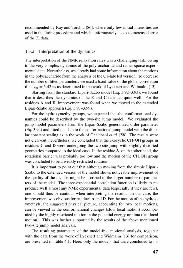
recommended by Kay and Torchia [86], where only few initial intensities areused in the fitting procedure and which, unfortunately, leads to increased errorof the T2 data.
4.3.2 Interpretation of the dynamics
The interpretation of the NMR relaxation rates was a challenging task, owingto the very complex dynamics of the polysaccharide and rather sparse experi-mental data. Nevertheless, we already had some information about the motionsin the polysaccharide from the analysis of the C1-labeled version. To decreasethe number of fitted parameters, we used a fixed value of the global correlationtime τM = 5.42 ns as determined in the work of Lycknert and Widmalm [13].
Starting from the standard Lipari-Szabo model (Eq. 3.92–3.93), we foundthat it describes the dynamics of the E and C residues quite well. For theresidues A and D, improvement was found when we moved to the extendedLipari-Szabo approach (Eq. 3.97–3.99).
For the hydroxymethyl groups, we expected that the conformational dy-namics could be described by the two-site jump model. We evaluated thejump model parameters from the Lipari-Szabo generalized order parameter(Eq. 3.94) and fitted the data to the conformational jump model with the dipo-lar constant scaling as in the work of Ghalebani et al. [58]. The results werenot clear-cut, nevertheless, we concluded that the exocyclic CH2OH groups inresidues C and D were undergoing the two-site jump with slightly distortedgeometries compared to the ideal case. In the residue A, on the other hand, therotational barrier was probably too low and the motion of the CH2OH groupwas concluded to be a weakly restricted rotation.
It is important to point out that although moving from the simple Lipari-Szabo to the extended version of the model shows noticeable improvement ofthe quality of the fit, this might be ascribed to the larger number of parame-ters of the model. The three-exponential correlation function is likely to re-produce well almost any NMR experimental data (especially if they are few),one should thus be cautious when interpreting the results. In our case, theimprovement was obvious for residues A and D. For the motion of the hydrox-ymethyls, the suggested physical picture, accounting for two local motions,can be viewed as the conformational changes (slow local motion) accompa-nied by the highly restricted motion in the potential energy minima (fast localmotion). This was further supported by the results of the above mentionedtwo-site jump model analysis.
The resulting parameters of the model-free motional analysis, togetherwith the data from the work of Lycknert and Widmalm [13] for comparison,are presented in Table 4.1. Here, only the models that were concluded to in-
47
terpret the motion of the particular residue most accurately are presented. Thefull analysis of our data, including errors, can be found in Paper IV.
The dynamics determined from the analysis of the C6 carbons is differentthan the one obtained for the C1 carbons, although some common trends canbe observed. Clearly, the Lipari-Szabo order parameters of the hydroxymethylgroups (C6) are lower than the corresponding values for the C1 carbons. Thismight indicate an additional motion (i.e. the conformational changes) influenc-ing the relaxation of the C6 sites. To draw a conclusion from the correlationtimes is more difficult. However, a trend is observed with a varying effectivelocal correlation time as a function of the sugar residue, where the neighboringresidues have local correlation times close to each others.
The relaxation study on the C6-labeled O-antigen revealed additional con-formational dynamics in the polymer, one may thus visualize the polysaccha-ride as “beads on a string” with flexible attachments to the beads.
Table 4.1: Comparison of the model-free motional parameters of residues A–Eas obtained for the [6-13C] and [1-13C] labeled O-antigen. LS stands for Lipari-Szabo model, ext. LS denotes the extended Lipari-Szabo model, LS + ex. meansLipari-Szabo with the exchange term Rex.
E A D B C[6-13C] labels
LS ext. LS ext. LS LSS2 = 0.12 S2 = 0.22 S2 = 0.37 S2 = 0.52
S2f = 0.84 S2
f = 0.84S2
s = 0.26 S2s = 0.44
τs = 381 ps τs = 555 ps τe = 413 psτe = 33.5 ps τ f = 15 ps
[1-13C] labelsE A D B CLS ext. LS LS LS LS + ex.
S2 = 0.62 S2 = 0.63 S2 = 0.64 S2 = 0.64 S2 = 0.62τs = 517 ps τe = 387 ps τs = 366 ps τe = 376 ps τe = 455 ps
Rex = 2.7 s−1
48

4.4 Hydrogen-bonded molecular clusters of ethanol
Hydrogen bonding is a phenomenon that has been extensively studied in re-cent years and yet not all of its aspects are fully understood. The hydrogen-bonded structures in low-molecular-weight alcohols have been subjected toa vast number of studies employing infrared spectroscopy [87–90], X-ray orneutron diffraction [91–93], NMR spectroscopy [94], as well as theoretical ap-proaches [20; 22; 95–99] and combinations of methods. A discussion of thefindings of some of these works is presented in the introduction of Paper V.
Despite of the long-lasting interest in this subject, the details of the struc-ture and dynamics of hydrogen-bonded species remain an open question. Thisfact was the motivation to open the issue of hydrogen bonding again and in-vestigate the hydrogen-bonded clusters of ethanol.
Alcohols are among the simplest systems that undergo classical hydrogen-bonding. Compared to the complex three-dimensional hydrogen-bonded net-works in water, alcohol clusters are known to have reduced dimensionality,preferring linear and cyclic topologies [100–103]. Therefore, ethanol providesa simple, yet realistic, model system which was a natural choice for our study.
Taking the advantage of the recent progress in NMR spectroscopy andcomputational techniques, we developed a novel method for investigation ofthe hydrogen bonding in liquid ethanol combining NMR diffusion experi-ments, hydrodynamic simulations and quantum chemical density functionaltheory (DFT) calculations. This method allowed us to report the average sizeof ethanol clusters in dilute solutions of ethanol throughout a wide range oftemperatures.
We decided to investigate two samples of ethanol dissolved in deuteratedhexane (concentrations 0.16 and 0.44 M) with an addition of TMS standard.
NMR diffusion measurements were quite straightforward method of choice,since the diffusion coefficient of ethanol clearly reflects the size of the hydrogen-bonded clusters in solution. It is important to mention that the clusters in liquidare not static structures. There is an evidence that reorganization of the hydro-gen bonds occurs on a timescale of picoseconds or faster [89; 101; 102]. Thisfact is also reflected in the 1H NMR spectrum, where a single peak of the OHgroups is observed even at very low temperatures (approx. 180 K).
The reorganization of the clusters is therefore much faster than the diffu-sion mixing time ∆ in NMR diffusion experiments (see Section 3.3), i.e. thehydrogen bonds are broken and reestablished many times during the periodin which the diffusion is observed. This means that NMR diffusion measure-ments detect a population-weighted average of the diffusion coefficient of allclusters present.
However, transforming the diffusion properties into an actual number of
49
molecules (i.e. the size of the clusters) proved to be an arduous task.The first problem was to transform the diffusion properties into the appar-
ent hydrodynamic radius of the clusters since the viscosities of the deuteratedhexane in our experimental temperature range are, to our best knowledge, notavailable in literature. We come back to this problem in Section 4.4.2.
The second difficulty was to obtain the size of an average cluster, assum-ing we know its diffusion coefficient. We decided to employ the hydrodynamiccalculations, using the HydroNMR program [104], on the ethanol cluster struc-tures previously calculated by the DFT method [105]. In this way it would bepossible to associate the diffusion properties of the clusters obtained from ex-periments to those from the diffusion simulations and thus interpret the resultsquantitatively in terms of the size of the clusters.
4.4.1 TMS/hexane calibration experiments and simulations
Special attention was given to ensure that HydroNMR (designed for calcula-tions on globular proteins) would give reliable results for small molecules aswell. HydroNMR uses Stokes-Einstein approach that requires caution whenused on systems where the size of the studied molecules is comparable to thesize of the solvent molecules.
Tetramethylsilane (TMS) dissolved in hexane appeared to be a good choiceof a “testing” system for the hydrodynamic simulations. Therefore, we pre-pared a calibration sample (TMS/hexane sample) containing 1 vol.% of TMSin non-deuterated hexane, whose viscosity is available in literature [106]. Theidea was to perform both experiments and calculations on TMS, compare theresults and possibly adjust input settings of the program. As an input of theHydroNMR program, the radius of the atomic spheres (AER) [107], that entersthe bead model [108] used in the program for hydrodynamic calculations, canbe specified. This parameter is of an empirical nature and we subjected it tooptimization.
The measurements on the TMS/hexane sample turned out to be an exper-imental challenge. Because of the use of non-deuterated solvent, the sam-ple was prepared in a coaxial tube with the outer tube containing deuteratedmethanol for the field-frequency lock. Thus the sensitivity was quite low,partly owing to the coaxial tube set, partly to the necessary low concentrationof TMS and to the fact that TMS signal appeared close (approx. 1 ppm) to themuch larger signal of hexane. Moreover, phase distortions, leading to baselinedistortions, occurred during the processing. This problem was finally resolvedby adding a spin-lock before acquisition into the diffusion pulse sequence (seeSection 3.3).
Nevertheless, we found the optimized settings for the HydroNMR that
50

gave diffusion properties of TMS corresponding to experimental data, assum-ing that this would allow for correct simulations of the ethanol clusters.
Later on, we attempted to confirm the optimized settings for the programby the use of another system, hexamethylene tetramin (HMTA) dissolved inD2O/DMSO solvent with three different compositions. This effort turned outto be unsuccessful, probably due to the hydration of HMTA, which appearedto have different hydrodynamic radius at different D2O/DMSO ratios.
4.4.2 Viscosity determination
Before we turned to the hydrodynamic simulations of ethanol clusters, we hadto deal with the problem of deuterated hexane viscosities. At this point, weutilized the measured diffusion coefficients of TMS in hexane again.
The viscosity of the deuterated hexane (with a low ethanol and TMS con-tent) was obtained by assuming same hydrodynamic radius of TMS in bothdeuterated and non-deuterated hexane, employing the procedure described indetail in Paper V. The differences in the viscosity between the non-deuteratedhexane and 0.16 M and 0.44 M ethanol samples containing deuterated hexanewas found to be in average 4 % and 5 %, respectively.
We attempted to employ also other approaches to obtain the viscosity of thedeuterated hexane. The capillary viscometer with a falling ball (Anton Paar)failed to provide reproducible results due to the too low viscosity. Moreover,it could not cover the whole temperature range of our experiments. Vibratingquartz fork method [109] seemed to be more promising but it would requirefurther technical development.
4.4.3 Average size of molecular clusters of ethanol
Knowing the viscosities of the ethanol/hexane-d14 samples, we could obtainthe hydrodynamic radii of the ethanol clusters from the experimental diffusioncoefficients. Furthermore, we performed the hydrodynamic simulations on theDFT calculated structures of ethanol ranging from monomer to octamer, bothlinear and (for clusters larger than dimer) cyclic topologies. By comparing theresults of the simulations with the experimental data, we were able to obtainthe temperature dependent average size of the ethanol clusters at both concen-trations (see Figure 4.3).
The hydrodynamic radius of ethanol obtained from the diffusion measure-ments generally decreases with increasing temperature. The more concen-trated solution is composed of somewhat larger clusters in the whole investi-gated temperature range with average sizes, roughly, from dimer to heptamer.
At low temperatures (lower than approx. 210 K) the experimental hydrody-namic radii of both the 0.16 M and the 0.44 M ethanol samples reach a plateau
51

corresponding to clusters of average size of hexamer and heptamer, respec-tively. At the temperatures higher than 300 K, clusters of average size close toa dimer are present in the 0.44 M sample. In contrast, in the 0.16 M sample,the decrease stops at approx. 308 K, indicating that only monomers are signif-icantly populated above this temperature. This experimental observation of alower bound of hydrodynamic radius of ethanol is in excellent agreement withthe calculated hydrodynamic radii. This fact strongly validates the employedmethodology with the use of HydroNMR on small molecules with the priorcalibration and reinforces our interpretation of the diffusion NMR data.
1 8 0 2 0 0 2 2 0 2 4 0 2 6 0 2 8 0 3 0 0 3 2 0 3 4 0 3 6 01 . 0
1 . 5
2 . 0
2 . 5
3 . 0
3 . 5
4 . 0
4 . 5
0 . 4 4 M e x p 0 . 1 6 M e x p
o c t a m e rh e p t a m e rh e x a m e rp e n t a m e rt e t r a m e rt r i m e r
d i m e r
Hydro
dyna
mic r
adius
[Å]
T e m p e r a t u r e [ K ]
m o n o m e r
Figure 4.3: The average hydrodynamic radius of ethanol clusters from exper-iments and hydrodynamic simulations of the DFT-calculated cluster structures(horizontal lines).
52
Acknowledgements
I did not maybe find the answer to the ultimate question of life, the universe,and everything, but I’m quite sure there are many questions of mine that havebeen answered during my PhD. And this would not be possible without all thepeople I have met on my way.
I would like to thank my supervisors Jozef Kowalewski and Jan Lang forcontinuous support and help throughout whole period of my PhD. Withoutthem, this would never come to a happy ending.
My thanks also belong to Zdenek Nemecek, dean of the Faculty of Mathe-matic and Physics at Charles University in Prague, and Stefan Nordlund, deanof the Faculty of Science at Stockholm University, who made the joint PhDproject between the two universities possible.
I have spent a half of my entire PhD studies at the Department of LowTemperature Physics in Prague. I would like to thank especially the head ofthe department Ladislav Skrbek, the head of the Radiofrequency Spectroscopydivision Helena Štepánková and my colleagues Pavel Srb, Vašek Rímal, RíšaReznícek, Mikuláš Peksa, Kája Kouril, Karel Bernášek, Vojta Chlan, MariánGrocký, and others for their scientific colaboration and help with problems ofany imaginable kind.
The other half of my PhD have been spent with the people at the De-partment of Materials and Environmental Chemistry in Stockholm. I wishto thank the head of the department Gunnar Svensson, my coworkers ZoltanTakacs and Emilie Steiner for scientific help and fruitful discussions, and mycolleagues and friends Guido Todde, Henrik Fahlquist, Samrad Shafeie, IlichIbarra, Daniel Eklöf, Alfredo Metere, Aleksander Jaworski, Jon Kapla, AmberMace, Alexander Mirzoev, Claudia Turdean-Ionescu, Mikhail Voznesenskiy,and others for making my time in Stockholm enjoyable.
I would also like to thank Torbjörn Astlind, Rolf Eriksson, Ann-Britt Rön-nell, and other members of the technical and administrative stuff for makingthe things run smoothly.
It would not be possible to run the NMR experiments if there weren’t forthe samples prepared at the Department of Organic Chemistry. I am especiallythankful to Göran Widmalm, Olof Engström, and Robert Pendrill for providingunique samples, only of their kind on earth.
My thanks go also to the University of Padova in Italy, particularly to An-

tonino Polimeno, Mirco Zerbetto, and Dmytro Kotsyubynskyy for a fruitfulscientific collaboration.
I also wish to thank my wonderful boyfriend Jožo and my family for all thelove, support and understanding.
Last but not least, I wish to thank all my climbing, frisbee, and volleyballfriends, who kept reminding me that there’s more to life than just science.
So long, and thanks for all the fish!
References
[1] A. COLLET, J. GABARD, J. JACQUES, M. CESARIO, J. GUILHEM, AND C. PASCARD. Synthe-sis and Absolute Configuration of Chiral (C3) Cyclotriveratrylene Derivatives. Crystal struc-ture of (M)-(–)-2,7,12-triethoxy-3,8,13-tris-[(R)-1-methoxycarbonylethoxy]-10,15-dihydro-5H-tribenzo[a,d,g]-cyclononene. J. Chem. Soc., Perkin Trans. 1, pages 1630–1638, 1981. 4
[2] J. GABARD AND A. COLLET. Synthesis of a (D3)-Bis(cyclotriveratrylenyl) Macrocage by Stere-ospecific Replication of a (C3)-Subunit. J. Chem. Soc., Chem. Commun., pages 1137–1139, 1981.
[3] J. CANCEILL, M. CESARIO, A. COLLET, J. GUILHEM, AND C. PASCARD. A New Bis-Cyclotribenzyl Cavitand Capable of Selective Inclusion of Neutral Molecules in Solution. Crys-tal Structure of its CH2Cl2 Cavitate. J. Chem. Soc., Chem. Commun., pages 361–363, 1985.
[4] J. CANCEILL, M. CESARIO, A. COLLET, J. GUILHEM, C. RICHE, AND C. PASCARD. Selec-tive Recognition of Neutral Molecules: 1H n.m.r. Study of the Complexation of CH2Cl2 andCH2Br2 by Cryptophane-D in Solution and Crystal Structure of its CH2Cl2 Cavitate. J. Chem.Soc., Chem. Commun., pages 339–341, 1986.
[5] CNCEILL AND A. COLLET. Two-Step Synthesis of D3 and C3h Cryptophanes. J. Chem. Soc.,Chem. Commun., pages 582–584, 1988. 4
[6] T. BROTIN AND J. P. DUTASTA. Cryptophanes and Their Complexes - Present and Future.Chem. Rev., 109:88–130, 2009. 4
[7] M. ZERBETTO, A. POLIMENO, D. KOTSYUBYNSKYY, L. GHALEBANI, J. KOWALEWSKI,E. MEIROVITCH, U. OLSSON, AND G. WIDMALM. An Integrated Approach to NMRSpin Relaxation in Flexible Biomolecules: Application to β -D-glucopyranosyl-(1→6)-α-D-mannopyranosyl-OMe. J. Chem. Phys., 131:234501, 2009. 6, 41, 42, 44, 45
[8] R. STENUTZ, A. WEINTRAUB, AND G. WIDMALM. The Structures of Escherichia coli O-Polysaccharide Antigens. FEMS Microbiol. Rev., 30:382–403, 2006. 7
[9] J. YOTHER. Capsules of Streptococcus pneumoniae and Other Bacteria: Paradigms forPolysaccharide Biosynthesis and Regulation. Annu. Rev. Microbiol., 65:563–581, 2011. 8
[10] C. WHITFIELD. Biosynthesis and Assembly of Capsular Polysaccharides in Escherichia coli.Annu. Rev. Biochem., 75:39–68, 2006.
[11] C. R. RAETZ AND C. WHITFIELD. Lipopolysaccharide Endotoxins. Annu. Rev. Biochem.,71:635–700, 2002. 8
[12] A. KJELLBERG, A. WEINTRAUB, AND G. WIDMALM. Structural Determination and Biosyn-thesis Studies of O-Antigenic Polysaccharide from the Enterohemorrhagic Escherichia coliO91 Using 13C-Enrichment and NMR Spectroscopy. Biochemistry, 38:12205–12211, 1999. 8
[13] K. LYCKNERT AND G. WIDMALM. Dynamics of the Escherichia coli O91 O-Antigen Polysac-charide in Solution as Studied by Carbon-13 NMR Relaxation. Biomacromolecules, 5:1015–1020, 2004. 9, 45, 47
[14] T. STEINER. The Hydrogen Bond in the Solid State. Angew. Chem. Int. Ed., 41:48–76, 2002. 9
[15] I. ALKORTA, I. ROZAS, AND J. ELGUERO. Non-Conventional Hydrogen Bonds. Chem. Soc.Rev., 27:163–170, 1998.
[16] C. L. PERRIN AND J. B. NIELSON. Strong Hydrogen Bonds in Chemistry and Biology. Annu.rev. Phys. Chem., 48:511–544, 1997.
[17] B. KOJIC-PRODIC AND K. MOLCANOV. The Nature of Hydrogen Bond: New Insights into OldTheories. Acta Chim. Slov., 55:692–708, 2008. 9
[18] G. JEFFREY AND W. SAENGER. Hydrogen Bonding in Biological Structures. Springer, Berlin,1991. 9
[19] G. JEFFREY. An Introduction to Hydrogen Bonding. Oxford University Press, 1997. 9
[20] L. SAIZ, J. A. PARDÓ, AND E. GUÀRDIA. Dynamics and Hydrogen Bonding in Liquid Ethanol.Mol. Phys., 97:897–905, 1999. 10, 49
[21] M. HÜLSEKOPF AND R. LUDWIG. Temperature Dependence of Hydrogen Bonding in Alcohols.J. Mol. Liq., 85:105–125, 2000.
[22] A. K. SUM AND S. I. SANDLER. Ab Initio Calculations of Cooperativity Effects on Clusters ofMethanol, Ethanol, 1-Propanol, and Methanethiol. J. Phys. Chem. A, 104:1121–1129, 2000. 10,49
[23] U. LIDDEL AND N. F. RAMSEY. Temperature Dependent Magnetic Shielding in Ethyl Alcohol.J. Chem. Phys., 19:1608, 1951. 11
[24] J. T. ARNOLD AND M. E. PACKARD. Variations in Absolute Chemical Shift of Nuclear Induc-tion Signals of Hydroxyl Groups of Methyl and Ethyl Alcohol. J. Chem. Phys., 19:1608, 1951.11
[25] M. H. LEVITT. Spin Dynamics: Basics of Nuclear Magnetic Resonance, Second Edition. JohnWiley & Sons, 2008. 12, 14
[26] J. KEELER. Understanding NMR Spectroscopy. John Wiley & Sons, 2010. 14
[27] J. KOWALEWSKI AND L. MÄLER. Nuclear Spin Relaxation in Liquids: Theory, Experiments andApplications. Taylor and Francis Group, 2006. 30, 31, 35
[28] A. ABRAGAM. The Principles of Nuclear Magnetism. Oxford University Press, 1961. 32
[29] C. P. SLICHTER. Principles of Magnetic Resonance. Springer Verlag, 1989. 12, 32
[30] F. BLOCH. Nuclear Induction. Phys. Rev., 70:460–467, 1946. 16
[31] A. EINSTEIN. Über die von der molekularkinetischen Theorie der Wärme geforderte Bewe-gung von in ruhenden Flüssigkeiten suspendierten Teilchen. Analen der Physik, 17:549–560,1905. 20
[32] E. STEJSKAL AND J. TANNER. Spin Diffusion Measurements: Spin Echo in the Presence of aTime-Dependent Field Gradient. J. Phys. Chem., 42(1):288–292, 1965. 20
[33] D. WU, A. CHEN, AND C. S. JOHNSON, JR. An Improved Diffusion-Ordered SpectroscopyIncorporating Bipolar-Gradient Pulses. J. Magn. Reson., 117:260–264, 1995. 21
[34] S. J. GIBBS AND C. S. JOHNSON, JR. A PFG NMR Experiment for Accurate Diffusion andFlow Studies in the Presence of Eddy Currents. J. Magn. Reson., 93:395–402, 1991. 21

[35] A. JERSCHOW AND R. MÜLLER. Suppression of Convection Artifacts in Stimulated-EchoDiffusion Experiments. Double-Stimulated-Echo Experiments. J. Magn. Reson., 125:372–375,1997. 21
[36] M. PELTA, H. BARJAT, G. MORRIS, A. DAVIS, AND S. HAMMOND. Pulse Sequences for High-Resolution Diffusion-Ordered Spectroscopy (HR-DOSY). Magn. Reson. Chem., 36:706–714,1998. 21
[37] G. BINCH. The Study of Intramolecular Rate Processes by Dynamic Nuclear Magnetic Reso-nance. Topics in Stereochemistry, 3:97–191, 1968. 23, 40
[38] V. RÍMAL, H. ŠTEPÁNKOVÁ, AND J. ŠTEPÁNEK. Analysis of NMR Spectra in Case ofTemperature-Dependent Chemical Exchange Between two Unequally Populated Sites. Con-cepts Magn. Reson. Part A, A38:117–127, 2011. 23, 40
[39] K. STOTT, J. KEELER, Q. N. VAN, AND A. J. SHAKA. One-Dimensional NOE ExperimentsUsing Pulsed Field Gradients. Magn. Reson. Chem., 125:302–324, 1997. 24
[40] S. MACURA, B. T. FARMER, AND L. R. BROWN. An Improved Method for the Determinationof Cross-Relaxation Rates from NOE Data. J. Magn. Reson., 70:1969–1992, 1986. 24
[41] H. T. HU AND K. KRISHNAMURTHY. Revisiting the Initial Rate Approximation in Kinetic NOEMeasurements. J. Magn. Reson., 182:173–177, 2006. 24
[42] I. SOLOMON. Relaxation Processes in a System of Two Spins. Phys. Rev., 99:559–565, 1955. 26
[43] J. KOWALEWSKI, M. EFFEMEY, AND J. JOKISAARI. Dipole-Dipole Coupling Constant for aDirectly Bonded CH Pair – A Carbon-13 Relaxation Study. J. Magn. Reson., 157:171–177,2002. 29
[44] E. HAHN. Spin Echoes. Phys. Rev., 80:580–595, 1950. 30
[45] H. CARR AND E. PURCELL. Effect of Diffusion on Free Precession in Nuclear Magnetic Reso-nance Experiments. Phys. Rev., 94:630–638, 1954. 31
[46] S. MEIBOOM AND D. GILL. Modified Spin-Echo Method for Measuring Nuclear RelaxationTimes. Rev. Sci. Instrum., 29:688–691, 1958. 31
[47] A. G. PALMER, N. SKELTON, W. J. CHAZIN, P. E. WRIGHT, AND M. RANCE. Suppression ofthe Effects of Cross-Correlation between Dipolar and Anisotropic Chemical Shift RelaxationMechanisms in the Measurement of Spin-Spin Relaxation Rates. Mol. Phys., 75:699–711, 1992.31
[48] L. E. KAY, L. K. NICHOLSON, F. DELAGLIO, A. BAX, AND D. A. TORCHIA. Pulse Se-quences for Removal of the Effects of Cross Correlation between Dipolar and Chemical-ShiftAnisotropy Relaxation Mechanisms on the Measurement of Heteronuclear T1 and T2 Values inProteins. J. Magn. Reson., 97:359–375, 1992. 31
[49] R. WANGSNESS AND F. BLOCH. The Dynamic Theory of Nuclear Induction. Phys. Rev., 89:728–739, 1953. 32
[50] A. REDFIELD. The Theory of Relaxation Processes. Adv. Magn. Reson., 1:1–32, 1965. 32
[51] A. KUMAR, R. C. R. GRACE, AND P. K. MADHU. Cross-Correlation in NMR. Prog. Nucl.Magn. Reson. Spectr., 37:191–319, 2000. 33
[52] L. G. WERBELOW AND D. M. GRANT. Intramolecular Dipolar Relaxation in Multispin System.Adv. Magn. Reson., 9:182–299, 1977. 34

[53] L. GHALEBANI, P. BERNATOWICZ, S. NIKKHOU ASKI, AND J. KOWALEWSKI. Cross-Correlatedand Conventional Dipolar Carbon-13 Relaxation in Methylene Groups in Small, SymmetricMolecules. Concepts Magn. Reson. Part A, 30A:100–115, 2007. 35
[54] V. A. DARAGAN AND K. H. MAYO. Motional Model Analyses of Protein and Peptide DynamicsUsing 13C and 15N NMR Relaxation. Prog. Nucl. Magn. Reson. Spectr., 31:63–105, 1997. 36
[55] V. A. JARYMOWYCZ AND M. J. STONE. Fast Time Scale Dynamics of Protein Backbones: NMRRelaxation Methods, Applications, and Functional Consequences. Chem. Rev., 106:1624–1671,2006. 36
[56] G. LIPARI AND A. SZABO. Model-Free Approach to the Interpretation of Nuclear MagneticResonance Relaxation in Macromolecules. 1. Theory and Range of Validity. J. Am. Chem. Soc.,104:4546–4559, 1982. 36
[57] G. LIPARI AND A. SZABO. Model-Free Approach to the Interpretation of Nuclear MagneticResonance Relaxation in Macromolecules. 2. Analysis of Experimental Results. J. Am. Chem.Soc., 104:4559–4570, 1982. 36
[58] L. GHALEBANI, D. KOTSYUBYNSKYY, AND J. KOWALEWSKI. NMR Relaxation InterferenceEffects and Internal Dynamics in γ-Cyclodextrin. J. Magn. Reson., 195:1–8, 2008. 36, 44, 47
[59] K. E. KÖVÉR, G. BATTA, J. KOWALEWSKI, L. GHALEBANI, AND D. KRUK. Internal Dynamicsof Hydroxymethyl Rotation from CH2 Cross-Correlated Dipolar Relaxation in Methyl-β -D-Glucopyranoside. J. Magn. Reson., 167:273–281, 2004. 36, 44
[60] N. R. SKRYNNIKOV, O. MILLET, AND L. E. KAY. Deuterium Spin Probes of Side-Chain Dy-namics in Proteins. 2. Spectral Density Mapping and Identification of Nanosecond Time-ScaleSide-Chain Motions. J. Am. Chem. Soc., 124:6449–6460, 2002. 37
[61] G. M. CLORE, A. SZABO, A. BAX, L. E. KAY, P. DRISCOLL, AND A. M. GRONENBORN. Devia-tions from the Simple Two-Parameter Model-Free Approach to the Interpretation of Nitrogen-15 Nuclear Magnetic Relaxation of Proteins. J. Am. Chem. Soc., 112:4989–4991, 1990. 37
[62] G. M. CLORE, P. C. DRISCOLL, P. T. WINGFIELD, AND A. M. GRONENBORN. Analysis of theBackbone Dynamics of Interleukin-1β Using Two-Dimensional Inverse Detected Heteronu-clear 15N - 1H NMR Spectroscopy. Biochem., 29:7387–7401, 1990. 37
[63] A. POLIMENO AND J. H. FREED. A Many-Body Stochastic Approach to Rotational Motions inLiquids. Adv. Chem. Phys., 83:89–206, 1993. 38, 41
[64] A. POLIMENO AND J. H. FREED. Slow Motional ESR in Complex Fluids: The Slowly RelaxingLocal Structure Model of Solvent Cage Effects. J. Chem. Phys., 99:10995, 1995. 38, 41
[65] H. M. MCCONNELL. Reaction Rates By Nuclear Magnetic Resonance. J. Chem. Phys., 28:430–431, 1958. 38
[66] J. LANG, J. J. DECHTER, M. EFFEMEY, AND J. KOWALEWSKI. Dynamics of an Inclusion Com-plex of Chloroform and Cryptophane-E: Evidence for a Strongly Anisotropic van der WaalsBond. J. Am. Chem. Soc., 123:7852–7858, 2001. 39
[67] Z. TOŠNER, J. LANG, D. SANDSTRÖM, O. PETROV, AND J. KOWALEWSKI. Dynamics of anInclusion Complex of Dichloromethane and Cryptophane-E. J. Phys. Chem. A, 106:8870–8875,2002. 40
[68] Z. TOŠNER, O. PETROV, S. V. DVINSKIKH, J. KOWALEWSKI, AND D. SANDSTRÖM. A 13CSolid-State NMR Study of Cryptophane-E:Chloromethane Inclusion Complexes. Chem. Phys.Lett., 388:208–211, 2004.

[69] O. PETROV, Z. TOŠNER, I. CSÖREGH, J. KOWALEWSKI, AND D. SANDSTRÖM. Dynamics ofChloromethanes in Cryptophane-E Inclusion Complexes: A 2H Solid-State NMR and X-rayDiffraction Study. J. Phys. Chem. A, 109:4442–4451, 2005.
[70] S. NIKKHOU ASKI, Z. TAKACS, AND J. KOWALEWSKI. Inclusion Complexes of Cryptophane-E with Dichloromethane and Chloroform: A Thermodynamic and Kinetic Study Using the1D-EXSY NMR Method. Magn. Reson. Chem., 46:1135–1140, 2008.
[71] S. NIKKHOU ASKI, A. Y. H. LO, T. BROTIN, J. P. DUTASTA, M. EDÉN, AND J. KOWALEWSKI.Studies of Inclusion Complexes of Dichloromethane in Cryptophanes by Exchange Kineticsand 13C NMR in Solution and the Solid State. J. Phys. Chem. C, 112:13873–13881, 2008. 39, 40
[72] Z. TAKACS, T. BROTIN, J.-P. DUTASTA, J. LANG, G. TODDE, AND J. KOWALEWSKI. Inclusionof Chloromethane Guests Affects Conformation and Internal Dynamics of Cryptophane-DHost. J. Phys. Chem. B, 116:7898–7913, 2012. 39, 40
[73] Z. TAKACS. Chloromethane Complexation by Cryptophanes: Host-Guest Chemistry Investigatedby NMR and Quantum Chemical Calculations. doctoral thesis, Stockholm University, 2012. 39
[74] E. MEIROVITCH, Y. E. SHAPIRO, A. POLIMENO, AND J. H. FREED. Protein Dynamics fromNMR: The Slowly Relaxing Local Structure Analysis Compared with Model-Free Analysis. J.Phys. Chem. A, 110:8366–8396, 2006. 41
[75] E. MEIROVITCH, Y. E. SHAPIRO, A. POLIMENO, AND J. H. FREED. Structural Dynamics ofBio-Macromolecules by NMR: The Slowly Relaxing Local Structure Approach. Prog. Nucl.Magn. Reson. Spectrosc., 56:360–405, 2010.
[76] Y. E. SHAPIRO, A. POLIMENO, J. H. FREED, AND E. MEIROVITCH. Methyl Dynamics of aCa2+-Calmodulin-Peptide Complex from NMR/SRLS. J. Phys. Chem. B, 115:354–365, 2011.
[77] E. MEIROVITCH, M. ZERBETTO, A. POLIMENO, AND J. H. FREED. Backbone Dynamics ofDeoxy and Carbonmonoxy Hemoglobin by NMR/SRLS. J. Phys. Chem. B, 115:143–157, 2011.
[78] E. MEIROVITCH, Y. E. SHAPIRO, M. ZERBETTO, AND A. POLIMENO. SRLS Analysis of 15NSpin Relaxation from E. coli Ribonuclease HI: The Tensorial Perspective. J. Phys. Chem. B,116:886–894, 2012. 41
[79] V. BARONE AND A. POLIMENO. Toward an Integrated Computational Approach to CW-ESRSpectra of Free Radicals. Phys. Chem. Chem. Phys., 8:4609–4629, 2006. 42
[80] M. ZERBETTO, A. POLIMENO, AND V. BARONE. Simulation of Electron Spin ResonanceSpectroscopy in Diverse Environments: An Integrated Approach. Comput. Phys. Commun.,180:2680–2697, 2009. 42
[81] M. ZERBETTO, M. BUCK, E. MEIROVITCH, AND A. POLIMENO. Integrated ComputationalApproach to the Analysis of NMR Relaxation in Proteins: Application to ps-ns Main Chain15N-1H and Global Dynamics of the Rho GTPase Binding Domain of Plexin-B1. J. Phys. Chem.B, 115:376–388, 2011. 42
[82] M. ZERBETTO, D. KOTSYUBYNSKYY, J. KOWALEWSKI, G. WIDMALM, AND A. POLIMENO.Stochastic Modeling of Flexible Biomolecules Applied to NMR Relaxation. I. Internal Dynam-ics of Cyclodextrins: γ-Cyclodextrin as a Case Study. J. Phys. Chem. B, 116:13159–13171, 2012.42, 44
[83] R. STENUTZ, I. CARMICHAEL, G. WIDMALM, AND A. S. SERIANNI. Hydroxymethyl GroupConformation in Saccharides: Structural Dependencies of 2JHH , 3JHH , and 1JCH Spin-SpinCoupling Constants. J. Org. Chem., 67:949–958, 2002. 42, 45
[84] V. BARONE, M. ZERBETTO, AND A. POLIMENO. Hydrodynamic Modeling of Diffusion TensorProperties of Flexible Molecules. J. Comput. Chem., 30:2–12, 2009. 42

[85] U. OLSSON, E. SÄWÉN, R. STENUTZ, AND G. WIDMALM. Conformation Flexibility and Dy-namics of Two (1→ 6)-Linked Disaccharides Related to an Oligosaccharide Epitope Expressedon Malignant Tumor Cells. Chem. Eur. J., 15:8886–8894, 2009. 45
[86] L. E. KAY AND D. A. TORCHIA. The Effects of Dipolar Cross Correlation on 13C Methyl-Carbon T1, T2, and NOE Measurements in Macromolecules. J. Magn. Reson., 95:536–547,1991. 47
[87] H. GRAENER, T. Q. YE, AND A. LAUBEREAU. Ultrafast Dynamics of Hydrogen Bonds DirectlyObserved by Time-Resolved Infrared Spectroscopy. J. Chem. Phys., 90:3413, 1989. 49
[88] K. M. MURDOCH, T. D. FERRIS, J. C. WRIGHT, AND T. C. FARRAR. Infrared Spectroscopy ofEthanol Clusters in Ethanol-Hexane Binary Solutions. J. Chem. Phys., 116:5717, 2002.
[89] K. J. GAFFNEY, I. R. PILETIC, AND M. D. FAYER. Orientational Relaxation and VibrationalExcitation Transfer in Methanol-Carbon Tetrachloride Solutions. J. Chem. Phys., 118:2270,2003. 49
[90] P. SASSI, F. PALOMBO, R. S. CATALIOTTI, M. PAOLANTONI, AND A. MORRESI. Distributionof H-Bonding Aggregates in tert-Butyl Alcohol: The Pure Liquid and Its Alkane Mixtures. J.Phys. Chem. A, 111:6020–6027, 2007. 49
[91] S. SARKAR AND R. N. JOARDER. Molecular Clusters and Correlations in Liquid Methanol atRoom Temperature. J. Chem. Phys., 99:2032, 1993. 49
[92] S. SARKAR AND R. N. JOARDER. Molecular Clusters in Liquid Ethanol at Room Temperature.J. Chem. Phys., 100:5118, 1994.
[93] T. YAMAGUCHI, K. HIDAKA, AND A. K. SOPER. The Structure of Liquid Methanol Revisited:a Neutron Diffraction Experiment at −80 ◦C and +25 ◦C. Mol. Phys., 96:1159–1168, 1999. 49
[94] T. D. FERRIS AND T. C. FARRAR. The Temperature Dependence of the Deuterium QuadrupoleCoupling Parameter and The Rotational Correlation Time of The OD Internuclear Vector inNeat Ethanol-d1. Mol. Phys., 100:303–309, 2002. 49
[95] V. DYCZMONS. Dimers of Ethanol. J. Phys. Chem. A, 108:2080–2086, 2004. 49
[96] S. M. MEJÍA, J. F. ESPINAL, A. RESTREPO, AND F. MONDRAGÓN. Molecular Interaction of(Ethanol)2-Water Heterodimer. J. Phys. Chem. A, 111:8250–8256, 2010.
[97] S. M. MEJÍA, J. F. ESPINAL, AND F. MONDRAGÓN. Cooperative Effects on The Struc-ture and Stability of (Ethanol)3-Water, (Methanol)3-Water Heterotetramers and (Ethanol)4,(Methanol)4 Tetramers. J. Mol. Struct. THEOCHEM, 901:186–193, 2009.
[98] J. DAVID, D. GUERRA, AND A. RESTREPO. Structural Characterization of the (Methanol)4Potential Energy Surface. J. Phys. Chem. A, 113:10167–10173, 2009.
[99] K. BLOCH AND C. P. LAWRENCE. Hydrogen Bond Lifetimes and Clustering of Methanol inCarbon Tetrachloride Solutions. J. Phys. Chem. B, 114:293–297, 2010. 49
[100] C. P. BROCK AND L. L. DUNCAN. Anomalous Space-Group Frequencies for MonoalcoholsCnHmOH. Chem. Mater., 6:1307–1312, 1994. 49
[101] J. GAFFNEY, I. R. PILETIC, AND M. D. FAYER. Hydrogen Bond Breaking and Reformationin Alcohol Oligomers Following Vibrational Relaxation of a Non-Hydrogen-Bond DonatingHydroxyl Stretch. J. Phys. Chem. A, 106:9428–9435, 2002. 49
[102] J. GAFFNEY, P. H. DAVIS, I. R. PILETIC, N. E. LEVINGER, AND M. D. FAYER. Hydrogen BondDissociation and Reformation in Methanol Oligomers Following Hydroxyl Stretch Relaxation.J. Phys. Chem. A, 106:12012–12023, 2002. 49

[103] J. B. ASBURY, T. STEINEL, AND M. D. FAYER. Hydrogen Bond Networks: Structure andEvolution after Hydrogen Bond Breaking. J. Phys. Chem. B, 108:6544–6554, 2004. 49
[104] J. G. DE LA TORRE, M. L. HUERTAS, AND B. CARRASCO. HYDRONMR: Prediction of NMRRelaxation of Globular Proteins from Atomic-Level Structures and Hydrodynamic Calcula-tions. J. Magn. Reson., 147:138–46, 2000. 50
[105] L. BENDA. Computational Study on Structure and Properties of Ethanol Clusters. diploma thesis,Charles University in Prague, 2006. 50
[106] E. B. GILLER AND H. G. DRICKAMER. Viscosity of Normal Paraffins near the Freezing Point.Ind. Eng. Chem. Res., 41:2067, 1949. 50
[107] B. CARRASCO AND J. G. DE LA TORRE. Hydrodynamic Properties of Rigid Particles: Com-parison of Different Modeling and Computational Procedures. J. Biophys., 76:3044–3057, 1999.50
[108] V. A. BLOOMFIELD, W. O. DALTON, AND K. E. VAN HOLDE. Frictional Coefficients of Multi-subunit Structures. I. Theory. Biopolymers, 5:135–148, 1967. 50
[109] H. DIVIŠOVÁ, J. LANG, M. ROTTER, AND D. SCHMORANZER. Measurement of Viscosity inSmall Volumes of Fluids by Tuning Fork Oscillators. EPJ Web of Conferences, 25:01008, 2012.51

![doc. Mgr. Marián Vanderka, PhD. - uniba.sk · [o4] 2015 Šuba, J.: Zmeny dynamických parametrov vertikálneho výskoku vo vzťahu k tréningovým ukazovateľom a športovému výkonu](https://static.fdocuments.net/doc/165x107/5e165bc23101584fe3619001/doc-mgr-marin-vanderka-phd-unibask-o4-2015-uba-j-zmeny-dynamickch.jpg)