
Doc URL //eprints.lib.hokudai.ac.jp/dspace/bitstream/2115...File Information Sarwar et al.pdf...
20
Instructions for use Title Ontogenetic development of pollen tetrads in Vaccinium smallii A. Gray (Ericaceae) Author(s) Sarwar, A.K.M. Golam; Ito, Toshiaki; Takahashi, Hideki Citation 日本花粉学会会誌 = Japanese journal of palynology, 58(2): 61-71 Issue Date 2012-12-31 Doc URL http://hdl.handle.net/2115/55508 Type article (author version) Additional Information There are other files related to this item in HUSCAP. Check the above URL. File Information Sarwar et al.pdf Hokkaido University Collection of Scholarly and Academic Papers : HUSCAP
-
Upload
nguyennhan -
Category
Documents
-
view
222 -
download
6
Transcript of Doc URL //eprints.lib.hokudai.ac.jp/dspace/bitstream/2115...File Information Sarwar et al.pdf...

Instructions for use
Title Ontogenetic development of pollen tetrads in Vaccinium smallii A. Gray (Ericaceae)
Author(s) Sarwar, A.K.M. Golam; Ito, Toshiaki; Takahashi, Hideki
Citation 日本花粉学会会誌 = Japanese journal of palynology, 58(2): 61-71
Issue Date 2012-12-31
Doc URL http://hdl.handle.net/2115/55508
Type article (author version)
Additional Information There are other files related to this item in HUSCAP. Check the above URL.
File Information Sarwar et al.pdf
Hokkaido University Collection of Scholarly and Academic Papers : HUSCAP

1) Date of submission of revised manuscript: September, 2012
2) Type of manuscript: Research Article
3) Title: Ontogenetic development of pollen tetrads in Vaccinium smallii A. Gray (Ericaceae)
Name(s) of author(s): A. K. M. Golam Sarwar1), Toshiaki Ito2) and Hideki Takahashi3)
Address(s): 1) Department of Crop Botany, Bangladesh Agricultural University, Mymensingh 2202,
Bangladesh
2) Research Faculty of Agriculture, Hokkaido University, N8 W8, Sapporo 060-8589,
Japan 3) The Hokkaido University Museum, N10 W8, Sapporo 060-0810, Japan
4) Running title: Pollen tetrad development in Vaccinium
5) Total pages for text: 12
6) Total number of table(s): 0
7) Total number of figure(s): 29 (in 4 plates)
8) Total page(s) for captions for Figures: 3
9) Corresponding Author: Professor Dr. Hideki Takahashi
The Hokkaido University Museum
Kita 10, Nishi 8, Sapporo 060-0810, Japan.
Tel. & Fax: +81-11-706-4508
E-mail: [email protected]

Ontogenetic development of pollen tetrads in Vaccinium smallii A. Gray
(Ericaceae)
The ontogenetic development of pollen tetrads in Vaccinium smallii was investigated
by light and transmission electron microscopy with special attention to the mode of exine
deposition. The deposition of the primexine matrix represented by a fibrillar surface coat
started during the cytokinesis of the microspore mother cell within a thick callose wall. The
deposition of dark lipoidal globules of sporopollenin precursor substances was observed
within the primexine matrix. After the disappearance of these globules, radially directed pro-
columellae appeared within the primexine matrix, and the primexine gives rise to the tectum,
columellae and foot layer after the callose dissolution. The position of future apertures was
observed at a thick zone and/or a thin layer of the primexine matrix. In both regions, the pro-
columellae, protectum and foot layer were not formed. Major binding mechanism of the
permanent mature pollen tetrads in V. smallii is attributed to 1) the early deposition of the
primexine matrix on the microspore mother cell undergoing cytokinesis, and 2) thin and
tenuous callose depositions, 3) the common primexine matrix, and 4) the cytoplasmic
channels between the adjoining microspores of the meiotic tetrad within a thick callose wall.
Key words: binding mechanism, ontogeny, pollen tetrads, primexine, Vaccinium
Introduction
The cosmopolitan family Ericaceae contains 4084 species and 121 genera (1), which comprises 8
subfamilies: Enkianthoideae, Monotropoideae (including Pyrolaceae), Arbutoideae, Cassiopoideae,
Ericoideae (including Empetraceae), Harrimanelloideae, Styphelioideae (including Epacridaceae) and
Vaccinioideae (2). Members of this family are highly diverse not only in life forms, leaf morphology and
inflorescence characteristics, but also in palynological features. Pollen grains of Ericaceae are dispersed
either as monads (mutually isolated grains; e.g., in Enkianthoideae) or compound grains. The latter can be
further categorized into pseudomonads, reduced tetrads with one or more aborting cells (e.g., in
Styphelieae of Styphelioideae), normal tetrads (commonly without viscin threads but occasionally with
them such as in Rhodoreae of Ericoideae), and polyads of indefinite number of tetrads (e.g., in
Chimaphila of Monotropoideae). Because of the large size of the family Ericaceae, our knowledge on its
pollen ultrastructure and development is still poor and therefore further studies are required.
Transmission electron microscopic (TEM) images of mature pollen exine in Ericaceae have been
published only for several genera (3 – 18). Available information on pollen wall development at the TEM

level is even more scarce and is restricted to three subfamilies of Ericaceae: Styphelioideae (Styphelia (19)),
Monotropoideae (Chimaphila, Orthilia, and Pyrola (20 – 22)), and Ericoideae (Calluna (23)).
Vaccinium L. of the subfamily Vaccinioideae, with about 500 species, is the third largest genus in
the Ericaceae (1). Although pollen morphology of this genus has been studied widely, the majority of
researchers used light microscopy (LM). Although scanning electron microscopy (SEM) has also been
used to study the mature pollen, a single study employed TEM ((13) and references therein). Our previous
study (13) of Vaccinium gave an outline of pollen morphology and the details of its taxonomic significance
in the sectional classification. However, information on the pollen tetrad development in Vaccinium,
especially on the exine development, is still lacking. Published works on embryology ((24) and references
therein) do not provide much information in this regard. Hence the objective of this paper is to fill the
existing knowledge gap through a detailed description of the main processes of the pollen tetrad
development of V. smallii, and to compare the results obtained with the already existing information from
the other genera of Ericaceae and angiosperm taxa.
Materials and methods
Flowers of Vaccinium smallii A. Gray were collected at different developmental stages from the
cultivated plants at the Botanic Garden of Hokkaido University, Sapporo, Japan. The anthers were fixed
in 2.5% glutaraldehyde and 1% para-formaldehyde in 50 mM phosphate buffer (pH 7.4), and post-fixed in
1% osmium tetroxide in the same buffer. Fixed materials were dehydrated in an ethanol series and
embedded in Epon 812 epoxy resin. Sections were cut on a Reichert-Jung Ultracut N ultramicrotome
using glass or diamond knives. In order to confirm the developmental stages thick sections (ca. 1 μm)
were collected on glass slides, stained with toluidine blue, and observed with a Zeiss compound
microscope (Axiophot, Germany) using bright-field optics. Thin sections (ca. 0.1 μm) were collected and
dried on formvar-coated copper slot grids, and double-stained with saturated 2% uranyl acetate (20 min)
and lead acetate (3 min) solution, and observed and photographed with a Hitachi H-800 transmission
electron microscope at 75 KV. Descriptive terminology used in this paper follows Takahashi and Sohma (25), Punt et al. (26), El-Ghazaly et al. (27), and Hesse et al. (28).
Results
Cytokinesis of microspore mother cell within the callose
The tetrasporangiate anther wall consisted of epidermis, two layers of hypodermal cells, and one,
two, or rarely three layers of tapetal cells (Fig. 1). The tapetal cells had electron-dense cytoplasm denser
than that of the sporogenous cells. Following the meiotic nuclear division, cytokinesis started with the

formation of cell plates between four meiotic nuclei of each microspore mother cell (MMC), enveloped in
a thick callose wall. Initially, the MMC had an elongated triangular or tetragonal shape (Figs. 1 and 2).
The primexine matrix represented by a fibrillar surface coat was deposited between the callose wall and
undulating plasmalemma of MMC forming four meiotic microspores (Fig. 3). Its thickness was not
uniform (Figs. 4 – 6). In some cases, dark lipoidal globules were observed within the fibrillar surface coat
on the plasmalemma of MMC (Figs. 7 and 8).
Microspore tetrad within the callose
Soon after the primexine development had begun, the radially directed pro-columellae traversed
the primexine matrix (Figs. 9 – 12). The cytoplasm was rich in ribosomes and contained dilated cisternae
of primarily rough endoplasmic reticulum (RER). Structure of the primexine matrix coating the
microspore surface appeared similar on distal (apocolpial) and proximal (intersporal) surfaces. Each tip of
the cylindrical cytoplasmic microprojections of the plasmalemma was situated at the bottom of the
respective pro-columella (Figs. 10 – 12). The pro-columellae (probacula) developed simultaneously all
around the microspore except for the future apertural regions.
The adjoining microspores were connected by cytoplasmic channels (cytoplasmic bridges) across
thin and tenuous callose layer in the intersporal space between microspores (Fig. 13). The adjoining cells
in each meiotic tetrad were also connected to each other by the common primexine matrix (Figs. 13 – 15).
Cytoplasmic channels occurred less frequently compared to the common primexine matrix.
Protectum was faintly observed at the outer surface of the primexine matrix (Figs. 12, 15 and 17).
At the probable future apertural regions, the primexine matrix was either completely absent (Fig. 16) or
quite thick (Fig. 17), and no pro-columellae, protectum and foot layer were observed in both cases. The
tapetal cytoplasm became more electron-dense and contained numerous RER. Lipoidal electron-dense
granules were found on the surface of the tapetal cells (Figs. 14 and 16).
Free microspore tetrad
After the dissolution of callose wall, the four microspores of the tetrad were connected and
surrounded by thick tectum, and the discontinuous common tectum was formed between the neighboring
four microspores (Fig. 18). There were many starch grains and lipid globules in the microspore cytoplasm.
The outer pollen wall was composed of tectum, columellae, foot layer, endexine, and intine. The outer
wall was thicker than or as thick as the inner wall (Fig. 18). At this stage, the endexine showed more
electron-density than the ectexine. Thick intine was deposited near the endoaperture (os) regions (Fig. 18).
Exine of the ectoaperture (colpus) region was characterized by the lack of tectum and columellae, very
thin foot layer, and thick endexine (Fig. 19). The inner exine (septal exine) was composed of a common
tectum, columellae, foot layer and thin endexine (Fig. 20). A transparent space was sometimes observed

at the central part of the common tectum between the microspores (Fig. 20). These spaces appear to
correspond to those occupied by thin and tenuous callose layer between four microspore cells within the
callose wall.
Microspore mitosis producing pollen tetrad
The microspore nucleus first divided to form a large central vegetative nucleus and a smaller
generative nucleus pressed against the outer pollen wall (Fig. 21). Subsequently, the cell wall was formed
producing two distinct cells; a larger vegetative cell and a smaller generative cell. All generative cells
observed were situated at the distal pole of the grains constituting the permanent tetrad and the partition
wall between vegetative and generative cells was connected with intine of distal pole (Fig. 22). The first
mitosis of whole grains in the anther sac might have taken place synchronously. At this stage, aperture
(colpus) region was characterized by the lack of ectexine (tectum, columellae and foot layer), and by thick
endexine and intine (Fig. 23).
Mature pollen tetrad
The cytoplasm of mature pollen grains was filled with many starch grains and lipid globules,
which made nuclei difficult to see (Fig. 24). Distal pollen wall of the mature grains was composed of
thick ectexine (thick tectum, thin columellae, and thick foot layer), thin endexine with more electron-
density, and intine (Fig. 25). The electron density of the ectexine decreased, the differentiation became
much more distinct between the ectexine and endexine. The exine was about 1 µm thick at the outer distal
regions. The common tectum between the neighboring grains within a mature tetrad were found to be
fragmentary, and thin columellae connected the thick foot layers of adjoining grains (Fig. 26). Total
thickness of inner common exine was only about 0.5 µm. The aperture (colpus) region was characterized
by the lack of ectexine, thick endexine, and thick intine with the membranous granular layer (Fig. 27).
In SEM, mature pollen grains were observed as united forming a compact tetrahedral tetrad, 3-
colpor(oid)ate, with an average of 43.9 µm in tetrad diameter (D) and 35.0 µm in grain diameter (d), and
the exine sculpture being coarsely rugulate to coarsely rugulate-psilate with secondary sculptures (Figs.
28 and 29).
Discussion
Ontogenetic timing
Among the members of the family Ericaceae, the meiosis in the MMC occurs during two
different periods: the flowering season, e.g. in V. smallii and the summer of the year prior to flowering,

e.g. in Rhododendron schlippenbachii (Sarwar and Takahashi, unpublished data). These results support
and confirm the previous finding (24).
Two major patterns of microsporogenesis, simultaneous and successive cytokinesis, have been
recognized, and the tetrad geometry was found to reflect which of these patterns actually occurs (29). The
result of present investigation supports and confirms that the simultaneous type of microsporogenesis
occurs in the genus Vaccinium as well as in the family Ericaceae ((4, 19 – 24), Sarwar and Takahashi,
unpublished data). Eudicots are characterized by simultaneous microsporogenesis (29).
Exine development
General phenomena at the initial stages of exine development in V. smallii, i.e. while the
microspores are still within the callose wall, are similar to those described in many angiosperms (30).
Moreover, new features in the exine development are the initiation of the primexine matrix represented by
a fibrillar surface coat (Figs. 1 – 8) during the cytokinesis stage of MMC following meiotic nuclear
division.
In the similar stage of development, the dark lipoidal globules were also observed within the
fibrillar coat between the plasmalemma of MMC and inner surface of callose wall (Figs. 7 and 8). Such
an occurrence of dark lipoidal globules during cytokinesis may be a peculiar phenomenon in higher
angiosperms. But, this feature is common in the primitive angiosperms and Cycadales ((31) and references
therein). Gabarayeva and Grigorjeva (31) suggested these dark lipoidal globules to be sporopollenin
precursor substances. Being abundant at the cytokinesis stage, they almost disappear by the early
microspore tetrad stage. It is clear that the disappearance of the globular form of this substance means
either its transformation to other form, not so easily recognized with TEM, or its consumption. Another
possibility is the dissociation these globule substances to microglobules as in Stangeria eriopus (31). These
globules may be related with the early and rapid deposition of primary exine in V. smallii, but their
position is different from the protectal knobs in Calluna (23).
The columellae are the first pollen wall components to form, and these remain distinct throughout
ontogeny in early tetrad stage, followed by the tectum and foot layer development (Figs. 9 and 18). The
appearance of radially oriented pro-columellae in V. smallii is similar in Pyrola (22) than in Calluna (23).
Recently, Gabarayeva (32) reviewed and synthesized the ideas about the formation of sporoderm
structure and discussed the probable role of ER and Golgi apparatus in the process of exine development.
Even so each tip of cylindrical cytoplasmic micro-projection is always situated at the bottom of each pro-
columella (Figs. 10 – 12 and 15). This phenomenon means some possible relations between cytoplasmic
activity and the deposition of pro-columellae.
While the meiotic tetrads are still enclosed in the callose wall, the pro-columellae spread
tangentially and eventually the tectum becomes faintly apparent on the outer surface of primexine matrix
(Figs. 12 and 15). During this stage, white lines are observed within foot layer in many plant species and
this white line seems to be the “common mode of sporopollenin deposition” (33 – 34). The white line
between the foot layer and endexine may have some function in the morphological separation of the
ectexine from the endexine (35). However, we did not observe any white line during the stage of
Vaccinium pollen development partly due to the lack of the timely developmental stages. These might be
also due to a packing and/or masking as a result of rapid deposition of the sporopollenin in V. smallii as
argued by Takahashi and Sohma (22).
After the callose dissolution, the chemical reaction of sporopollenin undergoes transformation. At
the beginning, accumulation of sporopollenin precursors results in an increase of the electron-density of
the exine elements (Figs. 18 – 23). This is then followed by the maturation of sporopollenin causing
decrease in the electron-density especially of ectexine elements (Figs. 24 – 27). Similar observations were
reported for the pollen ontogeny of Tarrena gracilipes (36) and taken together they corroborate the exine
substructure model described by Abadie et al. (37).
The endexine generally differs from the ectexine in the staining capability. Soon after the callose
dissolution, the endexine is formed and it becomes more electron-dense than the ectexine. This is
especially true for mature grains (Fig. 24).
The outer layer of intine shows more fibrillar and darker staining with debris of cytoplasmic
canals, but the inner layer exhibits less staining and homogenous nonfibrillar nature (Fig. 25). Similar bi-
layered intine has been reported for Asimina triloba (38) and some other taxa. The bi-layered intine is not
common in dicots, but it may play some role in pollen germination (39). Recently, Rowley and Skvarla (40)
reported and concluded that some nutrients and some other growth supporting materials and construction
substances may pass not only through the apertures but also through the pollen wall in living microspores
and pollen grains. Cytoplasmic canals may play an important role in this transport process through the
intine.
Aperture ontogeny
The position of future apertures becomes apparent early in the primexine development while the
meiotic tetrads are enclosed in the callose wall. The processes of aperture formation were reviewed by
Rowley (41) and different types of individual apertural induction processes have been described. A new
type of aperture development was found in Calluna (23). The future apertural region was characterized by
a thicker zone of fibrils (primexine matrix) than elsewhere. After the formation of nexine, apertures
become prominent as endexine is interbedded with oncus material and no endexine component is present
in the apertures of mature pollen (23). Ressayre et al. (42) reported that the additional callose deposits are the
best indicators to predict the future aperture sites in some species.

The process of aperture formation in V. smallii does not completely follow in a manner described
by the workers mentioned above. Although features of aperture development almost similar to those in V.
smallii has been reported for Pyrola (22), a few details differ enough to merit comment. In V. smallii, either
thin or completely absent primexine matrix (Fig. 16) and a thick zone of primexine matrix (Fig. 17) may
form in the regions which are destined to be apertures in the mature grains. Both regions lack the pro-
columellae, and later protectum and foot layer. Thick endexine and thick intine are observed in the
apertures (colpus) of mature pollen (Fig. 27). The future apertural region gets induced by thick deposition
of the primexine matrix without the formation of pro-columellae in Pyrola (22) and Calluna (23), and the
same mechanism is found in V. smallii in this study. So the apertural region destined by thick primexine
matrix deposition may be a common feature of the Ericaceae.
Hitherto, many research works point to the occurrence of cytoplasmic inclusions within the thick
layer of the intine, especially near the aperture. In V. smallii also, the cytoplasmic inclusions of what
appear to be cytoplasmic canals are found at the apertural region, especially at the oral region (Fig. 27).
Although the role of these structures is not yet clear, one view is that they may include the enzymes that
are important in the germination of the pollen grains (43).
Binding mechanism of pollen tetrad
Cytoplasmic channels between the adjoining microspores within the meiotic tetrad form the first
physical basis of the tetrad cohesion. Similar to the study of the pollen development in Pyrola (22), we also
observed the common primexine matrix on the plasmalemma surface of cytoplasmic channels between
microspores (Fig. 13). The prolonged sharing of the cytomictic channels between adjoining MMCs might
provide a good ground for the occurrence of the pollen grains united in groups more than four in number
in Chimaphila japonica (20).
The deposition of a fibrillar surface coat, the primexine matrix at very early stage, when the
cytokinesis starts between four meiotic nuclei of each MMC (Figs. 5 – 8), might be another important
feature for the binding mechanism of pollen tetrads in V. smallii. The mechanisms of mature tetrad
formation are discussed in detail by Takahashi and Sohma (22, 25).
Microspores in the tetrads after the callose dissolution are sometimes interconnected with each
other by the cytoplasmic channels (e.g. Calluna (23), Hedycarya (44)). This sharing of common cytoplasm is
also believed to play an important role in maintaining a close synchrony during the mitotic division of
nucleus within the tetrad. In V. smallii, we observed also the synchronous mitotic division within the
tetrad. In this material, however, the cytoplasmic channels disappear by the time of the onset of mitotic
division. The synchronization of mitotic events may not always be explained by the presence of the
cytoplasmic channels between the neighboring microspores in the tetrad. Furthermore, in the materials

considered, mitosis of nucleus takes place synchronously in all of the tetrads produced within the anther
sac. Similar observation was also reported for Pyrola (22).
Acknowledgements
We wish to express our sincere thanks to the Director and Curator of the Botanic Garden of
Hokkaido University for allowing us to collect the experimental materials. We are also grateful to Dr.
Gautam Pitambar for his suggestions to improve the English text. The first author (A.K.M.G.S.)
acknowledges the receipt of MEXT (Japanese Ministry of Education, Culture, Sports, Science and
Technology) Scholarship during the period of this study.

References
(1) Kron, K.A., and J.L. Lutyen: Origin and biogeographic patterns in Ericaceae: new insight from recent
phylogenetic analyses. In: Friis, Ib., and H. Balslev (eds.), Plant Diversity and Complexity Pattern
– Local, Regional and Global Dimensions. Biol. Skrifter 55. The Roy. Aca. Sci. Lett.,
Copenhagen. pp. 479–500 (2005).
(2) Kron, K.A., W.S. Judd, P.F. Stevens, D.M. Crayn, A.A. Anderberg, P.A. Gadek, C.J. Quinn, and J.L.
Luteyn: Phylogenetic classification of Ericaceae: molecular and morphological evidence. Bot.
Rev. 68, 335–423 (2002).
(3) Kim, K.H., S. Nilsson, and J. Praglowski: A note on the pollen morphology of the Empetraceae.
Grana 27, 283–290 (1988).
(4) Lutz, R.W., and R.D. Sjolund: Development of the generative cell wall in Monotropa uniflora L.
Plant Physiol. 52, 498–500 (1973).
(5) McGlone, M.S.: Pollen wall structure of the New Zealand species of Epacris (Epacridaceae). New
Zealand J. Bot. 16, 83–89 (1978a).
(6) McGlone, M.S.: Pollen structure of the New Zealand members of the Styphelieae (Epacridaceae). New
Zealand J. Bot. 16, 91–101 (1978b).
(7) Praglowski, J., and E. Grafström: The genus Carpodetus (Escalloniaceae): a pollenmorphological
enigma. Grana 24, 11–21 (1985).
(8) Rowley, J.R.: Why the endexine and ectexine differ in resistance to oxidation: Calluna as a model
system. Grana 40, 159–162 (2001).
(9) Ridgway, J.E.: Pollen germination in Monotropa. Ann. Missouri Bot. Gard. 57, 384–385 (1970).
(10) Sarwar, A.K.M. Golam: Pollen morphology of Ericaceae and its systematic significance. LAP
Lambert Academic Press, Germany. 302 pp (2011).
(11) Sarwar, A.K.M. Golam, and H. Takahashi: Pollen morphology and systematics in two subfamilies of
Ericaceae: Cassiopoideae and Harrimanelloideae. Bangladesh J. Plant Taxon. 16, 37–46 (2009).
(12) Sarwar, A.K.M. Golam, T. Ito, and H. Takahashi: Pollenkitt ropes of Notopora schomburgkii Hook.
f. (Ericaceae, Vaccinieae). Jpn. J. Palynol. 51, 65–68 (2005).
(13) Sarwar, A.K.M. Golam, T. Ito, and H. Takahashi: An overview of pollen morphology and its
relevance to the sectional classification of Vaccinium L. (Ericaceae). Jpn. J. Palynol. 52, 15–34
(2006).
(14) Sarwar, A.K.M. Golam, T. Ito, and H. Takahashi: Pollen of Ceratostema Jusseieu (Ericaceae,
Vaccinieae): Tetrads without septa. J. Plant Res. 119, 685–689 (2006).
(15) Sarwar, A.K.M. Golam, T. Ito, and H. Takahashi: An overview of pollen morphology in subfamily
Arbutoideae (Ericaceae), and its systematic significance. Jpn. J. Palynol. 54: 79-92 (2008).

(16) Takahashi. H.: Pollen morphology of Pyrola and its taxonomic significance. Bot. Mag., Tokyo 99,
137–154 (1986).
(17) Takahashi, H.: Pollen morphology and its taxonomic significance of the Monotropoideae (Ericaceae).
Bot. Mag., Tokyo 100, 385–405 (1987).
(18) Waha, M.: Ultrastructure and function of pollen connecting threads in Ericaceae and other
Angiosperm families. Plant Syst. Evol. 147, 189–203 (1984). (in German with English abstract).
(19) Ford, J.: Ultrastructure and chemical studies of pollen wall development in the Epacridaceae. In:
Brooks, J. et al. (eds.), Sporopollenin. Acad. Press, pp. 130–173 (1971).
(20) Takahashi, H.: Pollen development in Chimaphila japonica Miq. (Pyrolaceae). Sci. Rep. Tôhoku
Univ. Fourth Ser. 37, 263–272 (1979).
(21) Takahashi, H.: Pollen morphology and development of Orthilia secunda (L.) House (Pyrolaceae). J.
Fac. Agr. Hokkaido Univ. 63, 145 – 153 with 4 plates (1987).
(22) Takahashi, H., and K. Sohma: Pollen development in Pyrola japonica Klenze. Sci. Rep. Tôhoku Univ.
Fourth Ser. 38, 57–71 (1980).
(23) Dahl, A.O., and J.R. Rowley: Microspore development in Calluna (Ericaceae) – exine formation.
Ann. Sci. Nat. Bot. (Paris), 13e śerie 11, 155–176 (1991).
(24) Hermann, P.M., and B.F. Palser: Stamen development in the Ericaceae. I. anther wall,
microsporogenesis, inversion and appendages. Amer. J. Bot. 87, 934–957 (2000).
(25) Takahashi, H., and K. Sohma: Development of pollen tetrad in Typha latifolia L. Pollen Spores 46,
5–18 (1984).
(26) Punt, W., S. Blackmore, S. Nilsson, and A. Le Thomas: Glossary of Pollen and Spore Terminology.
LPP Contrib. Ser. 1, LPP Found., Utrecht. 71 pp (1994).
(27) El-Ghazaly, G., S. Huysmans, and E.T. Smets: Pollen development of Rondeletia odorata
(Rubiaceae). Amer. J. Bot. 88, 14–30 (2001).
(28) Hesse, M., H. Halbritter, R. Zetter, M. Weber, R. Buchner, A. Frosch-Radivo, and S. Ulrich: Pollen
Terminology – An Illustrated Handbook. Springer-Verlag, Wien. 261 pp (2009).
(29) Furness, C.A., P.J. Rudall, and F.B. Sampson: Evolution of microsporogenesis in angiosperm. Int. J.
Plant Sci. 163, 235–260 (2002).
(30) Stanley, R.G., and H.F. Linskens: Pollen: Biology, Biochemistry, and Management. Springer-Verlag,
Berlin. 307 pp (1974).
(31) Gabarayeva, N.I., and V.V. Grigorjeva: Exine development in Stangeria eriopus (Stangeriaceae):
ultrastructure and substructure, sporopollenin accumulation, the equivocal character of the
aperture, and stereology of microspore organells. Rev. Palaeobot. Palynol. 122, 185–218 (2002).

(32) Gabarayeva, N.I.: Principles and recurrent themes in sporoderm development. In: Harley, M.M., C.M.
Morton, and S. Blackmore (eds.), Pollen and Spores: Morphology and Biology. Royal Botanic
Garden, Kew. pp. 1–16 (2000).
(33) Rowley, J.R., and D. Southworth: Deposition of sporopollenin of lamellae of unit membrane
dimension. Nature (London) 213, 703–704 (1967).
(34) Dickison, H.G., and J. Heslop-Harrison: Common mode of deposition for the sporopollenin of sexine
and nexine. Nature (London) 220, 926–927 (1968).
(35) Rowley, J.R., and J.J. Skvarla: Pollen development in Epilobium (Onagraceae): Late microspore
stages (a review). Rev. Palaeobot. Palynol. 140, 91–112 (2006).
(36) Vinckier, S., and E. Smets: A histological study of morphogenesis in Tarenna gracilipes (Rubiaceae).
Grana 44, 30–44 (2005).
(37) Abadie, M., M. Hideux, and J.R. Rowley: Ultrastructural cytology of anther. II. Proposal for a model
of exine considering a dynamic connection between cytoskeleton, glycolemma and sporopollenin
synthesis. Ann. Sci. Nat. Bot. (Paris) 13, 1–16 (1986).
(38) Waha, M.: Sporoderm development of pollen tetrads in Asimina triloba (Annonaceae). Pollen Spores
29, 31–44 (1987).
(39) Ubera, J.L., P.H. Fernández, M.G. Schlag, and M. Hesse: Pollen and tapetum development in male
fertile Rosmarinus officinalis L. (Lamiaceae). Grana 34, 305–316 (1996).
(40) Rowley, J.R., and J.J. Skvarla: Pollen development in Epilobium (Onagraceae): from microspore
mitosis to formation of the intine. Grana 46, 130–139 (2007).
(41) Rowley. J.R.: Germinal aperture formation in pollen. Taxon 24, 17–25 (1975).
(42) Ressayre, A., L. Dreyer, S. Triki-Teurtroy, A. Forchioni, and S. Nadot: Post-meiotic cytokinesis and
pollen aperture pattern ontogeny: Comparison of development in four species differing in
aperture pattern. Amer. J. Bot. 92, 576–583 (2005).
(43) Heslop-Harrison, J., Y. Heslop-Harrison, R.B. Knox, and B. Howlett: Pollen-wall proteins:
“gametophytic” and “sporophytic” fractions in the pollen walls of Malvaceae. Ann. Bot. 37, 403–
412 (1973).
(44) Sampson, F.B.: Pollen tetrads of Hedycarya arborea J.R. Forst. et G. Forst. (Monimiaceae). Grana
16, 61–63 (1977).

Legends
Fig. 1. The anther wall composed of thick epidermis (Ep), two layers of hypodermal cells, one, two, or
rarely three layers of tapetal cells (T). Each microspore mother cell (MMC) during simultaneous
cytokinesis is enveloped in a thick callose wall (C). Bar = 10 µm.
Fig. 2. MMC during cytokinesis following meiotic nuclear divisions shows the cell plate formation
(arrows). Each mother cell with four haploid nuclei (N; of which two nuclei are seen in this
section) enveloped in a thick callose wall (C) is separated by the cell wall (W). Bar = 1 µm.
Fig. 3. Stage similar to that of Fig. 2 showing the beginning of deposition of the primexine matrix (*)
represented by the fibrillar surface coat on undulating plasmalemma of the MMC during
cytokinesis. Also seen are Golgi apparatus (arrow), vesicles, and mitochondria (Mt) in the
cytoplasm. Bar = 100 nm.
Fig. 4. MMC during cytokinesis with the cell plate formation (arrow), enveloped in a thick callose wall
(C). Nucleus (N). Bar = 1 µm.
Fig. 5. MMC during cytokinesis following the meiotic nuclear divisions showing the cell plate formation
(arrow). More continuous primexine matrix observed on the distal plasmalemma of the MMC. A
nucleus (N) has a nucleolus. Bar = 1 µm.
Fig. 6. Detail of Fig. 5 (rectangle). Surface of MMC showing the constant and somewhat thick fibrillar
surface coat, primexine matrix (*). Callose (C) and cell wall (W). Bar = 1 µm.
Fig. 7. MMC during cytokinesis with the cell plate formation (arrows). Electron-dense lipoidal globules
observed within the fibrillar primexine matrix on the plasmalemma of MMC within the callose
wall (C). Bar = 5 µm.
Fig. 8. Detail of Fig. 7 (rectangle box). Surface of MMC during cytokinesis, showing electron-dense
lipoidal globules (arrows) within the fibrillar primexine matrix. Bar = 1 µm.
Fig. 9. A microspore (MS) tetrad within a thick callose wall (C). Each microspore contains a haploid
nucleus (N) and many small vacuoles. Tapetal cell (T). Bar = 5 µm.
Fig. 10. Early exine showing the primexine matrix (*) and pro-columellae (arrows) at the tip of radial
cytoplasmic micro-projections. Bar = 0.5 µm.
Fig. 11. Oblique section of developing exine showing the primexine matrix (*) and pro-columellae at the
tip of radial cytoplasmic micro-projections (arrows) and/or vesicles (V). Mitochondria (Mt) and
vacuoles (Va) in the cytoplasm. Bar = 0.5 µm.
Fig. 12. Oblique section of developing exine showing the primexine matrix (*), pro-columellae (arrows)
and protectum (arrowheads). Vesicles (V) and rough ER in the cytoplasm. Bar = 0.5 µm.
Fig. 13. Adjoining microspores of the meiotic tetrad connected by both common primexine matrix (*) and
cytoplasmic channels (arrows). Thin and discontinuous callose layer (arrowhead) between the
adjoining primexine matrix. Many rough ER in the cytoplasm. Bar = 0.5 µm.
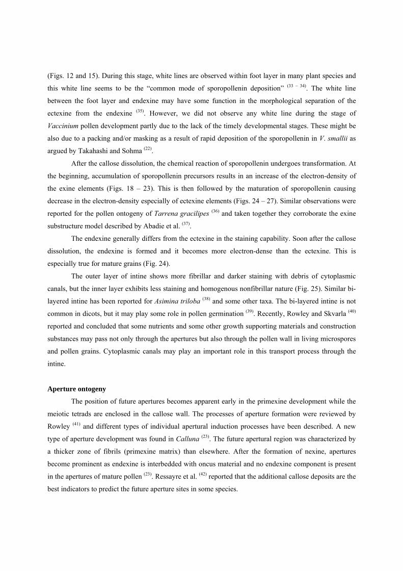
Fig. 14. Adjoining microspores of the meiotic tetrad connected by common primexine matrix (*). Dark
lipoidal granules (arrows) observed on inner surface of tapetum (T). Callose (C) and pro-
columellae, i.e., probacula (PB). Bar = 0.5 µm.
Fig. 15. The primexine matrix (*) connection between adjoining microspores. Vesicles (arrow) fusing
undulating plasmalemma. Developing exine is composed of primexine matrix (*), probacula (PB),
and faint protectum (PT). Callose (C). Bar = 0.5 µm.
Fig. 16. Possible future aperture region (arrow) destined by very thin or almost lack of primexine matrix.
Lipoidal electron-dense granules on the surface of the tapetal cells (T). Bar = 1 µm.
Fig. 17. The regions of thick primexine matrix (arrows), probably initial parts of future apertural regions.
Bar = 1 µm.
Fig. 18. A microspore tetrad after the dissolution of callose wall showing thick intine (I) near ora. Each
microspore contains a nucleus (N) with a nucleolus. Bar = 10 µm.
Fig. 19. A colpus region in the same stage of Fig. 18, characterized by the lack of ectexine (T: tectum, Co:
columellae, F: foot layer), thick endexine (En) (more electron-dense) and thick intine (I), Starch
grains (S). Bar = 1 µm.
Fig. 20. Septal pollen wall in the same stage of Fig. 18. Adjoining tectum is connected (*). A transparent
space (arrow) is observed at the central part of the common exine. Starch grains (S). Bar = 1 µm.
Fig. 21. The vegetative (VN) and generative cell nuclei (GN) separated after microspore mitosis, but cell
plate formation not complete yet. Each nucleus has a large nucleolus. Pollenkitt-like substance
(arrow) deposited on the outer surface of tectum. Starch grains (S). Bar = 5 µm.
Fig. 22. Generative cell (GC) formed at the distal region of the pollen grain. Partition wall connected with
intine (arrow). Starch grain (S) included in generative cell as well as in vegetative cell (VC). Bar
= 1 µm.
Fig. 23. Edge of aperture (colpus) region in the same stage of Fig. 22, characterized by very thin ectexine
(T: tectum, Co: columellae, and F: foot layer), thick depressed endexine (En), and thick intine (I).
Bar = 1 µm.
Fig. 24. A mature pollen tetrad showing many starch grains (S) and dark lipoidal globules (L) in the
cytoplasm. Electron density of the ectexine decreased. Aperture (os) regions (arrows)
characterized by the lack of ectexine, very thin endexine, and thick intine. Bar = 10 µm.
Fig. 25. A cross-section of exine at the distal area of mature pollen grains. Apocolpial pollen wall
composed of thick tectum (T), thin columellae (Co), thick foot layer (F), thin endexine (En), and
intine (I). Many starch grains (S) and lipoidal globules (L) in the cytoplasm. Bar = 1 µm.
Fig. 26. Tectum (arrows) becomes more fragmental in septal pollen wall between the adjoining three
grains of the mature pollen tetrad. Bar = 1 µm.

Fig. 27. Aperture (colpus) region of the mature pollen grain of the tetrad showing the lack of ectexine (T:
tectum, Co: columellae, and F: foot layer), thick endexine (En), and thick intine (I). Bar = 1 µm.
Fig. 28. A mature pollen tetrad showing an apocolpial region with three cloporate apertures.
Fig. 29. A mature pollen tetrad showing the adjoining mesocolpial regions of three pollen grains. Three
colporate apertures found.


