DNA based molecular motors - Uni Ulm
17
Physics of Life Reviews 6 (2009) 250–266 www.elsevier.com/locate/plrev Review DNA based molecular motors Jens Michaelis a,b,∗ , Adam Muschielok a , Joanna Andrecka a , Wolfgang Kügel a , Jeffrey R. Moffitt a a Department of Chemistry and Biochemistry and Center for Integrated Protein Science Munich (CIPS M ), Ludwig-Maximilians-Universität München, Butenandtstr 11, 81377 München, Germany b Center for Nanoscience (CeNS) and Center for Integrated Protein Science Munich (CIPS M ), Germany Received 23 September 2009; received in revised form 23 September 2009; accepted 23 September 2009 Available online 3 October 2009 Communicated by L. Perlovsky Abstract Most of the essential cellular processes such as polymerisation reactions, gene expression and regulation are governed by me- chanical processes. Controlled mechanical investigations of these processes are therefore required in order to take our understanding of molecular biology to the next level. Single-molecule manipulation and force spectroscopy have over the last 15 years been de- veloped into extremely powerful techniques. Applying these techniques to the investigation of proteins and DNA molecules has led to a mechanistic understanding of protein function on the level of single molecules. As examples for DNA based molecular machines we will describe single-molecule experiments on RNA polymerases as well as on the packaging of DNA into a viral capsid—a process that is driven by one of the most powerful molecular motors. © 2009 Elsevier B.V. All rights reserved. PACS: 82.35.Lr; 82.37.Np; 82.37.Rs; 82.39.Pj; 87.15.H Keywords: Single-molecule; Optical tweezers; RNA polymerase; Molecular motor; DNA packaging; DNA overstretching; Pol II; Backteriophage Contents 1. Introduction ........................................................................ 251 2. Single-molecule force spectroscopy ........................................................ 252 2.1. Force spectroscopy with an AFM cantilever .............................................. 252 2.2. Force spectroscopy using magnetic tweezers .............................................. 253 2.3. Force spectroscopy with optical tweezers ................................................ 253 3. Mechanics of DNA and RNA molecules ..................................................... 256 4. Viral DNA packaging .................................................................. 258 5. RNA polymerase ..................................................................... 260 * Corresponding author at: Department of Chemistry and Biochemistry and Center for Integrated Protein Science Munich (CIPS M), Ludwig- Maximilians-Universität München, Butenandtstr 11, 81377 München, Germany. E-mail address: [email protected] (J. Michaelis). 1571-0645/$ – see front matter © 2009 Elsevier B.V. All rights reserved. doi:10.1016/j.plrev.2009.09.001
Transcript of DNA based molecular motors - Uni Ulm

Physics of Life Reviews 6 (2009) 250–266
www.elsevier.com/locate/plrev
Review
DNA based molecular motors
Jens Michaelis a,b,∗, Adam Muschielok a, Joanna Andrecka a, Wolfgang Kügel a,Jeffrey R. Moffitt a
a Department of Chemistry and Biochemistry and Center for Integrated Protein Science Munich (CIPSM),Ludwig-Maximilians-Universität München, Butenandtstr 11, 81377 München, Germany
b Center for Nanoscience (CeNS) and Center for Integrated Protein Science Munich (CIPSM), Germany
Received 23 September 2009; received in revised form 23 September 2009; accepted 23 September 2009
Available online 3 October 2009
Communicated by L. Perlovsky
Abstract
Most of the essential cellular processes such as polymerisation reactions, gene expression and regulation are governed by me-chanical processes. Controlled mechanical investigations of these processes are therefore required in order to take our understandingof molecular biology to the next level. Single-molecule manipulation and force spectroscopy have over the last 15 years been de-veloped into extremely powerful techniques. Applying these techniques to the investigation of proteins and DNA molecules hasled to a mechanistic understanding of protein function on the level of single molecules. As examples for DNA based molecularmachines we will describe single-molecule experiments on RNA polymerases as well as on the packaging of DNA into a viralcapsid—a process that is driven by one of the most powerful molecular motors.© 2009 Elsevier B.V. All rights reserved.
PACS: 82.35.Lr; 82.37.Np; 82.37.Rs; 82.39.Pj; 87.15.H
Keywords: Single-molecule; Optical tweezers; RNA polymerase; Molecular motor; DNA packaging; DNA overstretching; Pol II; Backteriophage
Contents
1. Introduction . . . . . . . . . . . . . . . . . . . . . . . . . . . . . . . . . . . . . . . . . . . . . . . . . . . . . . . . . . . . . . . . . . . . . . . . 2512. Single-molecule force spectroscopy . . . . . . . . . . . . . . . . . . . . . . . . . . . . . . . . . . . . . . . . . . . . . . . . . . . . . . . . 252
2.1. Force spectroscopy with an AFM cantilever . . . . . . . . . . . . . . . . . . . . . . . . . . . . . . . . . . . . . . . . . . . . . . 2522.2. Force spectroscopy using magnetic tweezers . . . . . . . . . . . . . . . . . . . . . . . . . . . . . . . . . . . . . . . . . . . . . . 2532.3. Force spectroscopy with optical tweezers . . . . . . . . . . . . . . . . . . . . . . . . . . . . . . . . . . . . . . . . . . . . . . . . 253
3. Mechanics of DNA and RNA molecules . . . . . . . . . . . . . . . . . . . . . . . . . . . . . . . . . . . . . . . . . . . . . . . . . . . . . 2564. Viral DNA packaging . . . . . . . . . . . . . . . . . . . . . . . . . . . . . . . . . . . . . . . . . . . . . . . . . . . . . . . . . . . . . . . . . . 2585. RNA polymerase . . . . . . . . . . . . . . . . . . . . . . . . . . . . . . . . . . . . . . . . . . . . . . . . . . . . . . . . . . . . . . . . . . . . . 260
* Corresponding author at: Department of Chemistry and Biochemistry and Center for Integrated Protein Science Munich (CIPS M), Ludwig-Maximilians-Universität München, Butenandtstr 11, 81377 München, Germany.
E-mail address: [email protected] (J. Michaelis).
1571-0645/$ – see front matter © 2009 Elsevier B.V. All rights reserved.doi:10.1016/j.plrev.2009.09.001

J. Michaelis et al. / Physics of Life Reviews 6 (2009) 250–266 251
6. Conclusions and outlook . . . . . . . . . . . . . . . . . . . . . . . . . . . . . . . . . . . . . . . . . . . . . . . . . . . . . . . . . . . . . . . . 263Acknowledgements . . . . . . . . . . . . . . . . . . . . . . . . . . . . . . . . . . . . . . . . . . . . . . . . . . . . . . . . . . . . . . . . . . . . . . . . 264References . . . . . . . . . . . . . . . . . . . . . . . . . . . . . . . . . . . . . . . . . . . . . . . . . . . . . . . . . . . . . . . . . . . . . . . . . . . . . . 264
1. Introduction
DNA molecules are the carrier of all genetic information and have evolved both, as extremely stable entities inorder to preserve this genetic code, as well as in ways that allow for the controlled access of this information. Thus inaddition to understanding the biochemical information of a DNA molecule, it is also important to study its mechanicalbehaviour and characteristics, since it is these characteristics that control many of the biological processes that takeplace during gene expression.
To this end, mechanical studies of single biomolecules have in recent years revolutionised our understanding ofbiological complexes [1–6]. The most prominent methods for single-molecule force spectroscopy are the atomic forcemicroscope (AFM), magnetic tweezers and optical tweezers [7]. In single-molecule force spectroscopy AFMs havemainly been used for experiments which require high forces, i.e. to rupture bonds or unfold proteins [8]. Magnetictweezers allow for the study of proteins that twist DNA and proteins that are very sensitive to forces [9]. Opticaltweezers are probably the most widely used tool for studying the mechanics of molecular motors [10]. It has beenshown that forces as low as ∼10 fN [11] and distance changes as low as 0.1 nm [12–14] can be observed using opticaltweezers. Moreover, with the use of clever experimental geometries [15] or strong lasers, it is possible to exert forcesexceeding 100 pN. These forces and distances are in the range reported for molecular motors [16,17].
One of the most important applications of single-molecule force spectroscopy is to unravel the mechanics of DNAmolecules [18,19]. For any DNA the measured force-extension behaviour can be well described (up to forces of about50 pN) using a simplistic coarse grained polymer model. Moreover, at high forces or in situations when the DNAhelix is overwound (i.e. the number of bases per complete turn of the DNA helix is reduced) or underwound, theconformation of the DNA changes completely from the canonical B-DNA structure to a different, stable form in aspontaneous and cooperative manner which resembles a phase transition.
Understanding the mechanical behaviour of single DNA molecules becomes biologically important in the contextof DNA–protein interactions. Proteins that bind DNA or translocate along DNA oftentimes exert forces on the DNAsubstrate due to bending or unwinding [20,21]. Typical forces that are applied are in the range of 1–100 pN andtypical displacements that need to be observed can be as small as a single basepair (bp) which is only 0.34 nm.Physically one can understand these force- and distance-scales since the energies important for understanding theunderlying phenomena lie between the thermal energy kBT which in units of force times distance equals to 4.1 pN nmand the energy available through the chemical reaction. In the case of ATP hydrolysis at physiological conditions thefree energy liberated by the hydrolysis of a single ATP molecule by a single protein amounts to ∼100 pN nm forthe hydrolysis. The time-scales that are important to monitor such processes reach from the millisecond to secondregime. Single-molecule manipulation with optical (or magnetic) tweezers allows for investigations at these force-,distance-, and time-scales. Hence, optical tweezers are an ideal tool for the mechanistic investigations of these proteinnucleic acid interactions at the single-molecule level. The high sensitivity of the single-molecule experiments allowsfor a detailed analysis of this enzymatic behaviour and opens up the possibility to unravel the underlying molecularmechanisms. A huge variety of such DNA–protein interactions have been studied using single-molecule manipulationtechniques and the results are summarised in recent reviews [9,22]. Of particular interest are proteins that use thefree energy liberated by a chemical reaction (most often the hydrolysis of ATP) to either translocate along DNA [22],unwind DNA [23], change the topology of DNA [24] or catalyse a polymerisation reaction [3,25], thus constitutingmolecular motors.
In the literature one can find many excellent reviews on optical trapping and single-molecule force spectroscopy[10,26–29] and the purpose of this article is not to give another complete overview of the field. Instead, we focus onsome key aspects of current research and discuss experimental procedures as well as current challenges (Section 2).In particular we describe in detail the design of an optical tweezers apparatus that allows studying the mechanicalproperties of DNA molecules, such as the well-known force-extension behaviour and the overstretching transition(Section 3). This is a necessary pre-requisite for investigating the molecular mechanism of molecular motors that
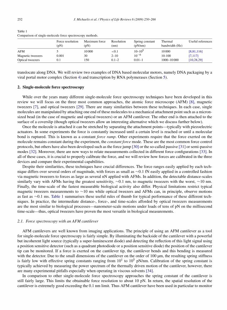
252 J. Michaelis et al. / Physics of Life Reviews 6 (2009) 250–266
Table 1Comparison of single-molecule force spectroscopy methods.
Force resolution(pN)
Maximum force(pN)
Resolution(nm)
Spring constant(pN/nm)
Thermalbandwidth (Hz)
Useful references
AFM 5 10 000 <0.1 10–105 10 000 [8,81,116]Magnetic tweezers 0.001 30 2–10 10−6 10-100 [7,117]Optical tweezers 0.1 150 0.1–2 0.01–1 1000–10 000 [10,28,29]
translocate along DNA. We will review two examples of DNA based molecular motors, namely DNA packaging by aviral portal motor complex (Section 4) and transcription by RNA polymerases (Section 5).
2. Single-molecule force spectroscopy
While over the years many different single-molecule force spectroscopy techniques have been developed in thisreview we will focus on the three most common approaches, the atomic force microscope (AFM) [8], magnetictweezers [7], and optical tweezers [29]. There are many similarities between these techniques. In each case, singlemolecules are manipulated by attaching one end of these molecules to a mechanical attachment point such as a micron-sized bead (in the case of magnetic and optical tweezers) or an AFM cantilever. The other end is then attached to thesurface of a coverslip (though optical tweezers allow an interesting alternative which we discuss further below).
Once the molecule is attached it can be stretched by separating the attachment points—typically with piezoelectricactuators. In some experiments the force is constantly increased until a certain level is reached or until a molecularbond is ruptured. This is known as a constant force ramp. Other experiments require that the force exerted on themolecule remains constant during the experiment, the constant force mode. These are the most common force controlprotocols, but others have also been developed such as the force jump [30] or the so-called passive [31] or semi-passivemodes [32]. Moreover, there are now ways to relate measurements collected in different force configurations [33]. Inall of these cases, it is crucial to properly calibrate the force, and we will review how forces are calibrated in the threedevices and compare their experimental capabilities.
Despite their similarities, these techniques have crucial differences. The force ranges easily applied by each tech-nique differs over several orders of magnitude, with forces as small as ∼0.1 fN easily applied in a controlled fashionvia magnetic tweezers to forces as large as several nN applied with AFMs. In addition, the detectable distance-scalessimilarly vary with AFMs having the greatest sensitivity, ∼0.1 nm, to magnetic tweezers with the worst, ∼10 nm.Finally, the time-scale of the fastest measurable biological activity also differ. Physical limitations restrict typicalmagnetic tweezers measurements to ∼10 ms while optical tweezers and AFMs can, in principle, observe motionsas fast as ∼0.1 ms. Table 1 summarises these useful rules of thumb for typical performance of these different tech-niques. In practice, the intermediate distance-, force-, and time-scales afforded by optical tweezers measurementsare the most similar to biological processes—nanometer-scale motions under loads of tens of pN on the millisecondtime-scale—thus, optical tweezers have proven the most versatile in biological measurements.
2.1. Force spectroscopy with an AFM cantilever
AFM cantilevers are well known from imaging applications. The principle of using an AFM cantilever as a toolfor single-molecule force spectroscopy is fairly simple. By illuminating the backside of the cantilever with a powerfulbut incoherent light source (typically a super-luminescent diode) and detecting the reflection of this light signal usinga position sensitive detector (such as a quadrant photodiode or a position sensitive diode) the position of the cantilevertip can be monitored. If a force is exerted on the cantilever tip, the cantilever bends and this bending is measuredwith the detector. Due to the small dimensions of the cantilever on the order of 100 µm, the resulting spring stiffnessis fairly low with effective spring constants ranging from 101 to 105 pN/nm. Calibration of the spring constant istypically achieved by measuring the power spectrum of the thermally driven motion of the cantilever, however, thereare many experimental pitfalls especially when operating in viscous solvents [34].
In comparison to other single-molecule force spectroscopy approaches the spring constant of the cantilever isstill fairly large. This limits the obtainable force resolution to about 10 pN. In return, the spatial resolution of thecantilever is extremely good exceeding the 0.1 nm limit. Thus AFM cantilever have been used in particular to monitor
J. Michaelis et al. / Physics of Life Reviews 6 (2009) 250–266 253
the rupture of bonds such as those between receptor and antibody [35], interactions between molecules and surfaces[36], unfolding of proteins due to the rupture of hydrogen bonds, salt bridges, etc. [37] and even to rupture covalentbonds [38–40]. By experimentally varying the force loading rate, a technique commonly referred to as dynamic forcespectroscopy, it is possible to unravel details of the underlying energy landscape such as the height of the energybarrier and the distance to the transition state [41–43]. Besides the rupture of bonds it is also possible to simply stretchmolecules in a repetitive fashion thus unravelling the force-extension behaviour e.g. of DNA [44] or RNA molecules,polypeptide chains [45], spectrin [46] and fibrin [47] fibers and membrane proteins [4] among many others.
In addition it has been shown that force-feedback circuits can be constructed for AFMs in order to hold moleculesat a constant force [48]. However, due to the limited force resolution of the AFM such a feedback is only possible atelevated forces of ∼10–20 pN and above, where most molecular motors have already stalled.
2.2. Force spectroscopy using magnetic tweezers
The experimental design of a magnetic tweezer is fairly simple. A super-paramagnetic bead is drawn into thehighest gradient of a magnetic field created by typically two permanent magnets. The motion of the bead is imagedonto a CCD camera via brightfield microscopy. The interference pattern that develops between light reflected from thenearby surface and from the bead can be exploited to sensitively measure the distance of the bead from the surface.By changing the position of the magnets with respect to the bead, the applied force can be changed. In parallel, themagnets can also be rotated, allowing for the application of torque to the molecule under study. Due to the relativelylow bandwidth and spatial resolution of the detection techniques used in typical magnetic tweezers, the distanceresolution is much lower than in optical tweezers or AFM. However, this limitation is an instrumentation issue only.It should be possible to use laser detection methods, as is done with optical tweezers, and obtain the same high spatialresolution, albeit with a different thermal bandwidth (Table 1).
There are three important features of magnetic tweezers which make them the method of choice for a variety ofdifferent applications. First, the very low spring constant of a magnetic tweezers of typically 10−6 pN/nm gives theopportunity to easily perform measurements at constant force, since movements in the range of 1 µm only resultin changes of forces on the order of 0.001 pN. Second, by rotating the magnets in a magnetic tweezers and usingattachments of the biomolecules which consist of many linkages, torsion can be exerted in a controlled manner. Ofcourse this requires the molecule to be torsionally constrained—i.e. it must have multiple attachment points so that itis not free to swivel around one of them, releasing the torque. The ability to apply torque is of particular importancefor experiments aimed at investigating proteins that alter the topology of the DNA by introducing supercoils [19,49].Third, since measurements merely amount to tracking of the interference patterns of the magnetic beads, it is possibleto monitor multiple experiments simultaneously in a single field of view, and thus allowing for a highly parallel dataacquisition. This is extremely important for measurements where the activity of the particular enzyme is rather low,which is the case for certain RNA polymerases [50], or when force measurements are combined with single-moleculefluorescence measurements [51] where the amount of useful collected data is reduced since two different single-molecule techniques are performed in parallel. Magnetic tweezers have been employed to characterise the mechanicalproperties of DNA and RNA molecules [19] as well as for the investigation of topoisomerases [52], DNA translocases[53], helicases [54], nucleosome remodelling complexes [55], viral packaging motors [51] and RNA polymerases [56].
2.3. Force spectroscopy with optical tweezers
Optical tweezers are by far the most versatile for investigating the behaviour of DNA based molecular motors.While the distance resolution of an optical tweezers can be as good as 0.1 nm, it is not quite up to par with that of anAFM. Despite the differences one can detect sub-piconewton forces and sub-nanometer motions, permitting detailedexperiments on enzymes where step-sizes down to a single bp are to be expected and forces need to be controlled onthe pN level. In fact (almost) all the DNA based molecular motor experiments described in this review were obtainedusing optical tweezers and while excellent technical reviews about optical tweezers exist [29,57,58], we consider itimportant to familiarise the reader with the technique, experimental procedures and challenges.
In optical tweezers a three-dimensional gradient in light intensity is used to trap small dielectric objects nearthe focus. For objects larger than the wavelength of light this effect can easily be understood using wave opticsand diffraction. Diffraction causes a change in momentum of the light transmitted through the dielectric object and

254 J. Michaelis et al. / Physics of Life Reviews 6 (2009) 250–266
Fig. 1. Schematics of dual-beam optical tweezers. (A). Two counter-propagating diode lasers are used to form a dual-beam optical trap. Theadvantage of such a setup is the high stiffness, due to efficient trapping in the axial direction, and the possibility to determine the amount oftransferred light momentum directly [15]. Abbreviations: BPA: beam profile adjuster, M: mirror, PBS: polarising beam-splitter, λ/2: half-waveplate, λ/4: quarter-wave plates, PSD: position sensitive detector, 3D-Stage: three-dimensional stepper motor driven position control, 2D-Piezo:two-dimensional linearised piezoelectric stage, LED: light emitting diode, LR : relay lens. (B) The force spectroscopy experiments are performedwithin a micro-flow chamber consisting of three channels that are connected by dispenser tubes downstream from the experimental region. Theupper and lower channels are used for the fast delivery of modified beads, while the central channel is used for tether assembly as well as for theactual experiments. A micro-pipette is glued to the chamber and acts as a mechanical anchor-point for microscopic beads which are attached to itstip via suction.
this change in momentum causes a restoring force to the object if not all intensities are balanced. The conceptuallyeasiest way to measure the force applied by optical tweezers is thus to measure the changes in light momentum [15].Practically this is often not possible since a high numerical aperture is required in order to form a stable trap in allthree dimensions and thus some of the trapping light may be scattered to higher numerical aperture after diffraction,where it typically cannot be collected by the optical system of the microscope. Thus the total amount of transferredlight momentum cannot be determined. This problem can be overcome by building a trap which consists of twocounter-propagating lasers, similar to arrangements that are used in magneto optical traps (Fig. 1(A)). This so-calleddual-beam optical tweezers apparatus has two characteristics that make it particularly useful for investigating DNAmechanics and DNA based molecular motors. First, the use of two counter-propagating beams leads to a very strongoptical trap, capable of exerting forces well beyond the over-stretching regime. Secondly, it allows for the in-situdetermination of the forces acting on a bead caught in the optical trap by direct measurement of the transferred lightmomentum [15] with a pN force resolution, far beyond what is possible with an AFM [44].
The system used for the experiments discussed here is similar to the one described by Smith et al. [15] and aschematic of the setup is shown in Fig. 1(A). The light of two diode lasers is each passed through a beam profile ad-juster, comprised of astigmatism correction prism assemblies and a spatial filter, to produce circular Gaussian beams,which are then directed into the centre of a flow chamber. The light is focussed and collected with high NA objectivesand detected on a pair of position sensitive detectors that monitor the image of the objective’s back focal plane. Theposition of both, the right microscope objective as well as the sample chamber are controlled by two flexure stagesand fine positioning of the chamber is achieved by linearised piezo-translators which can also be used to apply force-feedback. The two objective lenses are under-filled by the two counter-propagating laser beams. If the laser light isscattered by the object held at the centre of the trap, the high numerical aperture of the objective lens is used to collectall the scattered light (this holds for small deflections of the bead from the trap centre which typically correspondto forces up to ∼100 pN), thus allowing for the direct measurement of transferred light momentum. Therefore, onlycalibration of the instrument’s detection efficiency is required.

J. Michaelis et al. / Physics of Life Reviews 6 (2009) 250–266 255
Fig. 2. Power-spectrum calibration of optical tweezers. The graph shows the averaged power-spectrum of the deflection signal recorded duringtrapping of a ∼1.9 µm diameter bead. The flow chamber was oscillated with a frequency of 25 Hz and an amplitude of ∼89 nm, resulting in asharp peak in the power-spectrum. By fitting the amplitude of the peak in addition to the power-spectrum of the trap one can perform a directcalibration of the setup, which does not require a previous knowledge about the size of the trapped bead [59]. The fit determined a corner frequencyof 1565 Hz, a spring constant of 0.18 pN/nm and from the diffusion constant D, a bead diameter of 1.9 µm can be determined using the Einsteinrelation (D = kBT/γ , where γ is the drag coefficient of the bead), assuming a spherical bead size and a viscosity of the medium of 1 mPa s.
It is however, not necessary to measure directly the total amount of transferred light momentum since the detectorsignal is approximately proportional to the deflection, and by assumption of a harmonic trapping potential, also pro-portional to the force. The proportionality constants can be determined by calibration against known deflections andforces. In most applications of optical tweezers the thermal force is used. It causes the bead’s deflection from the trapcentre to fluctuate so that the motion of the bead is characterised by its drag coefficient and the stiffness of the trap.Unfortunately, both parameters are unknown and have to be determined experimentally, in addition to the constantthat converts detector signal to deflection. Since three unknowns must be estimated in total proper calibration needsat least three known parameters.
The best way to calibrate the instrument is to record the thermal fluctuations of the bead and to simultaneouslyoscillate the micro-fluidic chamber using a piezo-electric stage. The stage is calibrated by capacitive sensors and runin a closed loop mode. The readout of the capacitive sensors therefore determines the amplitude of the oscillation.The power spectrum of the measured signal consists of two parts, a broad background caused by thermal fluctuations,and a sharp peak caused by the forced oscillations imposed on the bead by the motion of the micro-fluidic chamber(Fig. 2). By comparing the amplitude of that peak to the oscillation amplitude and fitting the corner frequency of thepower-spectrum, one can determine the spring constant of the trap, the drag coefficient of the trapped object as wellas the proportionality constant between the deflection of the bead from the trap center (measured in meters) and thedetector readings (measured in arbitrary units) [59]. If one compares the forces determined by the oscillation/power-spectrum method to the measurement of momentum transfer one finds a good agreement within the experimentaluncertainties of ∼5%. It should be noted that both methods can be used even if no a priori knowledge of the size ofthe trapped object is available, making them insensitive to variations in bead size.
The described methodology of measuring forces between a mechanical element in this case a micro-pipette tipand an optical trap has severe drawbacks. Mechanical elements are suspect to mechanical noise such as vibrationsor thermal expansions. On the other hand precise position of the laser focus is altered by air density fluctuations thatare constantly occurring. Techniques have been developed to overcome such problems. For example using detectionlasers one can control the position of mechanical elements with 0.1 nm accuracy [60] and the position of the laser spotcan be kept constant by enclosing the apparatus in helium atmosphere [13] or coupling the laser through single modeoptical fibres and controlling the intensity that enters the microscope [61].
256 J. Michaelis et al. / Physics of Life Reviews 6 (2009) 250–266
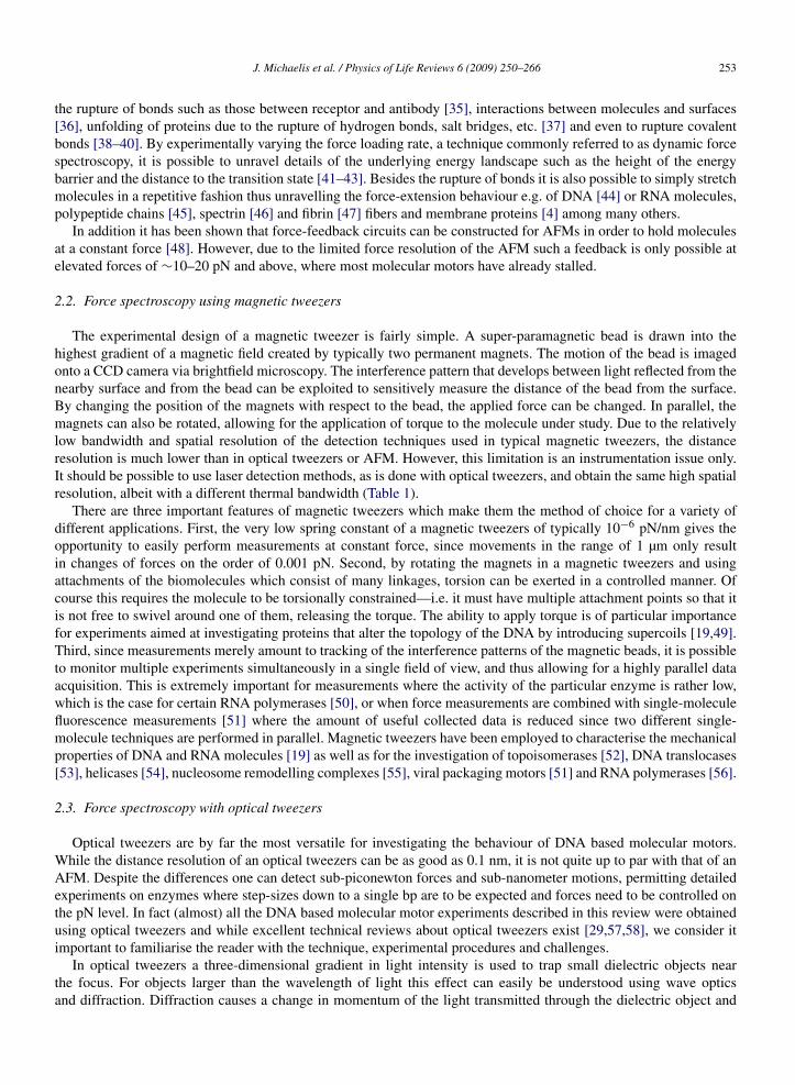
Fig. 3. Investigation of molecular motors in dumb-bell optical tweezers. As an example for an experiment in a dumb-bell optical tweezers a cartoonof a transcription experiment is shown. The polymerase is attached to a bead held in an optical trap. The end of the DNA tether is attached toa second bead held in a second optical trap. By manipulating the position of (at least) one of the optical traps the amount of load on the DNAmolecule and thus on the polymerase can be adjusted. Different attachment chemistries e.g. DIG/anti-DIG on one side and biotin/streptavidin (SA)on the other are used to ensure specificity. In order to achieve the highest resolution the light for the two optical traps is coming from the same laser.The laser light is simply split into two orthogonal polarisations which can be read out separately allowing for precise differential back focal planedetection [63].
A more elegant and simple solution to these problems is to design the optical tweezers such that measurementsare insensitive to these issues. The “dumb-bell” trap is such a design [13,62] (Fig. 3). By holding both ends of thebiological system with two optical traps it is possible to make much of the instrument common between the twotraps. Orthogonal polarisations can be used to form these traps and therefore the same laser and almost all of theoptical components and optical path can be shared. The advantage is that any noise introduced by these componentsor by air fluctuations along the optical paths will be common to the two optical traps, producing identical fluctuationsin both traps. Since the measurement is only sensitive to the distance between these traps, this extraneous noise iseffectively decoupled from the measurement. Further improvements can be made by correlating the motion of eachbead, allowing the separation of additional fluctuations [14,63]. Such a differential detection technique offers base-pair-scale resolution on the tens of milliseconds time-scale [32].
3. Mechanics of DNA and RNA molecules
Using single-molecule force spectroscopy one can manipulate single DNA molecules by tethering a fragment ofDNA between two beads. For this purpose the two ends of the DNA have to be marked with different tags, e.g. oneend can be biotinylated and the other labelled with digoxigenin (DIG), in order to bind to streptavidin and anti-DIGcoated beads, respectively. Simple molecular biology techniques can be used to cut any plasmid or viral DNA, andintroduce labelled nucleotides using a DNA polymerase [51] or labelled DNA oligomers in a ligation reaction. Due toits long length many experiments have been performed with the DNA from the bacteriophage λ [64].
Single-molecule force spectroscopy measurements in optical tweezers have to be performed in a serial fashionone molecule at a time, but information has to be extracted from histograms of many experiments. Therefore it isextremely important for the practicality of such experiments to employ micro-fluidic systems in order to allow for afast turnover between experiments. A simple design was developed by Bustamante and co-workers [65] (Fig. 1B).With such a design one can assemble DNA tethers between two immobilised beads in situ. For this procedure a bead(coated with antibodies) is loaded into the lower flow channel, trapped by the optical tweezers and transferred to theend of a micro-pipette tip. The micro-pipette is glued into the flow chamber and beads are immobilised to its endusing suction, thus providing a rigid mechanical attachment. Then, a second bead to which the DNA molecule isattached is caught by the optical tweezers near the outlet of the upper flow channel and brought into close proximityto the first bead. The strong affinity between antibodies attached to the bead and modified bases at the end of theDNA leads to the formation of a DNA tether, thus mechanically connecting the two beads. After tether formation,the molecule is stretched by moving the micro-pipette with respect to the position of the optical trap and the force-extension behaviour can be monitored (Fig. 4). For illustrative purposes a movie of this procedure can be found atwww.cup.uni-muenchen.de/pc/michaelis/movies.

J. Michaelis et al. / Physics of Life Reviews 6 (2009) 250–266 257
Fig. 4. Single-molecule overstretching of a ∼24 kbp fraction of lambda DNA. The DNA segment was tethered between a streptavidin coated bead(diameter ∼2.0 µm) and a bead labelled with anti-DIG (diameter ∼2.8 µm). The actual data recorded at 1 kHz are shown in black while the fit tothe extended WLC (force range of 10–50 pN) yielding a persistence length LP = 41 ± 5 nm, a contour length LC = 8638 ± 200 nm and an elasticcorrection factor K = 1143 ± 200 pN is shown in red. In order to use the extracted parameters for the analysis of single-molecule transcriptionexperiments, experiments were performed in the same buffer (50 mM HEPES pH 7.5, 40 mM (NH4)2SO4, 10 µM ZnCl2, 5% glycerol, 10 mMDTT, with 5 mM MgCl2). (For interpretation of the references to colour in this figure legend, the reader is referred to the web version of thisarticle.)
When stretching single DNA molecules using single-molecule force spectroscopy one can measure forces alreadyfor relatively low extensions (i.e. end-to-end distances) of the DNA molecule due to the conformational flexibility ofthe DNA [66]. In this regime the molecule behaves like an entropic spring [64]. Upon extension of the molecule themaximum number of its conformational states is reduced. Therefore, in order to extend the molecule, one has to putenergy into the system, thus resulting in a measurable force. It has been shown that the shape of the force-extensioncurve of the DNA molecule can be described accurately with the wormlike chain (WLC) model [64], in which theDNA is treated as a smoothly bendable rod. In addition, for higher forces above ∼5 pN one starts to observe enthalpiceffects caused by small mechanical deformations of the B-DNA structure [67]. However, at least for large DNAmolecules of several thousand bases in length, the force-extension behaviour of DNA molecules can be describedaccurately by introducing a Hookian term [68] resulting in the so-called extensible worm like chain model [68–71]:
(1)x = LC
(1 − 1
2
(kBT
FLP
)1/2
+ F
K
),
yielding the contour length LC , the persistence length LP and the enthalpic correction factor K for the molecule [18,26]. Note, that Eq. (1) is an analytical approximation for the true extensible WLC model [72]. Similar experimentshave also been done with double stranded RNA molecules [73]. Knowing the characteristic mechanical parametersfor describing the force-extension behaviour is a necessary pre-requisite for performing the experiments on molecularmotors such as the bacteriophage portal motor protein and the RNA polymerase which will be described in the nextsection.
Above 50 pN the force-extension curve cannot be described well with the extensible WLC model. In particular atforces of ∼65 pN one observes a force-plateau. Here, the molecule extends while the force remains (roughly) constant.This so-called over-stretching transition has been described thoroughly both experimentally and theoretically [69,74].Single-molecule force spectroscopy allows for the detailed analysis of the extension of the DNA during this over-stretching transition, yielding a characteristic length of the over-stretched DNA of about 170% of the contour lengthof B-DNA [69], as well as a characteristic helical repeat of ∼37 bp/turn (compared to 10.5 bp/turn for B-DNA) [49].These physical parameters provide constraints that must be upheld by a structural analysis of the overstretched state[70,75–77]. Moreover, experiments have successfully analysed ionic effects [65], sequence effects [44] as well as pHeffects [78,79] on the overstretching transition.

258 J. Michaelis et al. / Physics of Life Reviews 6 (2009) 250–266
The nature of the over-stretched state the so-called S-DNA is highly disputed. Experiments by Bloomfield and co-workers seem to indicate that over-stretching is merely a manifestation of DNA melting [78,80]. However, if S-DNAwere simply melted DNA the nature of a second plateau in the force-extension curves of double stranded DNA athigher forces, observed by AFM force spectroscopy [81] could not be explained. Moreover, recent Monte Carlosampling of the DNA over-stretching led to the conclusion that the observed experimental data can only be explainedif an S-DNA state different than simply a melted state occurs [82]. In fact, from such computations predictions havebeen made that could help to manifest the existence of S-DNA which still await experimental tests [83].
The B-S transition is not the only phase transition observed during mechanical studies of DNA molecules. Byapplying positive supercoils to the DNA in experiments where the DNA is torsionally constrained, one can destabilisethe DNA and drive it into a different conformation the so-called P-DNA [18,84]. P-DNA has a helicity of ∼2.6 bp/turn(compared to ∼10.5 bp/turn for B-DNA) and such a conformation can only be physically possible if the DNA basesare flipped to the outside. Ironically such a conformation was suggested by Linus Pauling [85] only months beforeWatson and Crick discovered the B-DNA structure [86]. In fact, combining the results of many experiments it is nowpossible to obtain a complete phase diagram of DNA where instead of pressure and temperature (which are typicallyconstant for biological systems) the important variables are force and torque [18]. At least one of these extreme formsof DNA, the left handed Z-DNA is known to exist in nature [87]. The B-Z transition occurs when DNA is under-wounda process that occurs quite often within the cell, e.g. during transcription by RNA polymerases.
4. Viral DNA packaging
One of the most interesting examples of DNA mechanics, which has triggered a large amount of theoretical mod-els, is the compaction of DNA inside of small compartments due to condensation by accessory proteins or due tothe action of a motor protein which exerts a mechanical force to push DNA into a preformed protein compartment.The latter is a central part of the life cycle of a large number of bacteriophage viruses, which package their viralgenome into a preformed protein capsid [88]. During packaging energetic penalties have to be overcome arising fromentropic, electrostatic and elastic contributions. Thus energy has to be provided in order to package DNA and thisenergy is liberated by a molecular motor which sits at the apex of the protein capsid and uses the free energy fromATP hydrolysis to power conformational changes which drive the DNA into the capsid. One of the best studied motorsystems is the portal packing motor from bacteriophage �29 [89]. The motor consists of three rings which are con-nected to each other and formed by five (or six) RNA molecules (termed pRNA), five (or six) ATPases called gp16and the so-called connector (gp10) which is a homododecamer [88]. While the structure of the homododecamer hasbeen solved using crystallographic studies [90], only low resolution cryo-EM structures exist for both the RNA andthe gp16 conformation at the apex of bacteriophage �29 proheads [91]. Therefore, only speculative models about theoverall mechanism and the structure of the complete motor assembly exist. However, recent single-molecule exper-iments using single-molecule photo-bleaching [92], single-molecule fluorescence polarisation [51] and in particularoptical tweezers [17,32,93–95] have determined many characteristics of the motor system and have thus helped torefine our picture of the molecular mechanism that underlies this powerful molecular motor. Here, we will reviewonly the findings of the optical tweezers measurements.
Packaging is investigated in the same geometry described in the previous section. Antibodies against the capsidprotein are used to immobilise the proheads onto beads, and a biotinylated DNA is used to hold the other end. Com-plexes are either assembled using pre-initiated complexes [17] or in-situ [93]. The portal motor packages the viralDNA in the pre-formed capsid with a remarkable processivity and speed. However, the velocity of the process ischanging over time (Fig. 5) an effect that has been attributed to the build up of a restoring force. The motor can pack-age DNA against external forces as high as 65 pN and simplistic models lead to the notion that during this packagingan internal pressure of ∼50 atm is developed, numbers which can also be obtained using physical models for DNAcompaction including entropic penalties, electrostatics and bending contributions [96,97]. Evidence for a pressure onthe order of tens of atmospheres also comes from ejection experiments under varying osmotic pressure [98]. Hence,the bacteriophage might act as a loaded spring and upon binding to a receptor on a target cell, the ring-like connectorcan open and the DNA is injected into the new cell. Since, the measured changes in packaging velocity with fillingfraction are quite low for the first half of the genome [93], this process can only account for a partial transfer of theDNA into the new cell. Most likely other molecular motors such as RNA polymerases take over and pull the DNAinto the new cell after the initial part of the DNA has been injected. This notion is strengthened by biochemical exper-

J. Michaelis et al. / Physics of Life Reviews 6 (2009) 250–266 259
Fig. 5. Viral DNA packaging. Packaging of DNA into a viral capsid by the portal motor complex of bacteriophage �29 was investigated using dual-beam optical tweezers. (A) A schematic of the experimental configuration is shown in the inset. The observed packaging rate varied as a functionof the percentage of genome packaged, due to a build-up of an internal opposing force (adapted from Smith et al. [89]). (B) Packaging is sloweddown by the applied external load. From experimental data obtained at various ATP concentrations one can calculate the equilibrium constant KM
and the maximum packaging velocity vmax (for saturating ATP) for different applied forces. KM and vmax both decrease with increasing force.(C) The ratio vmax/KM is independent of force. Therefore the catalytic rate depends on force and the effective binding rate does not (adapted fromChemla et al. [94]).
iments that tested the infectivity of the virus as a function of the position of a gene encoding for a RNA polymerasealong the viral genome [99].
Controlled experiments in optical tweezers also allowed to unravel the mechano-chemistry of the packaging pro-cess [94]. It could be shown that albeit several ATPases are assembled into a ring, there is no apparent cooperativityand motion stalls if one ATPase binds to a non-hydrolysable ATP analog. Moreover, the data also show that there is aload-dependence to the catalytic step of the enzyme and that the effective binding itself is force independent (Fig. 5).In addition, force dependent studies of the slippage frequency allowed for the identification of loosely bound statesof the motor thus further dissecting the mechano-chemical coupling. However, more direct insight into the molecularmechanism of this fascinating molecular machine comes from recent high-resolution measurements of the packagingprocess (Fig. 6) [32]. Using two optical traps and a differential detection scheme Moffitt et al. were able to directlyresolve individual bursts of activity of the enzyme. Most surprisingly these bursts amounted to fairly large displace-ments on the order of 10 bp. In their model multiple ATPases in the ring are in a loaded configuration (presumablybound to ATP or the product ADP) and translocation is achieved by a successive conformational change within theATPases of the ring. Support for this model comes from experiments performed at high loads, where substeps of the

260 J. Michaelis et al. / Physics of Life Reviews 6 (2009) 250–266
Fig. 6. High-resolution optical tweezers experiments reveal step size of packaging motor (adapted from Moffitt et al. [32]). (A) Schematic of theexperimental configuration used for the investigation of DNA packaging in a high-resolution dumb-bell optical tweezers setup. (B) Representativehigh-resolution packaging data obtained at a force of ∼8 pN showing distinct translocation dwells followed by packaging bursts of 10 bp in size.Data obtained at 250 (purple), 100 (brown), 50 (green), 25 (blue), 10 (black), 5 µM ATP (red) are shown. The time between dwells is increased whenthe ATP concentration is reduced. However, the packaging burst size and dwell-duration is independent of ATP concentration. (C) Representativepackaging data obtained at ∼40 pN and [ATP] = 250 µM. The data shows that at this high load the packaging bursts are broken up by severalsteps. (D) Pairwise distance analysis of the data obtained at ∼40 pN. The histogram shows distinct peaks with at 2.5 bp, 5 bp, 7.5 bp, etc. Thus, thebursts are made up of 2.5 bp steps. These steps become apparent at high loads since the high external force increases the waiting time between the2.5 bp steps. At forces below 30 pN these steps occur so fast, that they cannot be resolved given the limited time-resolution of the instrument. (Forinterpretation of the references to colour in this figure legend, the reader is referred to the web version of this article.)
10 bp bursts could be observed. Interestingly the size of these substeps was 2.5 bp indicating that either not the fullmovement of the ATPase is coupled to a DNA movement, or that the movement of the ATPase is independent ofthe repeat units of the DNA, an effect that has never been observed for an enzyme that actively translocates alongDNA. The ability to observe such unexpected step sizes illustrates the power of high-resolution optical trapping tech-niques and underscores the necessity for developments that improve the accuracy of measurements and characterisesystematic errors [32].
5. RNA polymerase
Another important molecular motor that has been investigated intensively over the last years is the (DNA de-pendent) RNA polymerase. The enzyme has a critical role in gene expression, since it is responsible for making allmRNA in the cell and mRNA production is a highly regulated process. The process of making RNA out of the templateDNA molecule is commonly called transcription and is divided into several phases known as transcription initiation,abortive transcription, promoter escape, transcription elongation and transcription termination. Here, we will describeexperiments performed using optical tweezers related to the latter two aspects. Conceptually the experimental setup

J. Michaelis et al. / Physics of Life Reviews 6 (2009) 250–266 261
Fig. 7. Single-molecule observation of Pol II transcription. The data were obtained in an opposing force mode depicted in Fig. 3. (A) Represen-tative single-molecule transcription event. The displayed data is filtered at 100 Hz (black) and 0.4 Hz (red) to reduce experimental noise. As thepolymerase moves along the DNA, the effective length of the DNA tether is reduced and thus the force is increasing. The right axis shows therecorded force (note, that the force axis is not linear) and the left axis shows the transcribed distance, calculated from the force using the extensibleWLC model. The velocity of the transcription is not homogeneous but interrupted by frequent pause. (B) A magnified section of the trace shownin (A) showing the polymerase entering and exiting from a pause. (For interpretation of the references to colour in this figure legend, the reader isreferred to the web version of this article.)
is quite similar to the one just described for the investigation of viral DNA packaging. Here, a RNA polymerase isimmobilised onto a bead held in an optical trap using antibodies [100] or using a biotinylated enzyme [101]. Mostexperiments have been performed on the bacterial RNA polymerase (RNAP) but recently experiments were also per-formed on the eukaryotic RNA polymerase II (Pol II) by the Bustamante lab [102,103] as well as ourselves [100](Fig. 7). The prokaryotic and eukaryotic enzymes, i.e. the engines of transcription, share a high sequence homologyand are structurally virtually identical; however, differences exist in the interaction of the polymerase with co-factorsduring all phases of transcription. As a general remark, the eukaryotic transcription has evolved to be extremely com-plex and many accessory proteins (referred to as transcription factors) are used in order to allow for a stringent controlof gene expression in the eukaryotic cell.
In order to observe transcription elongation of Pol II in an optical tweezer one needs to first assemble a transcriptionelongation complex. For the eukaryotic enzyme this is quite tricky, since initiation in-vivo requires many additionalproteins that are so far not available in large amounts at purities high enough to perform single-molecule experiments.However, elongation competent complexes can be assembled in-vitro without the need of protein co-factors by usingmismatched nucleic acid oligomers [104] or by simply adding first the template DNA–RNA hybrid to the polymerase,before incubation with the nontemplate strand [102,105,106]. In order to fix the DNA-molecular machine complex inbetween two beads in the optical tweezers, a linker is added to the protein complex by biotinylating a subunit of thepolymerase, namely the Rpb4/7 hetero-dimer (or Rpb3 [106]). The elongation complex needs to be ligated to a fairlylong DNA molecule which is then used as a tether between the two beads. In analogy to the stretching experimentsof bare DNA (see above), inside the chamber a Pol II-DNA tether is formed between two beads. During transcriptionthe polymerase decreases the length of the DNA tether (while increasing the length of the RNA transcript), resulting
262 J. Michaelis et al. / Physics of Life Reviews 6 (2009) 250–266
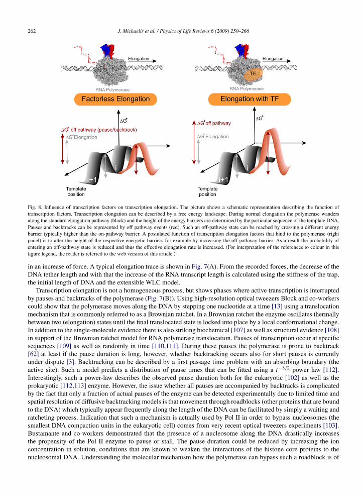
Fig. 8. Influence of transcription factors on transcription elongation. The picture shows a schematic representation describing the function oftranscription factors. Transcription elongation can be described by a free energy landscape. During normal elongation the polymerase wandersalong the standard elongation pathway (black) and the height of the energy barriers are determined by the particular sequence of the template DNA.Pauses and backtracks can be represented by off pathway events (red). Such an off-pathway state can be reached by crossing a different energybarrier typically higher than the on-pathway barrier. A postulated function of transcription elongation factors that bind to the polymerase (rightpanel) is to alter the height of the respective energetic barriers for example by increasing the off-pathway barrier. As a result the probability ofentering an off-pathway state is reduced and thus the effective elongation rate is increased. (For interpretation of the references to colour in thisfigure legend, the reader is referred to the web version of this article.)
in an increase of force. A typical elongation trace is shown in Fig. 7(A). From the recorded forces, the decrease of theDNA tether length and with that the increase of the RNA transcript length is calculated using the stiffness of the trap,the initial length of DNA and the extensible WLC model.
Transcription elongation is not a homogeneous process, but shows phases where active transcription is interruptedby pauses and backtracks of the polymerase (Fig. 7(B)). Using high-resolution optical tweezers Block and co-workerscould show that the polymerase moves along the DNA by stepping one nucleotide at a time [13] using a translocationmechanism that is commonly referred to as a Brownian ratchet. In a Brownian ratchet the enzyme oscillates thermallybetween two (elongation) states until the final translocated state is locked into place by a local conformational change.In addition to the single-molecule evidence there is also striking biochemical [107] as well as structural evidence [108]in support of the Brownian ratchet model for RNA polymerase translocation. Pauses of transcription occur at specificsequences [109] as well as randomly in time [110,111]. During these pauses the polymerase is prone to backtrack[62] at least if the pause duration is long, however, whether backtracking occurs also for short pauses is currentlyunder dispute [3]. Backtracking can be described by a first passage time problem with an absorbing boundary (theactive site). Such a model predicts a distribution of pause times that can be fitted using a t−3/2 power law [112].Interestingly, such a power-law describes the observed pause duration both for the eukaryotic [102] as well as theprokaryotic [112,113] enzyme. However, the issue whether all pauses are accompanied by backtracks is complicatedby the fact that only a fraction of actual pauses of the enzyme can be detected experimentally due to limited time andspatial resolution of diffusive backtracking models is that movement through roadblocks (other proteins that are boundto the DNA) which typically appear frequently along the length of the DNA can be facilitated by simply a waiting andratcheting process. Indication that such a mechanism is actually used by Pol II in order to bypass nucleosomes (thesmallest DNA compaction units in the eukaryotic cell) comes from very recent optical tweezers experiments [103].Bustamante and co-workers demonstrated that the presence of a nucleosome along the DNA drastically increasesthe propensity of the Pol II enzyme to pause or stall. The pause duration could be reduced by increasing the ionconcentration in solution, conditions that are known to weaken the interactions of the histone core proteins to thenucleosomal DNA. Understanding the molecular mechanism how the polymerase can bypass such a roadblock is of
J. Michaelis et al. / Physics of Life Reviews 6 (2009) 250–266 263
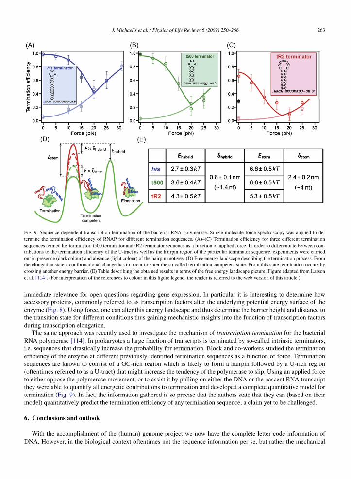
Fig. 9. Sequence dependent transcription termination of the bacterial RNA polymerase. Single-molecule force spectroscopy was applied to de-termine the termination efficiency of RNAP for different termination sequences. (A)–(C) Termination efficiency for three different terminationsequences termed his terminator, t500 terminator and tR2 terminator sequence as a function of applied force. In order to differentiate between con-tributions to the termination efficiency of the U-tract as well as the hairpin region of the particular terminator sequence, experiments were carriedout in presence (dark colour) and absence (light colour) of the hairpin motives. (D) Free energy landscape describing the termination process. Fromthe elongation state a conformational change has to occur to enter the so-called termination competent state. From this state termination occurs bycrossing another energy barrier. (E) Table describing the obtained results in terms of the free energy landscape picture. Figure adapted from Larsonet al. [114]. (For interpretation of the references to colour in this figure legend, the reader is referred to the web version of this article.)
immediate relevance for open questions regarding gene expression. In particular it is interesting to determine howaccessory proteins, commonly referred to as transcription factors alter the underlying potential energy surface of theenzyme (Fig. 8). Using force, one can alter this energy landscape and thus determine the barrier height and distance tothe transition state for different conditions thus gaining mechanistic insights into the function of transcription factorsduring transcription elongation.
The same approach was recently used to investigate the mechanism of transcription termination for the bacterialRNA polymerase [114]. In prokaryotes a large fraction of transcripts is terminated by so-called intrinsic terminators,i.e. sequences that drastically increase the probability for termination. Block and co-workers studied the terminationefficiency of the enzyme at different previously identified termination sequences as a function of force. Terminationsequences are known to consist of a GC-rich region which is likely to form a hairpin followed by a U-rich region(oftentimes referred to as a U-tract) that might increase the tendency of the polymerase to slip. Using an applied forceto either oppose the polymerase movement, or to assist it by pulling on either the DNA or the nascent RNA transcriptthey were able to quantify all energetic contributions to termination and developed a complete quantitative model fortermination (Fig. 9). In fact, the information gathered is so precise that the authors state that they can (based on theirmodel) quantitatively predict the termination efficiency of any termination sequence, a claim yet to be challenged.
6. Conclusions and outlook
With the accomplishment of the (human) genome project we now have the complete letter code information ofDNA. However, in the biological context oftentimes not the sequence information per se, but rather the mechanical

264 J. Michaelis et al. / Physics of Life Reviews 6 (2009) 250–266
nature of the DNA molecule, that is how easy it can be bend or twisted is important. In particular for proteins that bindto DNA, e.g. nucleosomes or DNA translocating enzymes. We have reviewed important methods to mechanicallymanipulate DNA such as AFM, magnetic tweezers and in particular optical tweezers and described the features ofthese techniques, achievements and shortcomings. With these methods one can accurately determine the mechanicalproperties of DNA. E.g. the observed force-extension behaviour can be described very accurately with an extensibleworm like chain model and phase transitions to other stable forms of DNA are observed under high tension or twist.With this knowledge about the mechanical properties of DNA molecules one can turn to the direct investigation of thebehaviour of molecular motors that travel along the DNA, such as the eukaryotic RNA polymerase or the viral DNApackaging motor. Therefore, single-molecule manipulation techniques are an important tool that allow for a betterunderstanding of molecular motors and in return a mechanistic understanding of important cellular processes such astranscription, replication [25], translation [115] and many others.
Acknowledgements
We would like to thank Florian Brückner and Patrick Cramer for providing us with Pol II enzyme. This work wasfunded by the Deutsche Forschungsgemeinschaft SFB 486 and SFB 646 as well as by the Nanosystems InitiativeMunich.
References
[1] Bustamante C, et al. Mechanical processes in biochemistry. Ann Rev Biochem 2004;73:705–48.[2] Li PT, Vieregg J, Tinoco I Jr. How RNA unfolds and refolds. Annu Rev Biochem 2008;77:77–100.[3] Herbert KM, Greenleaf WJ, Block SM. Single-molecule studies of RNA polymerase: Motoring along. Annu Rev Biochem 2008;77:149–76.[4] Engel A, Gaub HE. Structure and mechanics of membrane proteins. Annu Rev Biochem 2008;77:127–48.[5] Borgia A, Williams PM, Clarke J. Single-molecule studies of protein folding. Annu Rev Biochem 2008;77:101–25.[6] Valle F, Sandal M, Samori B. The interplay between chemistry and mechanics in the transduction of a mechanical signal into a biochemical
function. Phys Life Rev 2007;4(3):157–88.[7] Neuman KC, Nagy A. Single-molecule force spectroscopy: Optical tweezers, magnetic tweezers and atomic force microscopy. Nat Methods
2008;5(6):491–505.[8] Janshoff A, et al. Force spectroscopy of molecular systems—Single molecule spectroscopy of polymers and biomolecules. Angew Chemie
Internat Ed 2000;39(18):3213–37.[9] Seidel R, Dekker C. Single-molecule studies of nucleic acid motors. Curr Opin Struct Biol 2007;17(1):80–6.
[10] Molloy JE, Padgett MJ. Lights, action: Optical tweezers. Contemp Phys 2002;43(4):241–58.[11] Meiners JC, Quake SR. Femtonewton force spectroscopy of single extended DNA molecules. Phys Rev Lett 2000;84(21):5014–7.[12] Nugent-Glandorf L, Perkins TT. Measuring 0.1-nm motion in 1 ms in an optical microscope with differential back-focal-plane detection.
Opt Lett 2004;29(22):2611–3.[13] Abbondanzieri EA, et al. Direct observation of base-pair stepping by RNA polymerase. Nature 2005;438(7067):460–5.[14] Moffitt JR, et al. Differential detection of dual traps improves the spatial resolution of optical tweezers. Proc Natl Acad Sci USA
2006;103(24):9006–11.[15] Smith SB, Cui Y, Bustamante C. Optical-trap force transducer that operates by direct measurement of light momentum. Methods Enzymol
2003;361:134–62.[16] Maier B, et al. Single pilus motor forces exceed 100 pN. Proc Natl Acad Sci USA 2002;99(25):16012–7.[17] Smith DE, et al. The bacteriophage straight phi29 portal motor can package DNA against a large internal force. Nature 2001;413(6857):748–
52.[18] Bustamante C, Bryant Z, Smith SB. Ten years of tension: Single-molecule DNA mechanics. Nature 2003;421(6921):423–7.[19] Strick T, et al. Twisting and stretching single DNA molecules. Prog Biophys Mol Biol 2000;74(1–2):115–40.[20] Lionnet T, et al. DNA mechanics as a tool to probe helicase and translocase activity. Nucl Acids Res 2006;34(15):4232–44.[21] Allemand JF, Bensimon D, Croquette V. Stretching DNA and RNA to probe their interactions with proteins. Curr Opin Struct Biol
2003;13(3):266–74.[22] Hopfner KP, Michaelis J. Mechanisms of nucleic acid translocases: Lessons from structural biology and single-molecule biophysics. Curr
Opin Struct Biol 2007;17(1):87–95.[23] Pyle AM. Translocation and unwinding mechanisms of RNA and DNA helicases. Annu Rev Biophys 2008;37:317–36.[24] Charvin G, et al. Tracking topoisomerase activity at the single-molecule level. Annu Rev Biophys Biomol Struct 2005;34:201–19.[25] van Oijen AM. Single-molecule studies of complex systems: The replisome. Mol Biosyst 2007;3(2):117–25.[26] Bustamante C, et al. Single-molecule studies of DNA mechanics. Curr Opin Struct Biol 2000;10(3):279–85.[27] Bustamante C, Macosko JC, Wuite GJ. Grabbing the cat by the tail: Manipulating molecules one by one. Nat Rev Mol Cell Biol
2000;1(2):130–6.[28] Neuman KC, Block SM. Optical trapping. Rev Sci Instrum 2004;75(9):2787–809.
J. Michaelis et al. / Physics of Life Reviews 6 (2009) 250–266 265
[29] Moffitt JR, et al. Recent advances in optical tweezers. Annu Rev Biochem 2008;77:205–28.[30] Li PT, et al. Probing the mechanical folding kinetics of TAR RNA by hopping, force-jump, and force-ramp methods. Biophys J
2006;90(1):250–60.[31] Greenleaf WJ, et al. Passive all-optical force clamp for high-resolution laser trapping. Phys Rev Lett 2005;95(20):208102.[32] Moffitt JR, et al. Intersubunit coordination in a homomeric ring ATPase. Nature 2009;457(7228):446–50.[33] Dudko OK, Hummer G, Szabo A. Theory, analysis, and interpretation of single-molecule force spectroscopy experiments. Proc Natl Acad
Sci USA 2008;105(41):15755–60.[34] Pirzer T, Hugel T. Atomic force microscopy spring constant determination in viscous liquids. Rev Sci Instrum 2009;80(3):035110.[35] Moy VT, Florin EL, Gaub HE. Adhesive forces between ligand and receptor measured by Afm. Colloids and Surfaces A Physicochemical
and Engineering Aspects 1994;93:343–8.[36] Hugel T, et al. Elasticity of single polyelectrolyte chains and their desorption from solid supports studied by AFM based single molecule
force spectroscopy. Macromolecules 2001;34(4):1039–47.[37] Fernandez JM, Li H. Force-clamp spectroscopy monitors the folding trajectory of a single protein. Science 2004;303(5664):1674–8.[38] Grandbois M, et al. How strong is a covalent bond? Science 1999;283(5408):1727–30.[39] Schwaderer P, et al. Single-molecule measurement of the strength of a siloxane bond. Langmuir 2008;24(4):1343–9.[40] Schmidt SW, Beyer MK, Clausen-Schaumann H. Dynamic strength of the silicon–carbon bond observed over three decades of force-loading
rates. J Am Chem Soc 2008;130(11):3664–8.[41] Evans E, Ritchie K. Dynamic strength of molecular adhesion bonds. Biophys J 1997;72(4):1541–55.[42] Dudko OK, et al. Beyond the conventional description of dynamic force spectroscopy of adhesion bonds. Proc Natl Acad Sci USA
2003;100(20):11378–81.[43] Dudko OK. Single-molecule mechanics: New insights from the escape-over-a-barrier problem. Proc Natl Acad Sci USA 2009;106(22):8795–
6.[44] Clausen-Schaumann H, et al. Mechanical stability of single DNA molecules. Biophys J 2000;78(4):1997–2007.[45] Rief M, Gautel M, Gaub HE. Unfolding forces of titin and fibronectin domains directly measured by AFM. Adv Exp Med Biol 2000;481:129–
36; discussion 137–141.[46] Rief M, et al. Single molecule force spectroscopy of spectrin repeats: Low unfolding forces in helix bundles. J Mol Biol 1999;286(2):553–61.[47] Liu W, et al. Fibrin fibers have extraordinary extensibility and elasticity. Science 2006;313(5787):634.[48] Schlierf M, Li H, Fernandez JM. The unfolding kinetics of ubiquitin captured with single-molecule force-clamp techniques. Proc Natl Acad
Sci USA 2004;101(19):7299–304.[49] Bryant Z, et al. Structural transitions and elasticity from torque measurements on DNA. Nature 2003;424(6946):338–41.[50] Vilfan ID, et al. Reinitiated viral RNA-dependent RNA polymerase resumes replication at a reduced rate. Nucl Acids Res 2008;36(22):7059–
67.[51] Hugel T, et al. Experimental test of connector rotation during DNA packaging into bacteriophage phi29 capsids. PLoS Biol 2007;5(3):e59.[52] Koster DA, et al. Antitumour drugs impede DNA uncoiling by topoisomerase I. Nature 2007;448(7150):213–7.[53] Seidel R, et al. Real-time observation of DNA translocation by the type I restriction modification enzyme EcoR124I. Nat Struct Mol Biol
2004;11(9):838–43.[54] Dessinges MN, et al. Single-molecule assay reveals strand switching and enhanced processivity of UvrD. Proc Natl Acad Sci USA
2004;101(17):6439–44.[55] Lia G, et al. Direct observation of DNA distortion by the RSC complex. Mol Cell 2006;21(3):417–25.[56] Revyakin A, et al. Abortive initiation and productive initiation by RNA polymerase involve DNA scrunching. Science 2006;314(5802):1139–
43.[57] Greenleaf WJ, Woodside MT, Block SM. High-resolution, single-molecule measurements of biomolecular motion. Annu Rev Biophys
Biomol Struct 2007;36:171–90.[58] Neuman KC, Block SM. Optical trapping. Rev Sci Instrum 2004;75(9):2787–809.[59] Tolic-Norrelykke SF, et al. Calibration of optical tweezers with positional detection in the back focal plane. Rev Sci Instrum
2006;77(10):103101–10.[60] Carter AR, et al. Stabilization of an optical microscope to 0.1 nm in three dimensions. Appl Opt 2007;46(3):421–7.[61] Carter AR, Seol Y, Perkins TT. Precision surface-coupled optical-trapping assay with one-basepair resolution. Biophys J 2009;96(7):2926–
34.[62] Shaevitz JW, et al. Backtracking by single RNA polymerase molecules observed at near-base-pair resolution. Nature 2003;426(6967):684–7.[63] Moffitt JR, et al. High-resolution dual-trap optical tweezers with differential detection. In: Selvin P, Ha T, editors. Single-molecule
techniques—a laboratory manual. New York: Cold Spring Harbor Laboratory Press; 2008.[64] Bustamante C, et al. Entropic elasticity of lambda-phage DNA. Science 1994;265(5178):1599–600.[65] Baumann CG, et al. Ionic effects on the elasticity of single DNA molecules. Proc Natl Acad Sci USA 1997;94(12):6185–90.[66] Smith SB, Finzi L, Bustamante C. Direct mechanical measurements of the elasticity of single DNA-molecules by using magnetic beads.
Science 1992;258(5085):1122–6.[67] Gore J, et al. DNA overwinds when stretched. Nature 2006;442(7104):836–9.[68] Odijk T. Stiff chains and filaments under tension. Macromolecules 1995;28(20):7016–8.[69] Smith SB, Cui Y, Bustamante C. Overstretching B-DNA: The elastic response of individual double-stranded and single-stranded DNA
molecules. Science 1996;271(5250):795–9.[70] Lebrun A, Lavery R. Modelling extreme stretching of DNA. Nucl Acids Res 1996;24(12):2260–7.[71] Wang MD, et al. Stretching DNA with optical tweezers. Biophys J 1997;72(3):1335–46.
266 J. Michaelis et al. / Physics of Life Reviews 6 (2009) 250–266
[72] Spakowitz AJ, Wang ZG. Semiflexible polymer confined to a spherical surface. Phys Rev Lett 2003;91(16):166102.[73] Abels JA, et al. Single-molecule measurements of the persistence length of double-stranded RNA. Biophys J 2005;88(4):2737–44.[74] Cluzel P, et al. DNA: An extensible molecule. Science 1996;271(5250):792–4.[75] Strick T, et al. Phase coexistence in a single DNA molecule. Physica A 1999;263(1–4):392–404.[76] Leger JF, et al. Structural transitions of a twisted and stretched DNA molecule. Phys Rev Lett 1999;83(5):1066–9.[77] Sarkar A, et al. Structural transitions in DNA driven by external force and torque. Phys Rev E 2001;6305(5).[78] Rouzina I, Bloomfield VA. Force-induced melting of the DNA double helix. 2. Effect of solution conditions. Biophys J 2001;80(2):894–900.[79] Williams MC, et al. Entropy and heat capacity of DNA melting from temperature dependence of single molecule stretching. Biophys J
2001;80(4):1932–9.[80] Rouzina I, Bloomfield VA. Force-induced melting of the DNA double helix 1. Thermodynamic analysis. Biophys J 2001;80(2):882–93.[81] Clausen-Schaumann H, et al. Force spectroscopy with single bio-molecules. Curr Opin Chem Biol 2000;4(5):524–30.[82] Whitelam S, Pronk S, Geissler PL. There and (slowly) back again: Entropy-driven hysteresis in a model of DNA overstretching. Biophys J
2008;94(7):2452–69.[83] Whitelam S, Pronk S, Geissler PL. Stretching chimeric DNA: A test for the putative S-form. J Chem Phys 2008;129(20):205101.[84] Allemand JF, et al. Stretched and overwound DNA forms a Pauling-like structure with exposed bases. Proc Natl Acad Sci USA
1998;95(24):14152–7.[85] Pauling L, Corey RB. A proposed structure for the nucleic acids. Proc Natl Acad Sci USA 1953;39(2):84–97.[86] Watson JD, Crick FH. Molecular structure of nucleic acids; a structure for deoxyribose nucleic acid. Nature 1953;171(4356):737–8.[87] Ha SC, et al. Crystal structure of a junction between B-DNA and Z-DNA reveals two extruded bases. Nature 2005;437(7062):1183–6.[88] Jardine PJ, Anderson DL. DNA packaging in double-stranded DNA phages. In: Calendar R, editor. The bacteriophages. New York: Oxford
University Press; 2006. p. 49–65.[89] Smith DE, et al. The bacteriophage phi29 portal motor can package DNA against a large internal force. Nature 2001;413(6857):748–52.[90] Simpson AA, et al. Structure of the bacteriophage phi29 DNA packaging motor. Nature 2000;408(6813):745–50.[91] Morais MC, et al. Defining molecular and domain boundaries in the bacteriophage phi29 DNA packaging motor. Structure 2008;16(8):1267–
74.[92] Shu D, et al. Counting of six pRNAs of phi29 DNA-packaging motor with customized single-molecule dual-view system. EMBO J
2007;26(2):527–37.[93] Rickgauer JP, et al. Portal motor velocity and internal force resisting viral DNA packaging in bacteriophage phi29. Biophys J
2008;94(1):159–67.[94] Chemla YR, et al. Mechanism of force generation of a viral DNA packaging motor. Cell 2005;122(5):683–92.[95] Fuller DN, et al. Ionic effects on viral DNA packaging and portal motor function in bacteriophage phi29. Proc Natl Acad Sci USA
2007;104(27):11245–50.[96] Purohit PK, et al. Forces during bacteriophage DNA packaging and ejection. Biophys J 2005;88(2):851–66.[97] Purohit PK, Kondev J, Phillips R. Mechanics of DNA packaging in viruses. Proc Natl Acad Sci USA 2003;100(6):3173–8.[98] Kindt J, et al. DNA packaging and ejection forces in bacteriophage. Proc Natl Acad Sci USA 2001;98(24):13671–4.[99] Gonzalez-Huici V, Salas M, Hermoso JM. The push-pull mechanism of bacteriophage O29 DNA injection. Mol Microbiol 2004;52(2):529–
40.[100] Andrecka J, Muschielok A, Kügel W, Cramer P, Michaelis J. Unpublished data.[101] Yin H, et al. Transcription against an applied force. Science 1995;270(5242):1653–7.[102] Galburt EA, et al. Backtracking determines the force sensitivity of RNAP II in a factor-dependent manner. Nature 2007;446(7137):820–3.[103] Hodges C, et al. Nucleosomal fluctuations govern the transcription dynamics of RNA polymerase II. Science 2009;325(5940):626–8.[104] Kettenberger H, Armache KJ, Cramer P. Complete RNA polymerase II elongation complex structure and its interactions with NTP and
TFIIS. Mol Cell 2004;16(6):955–65.[105] Komissarova N, et al. Engineering of elongation complexes of bacterial and yeast RNA polymerases. Methods Enzymol 2003;371:233–51.[106] Galburt EA, Grill SW, Bustamante C. Single molecule transcription elongation. Methods 2009;48(4):323–32.[107] Bar-Nahum G, et al. A ratchet mechanism of transcription elongation and its control. Cell 2005;120(2):183–93.[108] Brueckner F, Cramer P. Structural basis of transcription inhibition by alpha-amanitin and implications for RNA polymerase II translocation.
Nat Struct Mol Biol 2008;15(8):811–8.[109] Herbert KM, et al. Sequence-resolved detection of pausing by single RNA polymerase molecules. Cell 2006;125(6):1083–94.[110] Neuman KC, et al. Ubiquitous transcriptional pausing is independent of RNA polymerase backtracking. Cell 2003;115(4):437–47.[111] Forde NR, et al. Using mechanical force to probe the mechanism of pausing and arrest during continuous elongation by Escherichia coli
RNA polymerase. Proc Natl Acad Sci USA 2002;99(18):11682–7.[112] Depken M, Galburt EA, Grill SW. The origin of short transcriptional pauses. Biophys J 2009;96(6):2189–93.[113] Mejia YX, et al. Thermal probing of E. coli RNA polymerase off-pathway mechanisms. J Mol Biol 2008;382(3):628–37.[114] Larson MH, et al. Applied force reveals mechanistic and energetic details of transcription termination. Cell 2008;132(6):971–82.[115] Wen JD, et al. Following translation by single ribosomes one codon at a time. Nature 2008;452(7187):598–603.[116] Hugel T, Seitz M. The study of molecular interactions by AFM force spectroscopy. Macromol Rapid Commun 2001;22(13):989–1016.[117] Neuman KC, Lionnet T, Allemand JF. Single-molecule micromanipulation techniques. Annu Rev Mat Res 2007;37:33–67.