DM Neuroimaging and Interventional Neuroradiology and Research/Academic/Board o… · The majority...
61
1 1 DM Neuroimaging and Interventional Neuroradiology program Department of Imaging Science and Interventional Radiology
Transcript of DM Neuroimaging and Interventional Neuroradiology and Research/Academic/Board o… · The majority...

1
1
DM Neuroimaging and Interventional Neuroradiology
program
Department of Imaging Science and Interventional Radiology

2
Index 1. AIMS AND OBJECTIVES
1.1 Aims...............................................................…........4-5
1.2 Objectives......................................................….…... 5-11 2. ACADEMIC PROGRAM COMMITTEE
2.1 Head of the Department.................................…........11
2.2 Program In-Charge.........................................…........12 .
2.3 Programme coordinator.......................................…...13
2.4 Research guide mentors.......................................….14 3. EDUCATIONAL STRATEGIES
3.1 CLINICAL TRAINING
Training modules...................................................…....... 16
3.2 CURRICULUM 3.2.1 Mandatory requirements.............................…..... 18 3.2.2 Structured teaching program.......................…..... 19
3.3 PORTFOLIO 3.3.1 The record of experience- the logbook..........…..... 22
3.3.2 Submission of Thesis.....................................….... 22
3.3.3 Conducting research.....................................…......23
3.4 Continuing professional development 3.4.1 Performance evaluation by faculty members…..... 23 3.4.2 Mid-term appraisal ......................................…...... 25
.
3. 4. 3 Personal development planning ..................…......25

3
3
3.5 Credit point system
Credit point system evaluation .........................................25
Data entry charts for credit system - Annexure:1
3.6 EXAMINATIONS AND MARKS...................................……..... 27
3.7 CERTIFICATION .......................................................……........ 37
Annexure 1 Performance evaluation charts............................... 28-35 Annexure 2 Credit point system and modules...................... 36-42 Annexure 3 Syllabus (Part I and Part II) .............................. 43-61

4
1.1 General Programme Aims The knowledge and practice of Neuroradiology comprise both imaging and
interventional procedures related to the brain, head and neck, spine and spinal cord,
and organs of special sense in adults and children. Special training and skills are
required to enable the Neuroradiologist to function as expert diagnostic - therapeutic
consultant, practitioner, teacher and researcher. The goal of the DM program is to
provide the senior residents with an organized, comprehensive, and supervised full-time
educational experience in the selection, interpretations, and performance of
neuroradiologic imaging and interventional procedures.
Guiding principles
The following six core competencies will be the guiding principles of this programme
A. Medical Knowledge
1. Acquire general medical knowledge and practice-specific knowledge
2. Know and critically evaluate current medical information
3. Understand and incorporate evidence-based decision-making
B. Patient Care
1. Medical interview and physical exam
2. Synthesis of clinical data, performance skills
C. Interpersonal and Communication Skills
1. Communicate effectively with other professionals and team members
2. Maintain comprehensive, legible medical records
D. Professionalism

5
5
1. Demonstrate self-awareness and knowledge of limits
2. Demonstrate high standards of ethical and moral behavior
3. Demonstrate reliability and responsibility
4. Demonstrate respect for patient’s dignity and autonomy
E. Practice-Based Learning and Improvement
1. Engage in ongoing learning to improve knowledge and skills
2. Analyze one’s practice to recognize strengths and deficiencies
3. Seek input to improve practice and quality care
F. Systems-Based Practice
1. Promote patient safety within the system
2. Provide value added and cost-effective care for patients
3. Promote health and prevention of disease and injury
4. Demonstrate effective practice management 1.2 Specific Objectives
Diagnostic Neuroradiology
1. Provide clear, concise, and informative reports that are clinically relevant. Senior residents
will notify referring clinicians of urgent and emergent findings in a timely fashion and
document appropriately. 2. Become proficient in the use of picture archiving computer systems (PACS), online
clinical document system, and other computer based modalities like voice recognition
dictation systems.

6
3. Demonstrate professional behavior at all times, adhering to ethical
principles and demonstrating sensitivity. Senior Residents will be cognizant
and respectful of patient confidentiality. 4. Critically evaluate the scientific literature and apply it to daily practice and
develop good habits of continuing medical education. 5. Play an active role in teaching of students, peers, and other members of the health care team.
6. Comply with the requirements of the Indian Medical Council, ICMR and Drug
Controller of India and other relevant guidelines like Health Insurance Portability and
Accountability Act (HIPAA) and FDA Interventional Neuroradiology
The majority of learning during the interventional radiology training is practice based.
That is most of the learning will center around specific cases and patients. This
involves the pre- operative patient assessment and work-up, technical aspects of the
actual procedure, post procedure patient care, communication with referring
physicians, and documentation skills. All these elements are critical to good practice. Six general competencies that cover all these skills are mentioned below. All residents
are expected to learn these skills during the course of their training. Although each
area of competency is separated for descriptive purposes, in actuality, they are less
discrete. At the end of his/her three year training each resident,
1. Should be well acquainted with the current literature on relevant aspects of the
basic, investigative, clinical and interventional neurosciences.
2. Should have acquired performance skills and ability to interpret relevant clinical
investigations.

7
7
3. Should be able to diagnose, plan investigations and treat common conditions in
the specialty by relevant current therapeutic methods.
4. Should have learned indications and performance skills of common
Neurointerventional procedures.
5. Should be acquainted with allied and general clinical disciplines to ensure
appropriate and timely referral.
6. Should be capable of imparting basic neuroradiological teaching, training and research.
The three year program consists of fundamental clinical evaluation, neurodiagnostic
and interventional training and research to allow for acquisition of graduated
experience in all aspects of Neuroradiology. Neuro-critical care experience is also
emphasized and there is extensive exposure to subspecialty services including,
neurovascular, neurooncology, head and neck radiology and pediatric
neuroradiology. Preparation of scientific manuscripts, review articles, book chapters and abstracts,
along with presentation skills are fostered with multi-disciplinary input and
mentorship. Leadership and administrative skills are learned as senior residents
progress in their training and are honed during the final year. Routine academic
activities will also include journal club, neuroradiology conference, clinical case
discussions and multi-disciplinary clinicopathological conferences. Residents will
also be encouraged to present papers at various scientific forums both at regional
,national and international levels.

8
Details of skills to be acquired in Diagnostic and Interventional
Neuroradiology and allied sciences. Medical knowledge:
• Demonstrate detailed knowledge of gross anatomy and embryology of the brain and
spine.
• Demonstrate in depth knowledge of sinus, facial bone, orbit, temporal bone, and
skull base anatomy.
• Discuss the compartments of the head and neck, their major components, and
most common pathology.
• Demonstrate proficiency in recognition of pathology and making differential diagnoses.
• Be familiar of MRI, CT, Doppler USG and Nuclear medicine (PET) studies of the
head and neck.
• Demonstrate proficiency in the performance and interpretation of myelograms
and cisternograms including CT Myelo/ Cisternograms. • Demonstrate understanding of MR sequences and advanced techniques such as
MRS, MR perfusion, fMRI, and DTI. • Demonstrate proficiency in interpreting CTA and CT perfusion.
• Demonstrate familiarity with 3D imaging.
• Develop thorough knowledge of the physical and physiological properties of
contrast agents used in CT and MR; including contraindications and
management of potential complications. • Develop skills in identifying indications and risks and actual preparation and
performance of angiography, endo- vascular neurointerventional procedures like
aneurysm coiling, AVM and Dural AV fistula embolization, Carotid and
intracranial angioplasty and stenting, intra-arterial thrombolysis andother
procedures like vertebroplasty, image guided biopsy, and simailar common
neurointerventional procedures. • Discuss modifications of studies based on expected and unexpected pathology.

9
9
• Demonstrate a thorough knowledge of the vascular anatomy of the CNS and
pathology as demonstrated by angiography.
• Demonstrate proficiency in the performance of catheterization of the
aorta, spinal arteries, and major cerebral vessels.
• Demonstrate proficiency in the performance of vascular and non-
vascular neurointerventions.
• Have exposure to intra-arterial treatment of acute stroke. Patient care:
• Screen, protocol, and supervise neuroimaging procedures. Be able to modify
imaging protocols based on identification of unexpected or novel findings.
• Supervise and screen patients for contrast administration especially for power injections.
When appropriate, screen for non- use of contrast in consultation with the
referring physicians or when essential, consent is obtained for Nephrogenic
Systemic Fibrosis/other adverse effects in high risk patient population.
• Supervise and screen patients for MR safety.
• Be able to evaluate patients for sedation and obtain informed consent.
• Learn the clinical and imaging indications for acute stroke intervention including
intra- arterial thrombolysis.
• Perform prioritization and triage for neuroradiologic emergencies.
• Make decisions to modify a procedure when unexpected pathology or
abnormalities occur, then follow through with the performance and
supervision of the procedure.

10
• Make decisions in the treatment of acute stroke patients in consensus with the
referring physicians.
• Make clinical decisions based on patients’ condition when consulting with the
patient, pre- or post – procedure.
• Write pre- and post – procedure orders.
• Be able to evaluate the clinical status of patients prior to, during and after the procedure.
Learn to recognize complications of these procedures and to initiate
appropriate treatment. Communication Skills
• Effectively obtain informed consent for procedures and sedation.
• Articulately review studies with referring physicians.
• Guide clinicians in the use of advanced neuroradiologic imaging techniques such as MR
perfusion, MR spectroscopy, functional MRI and advanced CT protocols.
• Present pre-reviewed cases in a complete, concise and accurate fashion during readout.
• Produce clear and concise radiology reports.
• Participate in teaching of residents and medical students. Professionalism
• Demonstrate altruism (putting the interests of patients and others above
own self- interest).
• Demonstrate compassion: Be understanding and respectful of the
patients, patient families, and staff and physicians caring for patients.
• Be honest with patients and all members of the health care team.
11
11
• Interact with others without discriminating on the basis of religious, ethnic,
sexual or educational differences and without employing sexual or other types
of harassment. • Demonstrate positive work habits, including punctuality and professional appearance.
• Demonstrate an understanding of broad principles of biomedical ethics.
• Demonstrate principles of confidentiality with all information transmitted during a
patient encounter. • Attend in-house and on-call duties as required by the rotations in
compliance with regulations • Formulate and get approvals for the research projects for thesis within the first 6
months of staring the training. 2. ACADEMIC PROGRAM COMMITTEE
2.1 Duties of the Head of the Department:
1. Overall supervision of the conduct of academic programs and evaluation process
in the department.
2. Assess the quality and adequacy of content of academic program.
3. Evaluate the progress of each student through the APC.
4. He / She will be member of the appraisal committee and will assess the remedial
measures taken to enhance performance of the resident/student.
5. Conduct of the external examination and supervision of conduct of internal
examinations.

12
2.2 Program In-Charge (PIC)
The Program In-Charge is accountable to the Head of the Department of IS&IR
the Board of studies and the Academic council. The Program In-Charge will
ensure that the formal teaching available in the program is organized, relevant, and
continually updated. Assistance and resources will be provided to faculty involved in
educational programs. The Program In-Charge acts as a liaison between the residents
and faculty, frequently in the role of resident advocate. Residents’ specific needs and
requests are to be dealt with compassionately and rationally. With the assistance of
faculty, the Program In- Charge is required to have an ongoing awareness of resident
performance. Concerns must be taken to the resident and the faculty in a timely
manner. Duties of the Program In-Charge, assisted by the residency training committee include:
1. Will be responsible for ensuring the implementation of academic programs as
envisaged by the BOS.
2. Assign equal number of academic programs for each resident for each year and
ensure it is conducted
3. Supervise the conduct of evaluation of academic programs by PC
4. Supervise the internal evaluation process
5. Organize external and internal examinations
6. Verify and validate entry of marks in the e-portfolio after it has been verified and
validated by PC.
7. Report to BOS/academic council, deficiencies, suggestions and feed back on the
upgraded curriculum and evaluation.

13
13
2.3 Programme Co-ordinator (PC):
The Programme coordinator will function as a personal educational supervisor for the
residents and will be accountable to the program In-Charge. The role of the
Postgraduate training coordinator as a supervisor is to support the clinical and
academic learning process throughout the training period. Specific duties of the programme co-ordinator include:
1. Circulate monthly academic roster of department and send a copy to the academic
division for its records
2. Maintain dossier for each student till the end of the course
3. Circulate and collect evaluation forms after each academic program
4. Maintain register of attendance in academic programs of both students and faculty.
5. Enter and validate entry of information and marks for each student for each program at the
end of each month in the e-portfolio.
6. E-portfolio entry, if made by dept secretary, should be verified and validated
7. Monitor log book entries
8. Organize appraisal meetings
9. Collect student feedback
10. Organize internal examination
11. Report to PIC periodically about progress and problems with implementation and resolve
them. Can seek guidance from academic council if needed
2.4 Research guide mentor: Each student should have a mentor in the department. A research mentor will be assigned
for the thesis and research projects. The Research mentor is the primary liaison

14
between residents and faculty for research and works to ensure that the research
requirements of the department are met. Specific duties
1. The mentor could be the thesis guide for post doctoral courses or any other
faculty member nominated by the APC.
2. Can guide the student in the selection of appropriate thesis topic, process of
submission to TAC and IEC, both procedures being mandatory.
3. Ensure participation and presentation in a national conference-mandatory
requirement
4. Guidance for publication of research paper
5. Review abstract submitted for conference and ensure that abstract is sent to
e-portfolio
6. Participate in appraisal meeting conducted by departmental academic
7. Committee and assist in planning remedial actions for candidates’ progress
8. Guide and counsel students in managing work and stress
9. Guidance students in planning their careers

15
15
3. EDUCATIONAL STRATEGIES
TRAINING SYSTEM
Exclusively on whole time in service basis, on residency pattern. ELIGIBILITY
Essential
M D (Radiodiagnosis) degree of an Indian University recognized by the Medical Council of
India or any other equivalent examination recognized for the purpose by the MCI. Mode of Selection
Once a year on All India basis, based entirely on merit (by a written test followed by
departmental assessment and Board interview). For written test (MCQ. type) 50% of
questions should be of level of MD (Radiodiagnosis) and the rest 50% will be from the
subspecialty. Those acquiring more than 50% marks in the theory will be eligible for
departmental assessment. Approximately three times of the number of post available
should be called for departmental assessment strictly on the basis of merit. Two times
the post available will be shortlisted for the final Board interview. 3.1 CLINICAL TRAINING
The clinical training program will be modular, wherein, the clinical modules will be clearly
defined. It has been designed to provide the candidate a comprehensive clinical training
in different aspects of Neuroradiology.

16
The Clinical training modules and their duration (months)
USG
C
MRI
DSA
OP Allied
Specialt
Outside posting
3 Yrs 3 6 6 11 6 3 1
Allied specialty Postings: Neurology including ICU 1 month, Neurosurgery including
ICU 1.5 months, Anesthesia 15 days.
TRAINING METHODS
1. Clinical teaching in the daily case discussions, teaching programmes, out patients
clinics and interventional labs.
2. Special teaching sessions like Neuroradiology rounds, Neuropathology rounds and
Neurology-Neurosurgery- Neuro- oncology case discussions.
3. Seminars, journal clubs, mortality, morbidity conferences and faculty lectures.
4. Treatment planning sessions.
5. Assisting and performing Neuro-diagnostic and intervetional procedures.
6. Paper presentations at conferences.
7. Preparation of manuscript for publication.
8. Training in an experimental / research activities and simulators. BASIC TOPICS (Detailed syllabus and syllabus for part I and part II are given in annexure 3)
• Neuroanatomy
• Neurophysiology
• Neuropathology & microbiology
• Neuropharmacology and Biochemistry
• Physical principles of imaging
• Instrumentation and Biomedical Engineering
• Stereotactic Radiotherapy and procedures
• Epidemiological Studies and Biostatistics

17
17
CLINICAL TOPICS (Detailed syllabus and syllabus for part I and part II are given in annexure 3)
• Diagnostic Neuroradiology: Principles and practice of clinical and applied
neuroradiology.
• Interventional Neuroradiology: Principles and practice of Interventional
Neuroradiology
• Relevant Neurology, Neurosurgery, Neurocritical care, Neuro-
ophthalmology, Otorhinolaryngology:- Principles theory and practice
• Experimental Neuroradiology
• Practice Management, Legal, and Socioeconomic issues in Neuroradiology.
RESEARCH
The resident should review and understand all principles of the scientific methods.
The resident should master the area of scientific inquiry involved in his
research. This knowledge should be adequate to serve as the basis for
future academic activity.
The resident should teach the basic principles of his research area to other
residents and faculty. The resident should master the principles of scientific
manuscript preparation during this rotation.
3.2 CURRICULUM
A well-defined preset curriculum will be implemented during the 3-year DM program.
The trainee residents are expected to actively participate in the academic activities
conducted during their training period. The curriculum will be announced by program In-
Charge of Neuroradiology division at the commencement of the training period.

18
The curriculum will include the following:
Structured teaching program: Attending and presenting lectures, seminars, symposia,
journal clubs, clinical case discussions, panel discussions, etc
Maintenance of the record of experience - the log book
Thesis/at least one project
3.2.1 Mandatory requirements to be met by all senior residents
I. Publication: ( credits 5)
At least one scientific paper as first author should be submitted for publication before the
end of the course and final evaluation. There will be uniform credit of 5 for satisfying this
requirement.
II. Conference attendance ( 5 credits)
There should be one paper/poster presented at a national, regional or international
conference. There will be equal credit of 5 for this.
The senior resident has to present certificate of participation to PC and submit abstract of
presentation to the PC and also ensure entry in e-portfolio
III. Statistics course for senior residents: (credits 5)
Students have to get minimum of 80% attendance for the mandatory course (20 hours) in
statistics. There will be an evaluation at the end of the course and marked for
performance. Marks will be entered in e-port folio.
IV.BMT wing posting for senior residents (3 credits)
2 week course and evaluation. 80% attendance is mandatory and there will be end of
course evaluation and marks given and entered in e-portfolio.
3.2.2 Structured teachingprogramme:

19
19
The responsibility for planning and implementing the annual academic program of DM
Neuroradiology will be shouldered by the program coordinator. A structured teaching
program will be implemented for the trainee residents, which will principally include,
although not restricted to lectures, seminars, symposia, clinical cases, journal clubs,
panel discussions, etc. Every DM resident will be informed about the annual academic
program at the beginning of the academic year and will be expected to present the
allotted topics. A faculty will be assigned the task of moderating the presentation topic
prepared by the trainee resident. Presentation articles for journal clubs are chosen by
residents under the guidance of the faculty moderator. Anyone wishing to suggest
particular papers for discussion, which is not included in the annual schedule, is
encouraged to forward the articles to the program coordinator. The residents ensure
that they remain punctual for attending the academic sessions. Attendance at academic
sessions is mandatory for residents, although it is understood that residents on call
duties may not be able to attend.

20
The academic schedule of the department will be as follows:
Academic sessions Day Time
1
Neuroradiology Seminar Consultant Class
Monday
8:00-9:00 am 3:00-4:00 pm
2 Journal club Tuesday 8:00-9:00 am
3
Neuroradiology- Pathology Case discussion Neurovascular meeting
Wednesday
8:00-9:00 am 4:00-5:00 pm
4
DAMIT – Technologists’ Seminar Stroke Meeting
Thursday
8:00-9:00 am 4:00-5:00 pm
6
PDCC Seminar
Friday
8.00-9.00 am
7
Neuroradiology Meet Neuropathology
Alt Saturday
8.00-10.00am 2:00-3:00 pm
8
Neuroradiology Case Discussion
All week days
9:00 –10: 00 am
3:00-4:00 pm
9
Clinical case / bedside discussion Neurology Case discussion
Alt Saturday
11:00 –12: 00 m
3:30- 4:30 pm

21
21
3.3 PORTFOLIO
A portfolio can be defined as a purposeful collection of information that demonstrates
development or evidences learning outcomes, skills or competencies.
Benefits of portfolio to the trainee: Effective portfolios provide a structure to guide the resident through the learning
process, enabling them to set and review personal goals, targets and objectives.
Portfolio-based learning helps residents use structured reflection to identify specific
learning needs.
Work-based assessments by the supervisors may demonstrate whether the trainee has attained practical skills appropriate to Neuroradiology specialty. This should be used
alongside a record of all completed procedures (logbook) to ensure the breadth and
quality of experience is represented. Portfolios can maintain record of procedures
and allow learning points to be noted alongside. This can be powerful as the learning
from successes and failures is reviewed by the trainee and supervisor. Benefits of portfolio for the program In-Charge and supervisors:
The curriculum functions like competency framework and is often used to define
the specific assessments, competencies and examinations that trainees must attain
at differing levels of training. The integration of curriculum within Portfolio can be a
valuable focus for discussion in educational supervision.
The portfolios provide potential to evaluate a doctor’s ability to reflect on practice
and learn from experience. Portfolios provide an infrastructure to record all
educational
and clinical supervision meetings, personal development plans, personal reflections
and a plan of development, along with skills and a record of work-based
assessments. This provides supervisors and program directors with a transparent
longitudinal record ensuring that a trainee’s progress can be reviewed as he/she
rotates from post to post.
22
3.3.1 The record of experience - the log book
Trainees will be expected to maintain a Log book of the clinical activities and
academic experiences. The log book is part of the portfolio, which should be
maintained in the form of a printed copy and an electronic version. The log book has the following functions:
It provides trainees with a personal record of all procedural and other training
experiences, which are requirements for satisfactory completion of the training
program.
It will be used by the Educational mentor to monitor the trainee’s experience to
ensure that it is appropriate.
Procedure of completing the logbook:
The trainee residents should enter the data regarding their clinical and academic
experiences in a format of weekly data entry chart. All residents are expected to
complete the weekly data entry charts at the end of the same week. Instructions for
completing the charts will be available with the format logbook.
The weekly data entry charts must be attested by the concerned consultants every
week. All charts must be preserved and produced for verification to the Program In-
Charge of DM Neuroradiology at the time of final preparation of logbook.
Resident doctors should create a logbook folder in their personal computer similar
to the format logbook. It will be a good practice to update the data in the logbook
folder at regular intervals. Completed log book should be submitted on or before of
September 30eth. 3.3.2 Submission of Thesis:
Submission of at least one thesis project is compulsory for the trainee DM
residents, who should design the project under guidance of the research mentor. It
will be recommended for the trainees to submit the thesis proposals within 3 months
after joining the program and necessary permissions and fundings should be sought
within 6 months of joining the programme.
The project must be approved by the institute technical advisory committee (TAC)
and the institute ethics committee (IEC).
Guidelines for preparation and submission for the TAC and IEC are available in the

23
23
institute web site
Submission of thesis for publication should be done at 30months and will be evaluated
by external examiners/experts. 3.3.3 Conducting research:
Research categories: The research projects may be of 2 categories: Clinical
research:
Residents can gain research experience either by joining departmental clinical
projects or collaborative studies with other departments. The project design may be
prospective or retrospective. Conducting retrospective analysis of large case series
may also be considered as clinical research.
All clinical research projects must be submitted to the Institute technical advisory
committee and institute ethics committee for approval.
Biomedical technology research:
One of the objectives of Institute is to enable the indigenous growth of biomedical
technology.
All trainee residents must complete a the introductory course in biomedical
technology wing of the institute (mandatory BMT wing posting for senior residents).
Biomedical technology research may be conducted under the guidance of research
mentor in collaboration with the scientist-engineers in the biomedical technology
wing. 3.4 CONTINUING PROFESSIONAL DEVELOPMENT:
3.4.1 Performance evaluation by faculty members:
Residents in Neuroradiology participate in the provision of radiology services both
inside and outside the diagnostic and interventional Neuroradiology suits. All resident
activities must be supervised by the responsible attending consultant . The degree of
this supervision must take into account the condition of each patient, the nature of the
radiology service, and the experience and capabilities of the resident (increasing
professional responsibility). At the discretion of the supervising consultant ,residents

24
may provide a range of patient care/intervention with minimal supervision. In all
cases, the supervising attending consultant must remain readily available to give
advice or assist the resident with urgent or routine patient care. Whether supervision
is direct or indirect, close communication between the resident and the responsible
supervising faculty is essential for safe patient care. The faculty members, who are
involved in supervising the training process, will provide their honest opinion on
performance evaluation of the trainees and report it to the program In-Charge.
Competency in following core areas will be assessed by the faculty members:
1. Patient care
2. Medical knowledge
3. Practice-based learning and improvement
4. Interpersonal and communication skills
5. Professionalism
6. Systems-based practice
The performance evaluation should be done by senior faculty members (Additional
Professors and Professors) and mentor of the resident.
Program In-Charge should allot a number to each senior faculty member to
document it on the chart. The number may be allotted in randomized fashion rather
than in the order of seniority. Individual faculty member should return the assessment
chart to the program In-Charge, without mentioning his/her name on it. It will help the
program
In-Charge in identifying the faculty; simultaneously maintaining confidentiality on the
matter.
The program In-Charge should summarize performance rating of individual faculty
before the process of appraisal.
Format for the performance evaluation is provided in ANNEXURE. 3.4.2 Mid-term Appraisal:
Good Medical Practice is central theme to appraisal. The appraisal should include
data on clinical performance, training and education, audit, concerns raised and
serious clinical complaints, application of relevant clinical guidelines, relationships
with patients and colleagues, teaching and research activities, and personal and
organizational effectiveness.

25
25
Appraisal process:
The appraisal committee will consist of 3 members:
Head of the department, Mentor Programme In-Charge of neuroradiology and two
Appraiser nominated by the Board of Studies, preferably a faculty from other
department. Every DM candidate will be appraised in the mid-term of every academic
year, preferably in the month of July. Process of appraisal is mentioned in
ANNEXURE. The Appraisal committee will submit its report card to the Board of
studies for further evaluation. 3.4.3 Personal development planning:
Personal development planning (PDP) is a structured and supported process
undertaken by individuals that enables them to reflect on their own learning,
performance and achievement. It allows the individual to plan personal, educational
and career development. It typically includes a statement of one's aspirations,
strengths or competencies, education and training, and stages or steps to indicate
how the plan is to be realized. A PDP is developed in agreement with the appraisal
committee every mid-term. The personal development plan should include identifying
and developing areas of special interest in the training process. 3.5 Credit point system:
In the system of credit-based evaluation of practicing doctors, physicians are
expected to maintain professional competence and upgrade their knowledge in
developing areas of their specialty that follows medical school and residency training.
Physicians use continuing medical education (CME) credit to measure their
participation in educational activities. The CME participation is translated into credit
hours, which is considered as one of the units of evaluation. Credit point system for
the evaluation continuous professional development of
resident doctors in DM (Neuroradiology) at SCTIMST will primarily involve
achievement of yearly target credit score.

26
Following parameters have been identified for the purpose of crediting. 1. Invasive procedures performed
2. Emergency duties attended
3. Departmental activities attended
4. Clinical biomedical technology research conducted
5. Attending academic events Resident doctors are expected maintain records pertaining to the departmental
activities, which will award them credit points. Please refer ANNEXURE for credit
point system. Out-of-bounds before appearing for examinations:
A period of one week before the final theory examination of Part II will be “out of
bounds” to senior residents. Attendance has to be marked on these days as usual.
The residents will be free of all duties during this period. Feedbacks from residents:
Without feedback from residents the department cannot make improvements. The
resident doctors are strongly encouraged to present their overall impressions
about the program every 6 months. Re-appraisal meetings will be conducted at 6
monthly intervals, to evaluate the progress.

27
27
3.6 EXAMINATIONS AND FINAL ASSESSMENT
Each DM candidate has to pass 2 examinations before the DM degree is awarded.
Part I examination will be conducted at the end of first 18 months of training.
Those unable to clear the Part I examination in first attempt may write the
examination after
6 months. Their training process in the department continues during those 6 months.
Those who have passed part I examination may appear for Final (part II)
examination at the end of the training program.
Inaddition to this, internal evaluation by the Academic division including theory
papers with limited syllabus will be conducted at 6 monthly intervals.
Distribution of marks in examinations Internal assesment - 200 marks
Part I theory – 200 marks (paper I and paper II), 50% required for pass
Part II theory – 200 marks (paper I and paper II), 50% required for pass
Part II practical examination – 400 marks, 50% required for pass
Case discussion – long case 100 marks, short case 100 marks (total 200
marks)
Spotters – 100 marks
Viva – 100 marks
Candidate has to score 50% for Part II theory and practicals separately to pass. 3.7 CERTIFICATION The final marks at the end of each year will be converted to absolute grades
Grade A = >80%, B=60 to 80%, C = 40 to 59%, D =<40%.
Total marks of internal and external examinations will given as transcript to student
with final grades for both.The final transcript of the student will have only the grades of each
activity, and not the individual marks for each session. The transcript will also specify the
credit weightage for that particular activity/ posting.
28
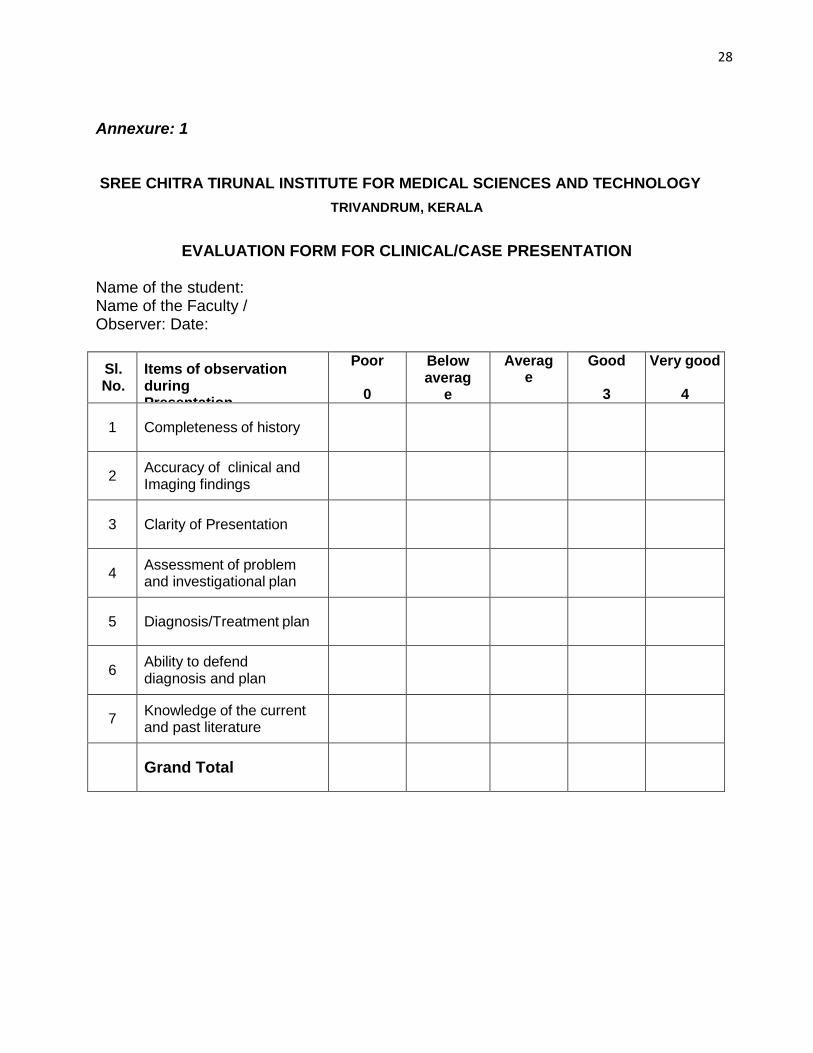
Annexure: 1
SREE CHITRA TIRUNAL INSTITUTE FOR MEDICAL SCIENCES AND TECHNOLOGY
TRIVANDRUM, KERALA
EVALUATION FORM FOR CLINICAL/CASE PRESENTATION
Name of the student: Name of the Faculty / Observer: Date:
Sl. No.
Items of observation during Presentation
Poor
0
Below averag
e
Average
Good
3
Very good
4
1 Completeness of history
2
Accuracy of clinical and Imaging findings
3
Clarity of Presentation
4
Assessment of problem and investigational plan
5
Diagnosis/Treatment plan
6
Ability to defend diagnosis and plan
7
Knowledge of the current and past literature
Grand Total

29
29
EVALUATION OF JOURNAL PRESENTATIONS
Name of the Student: Name of the Faculty / Observer: Date:
Sl. No.
Items of observation during Presentation
Poor
0
Below averag
e
Average
Good
3
Very good
4 1
Extent of understanding of scope & objectives of the paper
2
To critically evaluate methods, analysis and interpretations
3
Whether cross references have been consulted
4
Whether other relevant publications consulted
5
Ability to respond to questions on the paper / subject
6
Ability to defend the paper
7
Clarity of Presentation
8
Audio – Visual aids used
9
Ability to propose new research ideas based on study discussed
Total Score

30
EVALUATION OF SEMINAR PRESENTATIONS
Name of the student: Name of the Faculty / Observer: Date:
Sl. No.
Items of observation during Presentation
Poor
0
Below averag
e
Average
Good
3
Very good
4
1
Whether all relevant publications consulted
2
Understanding of the subject
3
Completeness of the preparation
4
Clarity of presentation
5
Current concepts coverage
6
Ability to answer the questions
7
Time scheduling
8
Appropriate use of Audio – Visual aids
9
Overall performance
10
Any other observation
Total Score

31
31
EVALUATION OF CLINICAL WORK IN WARD / OPD
Name of the student: Name of the Faculty / Observer: Date:
Sl. No.
Items of observation during presentation
Poor
0
Below averag
e
Average
Good
3
Very good
4
1 Regularity of attendance and punctuality
2 Presentations of cases
during rounds
3
Maintenance of case records
4
Investigations work up
5 Interaction with colleagues
and supporting staff
6 Teaching and training
junior colleagues
7
Bedside Manners
8 Rapport with patients
and family
9
Counseling Patient’s relatives for blood donation or postmortem and case follow
10
Overall quality of clinical work
Total Score

32
LOG BOOK
Table 1: Academic activities attended Name: Admission Year: College:
Date Type of activity Specify Seminar, Journal club, Presentation,
Particulars

33
33
LOG BOOK
Table 2: Academic presentations made by the students Name: Admission Year: College:
Date
Topic Type of activity Specify Seminar, Journal club, Presentation, UG

34
LOG BOOK Table 3: Diagnostic and Interventional procedures performed
Name: Admission Year: College:
Date
Name
I D No.
Procedure Category O, A, PA, PI*
Key: O - Washed up and observed
A - Assisted a more senior radiologist
PA - Performed procedure under the direct supervision of a senior radiologist
PI - Performed independently

35
35
MODEL OVERALL ASSESSMENT SHEET
Name of the college:
Academic Year:
Sl. No.
Particulars
Name of the student
A*
B*
C*
D*
E*
F*
G*
H*
I*
J*
1
Journal Review presentations
2
Seminars
3
Clinical work in wards
4
Clinical Presentation
5
Teaching skill practice
Total Score
Note: Use separate sheet for each year.
Signature of the HOD:
Signature of the Dean:
The above overall assessment sheet used along with the log book should form the
basis for certifying satisfactory completion of course of study, in addition to the
attendance require

36
Annexure: 2
CREDIT POINT SYSTEM FOR EVALUATION OF SENIOR RESIDENTS
Credit-based evaluation
The internal evaluation of the senior residents will be based on grading. The grading
will be based on the performance in each module with specified maximum credits against them.
The respective modules, with the maximum credits allotted against them, are given below.
SI No. Module Credits
1 Patient evaluation and management 75
2 Clinical evaluation, Imaging performance and Interpretation 75
3 Academic presentation 70
4 Invasive and interventional DSA procedures and decision making.
100
5 Publications and conference attendance (5 credits each) 10
6 Statistics course 5
7 BMT wing postings 3
8 Internal practical and theory evaluation 25
9 Outside training posting 20
10 Any outstanding activities 5
11 Project/thesis 12
Total 400

37
37
Module I : Patient evaluation and management (75 Credits)
I. Ward posting (30 credits)
The evaluation tools will be as follows,
1. Completion of admission and discharge summaries & at discharge patient
education and prescription.
2. Evaluation of his understanding of the clinical problem of all inpatients under his
charge and recognition using clinical laboratory parameters of patient’s progress,
deterioration or complications.
3. Identification of all clinical issues setting targets to be achieved at discharge.
4. Patient education and counseling especially with respect to post discharge life style,
diet, exercise, behavior modification & drugs and drug interactions.
5. Clinical appreciation of bedside signs and symptoms
6. Interpretation of all laboratory and invasive and noninvasive test results
7. Discharge Summary quality and completeness
8. Bedside procedures including catheterization, wound and puncture site care
2. Assessment of Outpatient training (25 credits).
1. Clinical cases seen and discussed with Consultant.
2. Completeness of case history writing and the plan of management along with
patient education and quality of prescription given to patient.
3. Interpretation of all routine investigation including Imaging & Laboratory reports.
4. Total evaluation / plan management strategy of patient on completed routine investigations.

38
5. Patients identified with new problem / worsening of existing clinical issues requiring
change of management plan and management discussed with consultant and also
presented to the Medical/Surgery Dept meetings charting out plan of management,
with all relevant investigations.
6. Identification of critically ill patients and channeling their acute management.
7. Inter-departmental consultations 3. ICU and Emergency (20 Credits)
1. This includes evaluation of patient management in the ICU (newly admitted.
transferred from wards, transferred after intervention procedure, etc) and charting
out plan of management and carrying out the same.
2. All emergency room visits of patients outside office hours / their evaluation
/charting out plan of management including ICU admissions and preparation for
emergency intervention/ surgery as indicated.
ICU training will include management of raised ICP, SAH/ICH, Vasospasm, stroke,
Spinal shock, Seizure, Ventilatory care, Fluid- Electrolyte care and CPR protocol etc.
Module II: Clinical evaluation, Imaging performance and Interpretation (75 credits)
1. Evaluation of MRI: Reporting and Analysis (40 credits)
Patient scheduling, evaluating, performing and reporting at least 500 MRIs in 3 years
1. To have a collection of at least 50 MRIs with all known abnormalities collected in a log
book. (Including all advanced Neuroimaging studies and their clinical, imaging and
histopathological follow up).
2. The evaluation will be based on the quality of the log book submitted.

39
39
2.Evaluation of CT: Reporting and Analysis (25 credits)
Patient scheduling, evaluating, performing and reporting at least 500 CT studies in 3 years
1. To have a collection of at least 50 CTs with all known abnormalities collected in a
log book. (Including all advanced Neuroimaging studies and their clinical,
imaging and /histopathological follow up).
2. The evaluation will be based on the quality of the log book submitted.
3. Evaluation of USG/Doppler: Reporting and Analysis (10 credits)
Analysis and reporting of at least 200 USG/ doppler cases with a collection of atleast 20
represatnative cases in the log book.
Module III : Academic Presentation (70 credits)
1. Journal Review ( Duration 30 min each) [20 credits]
Purpose of journal presentation it to instill qualities of enquiry and analysis of scientific
medical articles and to evaluate its relevance and impact in understanding pathophysiology of
disease or in clinical management. The resident can select recent articles of clinical relevance,
or consult the faculty to help select scientific articles with original research content for
presentation. The presentation should reflect the senior resident’s understanding of the problem
under discussion and the outcome and analysis of the results with regard to various aspects of
disease state and the clinical relevance. Two articles with brief exposition of the highlights of the
study and its clinical relevance and the take home message should be included. The senior
resident should include a short report of all the articles presented in print in the log, highlighting
the aim, methodology, patient recruitment criteria, results, discussion and implications for clinical
practice.

40
2. Seminar 45 min [20 credits]
It is intended to encourage extensive literature review on the topic and present the highlights of
the topic under review in a succinct manner with clear take home messages, but at the same
time the extensive literature search elevates the presenter as an authority on the topic. The topic
should be prepared as a review article with complete bibliography in a publishable format, along
with the topic presentation. The presentation and the write up are equally weighted.
3. Participation and presentations in Stroke and Neurovascular meet [6 credits] 4. Participation and presentations in Radiology-Neuropathology presentations [4 credits] 5. Participation and presentations in Neuroradiology meet [10 credits] 6. Clinical case discussion [10 credits]
The purpose of this exercise is to identify daily clinical problems confronted during the
routine hospitalization and management of patients, clinical problems significant enough to
influence patient management (diagnosis/therapy). The literature review will be up to date, and
will enable evidence based approach to patient management in different clinical scenarios.
The assessment will be based on the following parameters; review of the literature to chart out
evidence based management plan and to write up a short report on the clinical problems and the
current state of the art management and the level of evidence for such management option. The
oral presentation and the write up are equally weighted for purpose of evaluation.
Module IV: Invasive and interventional DSA procedures and decision making ( 100 credits)
Diagnostic Angiography atleast 50, Extracranial interventions 10, Intracranial and spinal Interventions 40
1. Pre-procedural work up and detailed knowledge of the case
2. Evaluation of available diagnostic images
3. Taking proper consent and investigations
4. DSA procedure Knowledge and execution ( as a First operator/ Assistant)
5. Post procedure care and orders
6. Follow up of patients

41
41
Module V. Publications and conference attendance (5 credits each).
At least one scientific paper as first author should be submitted for publication before the
end of the course and final evaluation. There will be uniform credit of 5 for satisfying this
requirement.
There should be one paper/poster presented at a national, state or international
conference. There will be equal credit of 5 for this.
The senior resident has to present certificate of participation to PC and submit abstract of
presentation to the PC and also ensure entry in e-portfolio
Module VI and VII: Statistics and BMT wing postings 1. Medical Statistics and research methodology (5 credits)
2. Biomedical Technology posting (3 credits)
Module VIII. Internal Examinations :Theory and Practicals (25 credits)
There will be internal practical and theory examinations, each having 100 marks during the 3-
year course. These examination will be conducted at every 6 months.
Module IX. Outside training posting (20 credits)
Candidates will be send to other academic institutes having diagnostic interventional
neuroradiologic training facilities for 1 month.
Module X. Outstanding activities : (5 credits) Additional credits will be awarded for additional publications, projects, national and
international conference presentations obtaining prizes, certificates of merits and awards
etc.

42
Module XI. Projects and thesis : (12 credits) 1. Mid-term evaluation of projects mandatory and will carry credits
2. Prospective / Retrospective Study
3. Ethical Committee clearance / Institute funding obtained
4. Contribution of candidates experience in the study
5. Descriptive data collection / Quantitative data subjected to statistical analysis.
6. Midterm Review: At 18 months of DM course: Aims and objectives, review of
literature, materials and methods ( exclusion / inclusion criteria), data collection and
presentation (% of target of the project) and preliminary data analysis.
7. Review at 30 months: Presentation of the full project as thesis and also in publishable
form, complete with statistical analysis, discussion, study limitations, conclusion, and
bibliography.
8. Overall impact of the project in adding to our knowledgebase, and patient management. Between
30-34 months, the project should be sent for publication to peer reviewed journals.
9. Presentation of the project work as scientific presentation at national level and at state
level- desirable. Additional publication ready manuscript should also be prepared.

43
43
ANEEXURE: 3
SYLLABUS
Part - 1 Basic sciences
1. Anatomy (gross and radiological anatomy)
a. Embryology of brain, spinal cord, and their vascular system
b. Basic correlative anatomy of the brain, spinal cord and peripheral nervous system
c. Blood supply of the brain and spinal cord
d. Embryology and anatomy of skull, face and head and neck.
e. Anatomy of the musculo skeletal system relevant to the disease of nervous system
f. Relevant embryology and anatomy of vascular system mainly related to aorta and brachio cephalic vessels.
2. Physiology
a. Basic and applied neurophysiology
b. Basics of hemodynamics
3.Pathology and microbiology
a. General and specific neuropathology
b. Applied use of electron microscopy and virology
c. Applied bacteriology, parasitology and virology
d. Neuro vascular pathology
e. Pathology of congenital malformations, neonatal and perinatal CNS disorders
f. Genetic and metabolic disorders of CNS
3.Biochemistry and pharmacology
a. Applied aspect of the brain chemistry in relation to neuroradiology
b. Pharmacology of drug action in relation to neuro radiology

44
c. Contrast media
d. Antihypertensives, antiplatelets, anti coagulants
e. Vasodilators and vaso constrictors
f. Embolic agents
g. Thrombolytic agents
h. Anesthetics and analgesics with respect to neuro imaging and interventions.
i. Anti epileptics
5. Physical principles of imaging
a. Image intensifier and flat pannel detector
b. Angiography and digital subtraction angiography
c. Spiral and multislice CT, CT perfusion imaging, and recent advances
d. Magnetic resonance imaging – hard wares, pulse sequences, MR spectroscopy, functional MRI, diffusion tensor imaging
e. Ultrasound, doppler and colour doppler ultrasound
f. Image processing in CT and MRI
g. PACS and digital radiography
h. Radionuclide scan, SPECT and PET
6. Instrumentation and bio medical engineering
a. Knowledge about the various imaging and interventional equipment in the department
b. Patient monitoring equipments and various life support systems
c. Catheters and other biomaterials used in interventional radiology
d. Radiation protection principles and devices
7. Steriotactic radiotherapy and steriotactic procedures
a. Principle and practice of steriotactic radiotherapy using x-ray source and cobalt source
b. Principle, theory and practice of steriotactic procedures on brain,spine and spinal cord
8. Epidemiological studies and biostatistics

45
45
Part - II
Clinical Sciences
a. Neuroradiology: Principles and practice of applied neuroradiology
b. Interventional neuroradiology: principles and practice of interventional neuroradiology
c. Neurology, neuro ophthalmology, neuro otorhino laryngology : principles, theory and practice
d. Neuro surgery : principles and theory
e. Experimental neuroradiology
f. Recent advances in neuroradiology.
Detailed syllabus for part I and part II
DIAGNOSTIC NEURORADIOLOGY
I. BRAIN AND COVERINGS
A. Normal anatomy
1. Skull, sutures 2. Major apertures 3. Hemispheres, cortex, gyri, sulci 4. Major fissures 5. Major cisterns 6. Basal ganglia, thalamus, hypothalamus, pituitary gland, pineal gland 7. Pons, cerebellum, cerebellopontine angle 8. Ventricles, choroid plexus 9. Vessels and major branches
B. Basic and Advanced Instrumentation
1. Computed tomography (CT)
a. Basic techniques b. Spiral scanning c. CT Angiography (CTA) d. CT perfusion e. Post processing

46
2. Magnetic resonance (MR)
a. Morphologic imaging Spin echo Gradient echo Inversion recovery Chemical shift imaging Suppression techniques High-speed imaging Diffusion/perfusion
b. MR Angiography (MRA) c. Spectroscopy d. Functional MRI c. Diffusion Tensor Imaging e. Post processing
3. Nuclear medicine
a. Planar scintigraphy b. SPECT c. PET
4. Ultrasound
a. General - techniques and physics b. Gray scale c. Duplex doppler d. Intra vascular Ultrasound
C. Congenital CNS lesions
1. Embryology of brain development 2. Normal variants 3. Disorders of organogenesis 4. Disorders of neuronal migration and sulcation 5. Disorders of diverticulation and cleavage 6. Posterior fossa cystic disorders 7. Disorders of histogenesis (phakomatoses)
D. CNS Infections 1. Imaging strategies 2. Pyogenic infections 3. Encephalitis 4. Granulomatous infections 5. Infections in the immunocompromised host
E. Neoplasms and other masses 1. Benign neoplasms of scalp or skull 2. Malignant neoplasms of scalp or skull 3. Intracranial tumor
a. Tumor classification by histology b. Tumor evaluation by location

47
47
F. Trauma 1. Imaging strategies CT/MR/skull films 2. Mechanisms 3. Primary vs. secondary 4. Focal lesions
Cortical contusions Diffuse axonal injury (DAI) -- shearing Subarachnoid hemorrhage (SAH) Subdural hemorrhage (SDH) Epidural hemorrhage (EDH) Parenchymal hemorrhage with differentials
5. Ages of hemorrhage by CT/MR 6. Intraventricular hemorrhage 7. Diffuse cerebral swelling and edema 8. Herniation syndromes 9. Skull fractures, types, complications 10. Vascular injuries—dissection, pseudoaneurysm, penetrating
injuries, lacerations, complications 11. Non-accidental trauma
G. White Matter Disease
1. Multiple sclerosis 2. Acute disseminated encephalomyelitis (ADEM) 3. Small vessel ischemic disease, hypertension, vasc. dis. 02140 4. Radiation/chemotherapy changes 5. Trauma (axonal injuries) 6. White matter changes in the elderly 7. Osmotic myelinolysis (central pontine myelinolysis) 8. Dysmyelinating disorders
H. Cerebrovascular disease
1. Ischemia and infarction 2. Spontaneous hemorrhage 3. Aneurysms 4. Cerebrovascular malformations 5. Angiography
I. Metabolic disease
1. Pituitary disorders 2. Thyroid disorders 3. Parathyroid disorders 4. Adrenal disorders 5. Intoxications, poisoning, metabolic encephalopathies 6. Diabetes mellitus

48
7. Pediatric disorders J. Generalized systemic disorders K. Hydrocephalus L. Cognitive imaging II. HEAD AND NECK
A. Deep spaces of the neck
1. Buccal space (anatomy, contents) 2. Sublingual space 3. Submandibular space 4. Parotid space (anatomy, contents) 5. Parapharyngeal space (anatomy, contents) 6. Masticator space (anatomy, contents) 7. Retropharyngeal space (anatomy, contents) 8. Perivertebral space (anatomy, contents) 9. Posterior cervical space (anatomy, contents) 10. Pharyngeal mucosal space (anatomy, contents) 11. Visceral space (anatomy, contents)
a. Larynx and hypopharynx(anatomy) b. Thyroid
12. Special issues a. Perineural spread b. Carotid encasement c. Bone and cartilage involvement
B. Orbit
1. Imaging techniques 2. Anatomy/embryology 3. Intra-ocular 4. Intraconal
a. Optic nerve b. Extraneural
5. Conal (Extra-ocular muscles) 6. Extra-conal masses 7. Extraorbital
a. Paranasal sinus tumors extending into orbit b. Paranasal sinus infections extending into orbit
8. Trauma C. Paranasal sinuses
1. Anatomy of paranasal sinuses 2. Congenital disease 3. Inflammation/infection 4. Benign sinus tumors 5. Malignant sinus tumors

49
49
D. Skull base 1. Anterior skull base (anatomy)
a. Invasion of lesions from above b. Intrinsic bone lesions c. Invasion of lesions from below
2. Central skull base (anatomy) a. Invasion of lesions from above b. Intrinsic bone lesions c. Invasion of lesions from below
3. Posterior skull base (anatomy) a. Invasions of lesions from above b. Intrinsic bone lesions c. Invasion of lesions from below
E. Temporal bone
1. Imaging techniques (Multi-planar CT/MR) 2. Anatomy/embryology 3. Congenital anomalies 4. Inflammatory disease 5. Tumors 6. Trauma 7. Pulsatile tinnitus
F. Cystic neck masses
1. Second brachial cleft cyst 2. Thyroglossal duct cyst 3. Cystic hygroma 4. Laryngocele, internal, external 5. Abscess 6. Ranula 7. Dermoid/epidermoid
III. SPINE
A. Anatomy and biomechanics
1. Vertebral bodies 2. Facet joints and transverse processes 3. Lamina and spinous processes 4. Support ligaments 5. Specific characteristics of cervical, thoracic, and lumbar segments 6. Cranio-vertebral and lumbo-sacral junctions 7. Normal stability and motion
50
B. Imaging modalities 1. Role and relative merit of noninvasive imaging
studies Plain radiography, CT, MR, nuclear medicine, PET
imaging 2. Role of invasive procedures
Myelography (including CT), angiography, biopsies, facet injections, nerve root blocks, discography, vertebroplasty
C. Developmental spine disease
1. Normal embryologic development of spine 2. Abnormalities of neurulation 3. Anomalies of caudal cell mass 4. Anomalies of development of the notochord 5. Congenital tumors 6. Syringohydromyelia 7. Craniovertebral anomalies 8. Dysplasia 9. Constitutional disorders
D. Inflammatory and demyelinating disease
1. Osteomyelitis 2. Discitis 3. Epidural and paravertebral abscess 4. Granulomatous infection 5. Meningitis (arachnoiditis) 6. Spinal cord lesions
E. Neoplastic disease
1. Neoplasms of bone 2. Intraspinal
Extradural Intradural extramedullary Intramedullary
F. Trauma 1. Mechanism of injury 2. Stable fractures and ligamentous injuries 3. Unstable injuries (involvement of the middle column and ligaments) 4. Traumatic disc herniation 5. Extrinsic cord compression 6. Cord contusion 7. Intra-spinal hemorrhage 8. Post-traumatic abnormalities 9. Postoperative findings
G. Metabolic, endocrine, toxic
1. Pituitary disorders 2. Thyroid disorders 3. Parathyroid disorder

51
51
4. Adrenal disorder 5. Osteoporosis 6. Osteomalacia, renal osteodystrophy 7. Intoxication, poisoning
H. Systemic disorders
1. Hematalogic disorders 2. Histiocytosis 3. Sphingolipidosis 4. Amyloid
I. Degenerative disease
1. Arthritis 2. Degenerative joint disorders 3. Degenerative disk order
a. Epidemiology and terminology b. Disc degeneration c. Disc herniation d. Spinal stenosis
J. Vascular lesions
1. Dural venous fistula 2. AVM 3. Cavernous angioma 4. Spinal cord ischemia and infarction 5. Vascular bone marrow lesions and ischemia
K. Cystic lesions
1. Extradural 2. Intradural extramedullary 3. Intramedullary
GENERAL VASCULAR CONSIDERATIONS
PATIENT CARE IN VASCULAR AND INTERVENTIONAL RADIOLOGY
Pre-procedural assessment and care Intraprocedural monitoring Post-procedural followup and care
General pharmacologic considerations Analgesia/anesthesia
Concious sedation Antibiotic therapy
Anticoagulation

52
Other VASCULAR DIAGNOSIS
Epidemiology of vascular disease Vascular anatomy: arterial and venous Embryology Normal anatomy Variant anatomy Anatomy of collateral pathways
VASCULAR PHYSIOLOGY, PATHOLOGY AND PATHOPHYSIOLOGY ARTERIAL SYSTEM
Normal histology/physiology/morphology Hemodynamics: normal and abnormal flow Vasoactive extrinsic/pharmacologic agents Normal response Disorders related to pharmacologic/extrinsic agent exposure Atherosclerosis Medial sclerosis Pathophysiology of arterial ischemia Aneurysms Thromboembolic disorders Dissection Congenital vascular disorders Vascular malformations Arterial effects of adjacent tissues/disorders Arterial infection Vascular alterations in neoplasia: vascular
supply of neoplasms, primary vascular neoplasms, vascular invasion by neoplasms
Vascular alterations in inflammatory diseases Systemic vascular disorders Primary systemic vascular disorders
vasculitides and others polyarteritis nodosa Takayasu’s arteritis
giant cell arteritis Buerger’s disease
Altered vascular pathology in systemic disease states Vascular trauma: injuries and vascular response to injury Mechanical injury: acute and chronic Thermal injury Arterial endothelium Alterations in coagulation status Hypercoagulable states Impaired coagulation Post-operative or post-interventional disorders Synthetic and endogenous grafts

53
53
Myointimal hyperplasia Other/unclassified
VENOUS CIRCULATION
Normal histology/physiology/morphology Hemodynamics: normal and abnormal flow Vasoactive extrinsic/pharmacologic agents Normal response Disorders related to pharmacologic/extrinsic agent exposure Thromboembolic disorders: acute and chronic Venous aneurysms Venous effects of adjacent tissues/disorders Congenital vascular disorders Vascular malformations Venous infection Vascular alterations in neoplasia
vascular drainage of neoplasms primary vascular neoplasms vascular invasion by neoplasms
Vascular alterations in inflammatory diseases Systemic vascular disorders Primary systemic vascular disorders Altered vascular pathology in systemic disease states Vascular trauma: injuries and vascular response to injury Mechanical injury--acute and chronic Thermal injury Venous endothelium Alterations in coagulation status Hypercoagulable states Impaired coagulation Post-operative or post-interventional disorders Synthetic and endogenous grafts Intimal hyperplasia
LYMPHATIC SYSTEM
Anatomy Lymphangiography Performance Interpretation Indications and contraindications Risks
CONTRAST REACTIONS, IODINATED AGENTS
Anaphylactoid reactions

54
Classification Prevention Ionic vs. nonionic agents Premedication Treatment Dose dependent reactions Acute and chronic renal effects Other
PROCEDURAL COMPLICATIONS
Puncture site complications Catheterization-related complications (apart from puncture site) Systemic/generalized complications
PHARMACOLOGY Vasodilators Vasoconstrictors Antihypertensives Anti epileptics Anticoagulants Antiplatelets Fibrinolytics
INTERVENTIONAL NEURORADIOLOGY
BASIC ANATOMY
GENERAL INTRODUCTION Vascular Anatomy and Biological Process Collateral circulation Principles in vascular patterns
SPINE AND SPINAL CORD ARTERIES AND VEINS Introduction Embryology,metameric supply and axial organization Fusion, desegmentation and failed fusion Spinal Arteries Dural Arteries Spinal cord arteries Spinal cord vein Spinal and extraspinal venous system
CRANIOCERVICAL JUNCTION
Pharyngo occipital system Arterial supply to the posterior fossa central nervous system
SKULL BASE AND MAXILLOFACIAL REGION

55
55
Embryology of the carotid system Arteries of middle ear and branches of the petrous Internal carotid artery Anatomy and variations of the Extracranial carotid artery branches
THE SKULL BASE AND EXTRADURAL ARTERIES
The cavernous sinus region Embryology and anatomy of arteries supplying the orbit Supply to adjacent meninges The transosseous Peripheral Nervous System arterial supply
INTRADURAL ARTERIES
General Aspects The Internal carotid artery divisions The caudal internal carotid artery division The Anterior choroidal artery The cranial internal carotid artery division
INTRACRANIAL VENOUS SYSTEM
Embryology Deepvenous system Superficial veins and dural sinuses Infratentorial veins Complex cerebrofacial venous drainages –Anamoly to abnormality CSF circulation
CLINICAL AND ENDOVASCULAR TREATMENT ASPECTS IN ADULTS
1. OCCLUSIVE VASCULAR DISEASE
Arterial Occlusive Disease Cerebrovascular Physiology Arterial Ischemic Stroke Atheroclerosis Ischemic stroke treatment Extracranial Carotid artery stenosis Carotid angioplasty and stenting Posterior circulation stroke and management Intracranial angioplasty and stenting
Vasospasm Venous Occlusive Disease
Etiology Incidence Clinical presentation and natural history Management and dural sinus thrombolysis
56
2. TUMORS Meningiomas Other bony and dural tumors Nasopharyngeal tumors Paragangliomas Other head and neck tumors Pretherapeutic evaluation surgical strategy Therapeutic strategies Technical Aspects of embolization Complications of Embolization.
3. TRAUMATIC ARTERIOVENOUS FISTULAE
Carotid Cavernous Fistula Etiology and Incidence Clinical presentation signs and symptoms Natural history and prognosis Pretherapeutic evaluation Therapeutic strategies Technical Aspects of embolization Risk and complications with embolisation Post embolisation care
Extracavernous Arteriovenous Fistulae Vertebral Arteriovenous Fistulae Common Carotid Fistulae External Carotid Fistulae Malignant Fistulae
4. ANEURYSMAL VASCULOPATHIES
From Aneurysm to Aneurysmal Vasculopathies Extradural Aneurysms Intradural Aneurysms Intradural Artery dissections Intradural Giant Saccular and Non saccular Arterial aneurysms Infectious, immune, and inflammatory Intradural aneurysms Flow related arterial aneurysms and other aneurismal vasculopathies Therapeutic management
5. DURAL ARTERIOVENOUS SHUNTS Incidence and etiology Pathophysiology and the Osteo-dural Complex Classifications Clinical presentation Natural history and prognosis Pretherapeutic Evaluation Strategy and Technical Aspects of Embolisation Risks and complications Post embolisation care and Surgical options
6. CEREBRALVASCULAR MALFORMATIONS General Considerations Incidence

57
57
Classification Clinical presentation Angioarchitecture Clinical and Angioarchitecture correlation Venous Variations Developmental Venous Anomalies Cavernomas Cerebral cavernous malformations Occult and cryptic malformations Sturge-Weber Syndrome
7. GOALS AND OBJECTIVES IN MANAGEMENT OF BRAIN AVMS Indications Pretherapeutic Evaluation Therapeutic management strategy Embolization :
Staging of embolization Partial targeted, partial palliative, curative Pre surgical embolisation Prior to radiosurgery embolisation Size reduction Emergency embolization
Surgical Excision Radiosurgery
SPINE
1. Spinal Arteriovenous Shunts 2. Spinal Angiography and Angiographic protocol 3. Parachordal AVF 4. Spinal cord Arteriovenous malformations
Incidence Classification Clinical presentation and aggravating factors Spinal cord hemodynamics and Angioarchitecture Clinical and Angioarchitecture correlation Natural History and prognosis Pretherapeutic evaluation Indications for treatment Technical aspects of embolization and Results Risks and complications of embolization Post embolisation management Surgery

58
Radiosurgery 5. Spinal Dural Arteriovenous Fistulae
Incidence Classification Clinical presentation and aggravating factors Clinical and Angioarchitecture correlation Natural History and prognosis Pretherapeutic evaluation Indications for treatment Technical aspects of embolization and Results Risks and complications of embolization Post embolisation management Surgery
6. Spinal Arteriovenous Metameric Syndromes 7. Tumors of spinal cord and spinal column 8. Spinal column
Vertebral body hemangiomas Benign Bony tumors Malignant Bony tumors Embolization and Vertebroplasty Osteoporosis and pain relief Degenerative disc disease
9. Spinal cord Hemangioblastomas
CERVICO-FACIAL VASCULAR MALFORMATIONS Hemangiomas and Phace syndromes
Incidence Classification Natural history Diagnostic and Pre-therapeutic evaluation Management
Arteriovenous malformations of face and scalp Incidence Classification Natural history Diagnostic and Pre-therapeutic evaluation Indications and timing of embolization Objectives and Technical aspect of embolization Complications of Embolization Surgery
Venous malformations Incidence Classification Natural history Diagnostic and Pre-therapeutic evaluation Sclerotherapy Risks and complications of sclerotherapy Post sclerotherapy Management
Lymphatic and other malformations Incidence

59
59
Classification Natural history Diagnostic and Pre-therapeutic evaluation Sclerotherapy Risks and complications of sclerotherapy Post sclerotherapy Management
CLINICAL AND ENDOVASCULAR TREATMENT ASPECTS IN PEDIATRIC AGE
Embryological and Anatomical Complement 1. GENERAL FEATURES OF INTRACRANIAL ARTERIOVENOUS DISEASE
Congestive cardiac failure Hydrodynamic equilibrium and disorder Melting brain syndrome Clinical evaluation scores
2. VEIN OF GALEN MALFORMATION Incidence
and etiology Natural History Prognosis Pretherapeutic evaluation Indications for treatment, Concept of Window period Objectives and Methods of treatment Technical aspects of Embolization Post embolisation management Surgery Radiosurgery Aneurysmal dilatation of Vein of Galen
3. PIAL ARTERIOVENOUS MALFORMATION/BRAIN AVMS
Incidence and etiology Clinical presentation Natural History and Prognosis Pretherapeutic evaluation Indications for treatment Objectives and Methods of treatment Technical aspects of Embolization Post embolisation management Surgery Radiosurgery
4. DURAL ARTERIOVENOUS SHUNTS
Classification Fetal changes in sinuses Dural Sinus malformation Infantile Dural Arteriovenous Shunt

60
Adult type Dural arteriovenous shunt Other dural shunts
5. INTRACRANIAL ANEURYSMS IN CHILDREN Incidence
and etiology Classification Locations Clinical presentation and natural history Outcomes Pretherapeutic evaluation Embolization Surgery
6. ARTERIAL ISCHEMIC STROKES AND VENOUS STROKES
Epidemiology Moya-Moya disease Arteritis Spontaneous dissections Clinical presentation and natural history Imaging and pretherapeutic evaluation Treatment and management Outcomes and prognosis
7. TRAUMA AND EPISTAXIS IN PEDIATRIC AGE GROUP
Tumor in naspharynx Traumatic CCF Traumatic dural sinus thrombosis Traumatic dissection Traumatic pseudoaneuryms
8. SPINAL VASCULAR LESIONS
Classification Spinal angiography and protocol for angiography Angioarchitecture and clinical correlation Natural History and prognosis Pretherapeutic evaluation Indication for treatment Objectives and goals for embolization. Risks and complications of embolization Surgery
9. CERVICO-FACIAL VASCULAR MALFORMATIONS
Hemangiomas and Phace syndromes Incidence Classification Natural history Diagnostic and Pre-therapeutic evaluation Management
Arteriovenous malformations of face and scalp Incidence Classification Natural history

61
61
Diagnostic and Pre-therapeutic evaluation Indications and timing of embolization Objectives and Technical aspect of embolization Complications of Embolization Surgery
Venous malformations Incidence Classification Natural history Diagnostic and Pre-therapeutic evaluation Sclerotherapy Risks and complications of sclerotherapy Post sclerotherapy Management
Lymphatic and other malformations Incidence Classification Natural history Diagnostic and Pre-therapeutic evaluation Sclerotherapy Risks and complications of sclerotherapy
Recommended text books
a. Magnetic Resonance Imaging of the Brain and Spine Scott W. Atlas
b. Diagnostic cerebral angiography Anne G. Osborn
c. Interventional Neuroradiology: Strategies and Practical Techniques J. J. Connors, Joan C. Wojak
Recommended text books / Periodicals
a. American Journal of Neuroradiology
b. Neuroradiology
c. Journal of magnetic resonance imaging
d. Neuro Imaging Clinics of North America
e. Neurology
f. Neurosurgery