Division of Molecular Biosciences, Biochemistry Building
16
Protein Folding Requires Crowd Control in a Simulated Cell Benjamin R. Jefferys ⁎ , Lawrence A. Kelley, and Michael J.E. Sternberg Division of Molecular Biosciences, Biochemistry Building, Imperial College London, South Kensington, London SW7 2AZ, UK. Abstract Macromolecular crowding has a profound effect upon biochemical processes in the cell. We have computationally studied the effect of crowding upon protein folding for 12 small domains in a simulated cell using a coarse-grained protein model, which is based upon Langevin dynamics, designed to unify the often disjoint goals of protein folding simulation and structure prediction. The model can make predictions of native conformation with accuracy comparable with that of the best current template-free models. It is fast enough to enable a more extensive analysis of crowding than previously attempted, studying several proteins at many crowding levels and further random repetitions designed to more closely approximate the ensemble of conformations. We found that when crowding approaches 40% excluded volume, the maximum level found in the cell, proteins fold to fewer native-like states. Notably, when crowding is increased beyond this level, there is a sudden failure of protein folding: proteins fix upon a structure more quickly and become trapped in extended conformations. These results suggest that the ability of small protein domains to fold without the help of chaperones may be an important factor in limiting the degree of macromolecular crowding in the cell. Here, we discuss the possible implications regarding the relationship between protein expression level, protein size, chaperone activity and aggregation. Abbreviation TM, template modelling Keywords macromolecular crowding; protein structure prediction; protein misfolding; protein aggregation; protein expression Introduction It is widely acknowledged that there are two fundamental questions in modelling protein folding: What is the biologically active conformation of a protein sequence? © 2010 Elsevier Ltd. This document may be redistributed and reused, subject to certain conditions. ⁎ Corresponding author. Fax: +44 (0) 20 7594 5789. [email protected]. This document was posted here by permission of the publisher. At the time of deposit, it included all changes made during peer review, copyediting, and publishing. The U.S. National Library of Medicine is responsible for all links within the document and for incorporating any publisher-supplied amendments or retractions issued subsequently. The published journal article, guaranteed to be such by Elsevier, is available for free, on ScienceDirect. Sponsored document from Journal of Molecular Biology Published as: J Mol Biol. 2010 April 16; 397(5): 1329–1338. Sponsored Document Sponsored Document Sponsored Document
Transcript of Division of Molecular Biosciences, Biochemistry Building
Protein Folding Requires Crowd Control in a Simulated Cell
Benjamin R. Jefferys⁎, Lawrence A. Kelley, and Michael J.E. SternbergDivision of Molecular Biosciences, Biochemistry Building, Imperial College London, SouthKensington, London SW7 2AZ, UK.
AbstractMacromolecular crowding has a profound effect upon biochemical processes in the cell. We havecomputationally studied the effect of crowding upon protein folding for 12 small domains in asimulated cell using a coarse-grained protein model, which is based upon Langevin dynamics,designed to unify the often disjoint goals of protein folding simulation and structure prediction. Themodel can make predictions of native conformation with accuracy comparable with that of the bestcurrent template-free models. It is fast enough to enable a more extensive analysis of crowding thanpreviously attempted, studying several proteins at many crowding levels and further randomrepetitions designed to more closely approximate the ensemble of conformations. We found thatwhen crowding approaches 40% excluded volume, the maximum level found in the cell, proteinsfold to fewer native-like states. Notably, when crowding is increased beyond this level, there is asudden failure of protein folding: proteins fix upon a structure more quickly and become trapped inextended conformations. These results suggest that the ability of small protein domains to foldwithout the help of chaperones may be an important factor in limiting the degree of macromolecularcrowding in the cell. Here, we discuss the possible implications regarding the relationship betweenprotein expression level, protein size, chaperone activity and aggregation.
AbbreviationTM, template modelling
Keywordsmacromolecular crowding; protein structure prediction; protein misfolding; protein aggregation;protein expression
IntroductionIt is widely acknowledged that there are two fundamental questions in modelling proteinfolding:
What is the biologically active conformation of a protein sequence?
© 2010 Elsevier Ltd.This document may be redistributed and reused, subject to certain conditions.⁎Corresponding author. Fax: +44 (0) 20 7594 5789. [email protected] document was posted here by permission of the publisher. At the time of deposit, it included all changes made during peer review,copyediting, and publishing. The U.S. National Library of Medicine is responsible for all links within the document and for incorporatingany publisher-supplied amendments or retractions issued subsequently. The published journal article, guaranteed to be such by Elsevier,is available for free, on ScienceDirect.
Sponsored document fromJournal of Molecular Biology
Published as: J Mol Biol. 2010 April 16; 397(5): 1329–1338.
Sponsored Docum
ent Sponsored D
ocument
Sponsored Docum
ent
How does a protein find that state in the cell?
These two problems have generally been approached using radically different methods. Themost successful methods for predicting the biologically active state are through finding ahomologue of the sequence to another sequence with known structure or through assembly offragments of structure when such homology cannot be detected. These are valuable tools forsuggesting the structure of a protein sequence where no experimental structure is available,and they consistently perform well at CASP (Critical Assessment of techniques for proteinStructure Prediction) experiments.
However, such methods provide little, if any, insight into the principles underlying the searchfor that state: the speed and reliability of folding, the stability and flexibility of the finalconformation, the effect of conditions in the cell, misfolding and aggregation and error-correcting processes. Molecular dynamics is used for investigating these elements of proteinfolding. Although progress is being made toward predicting protein structure throughmolecular dynamics, such methods are not yet among the most successful predictive tools andthe large computational resources required make their application to large-scale analysisimpractical.
Simplifications of molecular dynamics that reduce the number of particles modelled, simplifythe force field and use time-saving heuristics in the search method enable larger-scale analysesof protein folding. Ultimately, the goal is to study the mechanism of protein folding and obtainthe biologically active conformation with a unified model for protein structure, which predictsthe native state through accurate modelling of the folding process. We present here a proteinmodel developed for this purpose and demonstrate its effectiveness in both protein structureprediction and the study of the protein folding process. The structural model reduces thebackbone to a series of particles representing Cα atoms and particles representing the centroidof side-chain atoms. The folding model is a simplified iterative solution of Newtonian equationsof motion based upon Langevin dynamics, with linear elastic springs modelling the majorityof effects in the force field. Solution of this equation aims to simulate protein folding over timethrough a putative pathway. The Langevin equation includes a term for random bombardmentby implicit solvent; therefore, the generated folding pathway is dependent upon the seed for arandom number generator and multiple pathways must be produced to assess the ensemble ofconformations. Such a model can be used to investigate many aspects of protein folding,including the effect of conditions in the cell, such as macromolecular crowding.
The cell is a crowded and chaotic environment: it is estimated that between 10% and 40% ofthe cell's volume is occupied by macromolecules. However, complex and intricate biochemicalprocesses must be performed reliably in this environment. A minimum concentration ofmolecules in the cell is necessary for interacting partners to associate. There is also a maximumconcentration beyond which normal cellular function would be prevented due to restriction ofmolecular motion. Evidence suggests that crowding enhances protein stability, proteinassociation and chaperonin action but that it also increases protein aggregation and lowersdiffusion rates. The macromolecular concentration observed in cells may have been selectedto balance these factors, according to requirements in different cellular localities. In this study,we investigated the role of protein folding in this balance.
Previous work suggests that macromolecular crowding has a complex effect upon proteinfolding. Computational models and experimental work show that, at a crowding level similarto that of the cell as a whole, crowding can make proteins fold more rapidly and stabilise thenative state at secondary and tertiary levels. This effect is interpreted as being due to thereduction of conformation space by exclusion of extended conformations.
Jefferys et al. Page 2
Published as: J Mol Biol. 2010 April 16; 397(5): 1329–1338.
Sponsored Docum
ent Sponsored D
ocument
Sponsored Docum
ent
Computational studies of the effect of crowding upon protein folding commonly use a Gō-liketerm in the energy function for the protein, which favours interactions known to occur in thenative conformation. Clearly, such terms disqualify these methods as tools for predicting thefinal conformation. As a consequence of using a Gō-like potential, terms may be omitted fornon-native but favourable interactions that might be encountered on the way to the native state—interactions that might be important in finding the native state or in creating a non-nativelocal energy minimum. If the effect of crowding were to trap a protein in such a minimum,then this effect would not be observed with a Gō-like potential.
Simulations making use of molecular dynamics do not have this disadvantage. However, dueto computational limitations, they often study only the change in stability of proteins in theirnative state when subjected to crowding, and where proteins have been refolded from a partiallydenatured state, only very small proteins were studied. A study using molecular dynamics hasshown that crowding may destabilise the native state of the protein by forcing the solvent intoa more ordered state, reducing the entropic advantage of the native over otherconformations. Native stability and refolding rates are important aspects of the protein foldingproblem. However, in the cell, proteins are synthesized slowly into a crowded environment.While computational analysis has found that co-translational folding appears to have a smalleffect on protein folding rate and has only weakly imprinted motifs in structures that might beexpected if it did, experimental observations are more mixed. Slow synthesis may allowproteins to find a compact state more easily than from a denatured state, an effect that may beeven more important in a crowded environment. Additionally, computational and experimentalconstraints have limited previous studies to one or two structures and few crowding levels.This raises the possibility that effects observed are specific to the chosen conditions.
To address these issues, we have studied the effect of crowding using a protein model that doesnot include knowledge of long-range interactions, can predict protein tertiary structure withaccuracy comparable with that of the best current template-free methods, includes a simulationof the folding pathway during slow synthesis of a protein by a large, heavy ribosome and isfast enough to allow study of many proteins at several crowding levels. The aim of this workwas to find the upper limit of crowding in which proteins can successfully fold and comparethis with the level of crowding observed in the cell.
ResultsModel verification through structure prediction
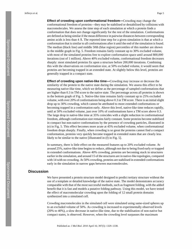
We tested our model by making predictions of the tertiary structure for a set of 30 small proteindomains (listed in Supplementary Material) that were chosen for the quality of experimentalstructure, size and fold class. The structure prediction protocol is described in Materials andMethods. Modelling is based upon the predicted secondary structure using psipred. Fivestructure predictions are made for each protein, and the best TM (template modelling) scoreand largest fraction of residues alignable to within 5 Å to the native are given in Fig. 1. TheTM score is a measure of structural similarity that is more sensitive to the similarity of proteinfragments than the more commonly used global RMSD (root-mean-square deviation). Themean TM score between randomly chosen Protein Data Bank structures is 0.17, and a TMscore exceeding 0.3 indicates a roughly native-like topology. It is unusual to exceed a TM scoreof 0.4 in template-free modelling. The best of the five predictions for 24 of the proteins has aTM score higher than 0.3, and for 4 of those proteins, the score is higher than 0.4. Predictionsand the best structures generated for 3 of the proteins are illustrated in Fig. 2.
These results mean this method is comparable with the best template-free structure predictionmethods. Each prediction is made from simulations using at most 40 CPU hours, a tiny fractionof the time that is used by many other successful template-free predictive methods. It is this
Jefferys et al. Page 3
Published as: J Mol Biol. 2010 April 16; 397(5): 1329–1338.
Sponsored Docum
ent Sponsored D
ocument
Sponsored Docum
ent
short processing time that has enabled this work studying protein folding in detail underhundreds of experimental conditions.
Our model iteratively generates putative folding pathways using basic physical principles.While we have not analysed how these pathways compare with experimental data that mightsuggest the natural folding process for a protein, the good results from predicting tertiarystructure using conformations sampled from these pathways suggest that they are reasonable.Although the amount of good-quality data regarding the mechanism of folding is growing, itis still small compared with the huge and reliable database of protein structures, and thereforethose data are used as a metric to assess the performance of our model.
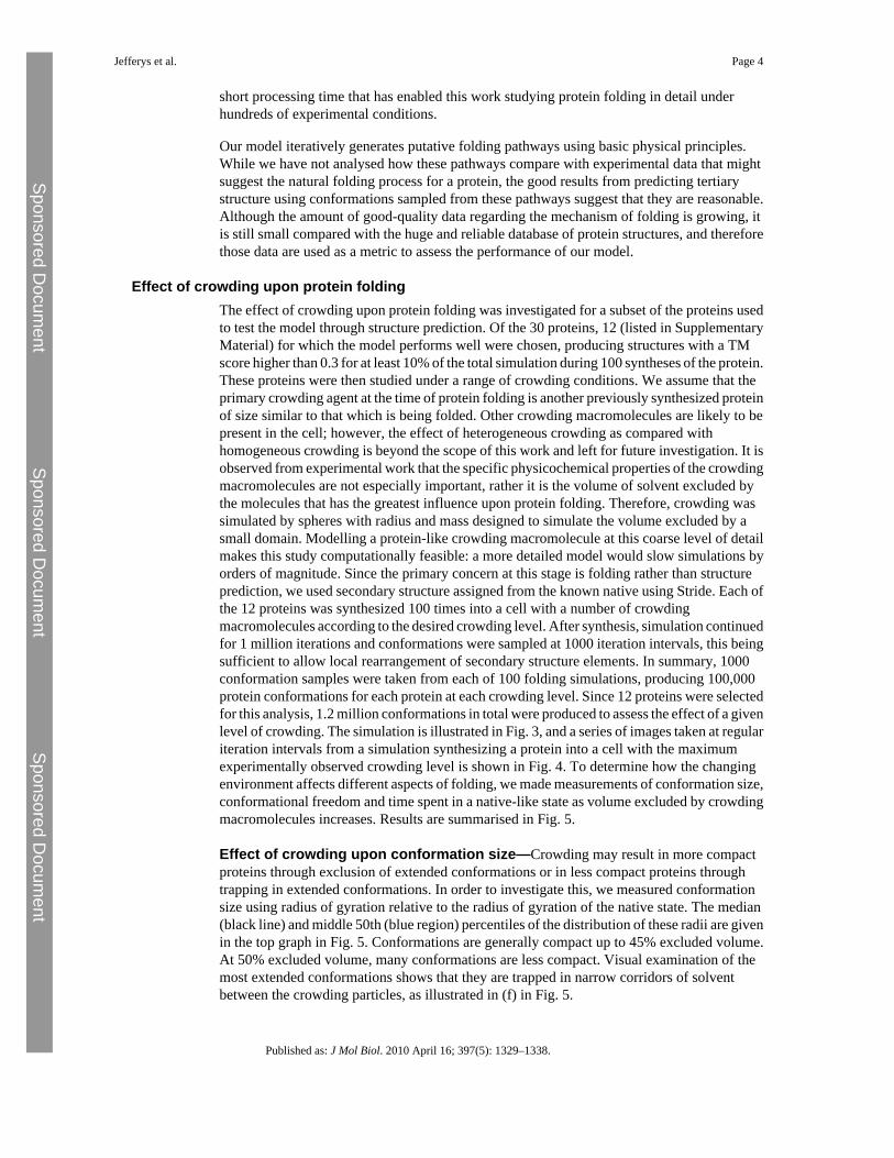
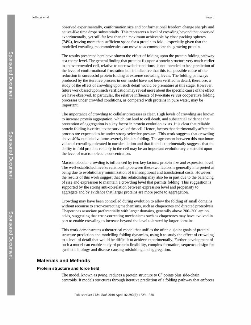
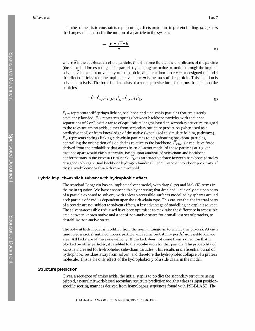
Effect of crowding upon protein foldingThe effect of crowding upon protein folding was investigated for a subset of the proteins usedto test the model through structure prediction. Of the 30 proteins, 12 (listed in SupplementaryMaterial) for which the model performs well were chosen, producing structures with a TMscore higher than 0.3 for at least 10% of the total simulation during 100 syntheses of the protein.These proteins were then studied under a range of crowding conditions. We assume that theprimary crowding agent at the time of protein folding is another previously synthesized proteinof size similar to that which is being folded. Other crowding macromolecules are likely to bepresent in the cell; however, the effect of heterogeneous crowding as compared withhomogeneous crowding is beyond the scope of this work and left for future investigation. It isobserved from experimental work that the specific physicochemical properties of the crowdingmacromolecules are not especially important, rather it is the volume of solvent excluded bythe molecules that has the greatest influence upon protein folding. Therefore, crowding wassimulated by spheres with radius and mass designed to simulate the volume excluded by asmall domain. Modelling a protein-like crowding macromolecule at this coarse level of detailmakes this study computationally feasible: a more detailed model would slow simulations byorders of magnitude. Since the primary concern at this stage is folding rather than structureprediction, we used secondary structure assigned from the known native using Stride. Each ofthe 12 proteins was synthesized 100 times into a cell with a number of crowdingmacromolecules according to the desired crowding level. After synthesis, simulation continuedfor 1 million iterations and conformations were sampled at 1000 iteration intervals, this beingsufficient to allow local rearrangement of secondary structure elements. In summary, 1000conformation samples were taken from each of 100 folding simulations, producing 100,000protein conformations for each protein at each crowding level. Since 12 proteins were selectedfor this analysis, 1.2 million conformations in total were produced to assess the effect of a givenlevel of crowding. The simulation is illustrated in Fig. 3, and a series of images taken at regulariteration intervals from a simulation synthesizing a protein into a cell with the maximumexperimentally observed crowding level is shown in Fig. 4. To determine how the changingenvironment affects different aspects of folding, we made measurements of conformation size,conformational freedom and time spent in a native-like state as volume excluded by crowdingmacromolecules increases. Results are summarised in Fig. 5.
Effect of crowding upon conformation size—Crowding may result in more compactproteins through exclusion of extended conformations or in less compact proteins throughtrapping in extended conformations. In order to investigate this, we measured conformationsize using radius of gyration relative to the radius of gyration of the native state. The median(black line) and middle 50th (blue region) percentiles of the distribution of these radii are givenin the top graph in Fig. 5. Conformations are generally compact up to 45% excluded volume.At 50% excluded volume, many conformations are less compact. Visual examination of themost extended conformations shows that they are trapped in narrow corridors of solventbetween the crowding particles, as illustrated in (f) in Fig. 5.
Jefferys et al. Page 4
Published as: J Mol Biol. 2010 April 16; 397(5): 1329–1338.
Sponsored Docum
ent Sponsored D
ocument
Sponsored Docum
ent
Effect of crowding upon conformational freedom—Crowding may change theconformational freedom of proteins—they may be stabilised or destabilised by collisions withmacromolecules. We assess the time step of each simulation at which a protein finds aconformation that does not change significantly for the rest of the simulation. Conformationsare defined as being similar if the mean difference in pairwise distances between correspondingamino acids is less than 4 Å. The reported time step for a given simulation is that at which aconformation that is similar to all conformations after it until the end of the simulation is found.The median (black line) and middle 50th (blue region) percentiles of this number are shownin the middle graph in Fig. 5. Freedom remains fairly constant up to 30% excluded volume,with most of the simulated proteins free to explore conformation space until around 800,000iterations (out of 1 million). Above 40% excluded volume, conformational freedom decreasessharply: most simulated proteins fix upon a structure before 200,000 iterations. Combiningthis with the observations on conformation size, at 50% excluded volume, it would seem thatproteins are becoming trapped in an extended state. At slightly below this level, proteins aregenerally trapped in a compact state.
Effect of crowding upon native-like time—Crowding may increase or decrease thesimilarity of the protein to the native state during the simulation. We assess this effect to bemeasuring native-like time, which we define as the percentage of sampled conformations thatare higher than 0.3 in TM score to the native state. The percentage across all proteins is shownin the bottom graph of Fig. 5. Native-like time remains fairly constant up to 25% excludedvolume, with over 20% of conformations being above 0.3 in TM score. There is an initial slightdrop up to 30% crowding, which cannot be attributed to more extended conformations orbecoming trapped in a conformation early. Above this level, native-like time reduces rapidly,until at 50% excluded volume, just over 10% of conformations have a TM score above 0.3.The large drop in native-like time at 35% coincides with a slight reduction in conformationalfreedom, although conformation size remains fairly constant. Some proteins become stabilisedin compact but non-native conformations by the presence of crowding particles, illustrated in(e) in Fig. 5. This effect becomes more acute at 45% excluded volume, when conformationalfreedom drops sharply. Finally, when crowding is so great the proteins cannot find a compactconformation, proteins very quickly become trapped in extended states that are clearly lesslikely to be similar to the native [illustrated in (f) in Fig. 5].
In summary, there is little effect on the measured features up to 20% excluded volume. Ataround 25%, native-like time begins to reduce, although not due to being fixed early or trappedin extended conformations. Above 40% crowding, proteins are becoming stuck in structuresearlier in the simulation, and around 15 of the structures are in native-like topologies, comparedwith 14 with no crowding. At 50% crowding, proteins are stabilised in extended conformationsearly in the simulation in narrow gaps between macromolecules.
DiscussionWe have presented a protein structure model designed to predict tertiary structure without theuse of a template or detailed knowledge of the native state. The model demonstrates accuracycomparable with that of the most successful methods, such as fragment folding, with the addedbenefit that it is fast and models a putative folding pathway. Using this model, we have testedthe effect of macromolecular crowding upon the folding of 12 small protein domainssynthesized into a simulated cell.
Crowding macromolecules in the simulated cell were simulated using same-sized spheres upto an excluded volume of 50%. As crowding is increased to experimentally observed levels(20% to 40%), a slow decrease in native-like time, due to the stabilisation of non-native butcompact states, is observed. However, when the crowding level surpasses the maximum
Jefferys et al. Page 5
Published as: J Mol Biol. 2010 April 16; 397(5): 1329–1338.
Sponsored Docum
ent Sponsored D
ocument
Sponsored Docum
ent
observed experimentally, conformation size and conformational freedom change sharply andnative-like time drops substantially. This represents a level of crowding beyond that observedexperimentally, yet still far less than the maximum achievable by close packing spheres(74%), leaving more than sufficient space for a protein to fold—especially given that themodelled crowding macromolecules can move to accommodate the growing protein.
The results presented here have shown the effect of folding upon the protein folding pathwayat a coarse level. The general finding that proteins fix upon a protein structure very much earlierin an overcrowded cell, relative to uncrowded conditions, is not intended to be a prediction ofthe level of conformational frustration but is indicative that this is a possible cause of thereduction in successful protein folding at extreme crowding levels. The folding pathwaysproduced by the iterative process in our model have not been verified in detail; therefore, astudy of the effect of crowding upon such detail would be premature at this stage. However,future work based upon such verification may reveal more about the specific cause of the effectwe have observed. In particular, the relative influence of two-state versus cooperative foldingprocesses under crowded conditions, as compared with proteins in pure water, may beimportant.
The importance of crowding to cellular processes is clear. High levels of crowding are knownto increase protein aggregation, which can lead to cell death, and substantial evidence thatprevention of aggregation is a key factor in protein evolution exists. It is clear that reliableprotein folding is critical to the survival of the cell. Hence, factors that detrimentally affect thisprocess are expected to be under strong selective pressure. This work suggests that crowdingabove 40% excluded volume severely hinders folding. The agreement between this maximumvalue of crowding tolerated in our simulation and that found experimentally suggests that theability to fold proteins reliably in the cell may be an important evolutionary constraint uponthe level of macromolecule concentration.
Macromolecular crowding is influenced by two key factors: protein size and expression level.The well-established inverse relationship between these two factors is generally interpreted asbeing due to evolutionary minimization of transcriptional and translational costs. However,the results of this work suggest that this relationship may also be in part due to the balancingof size and expression to maintain a crowding level that permits folding. This suggestion issupported by the strong anti-correlation between expression level and propensity toaggregate and by evidence that larger proteins are more prone to aggregation.
Crowding may have been controlled during evolution to allow the folding of small domainswithout recourse to error-correcting mechanisms, such as chaperones and directed proteolysis.Chaperones associate preferentially with larger domains, generally above 200–300 aminoacids, suggesting that error-correcting mechanisms such as chaperones may have evolved inpart to enable crowding to increase beyond the level tolerated by larger domains.
This work demonstrates a theoretical model that unifies the often disjoint goals of proteinstructure prediction and modelling folding dynamics, using it to study the effect of crowdingto a level of detail that would be difficult to achieve experimentally. Further development ofsuch a model can enable study of protein flexibility, complex formation, sequence design forsynthetic biology and disease-causing misfolding and aggregation.
Materials and MethodsProtein structure and force field
The model, known as poing, reduces a protein structure to Cα points plus side-chaincentroids. It models structures through iterative prediction of a folding pathway that enforces
Jefferys et al. Page 6
Published as: J Mol Biol. 2010 April 16; 397(5): 1329–1338.
Sponsored Docum
ent Sponsored D
ocument
Sponsored Docum
ent
a number of heuristic constraints representing effects important in protein folding. poing usesthe Langevin equation for the motion of a particle in the system:
(1)
where a⃗ is the acceleration of the particle, F⃗ is the force field at the coordinates of the particle(the sum of all forces acting on the particle), γ is a drag factor due to motion through the implicitsolvent, v⃗ is the current velocity of the particle, R⃗ is a random force vector designed to modelthe effect of kicks from the implicit solvent and m is the mass of the particle. This equation issolved iteratively. The force field consists of a set of pairwise force functions that act upon theparticles:
(2)
F⃗cov represents stiff springs linking backbone and side-chain particles that are directlycovalently bonded. F⃗bb represents springs between backbone particles with sequenceseparations of 2 or 3, with a range of equilibrium lengths based on secondary structure assignedto the relevant amino acids, either from secondary structure prediction (when used as apredictive tool) or from knowledge of the native (when used to simulate folding pathways).F⃗sc represents springs linking side-chain particles to neighbouring backbone particles,controlling the orientation of side chains relative to the backbone. F⃗vdw is a repulsive forcederived from the probability that atoms in an all-atom model of those particles at a givendistance apart would clash sterically, based upon analysis of side-chain and backboneconformations in the Protein Data Bank. F⃗hb is an attractive force between backbone particlesdesigned to bring virtual backbone hydrogen bonding O and H atoms into closer proximity, ifthey already come within a distance threshold.
Hybrid implicit–explicit solvent with hydrophobic effectThe standard Langevin has an implicit solvent model, with drag (−γv⃗) and kick (R⃗) terms inthe main equation. We have enhanced this by ensuring that drag and kicks only act upon partsof a particle exposed to solvent, with solvent-accessible surfaces modelled by spheres aroundeach particle of a radius dependent upon the side-chain type. This ensures that the internal partsof a protein are not subject to solvent effects, a key advantage of modelling an explicit solvent.The solvent-accessible radii used have been optimised to maximise the difference in accessiblearea between known native and a set of non-native states for a small test set of proteins, todestabilise non-native states.
The solvent kick model is modified from the normal Langevin to enable this process. At eachtime step, a kick is initiated upon a particle with some probability per Å2 accessible surfacearea. All kicks are of the same velocity. If the kick does not come from a direction that isblocked by other particles, it is added to the acceleration for that particle. The probability ofkicks is increased for hydrophobic side-chain particles. This results in preferential burial ofhydrophobic residues away from solvent and therefore the hydrophobic collapse of a proteinmolecule. This is the only effect of the hydrophobicity of a side chain in the model.
Structure predictionGiven a sequence of amino acids, the initial step is to predict the secondary structure usingpsipred, a neural network-based secondary structure prediction tool that takes as input position-specific scoring matrices derived from homologous sequences found with PSI-BLAST. The
Jefferys et al. Page 7
Published as: J Mol Biol. 2010 April 16; 397(5): 1329–1338.
Sponsored Docum
ent Sponsored D
ocument
Sponsored Docum
ent
accuracy of this method generally reduces where no homologue is found. A simple three-stateoutput (helix, extended/strand and coil) is used to assign secondary structure to the amino acidsin the poing model. The protein is then modelled in poing, for (6000l + 400,000) iterations,where l is the number of amino acids in the protein. Forty structures are sampled at equalintervals from iteration 6000l to the end of the simulation. As a computational shortcut to acompact structure, the protein is slowly synthesized by adding an amino acid to the C-terminusof the protein every set of 1000 iterations and tethering this growing end to the edge of a largeheavy sphere representing a ribosome. This process is repeated 150 times with different randomseeds for the generation of kicks from solvent in the Langevin equation, producing manyfolding trajectories. This produces the final pool of structures from which predictions are made.
Determination of structure predictions from generated conformations is performed in threestages. First, a mean of the contact maps of the backbone particles of the pool of structures isgenerated. The contact maps are calculated based upon backbone traces smoothed with awindow of nine amino acids. The contact maps disregard contacts between amino acids withinthe same secondary structure unit. Different distance cutoffs are used depending on thesecondary structure: 8 Å between amino acids that are both assigned as strand and 11 Å in allother cases. This reflects the fact that the backbones of hydrogen-bonded strands are closerthan other secondary structure elements. All 6000 structures are sorted according to theirsimilarity to this mean contact map—the contact map similarity score penalises a lack of contactwhere there should be one and rewards the presence of a contact where there should be one.
The top 20 structures selected by similarity to a consensus contact map are scored usingProQ, a neural net-based structure scoring program trained to predict the MaxSub score of aprotein as compared with its (unknown) native state. If any of the top 5 structures picked byProQ are very similar to one another, the one with the lowest score is eliminated and the nextstructure down is brought in to the top 5. Two proteins are judged to be very similar if morethan 90% of the residues can be aligned to less than 7 Å RMSD. This process is repeated untilthe top 5 represents a range of the best structures produced by poing, or there are no morestructures.
Model for protein synthesis into a crowded cellThis is illustrated in Fig. 3. The model consists of a simulated protein (described above) andcrowding macromolecule spheres inside a containing sphere and a heavy ribosome with asynthesis exit point located on its surface, with the exit point tethered to the centre of thecontaining sphere. The ribosome, crowding macromolecules and containing sphere all moveiteratively under forces applied through the Langevin equation, including the solvent model.Details of these elements are given below.
In poing, the ribosome is modelled with a heavy sphere of radius 100 Å. All simulated particlesare excluded from this sphere. The mass is set to make the ribosome an essentially immovableobject. The protein emerges from an exit tunnel on the surface of a ribosome, to which it istethered: this is modelled by a particle attached to the surface of the ribosome. This particlehas the same mass as the ribosome in order to approximate the moment of inertia of theribosome as a whole. The backbone particle at the growing end of a protein is attached to thisparticle. This set of strong springs and massive particles presents a major constraint to theprotein's freedom, which is designed to approximate the effect of the ribosome on proteinfolding.
Protein synthesis is modelled by periodically adding backbone and side-chain particles to theC-terminus chain at the position of the exit tunnel. The new backbone point is tethered to theexit point, and the old backbone point to which it is attached is no longer tethered (however,its location is still restricted by its backbone link to the new backbone particle). We used a
Jefferys et al. Page 8
Published as: J Mol Biol. 2010 April 16; 397(5): 1329–1338.
Sponsored Docum
ent Sponsored D
ocument
Sponsored Docum
ent
synthesis rate of one amino acid per 1000 time steps. This is an artificially fast synthesis rate,relative to the rate of protein collapse in our model; however, short tests have found that slowersynthesis rates do not alter our observations. This relatively fast rate was chosen as acomputational optimisation.
Macromolecular crowding is designed to simulate the crowding of a protein by other proteinsof a similar size. Previous work has suggested that the specific physicochemical properties ofthe crowding macromolecules are not especially important and that the principal effect isvolume exclusion. It would be computationally prohibitive and unnecessary to crowd a proteinwith other complete protein models if there is no requirement to simulate specific interactions.Therefore, in poing, a crowding macromolecule is reduced to a large, heavy sphere. Themacromolecules crowding a protein are assumed to be of size similar to the protein beingcrowded. The set of small domains used in this study is between 43 and 90 amino acids long.All particles in the protein model have the same mass, and each amino acid has two particlesassociated with it. This is simplified to a single crowding particle that models a 75-amino-acidprotein, of diameter 30 Å (the approximate size of a 75-amino-acid protein) and with mass 150times that of a single particle. The entire system of particles (excluding the ribosome) iscontained within a sphere of diameter 100 Å.
The ribosome exit point is tied to the centre of the crowding containment sphere with a spring,ensuring that the protein is synthesized into the centre of the crowding area. This ensures thatany observed effects upon folding from crowding are due to the crowding rather than boundary-specific effects that might be observed if the protein is synthesized at the boundary.
Appendix A Supplementary dataRefer to Web version on PubMed Central for supplementary material.
Appendix A Supplementary DataRefer to Web version on PubMed Central for supplementary material.
AcknowledgmentsThis work was supported by the Wellcome Trust and the Biotechnology and Biological Sciences Research Council.We thank Maxime Huvet and Mark Wass for valuable comments and suggestions.
References1. Liwo A. Khalili M. Scheraga H.A. Ab initio simulations of protein-folding pathways by molecular
dynamics with the united-residue model of polypeptide chains. Proc. Natl Acad. Sci. USA2005;102:2362–2367. [PubMed: 15677316]
2. Moult J. Fidelis K. Kryshtafovych A. Rost B. Hubbard T. Tramontano A. Critical assessment ofmethods of protein structure prediction—round VII. Proteins 2007;69:3–9. [PubMed: 17918729]
3. Zhang Y. Protein structure prediction: when is it useful? Curr. Opin. Struct. Biol. 2009;19:145–155.[PubMed: 19327982]
4. Das R. Qian B. Raman S. Vernon R. Thompson J. Bradley P. Structure prediction for CASP7 targetsusing extensive all-atom refinement with Rosetta@home. Proteins 2007;69:118–128. [PubMed:17894356]
5. Zhang Y. I-TASSER server for protein 3D structure prediction. BMC Bioinformatics 2008;9:40.[PubMed: 18215316]
6. Kelley L.A. Sternberg M.J.E. Protein structure prediction on the Web: a case study using the Phyreserver. Nat. Protoc. 2009;4:363–371. [PubMed: 19247286]
Jefferys et al. Page 9
Published as: J Mol Biol. 2010 April 16; 397(5): 1329–1338.
Sponsored Docum
ent Sponsored D
ocument
Sponsored Docum
ent
7. Soeding J. Biegert A. Lupas A.N. The HHpred interactive server for protein homology detection andstructure prediction. Nucleic Acids Res. 2005;33:W244–W248. [PubMed: 15980461]
8. Lucent D. Vishal V. Pande V.S. Protein folding under confinement: a role for solvent. Proc. Natl Acad.Sci. USA 2007;104:10430–10434. [PubMed: 17563390]
9. Shell M.S. Ozkan S.B. Voelz V. Wu G.A. Dill K.A. Blind test of physics-based prediction of proteinstructures. Biophys. J. 2009;96:917–924. [PubMed: 19186130]
10. DeBartolo J. Colubri A. Jha A.K. Fitzgerald J.E. Freed K.F. Sosnick T.R. Mimicking the foldingpathway to improve homology-free protein structure prediction. Proc. Natl Acad. Sci. USA2009;106:3734–3739. [PubMed: 19237560]
11. Levitt M. Warshel A. Computer simulation of protein folding. Nature 1975;253:694–698. [PubMed:1167625]
12. Ellis R.J. Macromolecular crowding: an important but neglected aspect of the intracellularenvironment. Curr. Opin. Struct. Biol. 2001;11:114–119. [PubMed: 11179900]
13. Rivas G. Ferrone F. Herzfeld J. Life in a crowded world. EMBO Rep. 2004;5:23–27. [PubMed:14710181]
14. Pielak G.J. A model of intracellular organization. Proc. Natl Acad. Sci. USA 2005;102:5901–5902.[PubMed: 15840719]
15. Long M.S. Jones C.D. Helfrich M.R. Mangeney-Slavin L.K. Keating C.D. Dynamicmicrocompartmentation in synthetic cells. Proc. Natl Acad. Sci. USA 2005;102:5920–5925.[PubMed: 15788532]
16. Minton A.P. Influence of macromolecular crowding upon the stability and state of association ofproteins: predictions and observations. J. Pharm. Sci. 2005;94:1668–1675. [PubMed: 15986476]
17. van den Berg B. Ellis R.J. Dobson C.M. Effects of macromolecular crowding on protein folding andaggregation. EMBO J. 1999;18:6927–6933. [PubMed: 10601015]
18. van den Berg B. Wain R. Dobson C.M. Ellis R.J. Macromolecular crowding perturbs protein refoldingkinetics: implications for folding inside the cell. EMBO J. 2000;19:3870–3875. [PubMed: 10921869]
19. Cheung M.S. Klimov D. Thirumalai D. Molecular crowding enhances native state stability andrefolding rates of globular proteins. Proc. Natl Acad. Sci. USA 2005;102:4753–4758. [PubMed:15781864]
20. Ai X. Zhou Z. Bai Y. Choy W.Y. 15N NMR spin relaxation dispersion study of the molecularcrowding effects on protein folding under native conditions. J. Am. Chem. Soc. 2006;128:3916–3917. [PubMed: 16551092]
21. Lu D. Liu Z. Wu J. Structural transitions of confined model proteins: molecular dynamics simulationand experimental validation. Biophys. J. 2006;90:3224–3238. [PubMed: 16461405]
22. Stagg L. Zhang S.Q. Cheung M.S. Wittung-Stafshede P. Molecular crowding enhances nativestructure and stability of alpha/beta protein flavodoxin. Proc. Natl Acad. Sci. USA 2007;104:18976–18981. [PubMed: 18024596]
23. Taketomi H. Ueda Y. Go N. Studies on protein folding, unfolding and fluctuations by computersimulation: I. The effect of specific amino acid sequence represented by specific inter-unitinteractions. Int. J. Pept. Protein Res. 1975;7:445–459. [PubMed: 1201909]
24. Samiotakis A. Wittung-Stafshede P. Cheung M.S. Folding, stability and shape of proteins in crowdedenvironments: experimental and computational approaches. Int. J. Mol. Sci. 2009;10:572–588.[PubMed: 19333422]
25. Li L. Mirny L.A. Shakhnovich E.I. Kinetics, thermodynamics and evolution of non-native interactionsin a protein folding nucleus. Nat. Struct. Biol. 2000;7:336–342. [PubMed: 10742180]
26. Sikorski A. Skolnick J. Dynamic Monte Carlo simulations of globular protein folding. Model studiesof in vivo assembly of four helix bundles and four member beta-barrels. J. Mol. Biol. 1990;215:183–198. [PubMed: 2398497]
27. Elcock A.H. Molecular simulations of cotranslational protein folding: fragment stabilities, foldingcooperativity, and trapping in the ribosome. PLoS Comput. Biol. 2006;e98:2.
28. Taylor W.R. Topological accessibility shows a distinct asymmetry in the folds of betaalpha proteins.FEBS Lett. 2006;580:5263–5267. [PubMed: 16979627]
Jefferys et al. Page 10
Published as: J Mol Biol. 2010 April 16; 397(5): 1329–1338.
Sponsored Docum
ent Sponsored D
ocument
Sponsored Docum
ent
29. Deane C.M. Dong M. Huard F.P.E. Lance B.K. Wood G.R. Cotranslational protein folding—fact orfiction? Bioinformatics 2007;23:i142–i148. [PubMed: 17646290]
30. Gay G.D.P. Ruiz-Sanz J. Neira J.L. Itzhaki L.S. Fersht A.R. Folding of a nascent polypeptide chainin vitro: cooperative formation of structure in a protein module. Proc. Natl Acad. Sci. USA1995;92:3683–3686. [PubMed: 7731965]
31. Fedorov A.N. Baldwin T.O. Cotranslational protein folding. J. Biol. Chem. 1997;272:32715–32718.[PubMed: 9407040]
32. Jones D.T. Protein secondary structure prediction based on position-specific scoring matrices. J. Mol.Biol. 1999;292:195–202. [PubMed: 10493868]
33. Zhang Y. Skolnick J. Scoring function for automated assessment of protein structure template quality.Proteins 2004;57:702–710. [PubMed: 15476259]
34. Jauch R. Yeo H.C. Kolatkar P.R. Clarke N.D. Assessment of CASP7 structure predictions for templatefree targets. Proteins 2007;69:57–67. [PubMed: 17894330]
35. Uversky V.N. Cooper E.M. Bower K.S. Li J. Fink A.L. Accelerated alpha-synuclein fibrillation incrowded milieu. FEBS Lett. 2002;515:99–103. [PubMed: 11943202]
36. Frishman D. Argos P. Knowledge-based protein secondary structure assignment. Proteins1995;23:566–579. [PubMed: 8749853]
37. Hales T.C. A proof of the Kepler conjecture. Ann. Math. 2005;162:1063–1183.38. Monsellier E. Chiti F. Prevention of amyloid-like aggregation as a driving force of protein evolution.
EMBO Rep. 2007;8:737–742. [PubMed: 17668004]39. Jansen R. Gerstein M. Analysis of the yeast transcriptome with structural and functional categories:
characterizing highly expressed proteins. Nucleic Acids Res. 2000;28:1481–1488. [PubMed:10684945]
40. Coghlan A. Wolfe K.H. Relationship of codon bias to mRNA concentration and protein length inSaccharomyces cerevisiae. Yeast 2000;16:1131–1145. [PubMed: 10953085]
41. Lu P. Vogel C. Wang R. Yao X. Marcotte E.M. Absolute protein expression profiling estimates therelative contributions of transcriptional and translational regulation. Nat. Biotechnol. 2007;25:117–124. [PubMed: 17187058]
42. Tartaglia G.G. Pechmann S. Dobson C.M. Vendruscolo M. Life on the edge: a link between geneexpression levels and aggregation rates of human proteins. Trends Biochem. Sci. 2007;32:204–206.[PubMed: 17419062]
43. Rousseau F. Serrano L. Schymkowitz J.W.H. How evolutionary pressure against protein aggregationshaped chaperone specificity. J. Mol. Biol. 2006;355:1037–1047. [PubMed: 16359707]
44. Teter S.A. Houry W.A. Ang D. Tradler T. Rockabrand D. Fischer G. Polypeptide flux throughbacterial Hsp70: DnaK cooperates with trigger factor in chaperoning nascent chains. Cell1999;97:755–765. [PubMed: 10380927]
45. Bukau B. Deuerling E. Pfund C. Craig E.A. Getting newly synthesized proteins into shape. Cell2000;101:119–122. [PubMed: 10786831]
46. Hartl F.U. Hayer-Hartl M. Molecular chaperones in the cytosol: from nascent chain to folded protein.Science 2002;295:1852–1858. [PubMed: 11884745]
47. Schlick, T. Molecular Modeling and Simulation. Springer-Verlag; New York, NY: 2002. p. 435-438.48. Berman H.M. Westbrook J. Feng Z. Gilliland G. Bhat T.N. Weissig H. The Protein Data Bank. Nucleic
Acids Res. 2000;28:235–242. [PubMed: 10592235]49. Wallner B. Elofsson A. Can correct protein models be identified? Protein Sci. 2003;12:1073–1086.
[PubMed: 12717029]
Jefferys et al. Page 11
Published as: J Mol Biol. 2010 April 16; 397(5): 1329–1338.
Sponsored Docum
ent Sponsored D
ocument
Sponsored Docum
ent
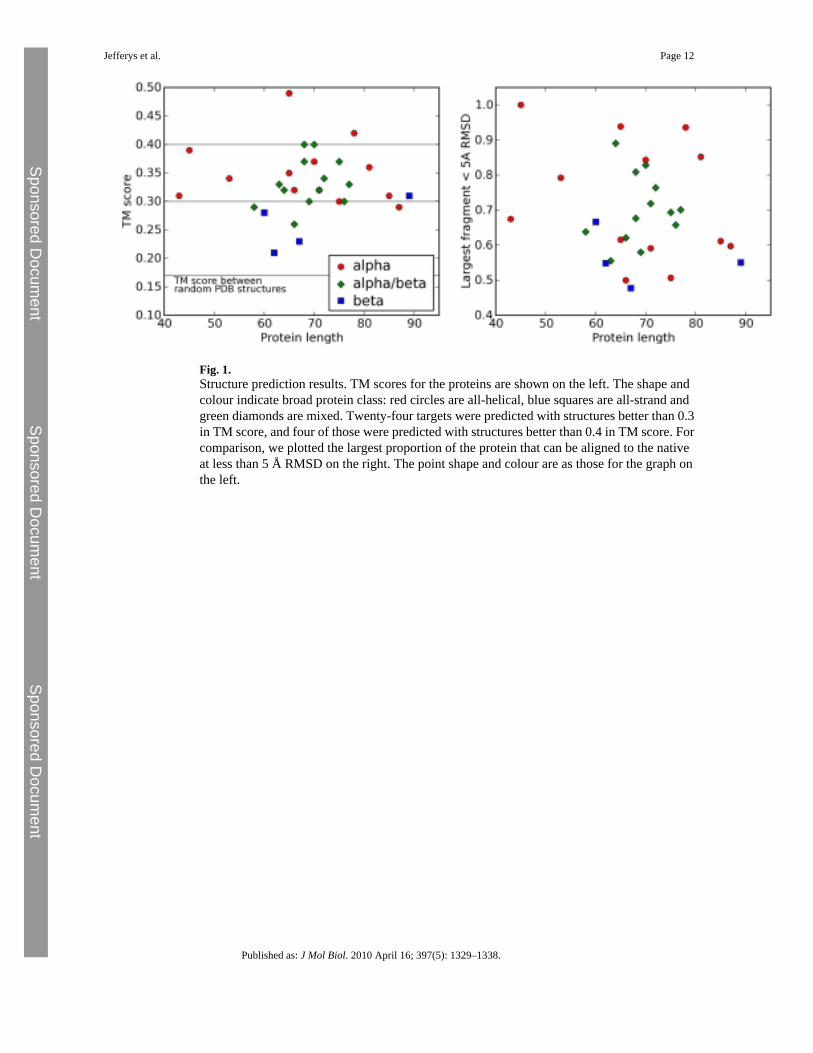
Fig. 1.Structure prediction results. TM scores for the proteins are shown on the left. The shape andcolour indicate broad protein class: red circles are all-helical, blue squares are all-strand andgreen diamonds are mixed. Twenty-four targets were predicted with structures better than 0.3in TM score, and four of those were predicted with structures better than 0.4 in TM score. Forcomparison, we plotted the largest proportion of the protein that can be aligned to the nativeat less than 5 Å RMSD on the right. The point shape and colour are as those for the graph onthe left.
Jefferys et al. Page 12
Published as: J Mol Biol. 2010 April 16; 397(5): 1329–1338.
Sponsored Docum
ent Sponsored D
ocument
Sponsored Docum
ent
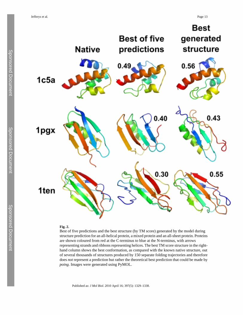
Fig. 2.Best of five predictions and the best structure (by TM score) generated by the model duringstructure prediction for an all-helical protein, a mixed protein and an all-sheet protein. Proteinsare shown coloured from red at the C-terminus to blue at the N-terminus, with arrowsrepresenting strands and ribbons representing helices. The best TM score structure in the right-hand column shows the best conformation, as compared with the known native structure, outof several thousands of structures produced by 150 separate folding trajectories and thereforedoes not represent a prediction but rather the theoretical best prediction that could be made bypoing. Images were generated using PyMOL.
Jefferys et al. Page 13
Published as: J Mol Biol. 2010 April 16; 397(5): 1329–1338.
Sponsored Docum
ent Sponsored D
ocument
Sponsored Docum
ent
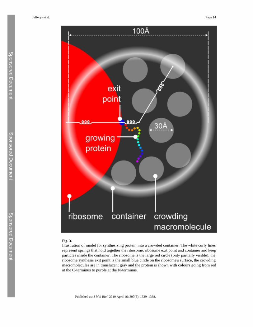
Fig. 3.Illustration of model for synthesizing protein into a crowded container. The white curly linesrepresent springs that hold together the ribosome, ribosome exit point and container and keepparticles inside the container. The ribosome is the large red circle (only partially visible), theribosome synthesis exit point is the small blue circle on the ribosome's surface, the crowdingmacromolecules are in translucent gray and the protein is shown with colours going from redat the C-terminus to purple at the N-terminus.
Jefferys et al. Page 14
Published as: J Mol Biol. 2010 April 16; 397(5): 1329–1338.
Sponsored Docum
ent Sponsored D
ocument
Sponsored Docum
ent
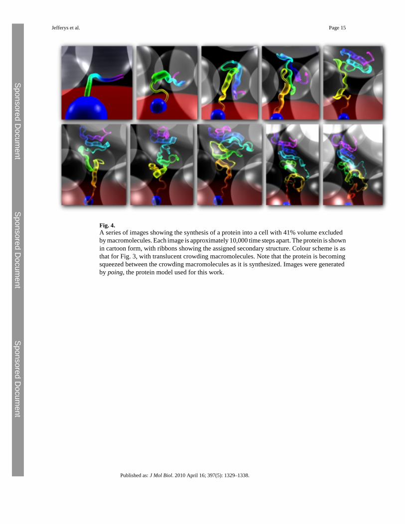
Fig. 4.A series of images showing the synthesis of a protein into a cell with 41% volume excludedby macromolecules. Each image is approximately 10,000 time steps apart. The protein is shownin cartoon form, with ribbons showing the assigned secondary structure. Colour scheme is asthat for Fig. 3, with translucent crowding macromolecules. Note that the protein is becomingsqueezed between the crowding macromolecules as it is synthesized. Images were generatedby poing, the protein model used for this work.
Jefferys et al. Page 15
Published as: J Mol Biol. 2010 April 16; 397(5): 1329–1338.
Sponsored Docum
ent Sponsored D
ocument
Sponsored Docum
ent
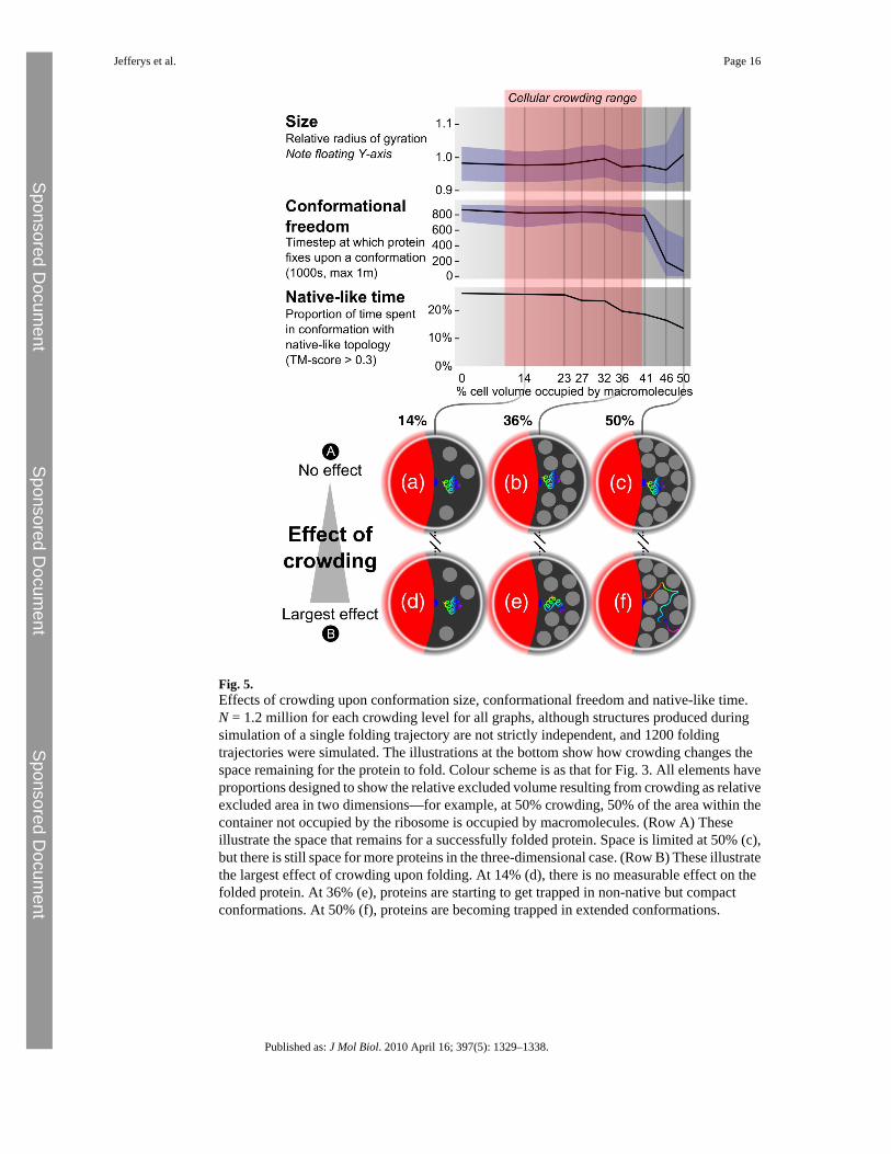
Fig. 5.Effects of crowding upon conformation size, conformational freedom and native-like time.N = 1.2 million for each crowding level for all graphs, although structures produced duringsimulation of a single folding trajectory are not strictly independent, and 1200 foldingtrajectories were simulated. The illustrations at the bottom show how crowding changes thespace remaining for the protein to fold. Colour scheme is as that for Fig. 3. All elements haveproportions designed to show the relative excluded volume resulting from crowding as relativeexcluded area in two dimensions—for example, at 50% crowding, 50% of the area within thecontainer not occupied by the ribosome is occupied by macromolecules. (Row A) Theseillustrate the space that remains for a successfully folded protein. Space is limited at 50% (c),but there is still space for more proteins in the three-dimensional case. (Row B) These illustratethe largest effect of crowding upon folding. At 14% (d), there is no measurable effect on thefolded protein. At 36% (e), proteins are starting to get trapped in non-native but compactconformations. At 50% (f), proteins are becoming trapped in extended conformations.
Jefferys et al. Page 16
Published as: J Mol Biol. 2010 April 16; 397(5): 1329–1338.
Sponsored Docum
ent Sponsored D
ocument
Sponsored Docum
ent