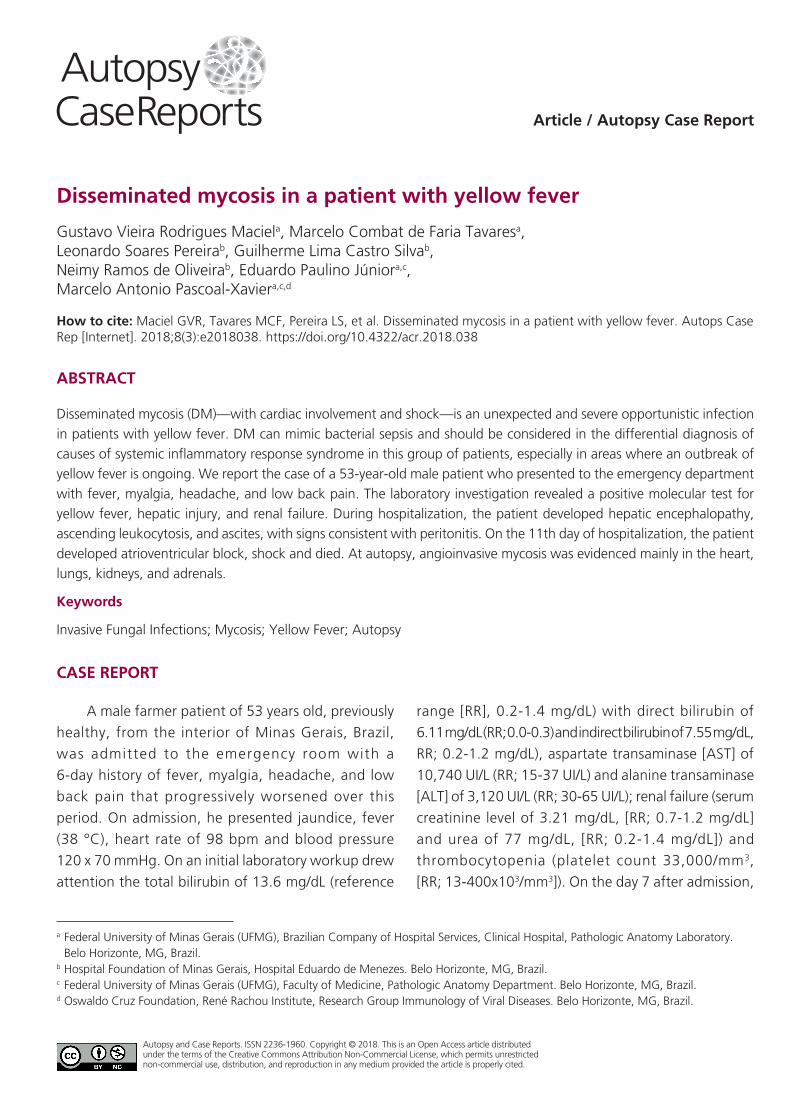
Disseminated mycosis in a patient with yellow...
8
Autopsy and Case Reports. ISSN 2236-1960. Copyright © 2018. This is an Open Access article distributed under the terms of the Creative Commons Attribution Non-Commercial License, which permits unrestricted non-commercial use, distribution, and reproduction in any medium provided the article is properly cited. a Federal University of Minas Gerais (UFMG), Brazilian Company of Hospital Services, Clinical Hospital, Pathologic Anatomy Laboratory. Belo Horizonte, MG, Brazil. b Hospital Foundation of Minas Gerais, Hospital Eduardo de Menezes. Belo Horizonte, MG, Brazil. c Federal University of Minas Gerais (UFMG), Faculty of Medicine, Pathologic Anatomy Department. Belo Horizonte, MG, Brazil. d Oswaldo Cruz Foundation, René Rachou Institute, Research Group Immunology of Viral Diseases. Belo Horizonte, MG, Brazil. Disseminated mycosis in a patient with yellow fever Gustavo Vieira Rodrigues Maciel a , Marcelo Combat de Faria Tavares a , Leonardo Soares Pereira b , Guilherme Lima Castro Silva b , Neimy Ramos de Oliveira b , Eduardo Paulino Júnior a,c , Marcelo Antonio Pascoal-Xavier a,c,d How to cite: Maciel GVR, Tavares MCF, Pereira LS, et al. Disseminated mycosis in a patient with yellow fever. Autops Case Rep [Internet]. 2018;8(3):e2018038. https://doi.org/10.4322/acr.2018.038 Article / Autopsy Case Report ABSTRACT Disseminated mycosis (DM)—with cardiac involvement and shock—is an unexpected and severe opportunistic infection in patients with yellow fever. DM can mimic bacterial sepsis and should be considered in the differential diagnosis of causes of systemic inflammatory response syndrome in this group of patients, especially in areas where an outbreak of yellow fever is ongoing. We report the case of a 53-year-old male patient who presented to the emergency department with fever, myalgia, headache, and low back pain. The laboratory investigation revealed a positive molecular test for yellow fever, hepatic injury, and renal failure. During hospitalization, the patient developed hepatic encephalopathy, ascending leukocytosis, and ascites, with signs consistent with peritonitis. On the 11th day of hospitalization, the patient developed atrioventricular block, shock and died. At autopsy, angioinvasive mycosis was evidenced mainly in the heart, lungs, kidneys, and adrenals. Keywords Invasive Fungal Infections; Mycosis; Yellow Fever; Autopsy CASE REPORT A male farmer patient of 53 years old, previously healthy, from the interior of Minas Gerais, Brazil, was admitted to the emergency room with a 6-day history of fever, myalgia, headache, and low back pain that progressively worsened over this period. On admission, he presented jaundice, fever (38 °C), heart rate of 98 bpm and blood pressure 120 x 70 mmHg. On an initial laboratory workup drew attention the total bilirubin of 13.6 mg/dL (reference range [RR], 0.2-1.4 mg/dL) with direct bilirubin of 6.11 mg/dL (RR; 0.0-0.3) and indirect bilirubin of 7.55 mg/dL, RR; 0.2-1.2 mg/dL), aspartate transaminase [AST] of 10,740 UI/L (RR; 15-37 UI/L) and alanine transaminase [ALT] of 3,120 UI/L (RR; 30-65 UI/L); renal failure (serum creatinine level of 3.21 mg/dL, [RR; 0.7-1.2 mg/dL] and urea of 77 mg/dL, [RR; 0.2-1.4 mg/dL]) and thrombocytopenia (platelet count 33,000/mm 3 , [RR; 13-400x10 3 /mm 3 ]). On the day 7 after admission,
Transcript of Disseminated mycosis in a patient with yellow...
Autopsy and Case Reports. ISSN 2236-1960. Copyright © 2018. This is an Open Access article distributed under the terms of the Creative Commons Attribution Non-Commercial License, which permits unrestricted non-commercial use, distribution, and reproduction in any medium provided the article is properly cited.
a Federal University of Minas Gerais (UFMG), Brazilian Company of Hospital Services, Clinical Hospital, Pathologic Anatomy Laboratory. Belo Horizonte, MG, Brazil.
b Hospital Foundation of Minas Gerais, Hospital Eduardo de Menezes. Belo Horizonte, MG, Brazil.c Federal University of Minas Gerais (UFMG), Faculty of Medicine, Pathologic Anatomy Department. Belo Horizonte, MG, Brazil.d Oswaldo Cruz Foundation, René Rachou Institute, Research Group Immunology of Viral Diseases. Belo Horizonte, MG, Brazil.
Disseminated mycosis in a patient with yellow fever
Gustavo Vieira Rodrigues Maciela, Marcelo Combat de Faria Tavaresa, Leonardo Soares Pereirab, Guilherme Lima Castro Silvab, Neimy Ramos de Oliveirab, Eduardo Paulino Júniora,c, Marcelo Antonio Pascoal-Xaviera,c,d
How to cite: Maciel GVR, Tavares MCF, Pereira LS, et al. Disseminated mycosis in a patient with yellow fever. Autops Case Rep [Internet]. 2018;8(3):e2018038. https://doi.org/10.4322/acr.2018.038
Article / Autopsy Case Report
ABSTRACT
Disseminated mycosis (DM)—with cardiac involvement and shock—is an unexpected and severe opportunistic infection in patients with yellow fever. DM can mimic bacterial sepsis and should be considered in the differential diagnosis of causes of systemic inflammatory response syndrome in this group of patients, especially in areas where an outbreak of yellow fever is ongoing. We report the case of a 53-year-old male patient who presented to the emergency department with fever, myalgia, headache, and low back pain. The laboratory investigation revealed a positive molecular test for yellow fever, hepatic injury, and renal failure. During hospitalization, the patient developed hepatic encephalopathy, ascending leukocytosis, and ascites, with signs consistent with peritonitis. On the 11th day of hospitalization, the patient developed atrioventricular block, shock and died. At autopsy, angioinvasive mycosis was evidenced mainly in the heart, lungs, kidneys, and adrenals.
Keywords
Invasive Fungal Infections; Mycosis; Yellow Fever; Autopsy
CASE REPORT
A male farmer patient of 53 years old, previously
healthy, from the interior of Minas Gerais, Brazil,
was admitted to the emergency room with a
6-day history of fever, myalgia, headache, and low
back pain that progressively worsened over this
period. On admission, he presented jaundice, fever
(38 °C), heart rate of 98 bpm and blood pressure
120 x 70 mmHg. On an initial laboratory workup drew
attention the total bilirubin of 13.6 mg/dL (reference
range [RR], 0.2-1.4 mg/dL) with direct bilirubin of
6.11 mg/dL (RR; 0.0-0.3) and indirect bilirubin of 7.55 mg/dL,
RR; 0.2-1.2 mg/dL), aspartate transaminase [AST] of
10,740 UI/L (RR; 15-37 UI/L) and alanine transaminase
[ALT] of 3,120 UI/L (RR; 30-65 UI/L); renal failure (serum
creatinine level of 3.21 mg/dL, [RR; 0.7-1.2 mg/dL]
and urea of 77 mg/dL, [RR; 0.2-1.4 mg/dL]) and
thrombocytopenia (platelet count 33,000/mm3,
[RR; 13-400x103/mm3]). On the day 7 after admission,
Disseminated mycosis in a patient with yellow fever
2-8 Autops Case Rep (São Paulo). 2018;8(3):e2018038
a positive molecular test for yellow fever was available. The patient was referred to the intensive care unit (ICU) of an Infectious Disease Referral Hospital because of the worsening clinical status characterized by mental confusion, which progressed to hepatic encephalopathy, ascites, respiratory failure, and bleeding at the puncture sites. An antimicrobial regimen was empirically started with piperacillin/tazobactam, and transfusion of fresh frozen plasma (800 mL), lactulose and glycerin enema were administered. The abdominal ultrasound revealed the presence of moderate ascites and signs of nephropathy, but the absence of bile ducts’ dilation and signs of chronic fibrosing liver disease. As the clinical course demanded, the patient was started on hemodialysis. Although no microorganism was isolated on cultures, the antibiotics were also empirically changed for meropenem, vancomycin, and polymyxin B due to the suspicion of peritonitis, ascending leukocytosis and severe hypotension requiring vasoactive drugs administration. Despite a normal echocardiography, on the D9 after
the ICU admission, the patient unexpectedly presented a Mobitz II second degree AV followed by a total atrioventricular block, cardiogenic shock, and death.
AUTOPSY PRESENTATION
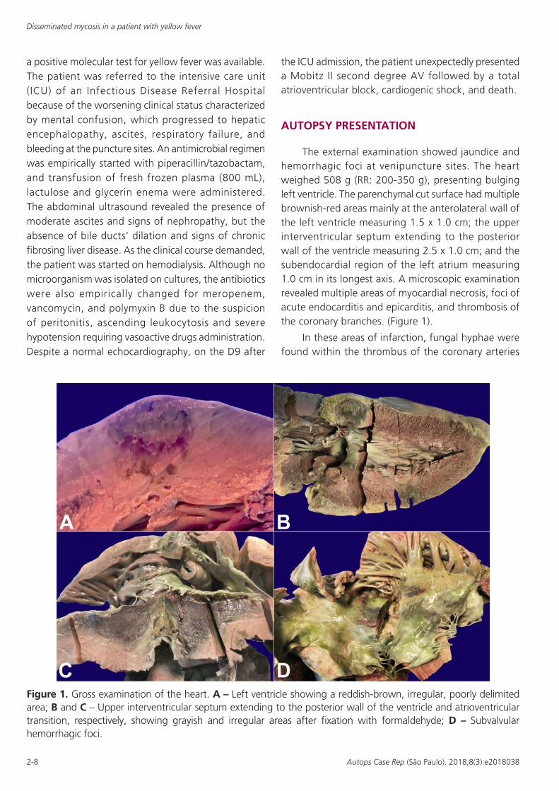
The external examination showed jaundice and hemorrhagic foci at venipuncture sites. The heart weighed 508 g (RR: 200-350 g), presenting bulging left ventricle. The parenchymal cut surface had multiple brownish-red areas mainly at the anterolateral wall of the left ventricle measuring 1.5 x 1.0 cm; the upper interventricular septum extending to the posterior wall of the ventricle measuring 2.5 x 1.0 cm; and the subendocardial region of the left atrium measuring 1.0 cm in its longest axis. A microscopic examination revealed multiple areas of myocardial necrosis, foci of acute endocarditis and epicarditis, and thrombosis of the coronary branches. (Figure 1).
In these areas of infarction, fungal hyphae were found within the thrombus of the coronary arteries
Figure 1. Gross examination of the heart. A – Left ventricle showing a reddish-brown, irregular, poorly delimited area; B and C – Upper interventricular septum extending to the posterior wall of the ventricle and atrioventricular transition, respectively, showing grayish and irregular areas after fixation with formaldehyde; D – Subvalvular hemorrhagic foci.

Maciel GVR, Tavares MCF, Pereira LS et al.
3-8Autops Case Rep (São Paulo). 2018;8(3):e2018038
and of the cardiac veins, which invaded the vessel walls and the adjacent myocardium (Figure 2).
The lungs were heavy (right lung: 845 g; left lung: 640 g [RR = 360-570 g each]), congested, and showed signs of bilateral collapse. The parenchymal cut surface was reddish with signs of hepatization at the bases. Microscopic examination revealed areas of recent
hemorrhagic infarction on the left, with corresponding purulent pleurisy. Microabscesses and alveolar edema were also found. Fungal hyphae were seen in these areas of infarction (Figure 3).
The kidneys were of average size and weight with a smooth external surface (right kidney: 245 g; left kidney: 250 g [RR for both – 230-440 g]).
Figure 2. Micrograph of the heart. A – Panoramic view of atrioventricular transition demonstrating extensive infarction area, besides hemorrhage, coronary branch thrombosis and subvalvular fibrinous deposit (H&E, 01X); B – Left ventricular myocardium showing well-established infarction, with necrosis of muscle fibers and prominent infiltrate of neutrophil polymorphs. The intramyocardial artery exhibits thrombosis and wall rupture (H&E, 10X); C – Intramyocardial artery and adjacent myocardium, both infiltrated by abundant fungal hyphae. There is thrombosis of arterial vessels and infarction of myocardium (Grocott’s stain, 20X).
Figure 3. Photomicrograph of the lungs. A – Focus of hemorrhagic necrosis in the lung parenchyma (H&E, 10X); B – Extensive alveolar collapse (H&E, 10X); C – Pulmonary edema and congestion (H&E, 20X).
Disseminated mycosis in a patient with yellow fever
4-8 Autops Case Rep (São Paulo). 2018;8(3):e2018038
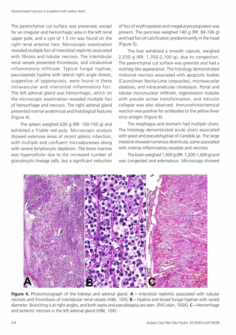
The parenchymal cut surface was preserved, except for an irregular and hemorrhagic area in the left renal upper pole, and a cyst of 1.5 cm was found on the right renal anterior face. Microscopic examination revealed multiple foci of interstitial nephritis associated with fibrosis and tubular necrosis. The interlobular renal vessels presented thrombosis, and intraluminal inflammatory infiltrate. Typical fungal hyphae, pauciseptate hyaline with lateral right angle shoots, suggestive of zygomycosis, were found in these intravascular and interstitial inflammatory foci. The left adrenal gland was hemorrhagic, which on the microscopic examination revealed multiple foci of hemorrhage and necrosis. The right adrenal gland presented normal anatomical and histological features (Figure 4).
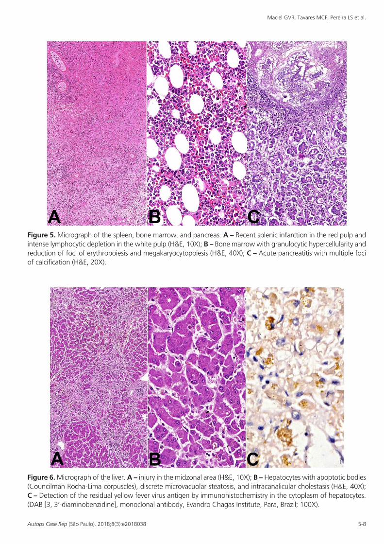
The spleen weighed 630 g (RR: 100-150 g) and exhibited a friable red pulp. Microscopic analysis showed extensive areas of recent splenic infarction, with multiple and confluent microabscesses along with severe lymphocytic depletion. The bone marrow was hypercellular due to the increased number of granulocytic-lineage cells, but a significant reduction
of foci of erythropoiesis and megakaryocytopoiesis was present. The pancreas weighed 140 g (RR: 84-106 g) and had foci of calcification predominantly in the head (Figure 5).
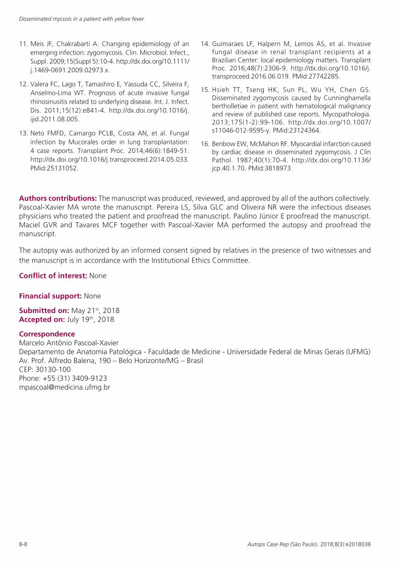
The liver exhibited a smooth capsule, weighed 2,030 g (RR: 1,350-2,100 g), due to congestion. The parenchymal cut surface was greenish and had a nutmeg-like appearance. The histology demonstrated midzonal necrosis associated with apoptotic bodies (Councilman Rocha-Lima corpuscles), microvacuolar steatosis, and intracanalicular cholestasis. Portal and lobular mononuclear infiltrate, regeneration nodules with pseudo acinar transformation, and reticulin collapse was also observed. Immunohistochemical reaction was positive for antibodies to the yellow fever virus antigen (Figure 6).
The esophagus and stomach had multiple ulcers. The histology demonstrated acute ulcers associated with yeast and pseudohyphae of Candida sp. The large intestine showed numerous diverticula, some associated with intense inflammatory exudate and necrosis.
The brain weighed 1,400 g (RR: 1,200-1,600 g) and was congested and edematous. Microscopy showed
Figure 4. Photomicrograph of the kidneys and adrenal gland. A – Interstitial nephritis associated with tubular necrosis and thrombosis of interlobular renal vessels (H&E, 10X); B – Hyaline and broad fungal hyphae with varied diameter. Branching is at right angles, and both septa and pseudosepta are seen. (PAS stain, 100X); C – Hemorrhage and ischemic necrosis in the left adrenal gland (H&E, 10X).
Maciel GVR, Tavares MCF, Pereira LS et al.
5-8Autops Case Rep (São Paulo). 2018;8(3):e2018038
Figure 6. Micrograph of the liver. A – injury in the midzonal area (H&E, 10X); B – Hepatocytes with apoptotic bodies (Councilman Rocha-Lima corpuscles), discrete microvacuolar steatosis, and intracanalicular cholestasis (H&E, 40X); C – Detection of the residual yellow fever virus antigen by immunohistochemistry in the cytoplasm of hepatocytes. (DAB [3, 3′-diaminobenzidine], monoclonal antibody, Evandro Chagas Institute, Para, Brazil; 100X).
Figure 5. Micrograph of the spleen, bone marrow, and pancreas. A – Recent splenic infarction in the red pulp and intense lymphocytic depletion in the white pulp (H&E, 10X); B – Bone marrow with granulocytic hypercellularity and reduction of foci of erythropoiesis and megakaryocytopoiesis (H&E, 40X); C – Acute pancreatitis with multiple foci of calcification (H&E, 20X).

Disseminated mycosis in a patient with yellow fever
6-8 Autops Case Rep (São Paulo). 2018;8(3):e2018038
sparse areas of edema, and discrete interstitial and perivascular lymphocytic infiltrate.
DISCUSSION
In 2018, there was a new outbreak of sylvatic yellow fever in Minas Gerais State -Brazil, where 570 suspected cases were investigated, of which 446 were confirmed with 33.6% of mortality rate (150 cases).1
The clinical presentation of yellow fever disease may vary from subclinical infection to systemic disease, with fever, nausea, vomiting, epigastric pain, hepatitis with jaundice, renal failure, hemorrhage, shock, and death in 20%-60% of cases, as the result of the virus and the host factors.2
The fatal outcome of yel low fever mainly correlates with liver damage, renal failure, hemorrhagic phenomena, and shock. Reportedly, hepatic and renal damages often lead to severe hemorrhagic diathesis, shock, and death in the period of intoxication of the infection.3 However, death can occur independently of the classic hemorrhagic manifestations. The case reported herein illustrates the clinical presentation of severe yellow fever, with hepatic and renal damages (without hemorrhagic manifestation), and fatal outcome with acute myocardial infarction after 25 days of the onset of symptoms.
In the context of the late fatal outcome, two essential aspects need to be discussed. First, the possibility of the viral persistence within the tissues prolonged the necroapoptotic action in the most vulnerable cells, such as hepatocytes, splenocytes, renal tubular cells, endothelium, and cardiomyocytes. Second, the immunosuppression caused by the sudden high viremia of the yellow fever virus. Both aspects directly and suddenly provoked by high viremia, and later caused by the dysfunction of both innate and adaptive immunity, favoring disseminated fungal infection.
In an excellent review, Monath and Barrett4 approached the pathogenesis and pathophysiology of yellow fever and reported viral necroapoptotic action in several tissues. They indicated that the viral virulence is highly dependent on the pairing of virus strain and host species, and mentioned that the virus virtually disappears from circulation after the onset
of the neutralizing antibodies between the fifth and tenth days of illness. Additionally, they assumed that the cardiomyopathy is directly related to the viral replication in the cardiomyocytes.
Thus, considering the hypothesis of the persistence of viral necroapoptotic action in the tissues as a direct cause of hepatic, renal, and cardiac damage, we performed the investigation of the histopathological changes characteristic of yellow fever along with viral antigens by immunohistochemistry. We found some histopathological features consistent with yellow fever and were able to demonstrate the focal presence of viral antigens in the liver. The remaining organs, specifically the kidneys and the heart did not exhibit the typical morphological characteristics of yellow fever.
These residual hepatic histopathological findings, indicative of yellow fever, were also observed by Francis et al.5 They reported necrosis, apoptosis, microvacuolar steatosis, portal and lobular lymphocytic infi ltrate between the 15th and 20th days of infection. However, they did not undertake the immunohistochemical analysis for viral antigen screening in the liver samples.
In contrast, and more striking in this fatal evolution, was the severe immunosuppression triggered by yellow fever in our patient. Despite the small number of published studies on the pathogenesis of yellow fever, the available data enable the interpretation of direct and indirect viral causes for this immunosuppression. The viral load may directly cause immunosuppression in cases of fatal yellow fever. In this setting, Chen et al.6 described a fatal case of yellow fever and indicated that the persistent viral infection damaged the immune system. They demonstrated that the viral load was 1.4 × 10⁴ copies per mL on day 3. Subsequently, the viral load decreased but maintained at a level ranging from 103 to 102 copies per mL from day 5 to day 9, the day of the patient’s death.
Indirectly, fatal yellow fever may cause an intense cytokine network signaling disorder as observed in sepsis and may induce post-infectious immunosuppression. The differentiated pro-inflammatory profile of the fatal cases of yellow fever, characterized by high levels of interleukin-6 (IL-6), monocyte chemoattractant protein (MCP)-1, interferon-inducible protein (IP)-10, tumor necrosis factor (TNF)-α, and IL-1 receptor antagonist (IL-1RA), corroborates to this observation.7 In addition, there is evidence of inhibition of the interferon

Maciel GVR, Tavares MCF, Pereira LS et al.
7-8Autops Case Rep (São Paulo). 2018;8(3):e2018038
(IFN)-mediated JAK-STAT (Janus kinase-signal transducer and activator of transcription) pathway of signal transduction by flavivirus non-structural (NS)-5 protein and impairment of the immune response to fungi.8,9
Thus, this cytokine network signaling disorder would increase susceptibility to zygomycosis—a highly invasive and emerging opportunistic fungal infection—in conditions of immunosuppression, such as hematological malignancy, prolonged neutropenia, diabetes mellitus, organ transplantation, AIDS, and immunosuppressive therapies. Globally, there is still a predominance of invasive fungal infections by candidiasis and aspergillosis in the immunosuppressed patient. However, the prevalence of zygomycosis has dramatically increased in recent decades. Also, mortality is extremely high in patients with disseminated disease, and gastrointestinal and pulmonary infections.10,11 In Brazil, the epidemiology of zygomycosis is rarely described, and are restricted to reports of rhinofacial, lung and kidney transplantation cases.12-14
This invasive fungal infection is histopathologically characterized by pauciseptate hyaline hyphae of Mucorales genus, which is different from the multiseptate hyaline hyphae (Aspergillus spp., Candida) and the pigmented hyphae (dematiaceous). Zygomycosis presents in many forms, including pulmonary, rhinocerebral, cutaneous, gastrointestinal, and disseminated forms, as well as severe cardiac involvement.15 In particular, the case described herein recalls all the cardiac morphological findings of the report by Benbow and McMahon,16 highlighting the myocardial infarction in disseminated zygomycosis.
CONCLUSION
The present autopsy shows the importance of the postmortem exam in patients with yellow fever. Two major diagnoses were made in this autopsy: (i) disseminated zygomycosis with acute myocardial infarction; and (ii) persistence of the yellow fever virus in the liver after 25 days of infection. Opportunistic fungal infection must be considered in the differential diagnosis of sepsis in the patient with severe yellow fever hospitalized in the intensive care unit. Also, immunosuppression and the possibility of viral persistence in the tissues, which prolongs the
necroapoptotic action in the most vulnerable cells (e.g., such as hepatocytes, renal tubular cells, endothelium, and cardiomyocytes), needs to be further investigated.
REFERENCES
1. Minas Gerais. Informe Epidemiológico da Febre Amarela [Internet]. Belo Horizonte: Secretaria de Estado de Saúde; 2018 [cited 2018 May 21]. Vol. 03/04, 3 Abr 2018. Available from: http://www.saude.mg.gov.br/component/gmg/story/10381-informe-epidemiologico-da-febre-amarela-03-04/
2. Monath TP, Vasconcelos PF. Yellow fever. J. Clin. Virol. 2015;64:160-73. http://dx.doi.org/10.1016/j.jcv.2014.08.030.
3. Quaresma JA, Pagliari C, Medeiros DB, Duarte MI, Vasconcelos PF. Immunity and immune response, pathology and pathologic changes: progress and challenges in the immunopathology of yellow fever. Rev Med Virol. 2013;23(5):305-18. http://dx.doi.org/10.1002/rmv.1752. PMid:23873723.
4. Monath TP, Barrett AD. Pathogenesis and pathophysiology of yellow fever. Adv Virus Res. 2003;60:343-95. http://dx.doi.org/10.1016/S0065-3527(03)60009-6. PMid:14689698.
5. Francis TI, Moore DL, Edington GM, Smith JA. A clinicopathological study of human yellow fever. Bull World Health Organ. 1972;46(5):659-67. PMid:4538039.
6. Chen Z, Liu L, Lv Y, et al. A fatal yellow fever virus infection in China: description and lessons. Emerg Microbes Infect. 2016;5(7):e69. http://dx.doi.org/10.1038/emi.2016.89. PMid:27406389.
7. ter Meulen J, Sakho M, Koulemou K, et al. Activation of the cytokine network and unfavorable outcome in patients with yellow fever. J Infect Dis. 2004;190(10):1821-7. http://dx.doi.org/10.1086/425016. PMid:15499539.
8. Best SM, Morris KL, Shannon JG, et al. Inhibition of interferon-stimulated JAK-STAT signaling by a tick-borne flavivirus and identification of NS5 as an interferon antagonist. J Virol. 2005;79(20):12828-39. http://dx.doi.org/10.1128/JVI.79.20.12828-12839.2005. PMid:16188985.
9. Laurent-Rolle M, Morrison J, Rajsbaum R, et al. The interferon signaling antagonist function of yellow fever virus NS5 protein is activated by type I interferon. Cell Host Microbe. 2014;16(3):314-27. http://dx.doi.org/10.1016/j.chom.2014.07.015. PMid:25211074.
10. Roden MM, Zaoutis TE, Buchanan WL, et al. Epidemiology and outcome of zygomycosis: a review of 929 reported cases. Nephrol Dial Transplant. 2005;41(5):634-53. PMid:16080086.
Disseminated mycosis in a patient with yellow fever
8-8 Autops Case Rep (São Paulo). 2018;8(3):e2018038
11. Meis JF, Chakrabarti A. Changing epidemiology of an emerging infection: zygomycosis. Clin. Microbiol. Infect., Suppl. 2009;15(Suppl 5):10-4. http://dx.doi.org/10.1111/j.1469-0691.2009.02973.x.
12. Valera FC, Lago T, Tamashiro E, Yassuda CC, Silveira F, Anselmo-Lima WT. Prognosis of acute invasive fungal rhinosinusitis related to underlying disease. Int. J. Infect. Dis. 2011;15(12):e841-4. http://dx.doi.org/10.1016/j.ijid.2011.08.005.
13. Neto FMFD, Camargo PCLB, Costa AN, et al. Fungal infection by Mucorales order in lung transplantation: 4 case reports. Transplant Proc. 2014;46(6):1849-51. http://dx.doi.org/10.1016/j.transproceed.2014.05.033. PMid:25131052.
14. Guimaraes LF, Halpern M, Lemos AS, et al. Invasive fungal disease in renal transplant recipients at a Brazilian Center: local epidemiology matters. Transplant Proc. 2016;48(7):2306-9. http://dx.doi.org/10.1016/j.transproceed.2016.06.019. PMid:27742285.
15. Hsieh TT, Tseng HK, Sun PL, Wu YH, Chen GS. Disseminated zygomycosis caused by Cunninghamella bertholletiae in patient with hematological malignancy and review of published case reports. Mycopathologia. 2013;175(1-2):99-106. http://dx.doi.org/10.1007/s11046-012-9595-y. PMid:23124364.
16. Benbow EW, McMahon RF. Myocardial infarction caused by cardiac disease in disseminated zygomycosis. J Clin Pathol. 1987;40(1):70-4. http://dx.doi.org/10.1136/jcp.40.1.70. PMid:3818973.
Authors contributions: The manuscript was produced, reviewed, and approved by all of the authors collectively. Pascoal-Xavier MA wrote the manuscript. Pereira LS, Silva GLC and Oliveira NR were the infectious diseases physicians who treated the patient and proofread the manuscript. Paulino Júnior E proofread the manuscript. Maciel GVR and Tavares MCF together with Pascoal-Xavier MA performed the autopsy and proofread the manuscript.
The autopsy was authorized by an informed consent signed by relatives in the presence of two witnesses and the manuscript is in accordance with the Institutional Ethics Committee.
Conflict of interest: None
Financial support: None
Submitted on: May 21st, 2018 Accepted on: July 19th, 2018
Correspondence Marcelo Antônio Pascoal-Xavier Departamento de Anatomia Patológica - Faculdade de Medicine - Universidade Federal de Minas Gerais (UFMG) Av. Prof. Alfredo Balena, 190 – Belo Horizonte/MG – Brasil CEP: 30130-100 Phone: +55 (31) 3409-9123 [email protected]