DISCUSSION SESSION: GROSS ANATOMY ONN BLOCK Sunday, …
32
DISCUSSION SESSION: GROSS ANATOMY ONN BLOCK Sunday, Feb 7, 2021 Discuss Branchial Arches, Neck
Transcript of DISCUSSION SESSION: GROSS ANATOMY ONN BLOCK Sunday, …
DISCUSSION SESSION: GROSS ANATOMY
ONN BLOCK
Sunday, Feb 7, 2021
Discuss Branchial Arches, Neck

BRANCHIAL ARCHESKnow – Branchial cartilages, muscles, nerves, pouches
Clinical Branchial Cleft Syndromes
ALSO NOT DERIVED FROM BRANCHIAL ARCHES Abnormalities of Thyroid development
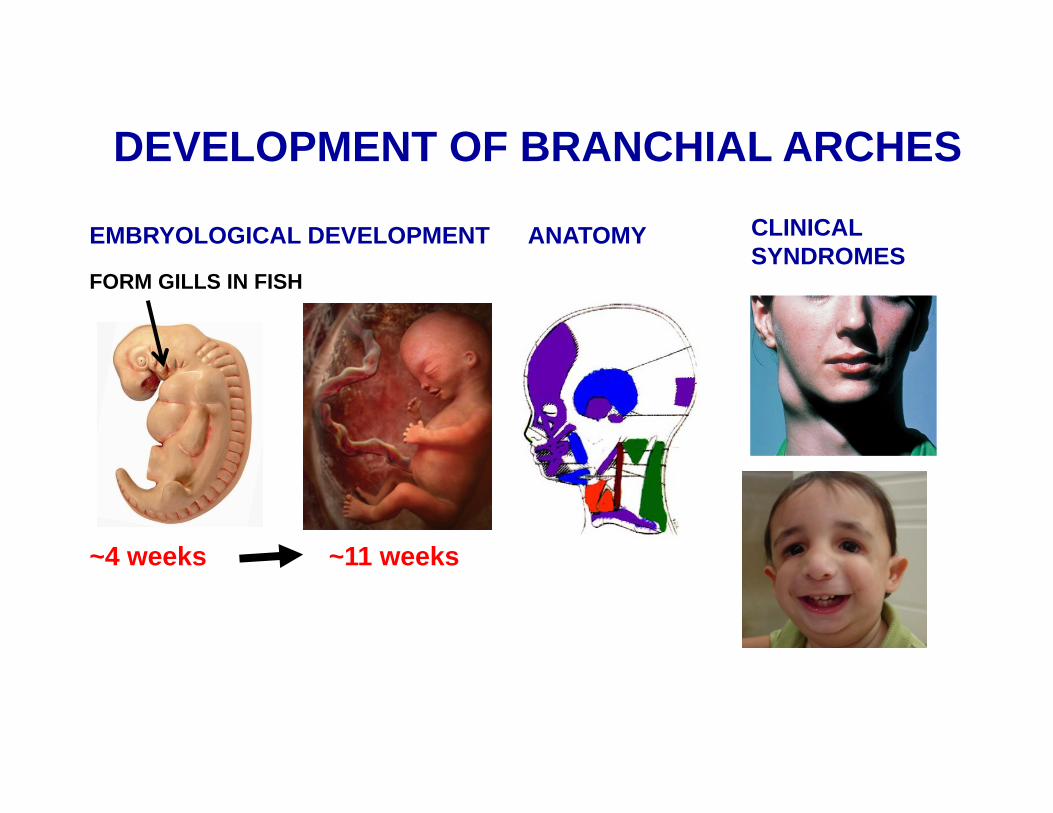
DEVELOPMENT OF BRANCHIAL ARCHES
~4 weeks ~11 weeks
FORM GILLS IN FISH
EMBRYOLOGICAL DEVELOPMENT ANATOMY CLINICALSYNDROMES

BRANCHIAL ARCHES HAVE CARTILAGES, MUSCLES, ARTERIES
FORM - CLEFTS ON OUTSIDE (ECTODERM)POUCHES ON INSIDE (ENDODERM)
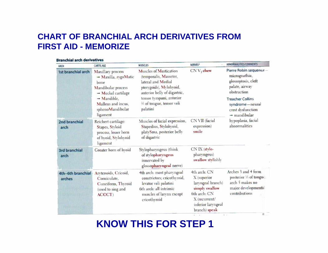
CHART OF BRANCHIAL ARCH DERIVATIVES FROM FIRST AID - MEMORIZE
KNOW THIS FOR STEP 1

BREAK DOWN TO COMPONENT IN LECTURE HANDOUT
CHART OF BRANCHIAL ARCH DERIVATIVES FROM FIRST AID - MEMORIZE
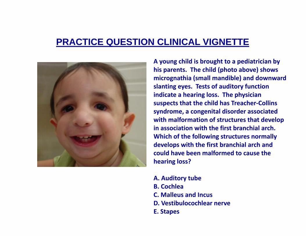
A young child is brought to a pediatrician by his parents. The child (photo above) shows micrognathia (small mandible) and downward slanting eyes. Tests of auditory function indicate a hearing loss. The physician suspects that the child has Treacher‐Collins syndrome, a congenital disorder associated with malformation of structures that develop in association with the first branchial arch. Which of the following structures normally develops with the first branchial arch and could have been malformed to cause the hearing loss?
A. Auditory tubeB. CochleaC. Malleus and IncusD. Vestibulocochlear nerveE. Stapes
PRACTICE QUESTION CLINICAL VIGNETTE

MUSCLES AND NERVES = BRANCHIOMOTOR MUSCLES FROM CRANIAL NERVES HANDOUT (INCANTATION)

FOCUS ON CLINICAL: BRANCHIAL POUCHES, GROOVES, MEMBRANES
NOTE: CLEFT = GROOVE

Only First Branchial Groove and Membrane Normally form Structures in Adult
First Membrane= Tympanic Membrane
First Pouch -AuditoryTube
BRANCHIAL GROOVES (CLEFTS) AND MEMBRANES
Ext. Aud.Meatus
First Groove - External Auditory Meatus
First Groove -External Auditory Meatus
RemainingGrooves merge - form CervicalSinus- Cervical Sinus normallyobliterated

EAR
FIRST GROOVE -Ext. Aud.Meatus
FIRST POUCH -AuditoryTube, Tympanic Cavity
First Membrane- Tympanic Membrane
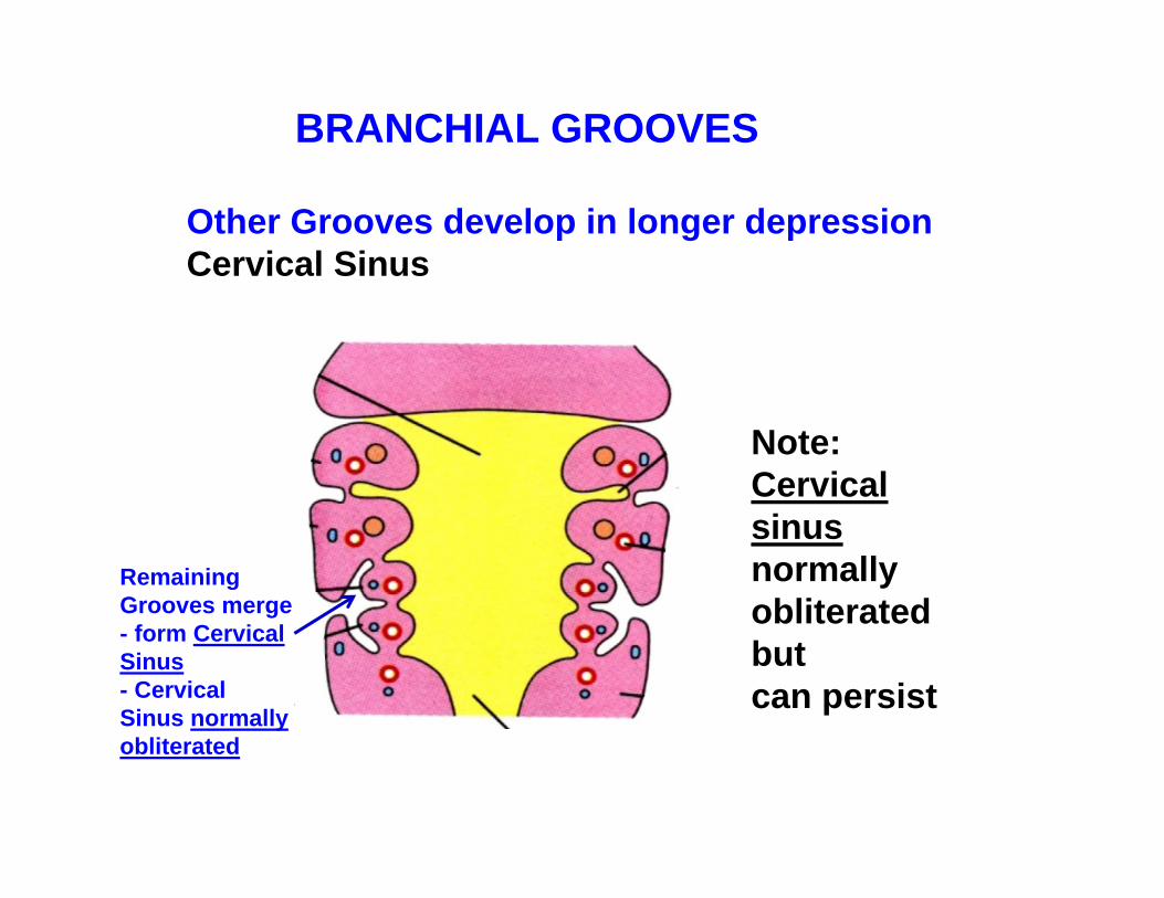
Other Grooves develop in longer depressionCervical Sinus
Note: Cervical sinus normally obliterated butcan persist
BRANCHIAL GROOVES
RemainingGrooves merge - form CervicalSinus- Cervical Sinus normallyobliterated
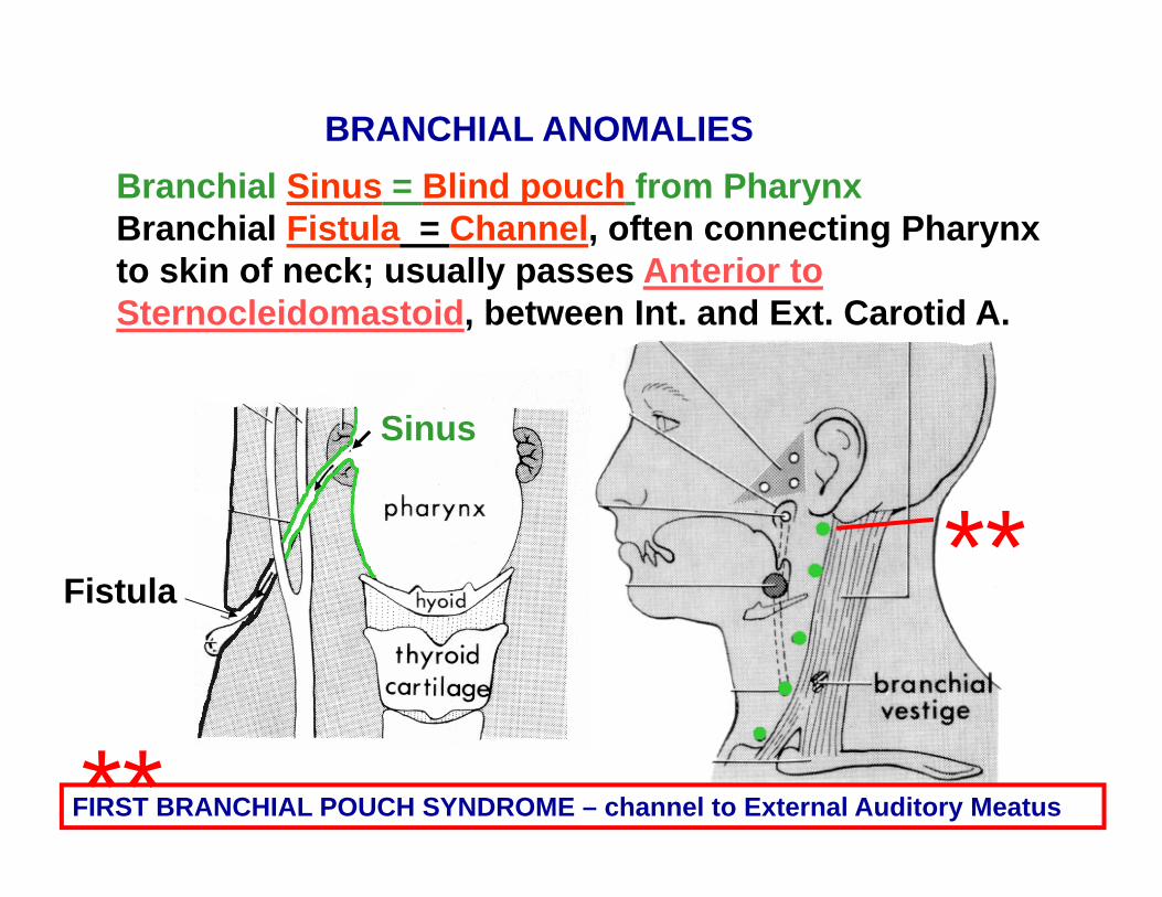
Branchial Sinus = Blind pouch from PharynxBranchial Fistula = Channel, often connecting Pharynx to skin of neck; usually passes Anterior to Sternocleidomastoid, between Int. and Ext. Carotid A.
BRANCHIAL ANOMALIES
Sinus
Fistula
FIRST BRANCHIAL POUCH SYNDROME – channel to External Auditory Meatus
**
**

A 24 year old woman develops a mass in her neck (see photo above). The mass is located immediately anterior to the sternocleidomastoid muscle. The physician suspects that this condition has result from a branchial cyst. During surgery, the mass is found to be connected to a tract that extends superiorly and medially. The tract is most likely to be connected to which of the following structures?
A. Middle meatus of the nasal cavityB. Pharyngeal tonsilC. Tonsillar fossa (palatine tonsils)D. Lingual tonsilE. Mandibular fossa
PRACTICE QUESTION CLINICAL VIGNETTE

SECOND BRANCHIAL POUCH SYNDROME
Branchial Fistula - drains to neck Branchial Cyst often remnantof Cervical Sinus
SECOND BRANCHIAL POUCH FORMS CRYPTS (LININGS) OF PALATINE TONSILS

2) Elongates to form Thyroid Diverticulum; descends ant. to hyoid bone and larynx3) Thyroglossal duct connects Diverticulum to Foramen cecum
DEVELOPMENT OF THYROID
1) Thyroid start as Median endodermal Thickening on floor of pharynx at future junction of anterior 2/3 and posterior 1/3 of tongue (marked by Foramen Cecum)
THYROGLOSSALDUCT
TONGUEhyoidbone

CONGENITAL MALFORMATIONS
Thyroglossal Duct Remnants -can form thyroid tissue (cysts) along path (midline, ant. tohyoid, larynx)
Pyramidal Lobe - 50%of people; attached tohyoid by fibrous strand;no clinical problems
LINGUAL THYROID* - gland in tongue
***

LINGUAL THYROID* - Thyroid gland in tongue
AT: Junction of anterior 2/3 and posterior 1/3 of tongue
***

NECKKnow Carotid Artery (Internal, External Carotid Arteries)Muscles: Torticollis, contracture of sternocleidomastoid, face directed to opposite sideWounds, surgery to neck damage Phrenic nervePyramidal lobe variant of Thyroid gland no clinical problems but important in thyroid surgeryCarotid angiogram Superior Thyroid artery

Lateral Compartment-lateral and posterior to pharynx
Contained in Carotid Sheath
1) Common and Internal Carotid arteries; 2) Internal jugular vein, 3) Vagus nerve
Note: Sympathetic chain is posterior to (NOT IN) Carotid Sheath
3. LATERAL COMPARTMENT - CAROTID SHEATH
CLINICAL **

A. MUSCLES OF NECK - NOT ATTACHED TO HYOID - move head and neck
1. STERNO-CLEIDOMASTOID
O - Two heads: 1) Manubrium of sternum; 2) Clavicle (L. root -cleido) - medial 1/3
I - Mastoid process of temporal bone
Act - bilateral - flex head; unilateral rotate head, face directed to opposite side
(MASTOID MOVES TOWARD STERNUM)
Inn - CN XI Accessory.
TORTICOLLIS –Contracture ofSternocleido-mastoid (congenital or acquired); face to opposite side
MOST IMPORTANT LANDMARK IN NECK
ACTION - PULL MASTOID TOWARD STERNUM
STERNAL NOTCH ON MANUBRIUM OF STERNUM
CLAVICLE
clavicularhead
sternalhead
**
TORTICOLLIS = twisted neck
SCALENUS ANTERIOR AND SCALENUS MEDIUS
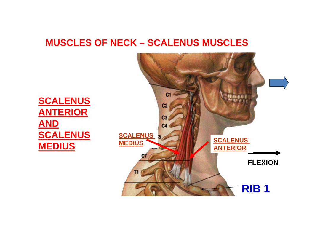
MUSCLES OF NECK – SCALENUS MUSCLES
FLEXION
SCALENUS ANTERIOR
SCALENUS MEDIUS
RIB 1
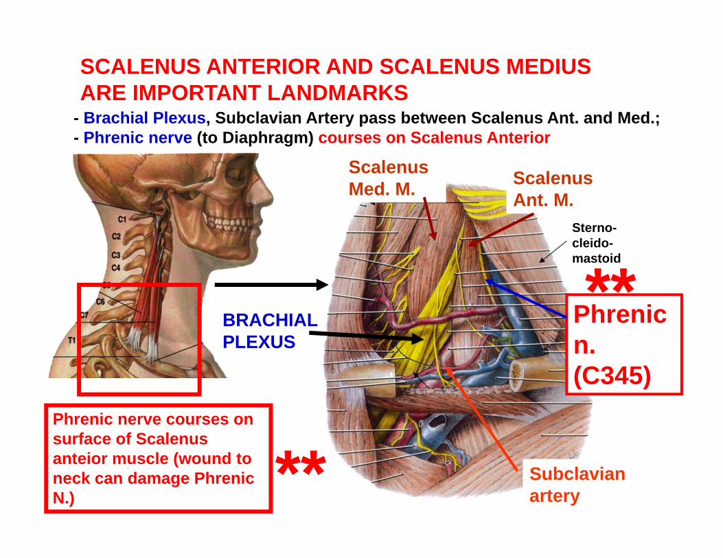
Scalenus Ant. M.
Phrenic n. (C345)
BRACHIALPLEXUS
Scalenus Med. M.
Subclavian artery
SCALENUS ANTERIOR AND SCALENUS MEDIUS ARE IMPORTANT LANDMARKS
- Brachial Plexus, Subclavian Artery pass between Scalenus Ant. and Med.; - Phrenic nerve (to Diaphragm) courses on Scalenus Anterior
Sterno-cleido-mastoid
**Phrenic nerve courses on surface of Scalenus anteior muscle (wound to neck can damage Phrenic N.) **

STERNOHYOID
OMOHYOID
THYROHYOID
STERNOTHYROID
ID MUSCLES - INFRAHYOID
deeper
NOSE
ORIENT -HEADTILTEDBACK

Large ExternalJugular V.
LargeAnteriorJugularV.
**EXTERNAL JUGULAR V. - ON SURFACE OF STERNOCLEIDOMASTOID; NOT IN CAROTID SHEATHINTERNAL JUGULAR V. - DEEP TO STERNOCLEIDOMASTOID; IN CAROTID SHEATH
VEINS OF NECK

4. FACIAL A -
BELOW THEN ON SURFACE OF MANDIBLE
3. LINGUAL A-TONGUE
2. ASCENDING PHARYNGEAL A-ASCENDS TO PHARYNX
1. SUPERIOR THYROID A-DESCENDS TO THYROID
5. OCCIPITAL A-POST SCALP
6. POST. AURICULAR A-POST TO EAR
EXTERNAL CAROTID ARTERY
CAROTIDBIFURCATION -AT UPPER BORDER OF THYROID CARTILAGE -LEVEL C4
Superficial Temporal A..Maxillary A..
Posteriorbranches
Anteriorbranches
Terminalbranches
**

1. COMMON CAROTID2. INTERNAL CAROTID3. ASCENDING PHARYNGEAL4. OCCIPITAL5. SUPERFICIAL TEMPORAL6. MIDDLE CEREBRAL7. ANTERIOR CEREBRAL8. MIDDLE MENINGEAL9. MAXILLARY10. FACIAL11. LINGUAL12. EXTERNAL CAROTID13. SUPERIOR THYROID
*- OPHTHALMIC ARTERYARISING FROM CAROTIDSIPHON
*POST.AURICULAR
NOSE
KNOW THISSLIDE
**WIGGLEWIGGLEWIGGLE
*
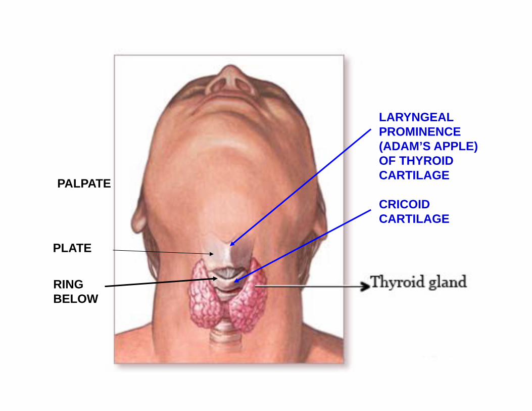
LARYNGEAL PROMINENCE (ADAM’S APPLE) OF THYROIDCARTILAGE
CRICOIDCARTILAGE
PLATE
RINGBELOW
PALPATE

THYROID GLAND
Right lateral lobe
Left lateral lobe
Isthmus -located below cricoid cartilage
Pyramidal lobe - when present often attached to hyoid bone by fibrous strand
Absence ofIsthmus
Normal variations common
*
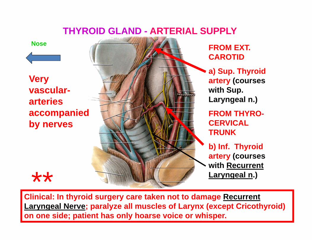
Very vascular-arteries accompanied by nerves
FROM EXT. CAROTID
a) Sup. Thyroid artery (courses with Sup. Laryngeal n.)
FROM THYRO-CERVICAL TRUNK
b) Inf. Thyroid artery (courses with Recurrent Laryngeal n.)
Clinical: In thyroid surgery care taken not to damage Recurrent Laryngeal Nerve; paralyze all muscles of Larynx (except Cricothyroid) on one side; patient has only hoarse voice or whisper.
THYROID GLAND - ARTERIAL SUPPLYNose
**
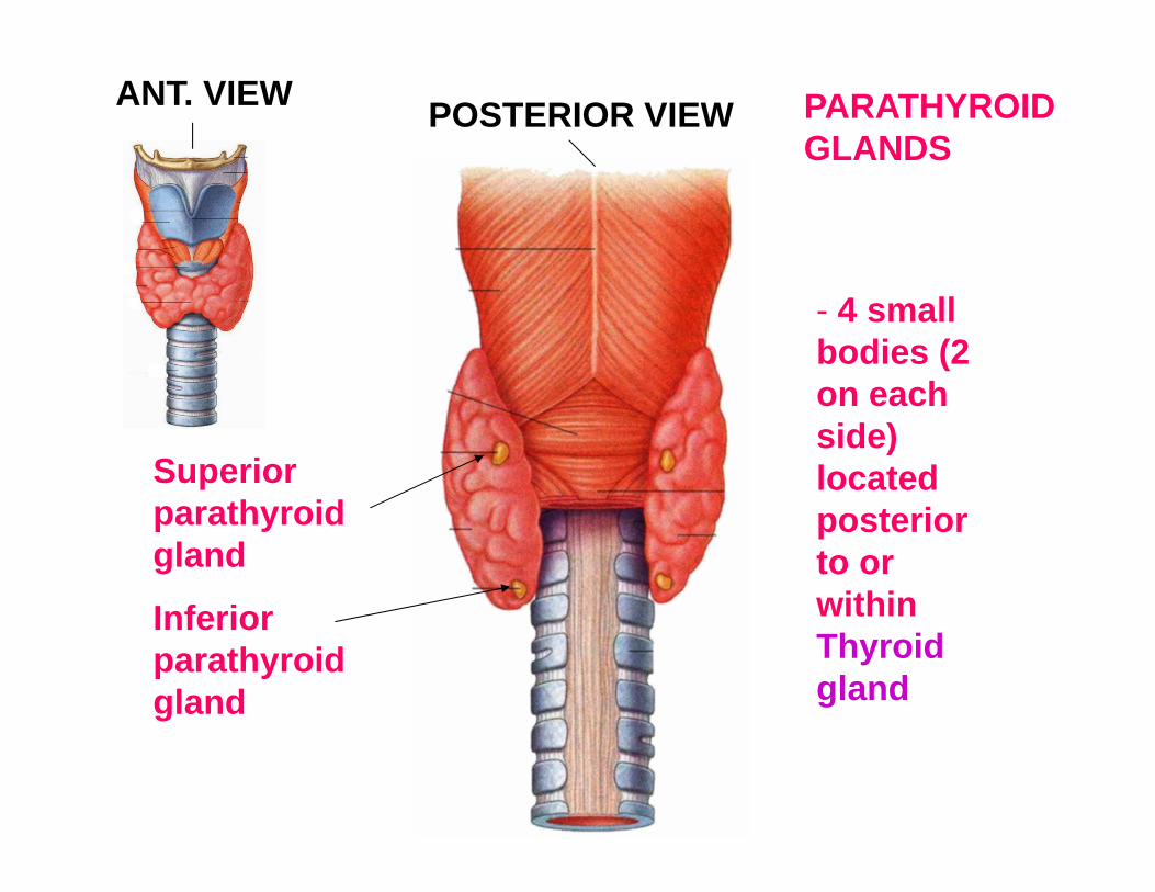
PARATHYROID GLANDS
- 4 small bodies (2 on each side) located posterior to or within Thyroid gland
Superior parathyroid gland
Inferior parathyroid gland
ANT. VIEW POSTERIOR VIEW
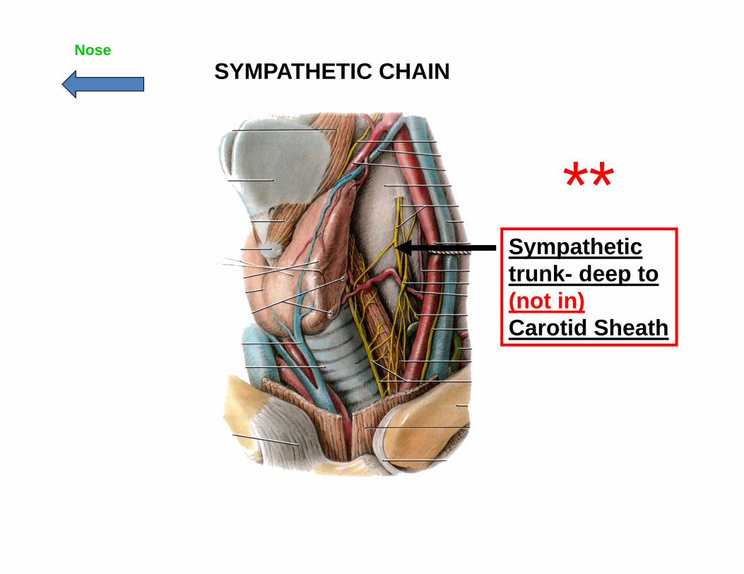
Sympathetic trunk- deep to (not in) Carotid Sheath
SYMPATHETIC CHAINNose
**