Dirofilariasis in humans presenting as a subcutaneous nodule
3
IP Archives of Cytology and Histopathology Research 2021;6(3):225–227 Content available at: https://www.ipinnovative.com/open-access-journals IP Archives of Cytology and Histopathology Research Journal homepage: https://www.achr.co.in/ Case Report Dirofilariasis in humans presenting as a subcutaneous nodule Srikousthubha MS 1, *, Mohandas AS 2 1 Dept. of Pathology, Shimoga Diagnostic Centre, Shimoga, Karnataka, India 2 Dept. of Surgery, Sri Raghavendra Nursing Home, Shimoga, Karnataka, India ARTICLE INFO Article history: Received 10-07-2021 Accepted 27-08-2021 Available online 16-09-2021 Keywords: Dirofilariasis Human subcutaneous ABSTRACT Human dirofilariasis is an uncommon zoonotic roundworm infection caused by worms belonging to dirofilaria species transmitted by zoo -anthropophilic blood sucking insects which is known to manifest as ocular infection or as a subcutaneous nodule usually in the upper part of the body. We present a case report of subcutaneous dirofilariasis in the left temporal region besides the eye brow in a 65yr old female, clinically diagnosed as epidermal cyst. Gross and histopathological evaluation revealed the presence of an adult worm with granulomatous inflammatory reaction. This is an Open Access (OA) journal, and articles are distributed under the terms of the Creative Commons Attribution-NonCommercial-ShareAlike 4.0 License, which allows others to remix, tweak, and build upon the work non-commercially, as long as appropriate credit is given and the new creations are licensed under the identical terms. For reprints contact: [email protected] 1. Introduction Human dirofilariasis is an uncommon zoonotic disease where man is an accidental host.About forty species of Dirofilaria are identified,among which only a few give rise to human infection. 1 Dirofilaria repens is the commonest species identified in India. The first reported case of human ocular dirofilariasis in India occurred in Kerala in 1976 and subcutaneous dirofilariasis was recorded in the same region in 2004.Most of the documented cases in India are of ocular dirofilariasis.Very few cases of subcutaneous dirofilariasis have been reported. 2 2. Case History A 65yr old female housewife from an agricultural background presented with a non tender nodule in the left temporal region besides the eyebrow of 2 months duration which had gradually progressed to the size of 2.5 x 2 cm. It was firm in consistency. Skin over the swelling was normal. A provisional diagnosis of epidermal cyst was made, * Corresponding author. E-mail address: [email protected] (Srikousthubha MS). excised and submitted for histopathological examination. Grossly, the specimen was grey white m 2.5 x 2 x 1.5 cm, cut section was solid grey white. On closer examination retrospectively revealed a thread like worm (Figure 1) Microscopic examination of Hematoxylin and Eosin stained sections showed granulation tissue with intense neutrophilic and eosinophilic infiltration, multinucleated giant cells, plasma cells. Cross section of a nematode parasite with a thick external cuticle, prominent circumferential muscle and cut section of intestine were recognized (Figure 1). Based on these findings the worm was identified as Dirofilaria repens. A diagnosis of Subcutaneous Dirofilariasis was made. Complete blood counts and Peripheral smear examination did not show eosinophilia. Serum Ig E levels were normal. Ophthalmologic examination was requested to look for ocular parasite which was negative. 3. Discussion Human dirofilariasis is a zoonosis caused by animal filarial parasite Dirofilaria species. D. repens, d.immitis, D. tenuis and D.urisi are the known species causing human infection. D.repens, a parasite of cats and dogs is most commonly https://doi.org/10.18231/j.achr.2021.050 2581-5725/© 2021 Innovative Publication, All rights reserved. 225
Transcript of Dirofilariasis in humans presenting as a subcutaneous nodule

IP Archives of Cytology and Histopathology Research 2021;6(3):225–227
Content available at: https://www.ipinnovative.com/open-access-journals
IP Archives of Cytology and Histopathology Research
Journal homepage: https://www.achr.co.in/
Case Report
Dirofilariasis in humans presenting as a subcutaneous nodule
Srikousthubha MS1,*, Mohandas AS2
1Dept. of Pathology, Shimoga Diagnostic Centre, Shimoga, Karnataka, India2Dept. of Surgery, Sri Raghavendra Nursing Home, Shimoga, Karnataka, India
A R T I C L E I N F O
Article history:Received 10-07-2021Accepted 27-08-2021Available online 16-09-2021
Keywords:DirofilariasisHumansubcutaneous
A B S T R A C T
Human dirofilariasis is an uncommon zoonotic roundworm infection caused by worms belonging todirofilaria species transmitted by zoo -anthropophilic blood sucking insects which is known to manifestas ocular infection or as a subcutaneous nodule usually in the upper part of the body. We present a casereport of subcutaneous dirofilariasis in the left temporal region besides the eye brow in a 65yr old female,clinically diagnosed as epidermal cyst. Gross and histopathological evaluation revealed the presence of anadult worm with granulomatous inflammatory reaction.
This is an Open Access (OA) journal, and articles are distributed under the terms of the Creative CommonsAttribution-NonCommercial-ShareAlike 4.0 License, which allows others to remix, tweak, and build uponthe work non-commercially, as long as appropriate credit is given and the new creations are licensed underthe identical terms.
For reprints contact: [email protected]
1. Introduction
Human dirofilariasis is an uncommon zoonotic diseasewhere man is an accidental host.About forty species ofDirofilaria are identified,among which only a few give riseto human infection.1 Dirofilaria repens is the commonestspecies identified in India. The first reported case of humanocular dirofilariasis in India occurred in Kerala in 1976 andsubcutaneous dirofilariasis was recorded in the same regionin 2004.Most of the documented cases in India are of oculardirofilariasis.Very few cases of subcutaneous dirofilariasishave been reported.2
2. Case History
A 65yr old female housewife from an agriculturalbackground presented with a non tender nodule in the lefttemporal region besides the eyebrow of 2 months durationwhich had gradually progressed to the size of 2.5 x 2 cm. Itwas firm in consistency. Skin over the swelling was normal.A provisional diagnosis of epidermal cyst was made,
* Corresponding author.E-mail address: [email protected] (Srikousthubha MS).
excised and submitted for histopathological examination.Grossly, the specimen was grey white m 2.5 x 2 x 1.5 cm,cut section was solid grey white. On closer examinationretrospectively revealed a thread like worm (Figure 1)Microscopic examination of Hematoxylin and Eosin stainedsections showed granulation tissue with intense neutrophilicand eosinophilic infiltration, multinucleated giant cells,plasma cells. Cross section of a nematode parasite with athick external cuticle, prominent circumferential muscle andcut section of intestine were recognized (Figure 1). Based onthese findings the worm was identified as Dirofilaria repens.A diagnosis of Subcutaneous Dirofilariasis was made.
Complete blood counts and Peripheral smearexamination did not show eosinophilia. Serum Ig Elevels were normal. Ophthalmologic examination wasrequested to look for ocular parasite which was negative.
3. Discussion
Human dirofilariasis is a zoonosis caused by animal filarialparasite Dirofilaria species. D. repens, d.immitis, D. tenuisand D.urisi are the known species causing human infection.D.repens, a parasite of cats and dogs is most commonly
https://doi.org/10.18231/j.achr.2021.0502581-5725/© 2021 Innovative Publication, All rights reserved. 225
226 Srikousthubha MS and Mohandas AS / IP Archives of Cytology and Histopathology Research 2021;6(3):225–227
Fig. 1: Gross image of the filiform worm in the excised specimen.
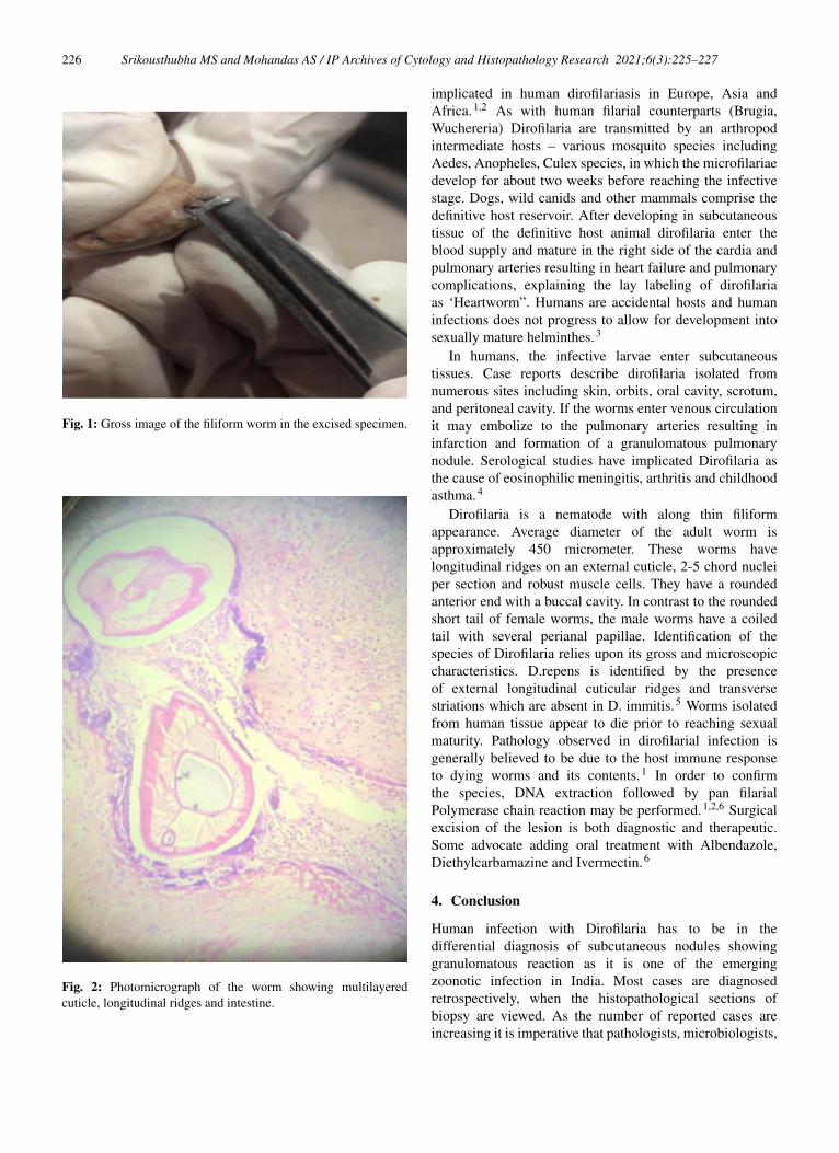
Fig. 2: Photomicrograph of the worm showing multilayeredcuticle, longitudinal ridges and intestine.
implicated in human dirofilariasis in Europe, Asia andAfrica.1,2 As with human filarial counterparts (Brugia,Wuchereria) Dirofilaria are transmitted by an arthropodintermediate hosts – various mosquito species includingAedes, Anopheles, Culex species, in which the microfilariaedevelop for about two weeks before reaching the infectivestage. Dogs, wild canids and other mammals comprise thedefinitive host reservoir. After developing in subcutaneoustissue of the definitive host animal dirofilaria enter theblood supply and mature in the right side of the cardia andpulmonary arteries resulting in heart failure and pulmonarycomplications, explaining the lay labeling of dirofilariaas ‘Heartworm”. Humans are accidental hosts and humaninfections does not progress to allow for development intosexually mature helminthes.3
In humans, the infective larvae enter subcutaneoustissues. Case reports describe dirofilaria isolated fromnumerous sites including skin, orbits, oral cavity, scrotum,and peritoneal cavity. If the worms enter venous circulationit may embolize to the pulmonary arteries resulting ininfarction and formation of a granulomatous pulmonarynodule. Serological studies have implicated Dirofilaria asthe cause of eosinophilic meningitis, arthritis and childhoodasthma.4
Dirofilaria is a nematode with along thin filiformappearance. Average diameter of the adult worm isapproximately 450 micrometer. These worms havelongitudinal ridges on an external cuticle, 2-5 chord nucleiper section and robust muscle cells. They have a roundedanterior end with a buccal cavity. In contrast to the roundedshort tail of female worms, the male worms have a coiledtail with several perianal papillae. Identification of thespecies of Dirofilaria relies upon its gross and microscopiccharacteristics. D.repens is identified by the presenceof external longitudinal cuticular ridges and transversestriations which are absent in D. immitis.5 Worms isolatedfrom human tissue appear to die prior to reaching sexualmaturity. Pathology observed in dirofilarial infection isgenerally believed to be due to the host immune responseto dying worms and its contents.1 In order to confirmthe species, DNA extraction followed by pan filarialPolymerase chain reaction may be performed.1,2,6 Surgicalexcision of the lesion is both diagnostic and therapeutic.Some advocate adding oral treatment with Albendazole,Diethylcarbamazine and Ivermectin.6
4. Conclusion
Human infection with Dirofilaria has to be in thedifferential diagnosis of subcutaneous nodules showinggranulomatous reaction as it is one of the emergingzoonotic infection in India. Most cases are diagnosedretrospectively, when the histopathological sections ofbiopsy are viewed. As the number of reported cases areincreasing it is imperative that pathologists, microbiologists,

Srikousthubha MS and Mohandas AS / IP Archives of Cytology and Histopathology Research 2021;6(3):225–227 227
surgeons, ophthalmologists and veterinarians are aware ofdirofilariasis. The development of specific and sensitivediagnostic tools for the most common specie D. repens andD immitis may help in early diagnosis.
5. Conflict of Interest
The authors declare that there are no conflicts of interest inthis paper.
6. Source of Funding
None.
References1. Human dirofilariasis: Clinical and epidemiological aspects. Theory and
practice of parasitic disease control. 2020;21:261–6.2. Khyriem A, Lynrah K, Lyngdoh W, Banik A. Subcutaneous
dirofilariasis. Indian J Med Microbiol. 2013;31(4):403–5.3. Simón F, Siles-Lucas M, Morchón R, González-Miguel J, Mellado I,
Carretón E, et al. Human and Animal Dirofilariasis: the Emergence of
a Zoonotic Mosaic. Clin Microbiol Rev. 2012;25(3):507–44.4. Kini R, Leena J, Shetty P, Lyngdoh R, Sumanth D, George L, et al.
Human dirofilariasis: an emerging zoonosis in India. J Parasit Dis.2013;39(2):349–54.
5. Padmaja P, Samuel R, Kuruvilla P, Mathai E. Subcutaneousdirofilariasis in southern India: a case report. Ann Trop Med Parasitol.2005;99(4):437–40.
6. Srinivasamurthy V, Rao S, Thejaswini M, Yoganand. Humansubcutaneous dirofilariasis. Ann Trop Med Public Health.2012;5(4):349.
Author biography
Srikousthubha MS, Pathologist
Mohandas AS, Surgeon
Cite this article: Srikousthubha MS, Mohandas AS. Dirofilariasis inhumans presenting as a subcutaneous nodule. IP Arch CytolHistopathology Res 2021;6(3):225-227.