An Unusual Presentation of Squamous Cell …...ao Chang SL, Chang ML, Chang AL, Chang BL, Chang LD...
3
Chang et al., Dermatol Case Rep 2017,2:3 Dermatology Case Reports D e r m a t o lo g y C a s e R e p o r t Open Access Case Report Volume 2 • Issue 3 • 1000123 Dermatol Case Rep, an open access journal *Corresponding author: Lawrence D. Chang, M.D. Advanced Plastic Surgery Center, 774 Christiana Road, Suite 101, New Castle County, Newark, DE 19713, USA, Tel: (302) 355-0005, E-mail: [email protected] Received: June 14, 2017; Accepted: June 27, 2017; Published: July 04, 2017 Citation: Chang SL, Chang ML, Chang AL, Chang BL, Chang LD (2017) An Unusual Presentation of Squamous Cell Carcinoma as a Subcutaneous Nodule Without Overlying Skin Changes: A Case Report. Dermatol Case Rep 2: 123. Copyright: © 2017 Chang SL, et al. This is an open-access article distributed under the terms of the Creative Commons Attribution License, which permits unrestricted use, distribution, and reproduction in any medium, provided the original author and source are credited. An Unusual Presentation of Squamous Cell Carcinoma as a Subcutaneous Nodule Without Overlying Skin Changes: A Case Report Steven L Chang 1 , Michael L Chang 1 , Alyssa L Chang 2 , Brian L Chang 3 and Lawrence D Chang 1 * 1 Advanced Plastic Surgery Center, New Castle County, Delaware, Newark, USA 2 Rowan University, Cooper University School of Medicine, New Jersey, USA 3 University of Pennsylvania, Perelman School of Medicine, Philadelphia, USA Abstract Cutaneous squamous cell carcinoma (SCC) is the second-most common skin cancer and can present with a wide variety of skin lesions. Nodular types have been described and are often characterized by hyperkeratinization, ulceration, and increased vascularity. This case study presents an elderly gentleman with a history of skin cancer who presented with a subcutaneous nodule with no overlying skin changes and was later diagnosed with SCC confined to the dermis without an epidermal origin. We present this case with the hope of increasing awareness of the wide variety of presentations of SCC and, in particular, subcutaneous forms of SCC, as these may be more dangerous and require more aggressive treatment. Keywords: Cutaneous squamous cell carcinoma; Nodular squamous cell carcinoma; Dermal squamous cell carcinoma; In-transit squamous cell carcinoma metastasis Abbreviations: Squamous Cell Carcinoma (SCC), Basal Cell Carcinoma (BCC) Introduction Cutaneous squamous cell carcinoma (SCC) arises from the squamous epithelia of the epidermis and is the second-most common skin cancer with a lifetime risk of about 10% [1-4]. Cutaneous SCC can arise de novo or evolve from a precursor lesion, such as actinic keratosis, bowenoid papulosis, or epidermodysplasia verruciformis [2,5-8]. While the appearance the appearance of cutaneous SCCs varies greatly, they generally present as firm, hyperkeratotic, ulcerated, skin-colored papules or plaques found on sun-exposed skin and may be itchy or painful [2,3]. Nodular types of SCC have also been described and are frequently confused with other types of nodules, including nodular basal cell carcinoma (BCC), hypertrophic intraepidermal carcinoma, fibroxanthoma, Merkel cell carcinoma, nodular melanoma, seborrheic keratosis, lipomas, etc [8]. e nodular type of SCC is commonly characterized by a keratin crust or scale, a central keratin mass, prominent vascularity or haemorrhage, and multiple colors.8 As with most other types of SCC, nodular SCC arises in the epidermis and then invades the dermis [9-10]. While nodular SCCs typically have some form of epithelial change, there have been a few reports of SCCs that present as subcutaneous nodules without significant epithelial manifestations [11-16]. Currently, it is believed that these SCCs are not primary skin cancers but rather metastatic: either from a visceral malignancy or as an in-transit metastasis from a prior SCC, similar to melanoma [11-16]. Regardless of the origin, the subcutaneous SCCs have been reported to be more aggressive and recur more frequently. It is therefore important to be aware of this type of SCC and treat it appropriately. Here, we present a case of subcutaneous SCC and review the existing literature on disease course and management. e patient initially presented with a subcutaneous nodule without overlying skin changes and was subsequently diagnosed with SCC limited to the dermis without involvement of the epidermis. We hope that this case report will help raise awareness of this atypical presentation of SCC so that it can be properly managed. Case Report KF is a 72-year-old Caucasian male with a history of BCC, SCC, type II diabetes mellitus, and Parkinson’s disease who presented with a tender lump in his leſt outer cheek that had been slowly growing over the past two months. Exam revealed a 2 × 1 cm subcutaneous mass with tenderness, erythema, and warmth but no drainage or overlying skin changes (Figure 1). KF underwent routine skin surveillance examination 9 months prior to presentation; at that time, there was no sign of a lump or other skin changes (Figure 2). At this time, it was thought that KF likely had an infected sebaceous cyst, so he was empirically treated with oral amoxicillin. However, 2 weeks later, the subcutaneous growth remained unchanged, though the symptoms of tenderness and erythema resolved. He was then referred to plastic surgery. Figure 1: Image of KF’s left cheek at initial presentation that demonstrated a 2x1 cm subcutaneous mass that was tender, erythematous, and warm to the touch though lacked significant epithelial changes.
Transcript of An Unusual Presentation of Squamous Cell …...ao Chang SL, Chang ML, Chang AL, Chang BL, Chang LD...
Research Article
Chang et al., Dermatol Case Rep 2017,2:3
Dermatology Case ReportsDe
rmatology Case Report
Open AccessCase Report
Volume 2 • Issue 3 • 1000123Dermatol Case Rep, an open access journal
*Corresponding author: Lawrence D. Chang, M.D. Advanced Plastic SurgeryCenter, 774 Christiana Road, Suite 101, New Castle County, Newark, DE 19713,USA, Tel: (302) 355-0005, E-mail: [email protected]
Received: June 14, 2017; Accepted: June 27, 2017; Published: July 04, 2017
Citation: Chang SL, Chang ML, Chang AL, Chang BL, Chang LD (2017) An Unusual Presentation of Squamous Cell Carcinoma as a Subcutaneous Nodule Without Overlying Skin Changes: A Case Report. Dermatol Case Rep 2: 123.
Copyright: © 2017 Chang SL, et al. This is an open-access article distributed under the terms of the Creative Commons Attribution License, which permits unrestricted use, distribution, and reproduction in any medium, provided the original author and source are credited.
An Unusual Presentation of Squamous Cell Carcinoma as a Subcutaneous Nodule Without Overlying Skin Changes: A Case ReportSteven L Chang1, Michael L Chang1, Alyssa L Chang2, Brian L Chang3 and Lawrence D Chang1*1Advanced Plastic Surgery Center, New Castle County, Delaware, Newark, USA2Rowan University, Cooper University School of Medicine, New Jersey, USA3University of Pennsylvania, Perelman School of Medicine, Philadelphia, USA
AbstractCutaneous squamous cell carcinoma (SCC) is the second-most common skin cancer and can present with a
wide variety of skin lesions. Nodular types have been described and are often characterized by hyperkeratinization, ulceration, and increased vascularity. This case study presents an elderly gentleman with a history of skin cancer who presented with a subcutaneous nodule with no overlying skin changes and was later diagnosed with SCC confined to the dermis without an epidermal origin. We present this case with the hope of increasing awareness of the wide variety of presentations of SCC and, in particular, subcutaneous forms of SCC, as these may be more dangerous and require more aggressive treatment.
Keywords: Cutaneous squamous cell carcinoma; Nodular squamouscell carcinoma; Dermal squamous cell carcinoma; In-transit squamous cell carcinoma metastasis
Abbreviations: Squamous Cell Carcinoma (SCC), Basal CellCarcinoma (BCC)
Introduction Cutaneous squamous cell carcinoma (SCC) arises from the
squamous epithelia of the epidermis and is the second-most common skin cancer with a lifetime risk of about 10% [1-4]. Cutaneous SCC can arise de novo or evolve from a precursor lesion, such as actinic keratosis, bowenoid papulosis, or epidermodysplasia verruciformis [2,5-8]. While the appearance the appearance of cutaneous SCCs varies greatly, they generally present as firm, hyperkeratotic, ulcerated, skin-colored papules or plaques found on sun-exposed skin and may be itchy or painful [2,3].
Nodular types of SCC have also been described and are frequently confused with other types of nodules, including nodular basal cell carcinoma (BCC), hypertrophic intraepidermal carcinoma, fibroxanthoma, Merkel cell carcinoma, nodular melanoma, seborrheic keratosis, lipomas, etc [8]. The nodular type of SCC is commonly characterized by a keratin crust or scale, a central keratin mass, prominent vascularity or haemorrhage, and multiple colors.8 As with most other types of SCC, nodular SCC arises in the epidermis and then invades the dermis [9-10].
While nodular SCCs typically have some form of epithelial change, there have been a few reports of SCCs that present as subcutaneous nodules without significant epithelial manifestations [11-16]. Currently, it is believed that these SCCs are not primary skin cancers but rather metastatic: either from a visceral malignancy or as an in-transit metastasis from a prior SCC, similar to melanoma [11-16]. Regardless of the origin, the subcutaneous SCCs have been reported to be more aggressive and recur more frequently. It is therefore important to be aware of this type of SCC and treat it appropriately.
Here, we present a case of subcutaneous SCC and review the existing literature on disease course and management. The patient initially presented with a subcutaneous nodule without overlying skin changes and was subsequently diagnosed with SCC limited to the dermis without involvement of the epidermis. We hope that this case report will help raise awareness of this atypical presentation of SCC so that it can be properly managed.
Case Report KF is a 72-year-old Caucasian male with a history of BCC, SCC,
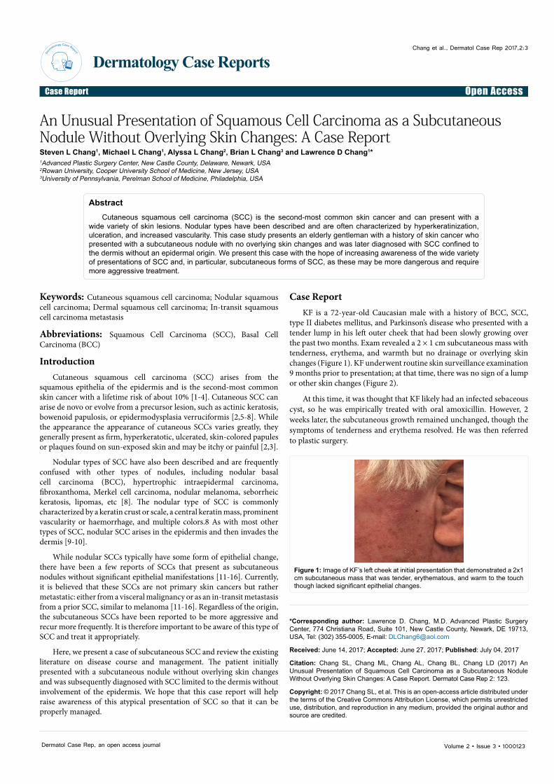
type II diabetes mellitus, and Parkinson’s disease who presented with a tender lump in his left outer cheek that had been slowly growing over the past two months. Exam revealed a 2 × 1 cm subcutaneous mass with tenderness, erythema, and warmth but no drainage or overlying skin changes (Figure 1). KF underwent routine skin surveillance examination 9 months prior to presentation; at that time, there was no sign of a lump or other skin changes (Figure 2).
At this time, it was thought that KF likely had an infected sebaceous cyst, so he was empirically treated with oral amoxicillin. However, 2 weeks later, the subcutaneous growth remained unchanged, though the symptoms of tenderness and erythema resolved. He was then referred to plastic surgery.
Figure 1: Image of KF’s left cheek at initial presentation that demonstrated a 2x1 cm subcutaneous mass that was tender, erythematous, and warm to the touch though lacked significant epithelial changes.
Citation: Chang SL, Chang ML, Chang AL, Chang BL, Chang LD (2017) An Unusual Presentation of Squamous Cell Carcinoma as a Subcutaneous Nodule Without Overlying Skin Changes: A Case Report. Dermatol Case Rep 2: 123.
Page 2 of 3
Volume 2 • Issue 3 • 1000123Dermatol Case Rep, an open access journal
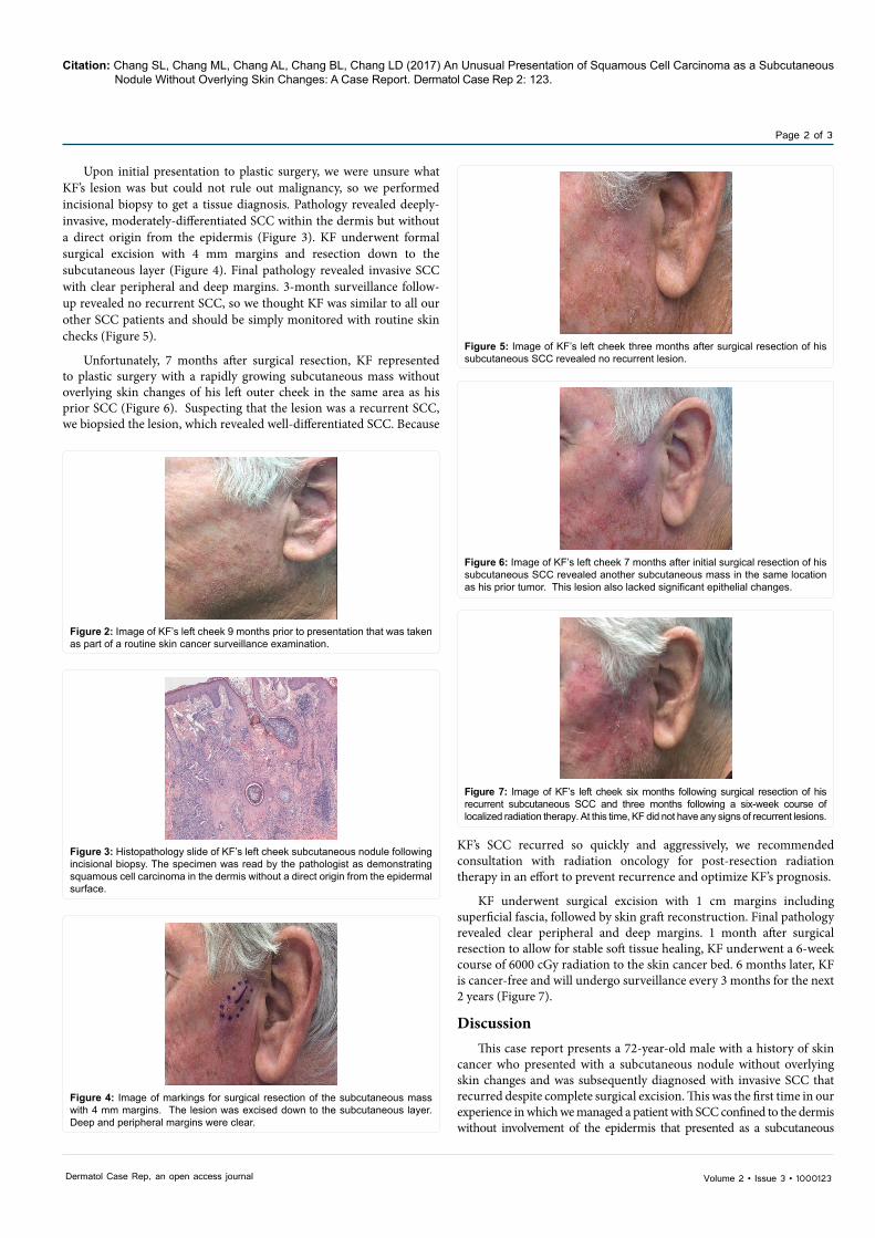
Upon initial presentation to plastic surgery, we were unsure what KF’s lesion was but could not rule out malignancy, so we performed incisional biopsy to get a tissue diagnosis. Pathology revealed deeply-invasive, moderately-differentiated SCC within the dermis but without a direct origin from the epidermis (Figure 3). KF underwent formal surgical excision with 4 mm margins and resection down to the subcutaneous layer (Figure 4). Final pathology revealed invasive SCC with clear peripheral and deep margins. 3-month surveillance follow-up revealed no recurrent SCC, so we thought KF was similar to all our other SCC patients and should be simply monitored with routine skin checks (Figure 5).
Unfortunately, 7 months after surgical resection, KF represented to plastic surgery with a rapidly growing subcutaneous mass without overlying skin changes of his left outer cheek in the same area as his prior SCC (Figure 6). Suspecting that the lesion was a recurrent SCC, we biopsied the lesion, which revealed well-differentiated SCC. Because
KF’s SCC recurred so quickly and aggressively, we recommended consultation with radiation oncology for post-resection radiation therapy in an effort to prevent recurrence and optimize KF’s prognosis.
KF underwent surgical excision with 1 cm margins including superficial fascia, followed by skin graft reconstruction. Final pathology revealed clear peripheral and deep margins. 1 month after surgical resection to allow for stable soft tissue healing, KF underwent a 6-week course of 6000 cGy radiation to the skin cancer bed. 6 months later, KF is cancer-free and will undergo surveillance every 3 months for the next 2 years (Figure 7).
Discussion This case report presents a 72-year-old male with a history of skin
cancer who presented with a subcutaneous nodule without overlying skin changes and was subsequently diagnosed with invasive SCC that recurred despite complete surgical excision. This was the first time in our experience in which we managed a patient with SCC confined to the dermis without involvement of the epidermis that presented as a subcutaneous
Figure 2: Image of KF’s left cheek 9 months prior to presentation that was taken as part of a routine skin cancer surveillance examination.
Figure 3: Histopathology slide of KF’s left cheek subcutaneous nodule following incisional biopsy. The specimen was read by the pathologist as demonstrating squamous cell carcinoma in the dermis without a direct origin from the epidermal surface.
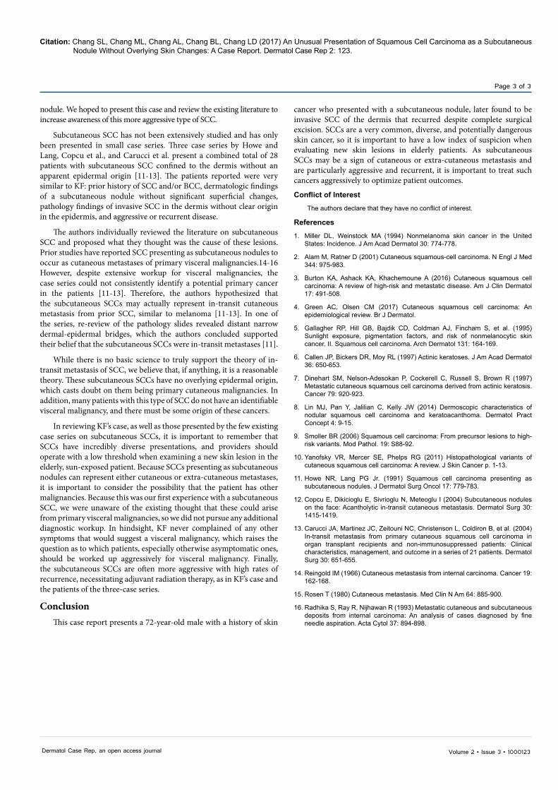
Figure 4: Image of markings for surgical resection of the subcutaneous mass with 4 mm margins. The lesion was excised down to the subcutaneous layer. Deep and peripheral margins were clear.
Figure 5: Image of KF’s left cheek three months after surgical resection of his subcutaneous SCC revealed no recurrent lesion.
Figure 6: Image of KF’s left cheek 7 months after initial surgical resection of his subcutaneous SCC revealed another subcutaneous mass in the same location as his prior tumor. This lesion also lacked significant epithelial changes.
Figure 7: Image of KF’s left cheek six months following surgical resection of his recurrent subcutaneous SCC and three months following a six-week course of localized radiation therapy. At this time, KF did not have any signs of recurrent lesions.
Citation: Chang SL, Chang ML, Chang AL, Chang BL, Chang LD (2017) An Unusual Presentation of Squamous Cell Carcinoma as a Subcutaneous Nodule Without Overlying Skin Changes: A Case Report. Dermatol Case Rep 2: 123.
Page 3 of 3
Volume 2 • Issue 3 • 1000123Dermatol Case Rep, an open access journal
nodule. We hoped to present this case and review the existing literature to increase awareness of this more aggressive type of SCC.
Subcutaneous SCC has not been extensively studied and has only been presented in small case series. Three case series by Howe and Lang, Copcu et al., and Carucci et al. present a combined total of 28 patients with subcutaneous SCC confined to the dermis without an apparent epidermal origin [11-13]. The patients reported were very similar to KF: prior history of SCC and/or BCC, dermatologic findings of a subcutaneous nodule without significant superficial changes, pathology findings of invasive SCC in the dermis without clear origin in the epidermis, and aggressive or recurrent disease.
The authors individually reviewed the literature on subcutaneous SCC and proposed what they thought was the cause of these lesions. Prior studies have reported SCC presenting as subcutaneous nodules to occur as cutaneous metastases of primary visceral malignancies.14-16 However, despite extensive workup for visceral malignancies, the case series could not consistently identify a potential primary cancer in the patients [11-13]. Therefore, the authors hypothesized that the subcutaneous SCCs may actually represent in-transit cutaneous metastasis from prior SCC, similar to melanoma [11-13]. In one of the series, re-review of the pathology slides revealed distant narrow dermal-epidermal bridges, which the authors concluded supported their belief that the subcutaneous SCCs were in-transit metastases [11].
While there is no basic science to truly support the theory of in-transit metastasis of SCC, we believe that, if anything, it is a reasonable theory. These subcutaneous SCCs have no overlying epidermal origin, which casts doubt on them being primary cutaneous malignancies. In addition, many patients with this type of SCC do not have an identifiable visceral malignancy, and there must be some origin of these cancers.
In reviewing KF’s case, as well as those presented by the few existing case series on subcutaneous SCCs, it is important to remember that SCCs have incredibly diverse presentations, and providers should operate with a low threshold when examining a new skin lesion in the elderly, sun-exposed patient. Because SCCs presenting as subcutaneous nodules can represent either cutaneous or extra-cutaneous metastases, it is important to consider the possibility that the patient has other malignancies. Because this was our first experience with a subcutaneous SCC, we were unaware of the existing thought that these could arise from primary visceral malignancies, so we did not pursue any additional diagnostic workup. In hindsight, KF never complained of any other symptoms that would suggest a visceral malignancy, which raises the question as to which patients, especially otherwise asymptomatic ones, should be worked up aggressively for visceral malignancy. Finally, the subcutaneous SCCs are often more aggressive with high rates of recurrence, necessitating adjuvant radiation therapy, as in KF’s case and the patients of the three-case series.
Conclusion This case report presents a 72-year-old male with a history of skin
cancer who presented with a subcutaneous nodule, later found to be invasive SCC of the dermis that recurred despite complete surgical excision. SCCs are a very common, diverse, and potentially dangerous skin cancer, so it is important to have a low index of suspicion when evaluating new skin lesions in elderly patients. As subcutaneous SCCs may be a sign of cutaneous or extra-cutaneous metastasis and are particularly aggressive and recurrent, it is important to treat such cancers aggressively to optimize patient outcomes.
Conflict of Interest
The authors declare that they have no conflict of interest.
References
1. Miller DL, Weinstock MA (1994) Nonmelanoma skin cancer in the UnitedStates: Incidence. J Am Acad Dermatol 30: 774-778.
2. Alam M, Ratner D (2001) Cutaneous squamous-cell carcinoma. N Engl J Med344: 975-983.
3. Burton KA, Ashack KA, Khachemoune A (2016) Cutaneous squamous cellcarcinoma: A review of high-risk and metastatic disease. Am J Clin Dermatol17: 491-508.
4. Green AC, Olsen CM (2017) Cutaneous squamous cell carcinoma: Anepidemiological review. Br J Dermatol.
5. Gallagher RP, Hill GB, Bajdik CD, Coldman AJ, Fincham S, et al. (1995)Sunlight exposure, pigmentation factors, and risk of nonmelanocytic skincancer. II. Squamous cell carcinoma. Arch Dermatol 131: 164-169.
6. Callen JP, Bickers DR, Moy RL (1997) Actinic keratoses. J Am Acad Dermatol36: 650-653.
7. Dinehart SM, Nelson-Adesokan P, Cockerell C, Russell S, Brown R (1997)Metastatic cutaneous squamous cell carcinoma derived from actinic keratosis.Cancer 79: 920-923.
8. Lin MJ, Pan Y, Jalilian C, Kelly JW (2014) Dermoscopic characteristics ofnodular squamous cell carcinoma and keratoacanthoma. Dermatol PractConcept 4: 9-15.
9. Smoller BR (2006) Squamous cell carcinoma: From precursor lesions to high-risk variants. Mod Pathol. 19: S88-92.
10. Yanofsky VR, Mercer SE, Phelps RG (2011) Histopathological variants ofcutaneous squamous cell carcinoma: A review. J Skin Cancer p. 1-13.
11. Howe NR, Lang PG Jr. (1991) Squamous cell carcinoma presenting assubcutaneous nodules. J Dermatol Surg Oncol 17: 779-783.
12. Copcu E, Dikicioglu E, Sivrioglu N, Meteoglu I (2004) Subcutaneous noduleson the face: Acantholytic in-transit cutaneous metastasis. Dermatol Surg 30:1415-1419.
13. Carucci JA, Martinez JC, Zeitouni NC, Christenson L, Coldiron B, et al. (2004)In-transit metastasis from primary cutaneous squamous cell carcinoma inorgan transplant recipients and non-immunosuppressed patients: Clinicalcharacteristics, management, and outcome in a series of 21 patients. Dermatol Surg 30: 651-655.
14. Reingold IM (1966) Cutaneous metastasis from internal carcinoma. Cancer 19: 162-168.
15. Rosen T (1980) Cutaneous metastasis. Med Clin N Am 64: 885-900.
16. Radhika S, Ray R, Nijhawan R (1993) Metastatic cutaneous and subcutaneous deposits from internal carcinoma: An analysis of cases diagnosed by fine needle aspiration. Acta Cytol 37: 894-898.